in migraine affect Ca
V2.1 channel regulation
by G proteins and SNAREs
Selma A. Serra Pascual
TESI DOCTORAL UPF / 2011
DIRECTORS DE LA TESI:
Dr.
José Manuel Fernández-Fernández
Dr. Miguel Á. Valverde
A mi madre, al meu pare y a Miguel.
En record del besavi Salvador
i de l’ avi Pere.
“¡Como el entomólogo a la caza de mariposas de vistosos matices, mi atención perseguía, en el vergel de la substancia gris, células de formas delicadas y elegantes, las misteriosas mariposas del alma, cuyo batir de alas quién sabe si esclarecerá algún día el secreto de la vida mental!”
¡Parece mentira que esta aventura haya llegado a su fin!
La realización de esta tesis ha supuesto una gran inversión personal y económica. Por lo segundo, agradezco al Ministerio de Ciencia e Innovación y a la Marató de TV3 el haber financiado nuestros proyectos científicos, y en concreto al Ministerio, el haberme concedido una beca. Me gustaría pensar que a pesar de la crisis que estamos atravesando los políticos y la sociedad sigan apostando por la investigación y el desarrollo en el futuro, pues en ello reside la clave fundamental para el progreso “sostenible”.
Son muchas las personas que han aportado su conocimiento e ideas y ofrecido su respaldo durante el desarrollo de mi tesis. Gracias a todos por haberme hecho crecer como científica y como persona, haber contribuido a realizar este sueño y compartido algunas tristezas pero también muchas alegrías.
En especial, quiero agradecer todo el cariño y el apoyo que siempre que recibido por parte de todos los compañeros y amigos del laboratorio de Fisio; a los que ya os fuisteis y a los que os quedáis. Con vosotros, no solo he compartido el lugar de trabajo sino también amistad, buen humor y momentos inolvidables dentro y fuera del laboratorio.
Quiero destacar la importante contribución de Cristina y Fanny a mi trabajo. Os doy las gracias a ambas por haberme permitido aprovechar y aprender tanto de vuestras habilidosas y sabias manos, pero sobretodo por haberme regalado vuestra alegría y risas contagiosas cuando más lo he necesitado.
Agradezco profundamente a mi director de tesis, Chema, el haberme enseñado tanto y tan bien, no solo sobre canales y electrofisiología, sino sobretodo a pensar y a discutir. Espero haber heredado tu gran capacidad de trabajo, perseverancia y paciencia infinitas ya que esas son, a mi parecer, las cualidades esenciales de un buen electrofisiólogo. Pero por encima de todo agradezco la confianza que has depositado en mí, la fuerza positiva transmitida en momentos complicados y el haberme brindado la oportunidad de aportar mis ideas e iniciativas propias al proyecto en tantas ocasiones.
Asimismo quiero destacar la trascendencia que ha tenido Miguel en mi vida profesional desde mucho antes de empezar a trabajar en este proyecto. Gracias por abrirme hace años las puertas de tu laboratorio, transmitirme la pasión por la “ciencia en directo”, y por apostar por mí como ser humano y como científica.
ABSTRACT
Familial hemiplegic migraine (FHM)-causing mutations in the gene encoding the P/Q Ca2+ channel α1A subunit (CACNA1A) normally locate in the pore or voltage
sensor regions and involve gain-of-channel function. We have studied the functional consequences of two new α1A mutations found in migraine patients.
Mutation Y1245C (which is first missense mutation found in a subject affected with childhood periodic syndromes that evolved into hemplegic migraine and the sole mutation described in any S1 segment of α1A subunit) produced an
overall gain of channel function by favouring its activation and lessening Gβγ subunit-dependent channel inhibition (even in response to physiological stimuli). FHM non-causative A454T mutation located in α1A I-II loop and associated with
attenuated migraine aura symptoms was found to alter CaVβ and SNARE
modulation of channel slow inactivation and drastically reduce channel-dependent secretory efficiency. Altogether, our results underscore S1 structural role in α1A voltage sensor function and unveil the importance of I-II loop in the
functional interplay between P/Q channels and SNARE proteins. Lastly, we propose that mutations in CACNA1A gene not only cause FHM but also may modify migraine phenotype.
RESUM
Mutacions causants de migranya hemiplègica familiar (MHF) en el gen que codifica per a la subunitat α1A del canal de Ca
2+
de tipus P/Q(CACNA1A) afecten les regions del porus o del sensor de voltatge i produeixen un guany de funció del canal. Hem analitzat funcionalment dues noves mutacions de la subunitat
α1A identificades en pacients migranyosos. La mutació Y1245C (la primera
mutació amb canvi de sentit identificada en un pacient afectat de síndromes periòdiques infantils que evolucionaren a migranya hemiplègica i l’única trobada en un segment S1 de la subunitat α1A) produeix un guany de funció global del
canal afavorint la seva activació i disminuint la inhibició per subunitats Gβγ (fins i tot en resposta a estímuls fisiològics). La mutació A454T ubicada al llaç I-II de la subunitat α1A, que no causa MHF però s’associa a una atenuació dels símptomes
de l’aura, altera la regulació de la inactivació lenta per subunitats CaVβ i
proteïnes SNARE, observant-se també una reducció dràstica de l’eficiència secretora depenent dels canals mutants. En conjunt, els nostres resultats subratllen el paper estructural del segment S1 en la funció del sensor de voltatge de la subunitat α1A i mostren la importància del llaç intracel·lular I-II en la
PREFACE
Migraine is one the most prevalent neurological disorders affecting around 12%
of the general population. However, the molecular and cellular origins of
migraine are among the most enigmatic in neuroscience. Most agree that
migraine susceptibility is inherited and that its clinical manifestation is strongly
modulated by both internal and external factors. In recent years we have
advanced considerably in our understanding of the mechanisms participating in
the generation of the migraine symptoms or the migraine attack, although many
open questions remain unresolved. We have learned that the trigeminovascular
system (TGVS) is responsible for the pain itself and that cortical spreading
depression (CSD), a wave of neuronal excitability throughout the brain cortex,
underlies the aura symptoms reported by many migraineurs. Still a matter of
debate is whether the CSD is also responsible for the triggering of the
neurovascular inflammation and the subsequent pain. Nevertheless, the
identification of the first causative genes for a mendelian subtype of migraine
with aura, i.e. familial hemiplegic migraine (FHM), pointed to an alteration in the
stimulus-secretion coupling at the neuronal synapses. Gain of function
mutations in the pore forming α1 subunit of CaV2.1 (P/Q-type) calcium channel (CACNA1A) were identified in families suffering from FHM.
CaV2.1 channel is a key player in the control of excitatory neurotransmitter
release in the central nervous system. It mediates Ca2+ entry at the synapse
following the arrival of an action potential, thereby triggering the fusion of the
neurotransmitter-containing vesicles with the plasma membrane and the
release of their content to the synapse cleft. The fact that gain of function
mutations in CaV2.1 channel were linked to FHM suggested that in those
patients excitatory synapses were also facilitated, which in turn would explain
the generation of the CSD. Once again, despite our substantial advance in the
understanding of the mechanism related to FHM, several questions related to
both the molecular mechanisms and the clinical phenotype arose. For example,
relates to the fact that different patients carrying the same migraine causative
mutation within a family do not present the same clinical phenotype. Other
questions relate to the molecular mechanism that generates the gain of function
at the protein level. Most reported mutations in CaV2.1 are located to the pore
or the voltage sensor regions of the channel, thereby affecting the core
properties of the channel. But, are there mutations affecting other regions of
the channel that mediate its interaction with regulatory proteins or even the
secretory machinery itself? This Thesis is an attempt to cast light on at least
ABBREVIATIONS
ABP = AID-binding pocket
AID = α1-interacting domain AP = action potential
APW = action potential-like waveforms
ATP1A2/ATP1A2 = gene encoding/α2 subunit of the Na +
, K+ ATPase
BBB = blood brain barrier
BF = blood flow
BID = β-interaction domain
BOLD = blood oxygenation level-dependent
BoNtC1 = Botulinum neurotoxin type C1
BPT = benign paroxysmal torticollis
BPV = benign paroxysmal vertigo
CACNA1A/CACNA1A= gene encoding/pore-forming α1 subunit of voltage-gated
CaV2.1 or P/Q-type Ca 2+
channels
[Ca2+] = concentration of Ca2+ ions
CaV X= voltage-gated Ca2+ channel
CaVβ = voltage-gated Ca 2+
channel beta subunit
CGRP = calcitonin gene-related peptide
CNS = central nervous system
CPS = childhood periodic syndromes
CSD = cortical spreading depression
DHP = dihydropyridine
DTT = dithiothreitol
EPSP = excitatory postsynaptic potential
fMRI = functional magnetic resonance imaging
FHM = familial hemiplegic migraine
FS = fast-spiking
GK = guanylate kinase
HVA = high voltage activated
KI = knockin
LVA = low voltage activated
MA = migraine with aura
MAGUK = membrane-associated guanylate kinases
MIDAS = metal-ion-dependent adhesion site
MO = migraine without aura
MPC = mouse pheochromocytoma
NO = nitric oxide
PAG = periaqueductal grey region
PECT or PET = photon emission computed tomography
PPF = paired-pulse facilitation
SCGN = superior cervical ganglion neuron
SCNA1A/SCNA1A = gene encoding/pore-forming α1 subunit of NaV1.1 channels
SH3 = Src homology 3
SHM = sporadic hemiplegic migraine
SM = Sec1/Munc 18-like
SNARE = soluble N-sensitive factor attachment receptor
SP = substance P
SSN = superior salivatory nucleus
Synprint = synaptic protein interaction
TBA = tetrabutylammonium
TGVS = trigeminovascular system
TNC = trigeminal nucleus pars caudalis
VWA = Von Willebrand factor A
Pg.
ABSTRACT/RESUM………..…….... vii
PREFACE……….……… ix
ABBREVIATIONS……….. xi
I. INTRODUCTION………..………... 1
1. VOLTAGE-GATED CALCIUM CHANNELS………... 3
1.1. Importance of Electrical-to-Chemical Transduction...…. 3
1.2. First Steps in Ca2+ Channel Discovery……... 4
1.3. Voltage-gated Ca2+ Current Types……….. 7
1.4. Structure of Voltage-gated Ca2+ Channels and Regulation by Auxiliary Subunits... 9
1.4.1. The Pore-forming α1 Subunit……….... 10
1.4.2. Auxiliary Ca2+ Channel Subunits...……… 12
α2δ Subunits………..…... 12
γ Subunits………..….…… 14
β Subunits………..……… 15
1.4.3. Molecular Determinants of Voltage-dependent Inactivation in HVA Ca2+ Channels……….……….…. 22
2. NEURONAL VOLTAGE-GATED Ca2+ CHANNEL ROLE IN SYNAPTIC TRANSMISSION……… 25
2.1. The Presynaptic CaV2 Channel Signalling Complexes……... 25
2.2. Regulation of CaV2 Channels by G Protein………... 26
2.3. Regulation of CaV2 Channels by SNARE Proteins………... 31
2.3.1. Impact of SNARE-synprint Interaction on Synaptic Transmission………..……….. 38
3. NEUROBIOLOGY OF MIGRAINE………... 43
3.1. Genetic Component of Migraine...……… 43
3.1.1. Comorbidity…….……….………. 47
3.2. Introduction to Migraine Pathophysiology...…………...………… 48
3.2.1. Primary Cause of the Migraine Headache: The Trigeminal Pathway……….………. 49
The Role of the Cerebral Cortex………... 52
The Involvement of the Brainstem ………. 57
3.2.2. Pain Mechanisms………... 58
3.2.3. An Integrated Model for Migraine Pathophysiology…….… 59
3.3. Familial Hemiplegic Migraine……….……… 61
3.4.1. Functional Consequences of FHM1 Mutations….……… 68
II. OBJECTIVES………..……….… 75
III. RESULTS..………..………... 79
1. ARTICLE A……….… 81
2. ARTICLE B………. 97
SUPPORTING INFORMATION…….……….. 105
IV. DISCUSSION .………..……… 113
1. GENOTYPE-PHENOTYPE CORRELATIONS IN FHM1……….... 115
2. FHM1 MUTATIONS AFFECT DIFFERENT BIOPHYSICAL ASPECTS OF CaV2.1 CHANNEL ACTIVITY AND REGULATION………... 116
2.1. Localization of FHM1 Mutations on α1A Subunit……….. 116
2.2. Biophysical Consequences of FHM1 Mutations on CaV2.1 Channel Activation and Channel Expression……….………… 118
2.2.1. Functional Effects of FHM1 Mutations on the Modulation of CaV2.1 Channel Gating by G Proteins…....… 119
2.3. Biophysical Consequences of FHM1 Mutations on Voltage-dependent CaV2.1 Channel Inactivation... 123
2.3.1. Modulation of Slow Inactivation by SNARE Proteins….….. 129
Consequences of an Impaired Coupling between CaV2.1 Channels and the Exocytotic Machinery...………. 133
3. RELEVANCE OF Y1245C AND A454T MUTATIONS IN MIGRAINE PATHOPHYSIOLOGY………. 138
V. CONCLUSIONS………..………. 143
VI. ANNEX ……….………. 147
1. ARTICLE C……… 149
[image:16.499.91.415.77.510.2]1.
VOLTAGE-GATED CALCIUM CHANNELS
1.1. Importance of Electrical-to-Chemical Transduction
Calcium (Ca2+) ion is a ubiquitous universal intracellular messenger of electrical signalling implicated in most if not all cellular processes.
Increases in cytosolic free Ca2+ concentration ([Ca2+]i) require a tight
spatiotemporal control for proper functioning of most cells1, and aberrant Ca2+ homeostasis can rapidly result in cell death2. Basically, changes in [Ca2+]i in cells are due to activation of Ca
2+
entry pathways in
the plasma membrane or by activation of release Ca2+ channels in the intracellular stores, and countered by transporters acting as Ca2+ pumps1. Voltage-gated Ca2+ channels are a preeminent class of large heteromeric plasma membrane proteins providing Ca2+ influx in response to changes in membrane potential3-5. They are members of a gene superfamily of transmembrane ion channel proteins that includes voltage-gated
potassium (K+) and sodium (Na+) channels6.
Ca2+ entry through voltage-gated Ca2+ initiates and controls a host of intracellular events such as muscle contraction, hormone secretion,
synaptic transmission, and gene expression2. Accordingly, voltage-gated Ca2+ channel activity is essential to couple electrical signals in the cell surface to chemical messages in excitable cells, leading to the nearly
universal rule that their presence is precisely what defines an excitable
cell7.
Mutations of genes encoding voltage-gated Ca2+ channels identified in humans, mice and other organisms have been implicated in the aetiology
of a diverse group of paroxystic (or episodic) neurological and muscular
diseases commonly known as channelopathies. In particular, mutations
channels, are associated to familial hemiplegic migraine and some forms
of ataxia. The study of the functional consequences of these mutations,
in addition to the formerly vast knowledge about Ca2+ channels obtained by the work of numerous investigators in the past few decades, not only
has allowed the unravelling of the nature of these complex pathologies
but has taught us many lessons about the physiology of the nervous
system.
Over the length of this introduction, I shall invite you to take part of an
enthralling trip that starts from the obscure -yet intriguing- times of Ca2+ channel discovery, crosses the confines of our current understanding
about Ca2+ channel structure and regulation by some of its faithful partners, and ends aiming to picture the most we have learned from
P/Q-type Ca2+ channels about migraine.
1.2. First Steps in Ca
2+Channel Discovery
Despite the pivotal role of Ca2+ channels in so many cellular processes, as well as the near-ubiquitous presence of different types of Ca2+ channels in excitable cells, they were surprisingly latecomers to cellular
electrophysiology (the early work on Ca2+ channel discovery has been reviewed in3,5,8).
In the early studies of neural circuits in the squid giant axon, excitability
became rightfully synonymous with the sodium (Na+) channel, on the minds of Nobel Prize winners Alan Hodgkin, Andrew Huxley, and Bernard
Katz (Figure 1A). In 1953, soon after the “sodium theory” of the action
potential (AP) was established, Paul Fatt and Bernard Katz found,
accidentally, an exception while studying neuromuscular transmission in
replaced by tetrabutylammonium (TBA)) the muscle still generated APs9. However, they were quite circumspect about concluding that Ca2+ was the charge carrier: “The mechanism of the AP, and the species of ions
involved in the movement of charge across the membrane, remain a
puzzling problem […] (i)it may be that TBA remains adsorbed to the fibre
surface, but is mobilized during excitation and temporarily transferred
into the cell interior; (ii) alternatively, influx of calcium or magnesium, or
outflux of some internal anion may be responsible of transport of
charge.”
The mystery of this new form of excitability was considerably unveiled by
Fatt and Ginsborg (1958) who were able to record AP mediated by
different divalent ions in crustacean muscle leading to a cautious but
clear statement that the movement of Ca2+ itself could support APs across the membrane10. Another important contributor to the birth of the field of Ca2+ channels was Susumu Hagiwara (Figure 1B) who carried out an in-depth characterization of Ca2+ conductances in various invertebrate tissues. He is well-known for his studies on “Ca2+ spikes” in barnacle giant muscle fibers11, and also because he provided the first evidences of the existence of various Ca2+ channel types in many invertebrate preparations like starfish eggs12.
Work on mammalian tissues began later in parallel with the invertebrate
work. Ca2+ APs and subsequently Ca2+ currents were identified first in mammalian skeletal and cardiac muscle, and then in all excitable cells.
Worth noting are the first voltage-clamp recordings of Ca2+ currents in cardiac myocytes performed by Herald Reuter in 1967 (Figure 1C) who
The use of pharmacological approaches has represented a key step to
understand the function and to identifying the existence of multiple
subtypes of Ca2+ currents. A clear example was the application of verapamil or nifedipine drugs, belonging to phenylalkylamines and
1,4-dihydropyridines (DHPs) class of molecules respectively, as “calcium
antagonists” (i.e. any drug that blocked excitation-contraction) in the
mid-1960s by Albert Fleckenstein (Figure 1D) and others (reviewed in14). The various class of “calcium antagonists” were found to block in fact
specific Ca2+ currents with differential selectivity in cardiac and smooth muscle, and this forms the basis of their well-known therapeutic role in a
number of cardiovascular syndromes.
These drugs also proved to be invaluable for the isolation of purified
[image:22.499.167.332.356.572.2]voltage-gated Ca2+ channels, an essential step on the path of the cloning era.
Figure 1. Some major figures in the early Ca2+ channels’ era. A) Sir Bernard Katz.
1.3. Voltage-gated Ca
2+Current Types
Multiple Ca2+ currents in different cell types have been identified following physiological and pharmacological criteria and an alphabetical
nomenclature has evolved for the distinct classes of Ca2+ currents4,15. In the early 1980s, voltage-gated Ca2+ currents were distinguished between low- and high-voltage activated (LVA and HVA) through several
electrophysiological studies mostly in biological systems less obscure
than starfish eggs.
In cardiac, smooth, and skeletal muscle, the major Ca2+ currents were defined by high voltage of activation, slow voltage-dependent
inactivation, and specific inhibition by DHPs, phenylalkylamines and
benzothiazepines16. These Ca2+ currents were designated as L-type, as they were long-lasting and showed large unitary conductance when
barium (Ba2+) was the charge carrier17.
In neurons, new components of the HVA Ca2+ current showed clear differences from L-type. On the one hand, there was a conductance that
required much more negative potentials to be activated, inactivated
rapidly, deactivated slowly, had a small single-channel conductance, and
was insensitive to DHPs18,19. These type of currents were designated LVA Ca2+ currents18 or T-type for their transient kinetics and tiny unitary Ba2+ currents19 and were also found in heart cells20. On the other hand, whole-cell voltage-clamp and single-channel recordings revealed additional Ca2+ currents which were largely specific to neurons, had an intermediate
conductance to Ba2+, voltage-dependence and rate of inactivation indicating that they were neither T- or L-type, and thus were named
L-type channel antagonists but blocked specifically by the toxin derived
from marine snail venom ω-conotoxin-GVIA (ω-CTx-GVIA)19,21.
Further studies with the invaluable help of other peptide toxins revealed
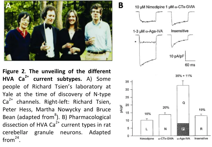
three additional Ca2+ currents: 1) P-type Ca2+ currents, first recorded in Purkinje neurons22 and exquisitely sensitive to the funnel web spider toxin peptide ω-agatoxin-IVA (ω-Aga-IVA)23, 2) Q-type Ca2+ currents, identified in cerebellar granule cells and also sensitive to ω-Aga-IVA24, and 3) R-type Ca2+ currents, also found in cerebellar granule neurons where they remained as a residual current resistant to the combined
application of nimodipine (a type of DHP), ω-CTx-GVIA and ω-Aga-IVA24 (Figure 2B).
Figure 2. The unveiling of the different HVA Ca2+ current subtypes. A) Some people of Richard Tsien’s laboratory at Yale at the time of discovery of N-type Ca2+ channels. Right-left: Richard Tsien, Peter Hess, Martha Nowycky and Bruce Bean (adapted from8). B) Pharmacological dissection of HVA Ca2+ current types in rat cerebellar granule neurons. Adapted from24.
The first full-length Ca2+ channel gene to be cloned from brain tissue25,26 encoded α1A subunit. Because the messenger for this Ca
2+
[image:24.499.81.420.317.552.2]abundant in cerebellum some researchers thought that it belonged to
P-type channel5.
However, when α1A was expressed in Xenopus oocytes, Ca 2+
currents
diverged greatly from native P-type ones and rather resembled those
from Q-type5,24. Several lines of evidence have been needed to convincingly establish that both P- and Q-type currents arise from α1A
5
.
Thus, the currently accepted terminology of “P/Q-type” channel is indeed
more appropriate, as the initial distinctions between P and Q
components mostly accounted for splice variation in α1A gene 27
.
1.4. Structure of Voltage-gated Ca
2+Channels and Regulation
by Auxiliary Subunits
The thrilling race towards voltage-gated Ca2+ channel cloning era has involved an enormous work from important laboratories together with
the inestimable contribution of DHP drugs and neurotoxins, key
molecular tools in the initial steps of Ca2+ channel purification.
Based on the biochemical purification of the L-type channel from skeletal
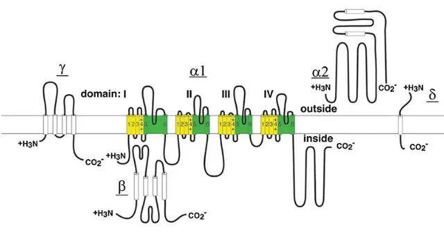
muscle28, members of the HVA Ca2+ channels are composed of four subunits: the pore-forming α1 subunit, and the auxiliary α2δ, β and γ
subunits 4 (see Figure 3, adapted from29). Conversely, there is not significant evidence supporting the association of LVA Ca2+ channels to auxiliary subunits30.
Auxiliary subunits have to meet the following criteria to be considered as
such: (1) existence in purified channel complexes (2) direct interaction
and/or trafficking of the α1 subunits and (4) stable stoichiometric
association with the α1 subunit 31
.
1.4.1. The Pore-forming
α
1Subunit
The α1 subunit, the principal determinant for Ca 2+
channel function, is the
largest (190-200 kDa) subunit, and it incorporates the conduction pore,
the voltage sensor and gating apparatus, and most of the known sites for
channel regulation by second messengers, drugs and toxins32.The α1
subunit of voltage-gated Ca2+ channels is composed of about 2000 amino acid residues organized in four homologous domains (I-IV). Each domain
consists of six transmembrane α helices (S1-S6) and a membrane
[image:26.499.88.408.358.528.2]associated pore (P) loop between S5 and S6.
Intensive structure and function studies of the related pore-forming
subunits of Na+, Ca2+, and K+ channels have led to identification of their principal functional domains6. The S1 through S4 serves as the voltage sensor module whereas transmembrane S5 and S6 in each domain and
the P loop between them form the pore module (Figure 3, yellow and
green, respectively). The N- and C-terminal regions as well as the large
intracellular loops between α1 domains serve as a signalling platform for
[image:27.499.86.418.299.529.2]channel gating regulation.
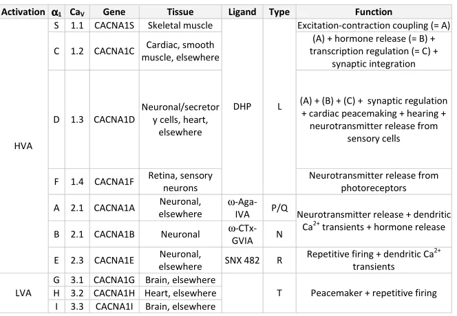
Table 1: Voltage-gated Ca2+ channel classification. Nomenclature of α1 subunits,
genes encoding CaV channels, main tissue localization, type of current,
pharmacology and principal functions are described.
Activation αααα1 CaV Gene Tissue Ligand Type Function
S 1.1 CACNA1S Skeletal muscle Excitation-contraction coupling (= A)
C 1.2 CACNA1C Cardiac, smooth
muscle, elsewhere
(A) + hormone release (= B) + transcription regulation (= C) +
synaptic integration
D 1.3 CACNA1D
Neuronal/secretor y cells, heart,
elsewhere
(A) + (B) + (C) + synaptic regulation + cardiac peacemaking + hearing + neurotransmitter release from
sensory cells
F 1.4 CACNA1F Retina, sensory
neurons
DHP L
Neurotransmitter release from photoreceptors
A 2.1 CACNA1A Neuronal,
elsewhere
ω
-Aga-IVA P/Q
B 2.1 CACNA1B Neuronal ω
-CTx-GVIA N
Neurotransmitter release + dendritic
Ca2+ transients + hormone release
HVA
E 2.3 CACNA1E Neuronal,
elsewhere SNX 482 R
Repetitive firing + dendritic Ca2+
transients G 3.1 CACNA1G Brain, elsewhere
H 3.2 CACNA1H Heart, elsewhere LVA
I 3.3 CACNA1I Brain, elsewhere
T Peacemaker + repetitive firing
In total, 10 different α1 subunit genes have been cloned and their
function has been characterized by expression in mammalian cells or
has divided voltage-gated Ca2+ channels into three structurally and functionally related families: CaV1, CaV2 and CaV3
33
(see Table 1, redrawn
from29,33,34).
1.4.2. Auxiliary Ca
2+Channel Subunits
α
2δ
Subunits
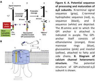
This subunit is encoded by a single gene, whose polypeptide product is
post-translationally cleaved to form a highly-glycosylated extracellular
143 KDa α2 and transmembrane 27 KDa δ subunit linked by a disulfide
bridge35. There are 4 genes that code for the α2δ family (i.e. α2δ-1-4)
each of which can produce several alternatively spliced isoforms.
Although initial studies suggested that α2δ subunit has a transmembrane
segment36, strong evidence now supports that in fact α2δ subunits are
glycosylphosphatidylinositol (GPI)-anchored rather that transmembrane
subunits37 (see Figure 4A, adapted from38).
The α2δ subunits have been shown to increase HVA currents about
threefold probably by enhancing their forward trafficking to the
plasma-membrane36. Moreover, they can also influence the biophysical properties of some Ca2+ channels, including inactivation kinetics and voltage-dependence35.
The mechanism(s) involved in α2δ modulation of CaV1 and CaV2 channel
trafficking and gating is still not completely understood. It was first
proposed that α2δ subunits may exert those functions via binding to
extracellular regions of α1 Domain III36,39. More recently, it has been
the Von Willebrand Factor A (VWA)1 domain of α2δ subunits is required
for the correct trafficking of α1 subunit to the plasma membrane 40
(see
Figure 4B, adapted from38).
The α2δ subunits are expressed in different tissues including heart, brain
(e.g. α2δ-1 and α2δ-2 expression is particularly high in the cerebellum),
and skeletal muscle, among others, being α2δ-4 the only subunit absent
in brain35.
Figure 4. A. Potential sequence of processing and maturation of
α αα
α2δδδδ subunits. N-terminal signal
sequence (gray), C-terminal hydrophobic sequence (red), α2
sequence (black), and δ sequence (white) are depicted. The ϖ-amino acid to which the GPI anchor is attached is indicated in purple. The GPI-anchor itself consists of ethanolamine (orange), three mannose rings (blue), glucosamine (pink) and inositol (yellow), attached to fatty acid side chains. B. Diagram of calcium channel heteromeric structure. The potential structure of GPI-anchored α2δ
subunit is shown.
The loss of α2δ subunits, as in the naturally occurring α2δ-2 knockout
(KO) strains of epileptic and ataxic mice,ducky and ducky2J 41 results in a
1
[image:29.499.88.415.224.505.2]reduction of Ca2+ currents in Purkinje neurons, and has marked effects on Purkinje cell function42.
Furthermore, gabapentinoid drugs, used in the treatment of epilepsy,
neuropathic pain and migraine, have been shown to bind α2δ-1 and α2δ
-2 and impair their trafficking. As a consequence, both α2δ and CaV2.1 Ca 2+
channel cell-surface expression is disrupted43.
γ
Subunits
The γ subunit impact on CaV2 channel family has not been as extensively
studied as the other auxiliary subunits probably due to the
muscle-preferential expression of γ1, isolated from the purification of skeletal
L-type Ca2+ channels28, and the absence of neuronal γ subunit in the initial purification of CaV2.2
44
. However, a more recent genetic study revealed
the existence of an additional γ2 subunit isoform from brain whose lack
of expression is responsible for the epileptic and ataxic phenotype of the
stargazer mouse45. Subsequently, six new γ subunits (also called stargazing-like proteins) have been identified and all eight have been
categorized into two subgroups: skeletal γ (γ1 and γ6) and neuronal γ (γ2-5
and γ7-8)46.
All γ subunits (y1-8) are ~32 KDa proteins characterized by four predicted
transmembrane domains, intracellular N- and C-termini, and the first
extracellular loop that includes an N-glycosilation site, and a pair of
cystein residues that may form a disulfide link. Particularly, neuronal γ
subunits display a longer C-terminus that contains PDZ2 binding motifs47 that could be important for physical and/or functional association of
2
these γ subunits with the Ca2+ channels and other proteins, such as AMPA receptors48. Alternatively, the N-linked glycosilation site or several potential sites for serine or threonine phosphorylation contained in γ
subunits may have an important role in the folding of γ subunits, and/or
in the targeting of proteins interacting with the γ subunit46.
Neuronal γ subunits are expressed in diverse areas of the central nervous
system (CNS), particularly in cerebellum and cerebral cortex46.
A relatively small number of functional assays compared with that of
other auxiliary subunits have been performed with neuronal γ subunits
and the results concerning functional effects of γ subunits on neuronal
CaV channels remain controversial 49-51
. Thus, although neuronal γ
subunits may have important functions both in physiological and
pathophysiological conditions in the CNS, more evidence is needed in
order to consider γ subunits as “true” auxiliary subunits of CaV2 channel
members.
β
Subunits
Voltage-gated Ca2+ channel β (CaVβ) subunits are intracellular essential
components of voltage-gated Ca2+ channels that profoundly affect multiple α1 subunit properties such as voltage-dependent activation,
inactivation rates, G-protein modulation, drug sensitivity, cell surface
expression, etc. 52,53. However, among all CaVβ actions I will focus on their
impact in CaV channel trafficking and regulation of voltage-dependence
of activation and inactivation. The impact of CaVβ on CaV channel
regulation by G-proteins as well as the interplay of the different
based on the current data and how they could be linked into a general
model of CaV channel inactivation will be also discussed during this
introduction.
Four β subunit genes have been cloned (β1-4), each encoding a number of
splice variants52. Comparison of the CaVβ subunit sequence variants has
led to the description of five domains (D1-D5), based on sequence
similarities, with D2 and D4 being highly conserved among the four β
subunits and domains D1 (corresponding to protein N-terminus) , 3 and 5
(corresponding to protein C-terminus) showing exon varibility53. All CaVβ subunits potentiate HVA Ca
2+
channel currents and hyperpolarize
the voltage-dependence of activation. However, the mechanism by
which CaVβ increase CaVα1 current amplitude remains controversial,
either attributed to increased trafficking, increased maximum open
probability, or both53,54. On the one side, some research groups have showed that CaVβ subunits increased localisation of functional channels
at the plasma membrane and suggested a “chaperone-like” effect of
CaVβ subunits55,56, probably by releasing CaVα1 subunits from the
endoplasmic reticulum56. On the other side, another group has argued that the effect of CaVβ subunits on CaVα1 subunits is to improve the
coupling between voltage sensor movement and pore opening
manifested as an increase in the ionic-current to charge-movement ratio
(I/Q) rather than to increase the gating charge movement which would
imply an increased number of functional channels inserted in the plasma
membrane57-59. One of the reasons for these discrepancy may be attributed to the heterologous expression system used in some of the
studies58,59. Xenopus oocytes have showed to express endogenous CaVβ
voltage-dependence processes (as both effects could be equally
mediated by CaVβ), since there is a difference in the
concentration-dependence of these two processes60.
All CaVβ subunits, except the splice variant β2a, accelerate channel
inactivation rate and left-shift voltage-dependence of steady-sate
inactivation curves61,62. CaVβ2a, which is indistinguishable from its
homologues in terms of CaVα1 modulation of activation, slows Ca 2+
channel inactivation rate and right-shifts voltage-dependence of
steady-state inactivation61-64. These differential regulatory properties have been classically associated with the palmitoylation of two cysteins at its
N-terminus that renders CaVβ2a membrane tethered65,66.
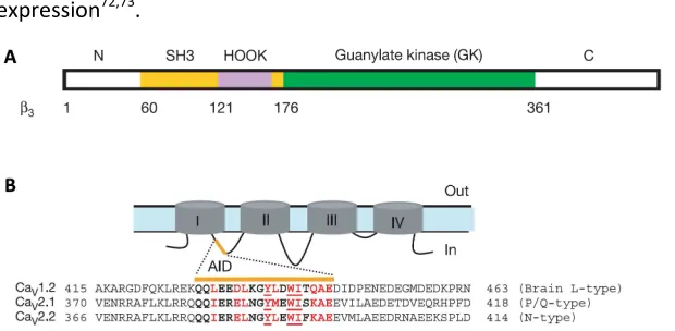
A molecular modelling study based on protein sequence homology
showed that CaVβ subunits consist of two conserved protein-protein
interaction domains; a Src homology 3 (SH3) domain and a guanylate
kinase (GK) domain, corresponding to D2 and D4 conserved regions
respectively67, suggesting that CaVβ belong to the family of
Membrane-Associated Guanylate Kinases (MAGUKs)53 (see Figure 5A, adapted from68). MAGUKs are a protein scaffold family that organizes signalling components near membranes, usually contain N-terminal PDZ domain(s),
followed by a SH3 domain, a flexible linker termed the HOOK, and a
nucleotide kinase domain69. The presence of MAGUK-like domains in CaVβ subunits suggest that they might have a role in scaffolding multiple
signalling pathways around the channel70 (see below).
All CaVβs have a primary high affinity binding site on CaVα1 subunits of 18
amino acids located in the cytoplasmic loop between domains I and II (I-II
loop) of CaVα1 subunits, referred to as the α1-interaction domain (AID),
sequence of 41 conserved amino acids (located in the N-terminus of the
second conserved domain of CaVβs and termed β-interaction domain
(BID)) was required both to bind AID and to influence CaVα1 subunit
expression72,73.
Figure 5. CaVββββ subunit domains and AID sequence alignment. A. Schematic
diagram of CaVβ3 subunit domain organization based on the crystal structure. B.
Schematic domain organization of α1 and alignment of the 49-amino-acid
sequence of I-II loop from CaV1.2c (M67515), CaV2.1 (X57477) and CaV2.2
(D14157). Residues forming the AID are in bold and those involved in interactions with the CaVβ subunit are in red, with the three most critical residues underlined.
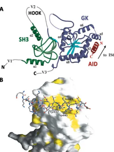
Three recent X-ray crystallographic studies have now solved the core
(SH3-HOOK-GK) domains of three CaVβ subunits, alone and/or in complex
with AID, providing new interesting structural and functional insights
about AID-CaVβ interaction68,74,75. Crystal structures have showed that
AID in complex with CaVβ folds into an amphiphatic α-helix 68,74,75
and that
AID transition from coil to helix is forced by the association to CaVβ 75
.
Conserved aromatic residues face to one side of the helix and strongly
interact with a deep hydrophobic groove located in the GK domain of
CaVβs now dubbed the AID-binding pocket (ABP) 68,74,75
rather than with
CaVβ-BID domain as previously reported 72
[image:34.499.87.397.135.287.2]from54,74). From these results one could speculate that GK module itself may preserve some if not all the modulatory capabilities of the full CaVβ
subunit. Alternatively, it is worth to note that as seen from the crystals
AID occupies only a very small area of CaVβ core and therefore, much of
its surface is free and available for interactions with other regions of α1
or other proteins. In partial favour to the first view, whole-cell recordings
in Xenopus oocytes confirmed that CaVβ3-GK module alone was sufficient
at least to potentiate CaV2.1 channel currents 68
. Similarly, CaVβ-GK
constructs had previously shown to significantly stimulate CaVcurrent
although not to the same extent as wild-type (WT) CaVβ, while long
deletions on GK domain caused significant loss of CaVcurrents 72
.
However, it is a still a matter of great debate whether GK module suffices
to recapitulate all the aspects of CaVβ-dependent modulation of CaVα1
subunits and the putative participation of the other domains of CaVβs on
this modulation via alternative contacts (of lower affinity than those with
AID) with CaVα1 subunits.
Figure 6. Structure of the CaVββββ2a–CaV1.2 AID complex.
A. Ribbon diagram of the complex. Dashed lines indicate regions absent from the structures. B. Surface representation of the CaVβ3
[image:35.499.90.273.393.636.2]A host of biochemical and functional data previous to crystallization of
CaVβ-AID complex has linked CaVβ short variable regions as well as CaVα1
cytoplasmic regions (in addition to I-II loop) to the CaVβ-mediated
inactivation process. The first evidences that the CaVβ N-terminal region
was an important determinant for inactivation were supported by the
fact that CaVβ2a, the solely non-inactivating CaVβ, has N-terminal
modification whose mutation reverses its phenotype66. Additional data has favoured the idea that not only CaVβ N-terminal
61,76,77
but also HOOK
regions77,78 are linked to CaVβ modulation of inactivation. Concerning
CaVα1, contacts between N- and C-terminus of α1A subunit with the low
conserved C-terminus of CaVβ4 have shown to be important for the
regulation of inactivation79-81. Similarly, N-terminus of α1B was shown to
contribute to CaVβ2a-mediated retardation of voltage-dependent
inactivation82. All these data suggest the existence of isoform-specific interactions other than CaVβ-GK with AID that provide the unique
regulatory characteristics for each α1- CaVβ pair.
Soon after the crystal structures of CaVβ core interacting with AID motif
came up, important works combining fine molecular, biochemical, and
electrophysiological approaches have prompted to more accurately
define the exact molecular determinants of CaVβs (and in one case also
of CaVα1S) that account for CaVβ-α1 functional interaction leading to
contradictory but quite intriguing conclusions.
Some authors have seen that GK module binding to AID is necessary and
sufficient to support Cavβ chaperone role in the increase of channel
surface expression83,84. However, the specific CaVβ roles on gating are
supported by secondary contacts of Cavβ N-term 83
Others have demonstrated that CaVβs require an interaction between
SH3 and GK modules to recapitulate regulatory functions on functional
cell surface channel expression85 or inactivation properties86.
Additional striking experiments have shown that CaVβs regulation of
surface expression and channel gating properties are AID-independent
and require SH3 module binding to a different from AID site within the
distal part of I-II loop of CaVα1. This new scenario might place CaVβ
-GK-AID binding as a mere pre-requisite that orientates and concentrates
CaVβs near α1 to permit an additional interaction between CaVβ-SH3 and
α1-I-II loop that underlies the particular regulation seen with each CaVβ
-α1 pair 87
.
Recently, a nice work has shed new light on the matter demonstrating
that in fact just CaVβ-GK module suffices to modulate channel gating 88
. In
this report, refolded GK domains from two functionally distinct Cavβs, β1b
and β2a (which are know to facilitate or inhibit channel inactivation
respectively), were found to equally inhibit inactivation88. Moreover, they showed that slow GK inactivation pattern switched to fast during channel
biogenesis suggesting that GK module acts as a brake to impair
voltage-dependent inactivation and that facilitation of inactivation requires
additional structural determinants acquired by post-translational
modifications that in the case of β2a may not occur because
palmitoylation sequesters it to other membranous compartments early
in the course of biogenesis88. Finally, they suggest that whereas GK regulates Ca2+ entrance, the SH3 domain may link channel activity to other cellular processes by binding to additional proteins88.
In line with this hypothesis there has recently been established that CaVβ
subunits can exert α1-independent functions that may or may not affect
same group has demonstrated that CaVβ-SH3 can interact with dynamin
(a GTPase involved in receptor-mediated endocytosis) and reduce the
number of CaV channels in the plasma membrane but only when its
binding to AID is prevented89. Associaton of CaVβ subunits to other small
GTPases had been previously shown to decrease CaV channels at the
plasma membrane by inhibiting CaVβ association to α190. All these CaVβ
new functions are further supported by the already demonstrated
physiological unbinding of CaVβ subunits from α1 60,91,92
.
1.4.3
Molecular
Determinants
of
Voltage-dependent
Inactivation in HVA Ca
2+Channels
Voltage-dependent inactivation is a key regulatory mechanism for the
control of intracellular Ca2+ increases in response to AP and is, therefore, believed to play an important role in Ca2+ signalling and neuronal excitability93,94.
In voltage-gated K+ and Na+ channels the molecular determinants that govern fast inactivation involve pore block by a cytoplasmic gating
particle: the N-terminus of the four α subunits in the case of tetrameric
K+ channels (i.e. “ball and chain” or “N” inactivation) or the III-IV loop of the α subunit in the case of Na+ channels (i.e. “hinged lid” inactivation)94. In the case of voltage-gated Ca2+ channels the general inactivation mechanism may be reminiscent of that of K+ or Na+ channels, but its molecular determinants appear to be more widely spread over the α1
subunit and show a much more complex regulation93,94.
Work from a number of laboratories has implicated both cytoplasmic
process. The importance of pore-lining S6 transmembrane helices in Ca2+ channel inactivation process has been first deduced from chimeric
studies where substitution of different S6 segments from different α1
domains between CaV channels showing different inactivation kinetics
was sufficient to induce a change in the inactivating phenotype95,96. Further evidence has arisen from the observed alteration of inactivation
kinetics due to mutations located in the IIS6, IIIS6 and IVS6 regions of
CaV2.1 channels 97,98
. Taken together, these findings indicate that all four
S6 segments of α1 subunit contribute to the inactivation process.
Furthermore, all four transmembrane domains have shown to contribute
in determining the overall voltage-dependence inactivation99. In sum, these evidences support a “pore collapse” model of inactivation that may
originate from a global structural change of the four domains of α1
subunit where the S6 segments, arranged as an inverted “tepee”
analogous to the crystal structure of KcsA channel100, would have a key role in inactivation gating by determining the pore constriction. In this
model, cytoplasmic regions of α1 such as the I-II loop (which, as
previously mentioned, has been intensively linked to the regulation of
inactivation by CaVβ subunits) may further contribute to the inactivation
process by affecting the orientation of pore-forming segments and/or
restrict their mobility93.
In contrast, a number of additional observations provide support for a
classic “hinged-lid-type” mechanism of Ca2+ channel inactivation that would involve the participation of α1 cytoplasmic regions
94
. In this
respect, the fact that CaVβ subunits (the major determinants governing
Ca2+ channel gating properties) bind to the α1 I-II loop 71
suggest a crucial
role of this cytoplasmic region as a crossroad for gating channel
channel inactivation. For instance, insertion of a single valine residue in
the CaV2.1 I-II loop dramatically slows the rate of inactivation of this
channel, shifting it from a Q-type to a P-type phenotype27. Chimeric substitutions between different CaV channels and site-directed
mutagenesis of the I-II loop result in significant changes in the rates of
inactivation94. Furthermore, overexpression of I-II loop peptides accelerates the inactivation kinetics of expressed CaV2.1 channels
102
.
Taken together, these findings suggest a role of the I-II loop as a putative
hinged-lid gating particle. In this model, S6 segments (which probably
line the inner mouth of the pore) could form part of the docking site for
the inactivation gate94. Mobility restriction of the I-II loop by interactions with CaVβ subunits or other cytoplasmic domains would differentially
regulate inactivation94. Accordingly, it has been suggested that the exclusive ability of CaVβ2a to prevent inactivation may be attributed to
anchoring of this subunit to the plasma membrane, that would be
expected to restrict I-II loop mobility toward the pore76,94. However, this hypothesis might need to be contrasted with current molecular and
functional data indicating that inhibition of inactivation is an intrinsic
property of all CaVβ-GK domains 88
.
As previously mentioned, in addition to the I-II loop, other α1 cytoplasmic
regions such as the N-terminus82, the II-III103, the III-IV loop104, and the C-terminus81 have been also linked to inactivation. Interestingly, it has been proposed that regulation of channel inactivation is mediated by an
interaction between I-II and III-IV loops of the CaV2.1 channel and that
CaVβ subunits modulate inactivation by modifying this interaction via
binding to AID105. In contrast, the contribution of N- and C-terminal regions to inactivation may arise indirectly from their interaction with
CaVβs 81,82,105
The extent to which these cytoplasmic regions either restrict S6 mobility
(i.e. in the “pore collapse” model) or I-II loop mobility (i.e. in the
“hinged-lid” model) to control channel gating is further increased by the reported
interactions between the various cytoplasmic regions and between those
cytoplasmic regions and other regulatory proteins (e.g. CaVβs, SNARE
proteins, etc.) adding additional steps of intricacy to the two proposed
models of Ca2+ channel inactivation93,94.
2. NEURONAL VOLTAGE-GATED Ca
2+CHANNEL ROLE IN
SYNAPTIC TRANSMISSION
2.1. The Presynaptic Ca
V2 Channel Signalling Complexes
CaV2.1 (P/Q) channels are located in presynaptic terminals and
somatodendritic membranes throughout the mammalian brain106 and although at many central synapses N- and R-type Ca2+ channels also cooperate in controlling neurotransmitter release, they display a
preferential role in initiating AP-evoked neurotransmitter release
because of a more efficient coupling to the exocytotic machinery107-109. The somatodendritic CaV2.1 channel localization points to additional
postsynaptic roles (e.g. control of Ca2+-dependent gene expression110). Ca2+ entering neurons by the opening of voltage-gated Ca2+ channels in response to APs forms a transient Ca2+ microdomain in the presynaptic nerve terminal111,112. Vesicle fusion and neurotransmitter release is initiated within 200 µs after the arrival of the AP and requires a brief (<1
Presynaptic proteins of the vesicle-docking/fusion machinery, including
plasma membrane SNARE proteins (i.e. syntaxin 1A and SNAP-25) and
synaptotagmin must be located near Ca2+ channels in order to receive the Ca2+ signal. In many cases, this close localization is achieved by direct interaction with the intracellular domains of Ca2+ channels, which serve as signal transduction platforms for cytosolic Ca2+ signalling4. The signalling complexes of presynaptic Ca2+ channels contain SNARE proteins involved in exocytosis, G proteins involved in feedback regulation of Ca2+ channels, and many Ca2+-binding proteins involved in regulation of channel activity and initiation of Ca2+-dependent responses.
2.2. Regulation of Ca
V2 Channels by G Proteins
For the neuronal CaV channels, particulary N- and P/Q-types, a major
mechanism of inhibitory modulation occurs via activation of
heterotrimeric G proteins by seven transmembrane G protein-coupled
receptors (GPCRs). GPCRs activation was first found to reduce AP
duration, and subsequently this effect was found to result from inhibition
of presynaptic voltage-gated Ca2+ channels in many types of neurons34. GPCRs in presynaptic nerve terminals bind released neurotransmitters
and provide a negative feed-back to inhibit N- and P/Q-type and thereby
reduce synaptic transmission114. G protein negative regulation of neurotransmitter release is very potent because of the power law
relationship between Ca2+ influx and synaptic transmission115. Most neurotransmitters inhibit Ca2+ currents in this manner, including acetylcholine, glutamate, GABA, biogenic amines, and many
neuropeptides. Autoreceptors in one nerve terminal bind
protein-coupled receptors in the same nerve terminal may respond to
neurotransmitters released by nearby nerve terminals from other
neurons29.
The key features that typify this inhibition are a positive shift in the voltage
dependence and a slowing of channel activation116. These effects are relieved by strong depolarization resulting in facilitation of Ca2+ currents because of a shift between two channel states with different gating
properties: “reluctant” and “willing” to open states116. Faster removal of inhibition can also be induced by the application of a strong depolarizing
prepulse immediately before the test pulse117. Looking from a more physiological point of view, given that G protein inhibition is
voltage-dependent, synaptic release may be strongly inhibited for a single AP, but
inhibition relieved by a train of APs118. This prediction has been positively confirmed in microisland cultures of hippocampal neurons in which
autapses are formed by single hippocampal pyramidal neurons119. In this preparation, trains of AP-like stimuli relieved the inhibition of synaptic
transmission caused by activation of GABA-B receptors resulting in
facilitation of synaptic transmission by 1.5 fold, which was subsequently
blocked by selective blockade of P/Q-type Ca2+ channels with peptide neurotoxins119.
The above described modulation of CaV2 channels in most native tissues is
usually mediated by heterotrimeric G protein subunits released from the
pertussis toxin-sensitive Gi/Go class 114
. At the molecular level, although G
protein α subunits confer specificity in receptor coupling,
voltage-dependent inhibition of Ca2+ channels is a membrane-delimited event mediated by G protein βγ dimers (Gβγ) who, by itself, are able to mimic
GPCR agonists-mediated inhibition when transfected either in primary
Relief from G-protein inhibition by strong depolarization causes a
dissociation of Gβγ from the channel α1 subunit, a highly voltage- and
time-dependent kinetic process commonly termed as facilitation development.
Furthermore, re-establishment of inhibition after a “facilitating” prepulse,
during a period at the holding potential, is likely to result from rebinding of
Gβγ to the α1subunit, and can be measured as facilitation decay rate.
Whether these two kinetic processes actually involve physical dissociation
and reassociation to the α1 subunit remains to be established34.
Direct measurements of ionic and gating currents in HEK293 cells
transfected with CaV2.2 channel after G protein activation have shown
slowness of the latter and a induction of ~20 mV separation between the
voltage-dependent activation of gating charge movement and ionic current
suggesting that the underlying biophysical mechanism of Gβγ is to restrain
the movement of the S4 voltage sensors of CaV2 channels and the
transduction of voltage-sensor activation into channel opening122. Prolonged depolarization to more positive membrane potentials can
overcome this effect by forcing voltage sensor movement and thus favour
Ca2+ channel activation4.
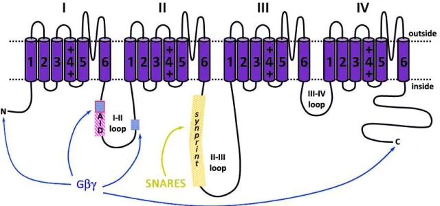
The effects of Gβγ subunits might be mediated by binding to one or more
sites on the Ca2+ channel α1 subunit (see Figure 7, redrawn from29). Possible sites of Gβγ subunit interaction with CaV channels have been
extensively studied by construction and analysis of channel chimeras, by
G protein-binding experiments, and by site-directed mutagenesis and
expression. Initially, most data pointed to the α1 I-II loop as an essential
determinant for Gβγ-mediated modulation101,123,124. Overlay and mutagenesis assays showed the existence of two Gβγ putative binding
sites in this α1 intracellular domain101,123: one inside AID sequence with a
domain comprised of several microsites in the distal part of the I-II
loop123. Peptide competition as well as mutagenesis of these sites prevented channel inhibition by Gβγ101,123,124.
Figure 7. Interaction sites of Gβγβγβγβγ and SNARE proteins on αααα1 subunit. Gβγ
subunits interact with a QQIER amino acid motif inside the AID sequence and a more distal second domain (blue wefted squares) in the I-II loop, the N- and the C-terminus. The SNARE proteins interact with the synprint site (amino acids 718-963 of the α1B subunit and amino acids 722-1036 of the BI isoform of α1A bind
both syntaxin 1A and SNAP-25, while amino acids 724-981 of the rbA isoform of
α1A only interact with SNAP-25) located inside the II-III loop.
However, further studies using chimeras containing different segments of
channels with different levels of G protein-mediated inhibition (i.e. CaV2.2
are strongly inhibited while CaV2.1 present less strong inhibition) or
chimeras between those and CaV1.2 channels which are not inhibited by G
protein at all found that I-II loop was either not essential for G protein
modulation or not the most critical region125-127. Instead, N-terminus of CaV2
α1 subunits was found to be more important in mediating G protein
modulation126,128. Specifically, an 11-amino acid motif YKQSIAQRART in CaV2.2 N-terminus (that is also highly conserved in CaV2.1 and CaV2.3) was
shown to be essential for G protein modulation, and mutation of either YKQ
[image:45.499.88.408.148.298.2]Additional studies using chimeric channels have either implicated125,129 or ruled out130 α1C-terminus region as a putative structural determinant and/or binding site to promote G protein mediated inhibition.
The fact that Gβγ share common molecular determinants with CaVβ
subunits for its functional interaction with α1 subunits as well as the notion
that their effects upon CaV2 channels could be considered as opposite has
led to many investigators to explore the possibility that CaVβ subunits might
be involved in G protein modulation and compete with Gβγ for the binding
to α134. In initial studies in native neurons, antisense oligonucleotides against CaVβs resulted in enhanced G protein-mediated inhibition of Ca
2+
currents leading to the conclusion that CaVβ counteracted Gβγ action 131
.
Additional experiments in Xenopus oocytes showed less or even complete
loss of G protein inhibition following coexpression of a CaVβ subunit further
reinforcing that hypothesis129, although these studies only examined inhibition at a single potential and need to be interpreted with caution
because of the presence of endogenous CaVβ subunits 34
. Further studies
studying the voltage-dependence of G protein inhibition in the presence or
absence of coexpressed CaVβs in Xenopus oocytes showed that the voltage
at which G proteins produced maximum amount of inhibition was displaced
by CaVβ subunits suggesting that the functional antagonism between Gβγ
and CaVβ rather than competitive is dynamic and depends on the
membrane voltage132.
Recent studies have demonstrated that abrogation of CaVβ interaction with
CaV2 α1 by AID mutagenesis do not affect the availability of Gβγ inhibition
but prevent current facilitation by a depolarizing prepulse133, and in a similar way, in the absence of CaVβs, coexpression of CaVβ-GK domain
inhibition134. Additionally, FRET studies have demonstrated that CaVβ andGβγ are able to bind α1 at the same time
135
. Taken together, these
results suggest that CaVβ subunits do not displace Gβγ but instead
modulate the voltage dependence of G protein modulation process via
their GK domain134.
2.3 Regulation of Ca
V2 Channels by SNARE Proteins
Neurotransmitter exocytosis is a highly regulated, multi-step process that
may occur through a host of molecular mechanisms that culminate with the
fusion of transmitter-containing vesicles at the presynaptic active zone of
nerve terminal membranes136. Active zones are small (i.e. 200-300 nm) specialized presynaptic membrane compartments that contain two
essential "gates" for neurotransmission: one for Ca2+ entry, which is the voltage-gated Ca2+ channel, and the other for neurotransmitter exit, which is the synaptic vesicle fusion site. The performance of a synapse depends on
the number of vesicles docked and primed at those sites to be immediately
available for fusion (i.e. the size of the ready releasable pool) following the
arrival of an AP137.
One crucial step of neurotransmitter release depends on the formation of
the SNARE (soluble N-sensitive factor attachment receptor) complex
(spatially and temporally organized by SM (Sec1/Munc 18-like) proteins)
that tethers neurotransmitter-laden vesicles close to the site of Ca2+ entry and promotes its priming138,139. In response to AP-induced Ca2+ influx the SNARE complex suffers a conformational change that facilitates the fusion
between vesicle and nerve terminal membranes to release
neurotransmitter into the synapse. The minimal determinants of this
synaptobrevin and target plasma membrane SNARE proteins (t-SNAREs)
syntaxin 1A and synaptosomal-associated protein 25 (SNAP-25). Each
contributes one (synaptobrevin and syntaxin) or two (SNAP-25) α-helices to
a four-helix bundle of such high affinity that the resulting complex is
resistant to denaturation by sodium dodecyl sulfate (SDS) (see Figure 8A,
redrawn from web.mit.edu).
Formation of SNARE complex is absolutely required for vesicle fusion
competence, but increasing evidence suggest that SNARE proteins may also
be involved in vesicle recruitment140. In fact, recruitment of synaptic vesicles to sites where Ca2+ channel cluster, rather than fusion competence, is a limiting step for rapid neurotransmitter release in response to
presynaptic APs140.
Biochemical studies demonstrate that both CaV2.1 and CaV2.2 channels
colocalize densely with syntaxin 1 at the presynaptic nerve terminals106 and can be isolated as a complex with SNARE proteins141,142. Pull-down and binding assays demonstrated that plasma membrane SNARE proteins
syntaxin 1A and SNAP-25, but not the synaptic vesicle SNARE
synaptobrevin, specifically interact with the CaV2.2 channel by binding to
the II-III loop of the α1B subunit via an 87 amino acid sequence termed
synaptic protein interaction (synprint) site (amino acids 718-963 of
α1B) 143,144
(see Figure 7, redrawn from29).
This interaction is Ca2+ dependent, with a maximal binding at 20 µM Ca2+ and reduced binding at lower or higher concentrations144, suggesting sequential steps of association and dissociation of SNARE proteins with
CaV2.2 channels as a function of Ca 2+
concentration29. CaV2.1 channels also
have an analogous synprint site, and different alternative splicing channel
isoforms have distinct interactions with syntaxin 1A and/or SNAP-25 that do
Figure 8.A. Cartoon depicting the SNARE core complex and Synaptotagmin. Note the four helix boundle formed by syntaxin (red), SNAP-25 (green) and synaptobrevin (blue).Transmembrane domains of syntaxin and synaptobrevin are indicated in yellow. Ca2+ ions bound to synaptotagmin C2 domains are shown in red. B. Topology and ribbon diagrams of syntaxin 1A in “closed” conformation. The Habc domain is shown in red, the Habc/H3 linker in orange and the H3 region in purple.
The BI isoform (previously cloned from rabbit brain but also present in rat
brain) of α1A binds both syntaxin 1A and SNAP-25 via two adjacent
segments of the II-III loop between amino acids 722 and 1036 while only
interaction with SNAP-25 can be detected for the rbA isoform of α1A
(previously cloned from rat brain but also present in rabbit brain) in its
correspondent amino acid region (amino acids 724-981)145(see Figure 7, redrawn from29).
Other synaptic proteins with a relevant role in the exocytotic process are
also able to interact with CaV2 channels. The synaptotagmins are a