Estudi de la regeneració miocàrdica en la
miocardiopatia alcohòlica i la seva relació amb el dany
funcional i estructural miocàrdic, activació d’apoptosi
i activitat miostatina
Meritxell Lluís Padierna
ADVERTIMENT. La consulta d’aquesta tesi queda condicionada a l’acceptació de les següents condicions d'ús: La difusió d’aquesta tesi per mitjà del servei TDX (www.tdx.cat) ha estat autoritzada pels titulars dels drets de propietat intel·lectual únicament per a usos privats emmarcats en activitats d’investigació i docència. No s’autoritza la seva reproducció amb finalitats de lucre ni la seva difusió i posada a disposició des d’un lloc aliè al servei TDX. No s’autoritza la presentació del seu contingut en una finestra o marc aliè a TDX (framing). Aquesta reserva de drets afecta tant al resum de presentació de la tesi com als seus continguts. En la utilització o cita de parts de la tesi és obligat indicar el nom de la persona autora.
ADVERTENCIA. La consulta de esta tesis queda condicionada a la aceptación de las siguientes condiciones de uso: La difusión de esta tesis por medio del servicio TDR (www.tdx.cat) ha sido autorizada por los titulares de los derechos de propiedad intelectual únicamente para usos privados enmarcados en actividades de investigación y docencia. No se autoriza su reproducción con finalidades de lucro ni su difusión y puesta a disposición desde un sitio ajeno al servicio TDR. No se autoriza la presentación de su contenido en una ventana o marco ajeno a TDR (framing). Esta reserva de derechos afecta tanto al resumen de presentación de la tesis como a sus contenidos. En la utilización o cita de partes de la tesis es obligado indicar el nombre de la persona autora.
For Peer Review
! " #$% & #$ '# ( ) " % & ) $# # $ ) !%
*#$% % #+, & -! ( ) !% & # %#
# . & #$/ ( " 0 & #" $ #% * # %#1
!% .#$ 2 * 0 $ # %
%# & # ( ) " % & ) $# # $ ) !% # $! )& $ %( ) " % & ) $# # $ ) !% !$0 % , $-!#+& . $ ( ) " % & ) $# # $ ) !%
3 4 & & #$ & 5 ,67
For Peer Review
EVALUATION OF MYOCYTE PROLIFERATION IN ALCOHOLIC
CARDIOMYOPATHY. TELOMERASE ENZYME ACTIVITY (TERT)
COMPARED TO Ki-67 EXPRESSION.
RUNNING HEAD:
MYOCYTE PROLIFERATION in ALCOHOL CARDIOMYOPATHY
Meritxell Lluís M.D., Ph.D.1; Joaquim Fernández-Solà M.D., Ph.D1,2; Sergi Castellví Ph.D.3; Emilio Sacanella M.D., Ph.D1,2; Ramón Estruch M.D.,Ph.D.1,2, Alvaro Urbano-Márquez1
1
Alcohol Research Unit. Hospital Clínic. Institut d’Investigació August Pi i Sunyer (IDIBAPS). Department of Medicine University of Barcelona. Spain
2
CIBEROBN Fisiopatologia de la Obesidad y la Nutrición. Instituto de Salud Carlos III, Spain.
3
Gastroenterology Department, Institut de Malalties Digestives i Metabòliques, Hospital Clínic, CIBEREHD, IDIBAPS, University of Barcelona, Barcelona, Catalonia, Spain.
Department of Medicine. University of Barcelona. Catalonia. Spain.
ABSTRACT: 259 words
TEXT: 4,396 words
TABLES: 2
FIGURES: 2
Submit correspondence to:
Joaquim Fernandez-Solà M.D.
Department of Internal Medicine. Alcohol Unit
Villarroel 170. 08036. BARCELONA- España
Phone and FAX: 00.34.93. 2279365
E-mail:jfernand@clinic.ub.es
Page 1 of 29 Manuscripts submitted to Alcohol and Alcoholism
For Peer Review
ABSTRACT
Aims: Although human heart was classically considered a terminal organ, recent studies
have reported a myocyte proliferation response versus some aggressions. Excessive
ethanol consumption induces development of cardiomyopathy (CMP) through myocyte
apoptosis. We evaluated myocyte proliferation response in the heart of chronic alcoholic
donors with telomerase activity (TERT) compared to Ki-67 nuclear expression.
Methods: Heart samples were prospectively obtained from organ donors on life
support. We included donors wit 1) High lifetime alcohol consumption (n=15),
2)Longstanding hypertension (n=14), 3) Other causes of CMP (valve, coronary or
idiopathic) (n=8), and 4) Previously healthy donors (n=6). Groups 2 and 3 were
subdivided according to the presence of CMP. Evaluation comprised parameters of
ethanol consumption, left ventricular (LV) function by chest X ray and 2-D
echocardiography and histology and immunohistochemical studies. Myocyte
proliferation was evaluated using an assay for Ki67 expression and measuring
telomerase gene activity by real-time PCR.
Results: Fourty–three donors were included in the study, 35 having CMP. Nuclear
Ki-67 activity was low in healthy controls and significantly increased in the other groups,
mainly in those with CMP. Alcoholics with CMP had a non-significantly lower
proliferation response than the other CMP groups. No proliferation activity was
detected with TERT in any case.
Conclusions: Heart Ki-67 proliferation activity increases in organ donors with CMP,
independently of its origin. Alcoholics presented non-significant lower myocyte
proliferation capacity compared to the other groups of CMP. TERT activity was not a
useful marker of proliferation in this model. Ki-67 is a better procedure to evaluate
proliferation than TERT expression in alcohol–induced heart damage.
Page 2 of 29 Manuscripts submitted to Alcohol and Alcoholism
For Peer Review
Key words: Alcohol, Cardiomyopathy, Telomerase, TERT, Ki-67.
INTRODUCTION
Recent scientific knowledge has shown that the human heart is not a terminal organ
(Anversa and Kajstura,1998; Nadal-Ginard et al, 2003a; Quaini et al, 2004; Buja and
Vela 2008). Heart exhibits plastic response versus diverse physiologic or pathologic
stimuli (Hill and Olsen, 2008; Kajstura et al, 2004; Gerder, 2002). Cardiac myocyte
renewal has been observed in different animal species (Soonpaa and Field, 1998;
Jopling, 2010), and also in human myocardial areas surrounding necrotic tissue
(Beltrami et al, 2001; Urbanek et al, 2005). Therefore, a relationship between myocyte
necrosis and subsequent proliferative response has been established (Anversa and
Nadal-Ginard, 2002; Beltrami et al, 2003; Anversa et al, 2004).
In last decades, a dose-dependent relationship between excessive alcohol consumption
and diffuse myocardial damage has been corroborated (Urbano-Márquez et al, 1998;
Molina et al, 2002; Piano, 2002; Nicolás et al, 2002; Urbano-Márquez and
Fernández-Solà, 2004). Mechanisms underlying this effect are diverse, with direct induction of
apoptosis and myocyte cell death by ethanol (Jänkalä et al, 2002; Molina et al, 2002;
Fernández-Solà et al, 2006). However, not all excessive alcohol consumers develop
significant myocardial damage, with some discordance between the high prevalence of
alcohol consumption and the relative low incidence of alcoholic CMP (Fernández-Solà
et al 2002; Urbano-Márquez et al, 1995). It has been suggested that some mechanisms
may compensate the degree of alcohol-induced myocyte damage (Urbano-Márquez and
Fernandez-Solà, 2004).
We previously analyzed factors influencing death and proliferation of cardiac myocytes
and observed a higher apoptosis index and myostatin activity in alcoholic compared to
Page 3 of 29 Manuscripts submitted to Alcohol and Alcoholism
For Peer Review
healthy donors (Fernández-Solà et al, 2006). Alcoholics with CMP showed higher
apoptotic and proliferative Ki-67 activity compared to their partners without CMP.
Chronic ethanol consumption increases myostatin activity, a factor that favors loss of
cardiac myocytes and impairs their proliferation (Fernández-Solà et al, 2008).
Several morphological, immunohistochemical and molecular markers have been used to
evaluate myocyte proliferation (Leri et al, 2001). Ki67 is a nuclear antigen only
expressed when cells are in the replication cycle. It is commonly used as a marker of
cell proliferation either in tumoral (Magdelénat, 1992) or non-tumoral tissues including
cardiac myocytes (Kajstura et al, 1998; Ginard et al, 2003b; Anversa and
Nadal-Ginard, 2002). Telomerase is a DNA polymerase constituted by two RNA subunits
(telomerase-RT) (TERT) that maintains the telomere length stable and protects
chromosomes from degradation and molecular recombination. When telomerase activity
decreases, telomere length shortens to a critical size, activates cell apoptosis and
stimulates TERT expression as a compensatory mechanism. This process
physiologically appears along senescence (Anversa et al, 2005), being implicated in
ischemia (Serrano and Andres, 2004), heart failure (Leri et al, 2003), ventricular
hypertrophy (Urbanek et al, 2003), infertility and sarcopenia (Blasco, 2005). In contrast,
telomerase exhibits a high activity in progenitor stem cells (Djojosubroto et al, 2003)
and in tumoral cells, (Blasco, 2005).
All these facts led us to consider the possible increase in telomerase activity and
proliferation response in the setting of alcoholic CMP. This myocyte proliferation
would partially compensate the ethanol-induced myocyte loss (Nadal-Ginard et al,
2003a; Hotchkiss, 2009). Differences in the myocyte proliferation response may explain
discordances in the relation between ethanol intake and the degree of cardiac lesion.
Page 4 of 29 Manuscripts submitted to Alcohol and Alcoholism
For Peer Review
In the present study we evaluated proliferation mechanisms similar to those described in
ischemic CMP in the heart of chronic alcoholics. In a prospective case-control study
using heart samples from human donors we assessed the effect of alcohol consumption
on myocyte proliferation, evaluated by cardiac myocyte telomerase activity and
expression of Ki67 nuclear antigen.
MATERIALS AND METHODS
Patient selection
Over a two-year period (September 2006 to November 2008), we consecutively studied
hearts from subjects who had brain death either of traumatic or cerebrovascular origin,
and had been considered suitable as organdonors by the transplant team of the Hospital
Clínic of Barcelona.
Of 127 cadaveric donors younger than 70 years of age, 43 hearts were not suitable for
transplantation. Of these latter organs, we selected 14 with chronic hypertension, 15
cases with a history of ethanol intake (≥ 60 g/day, longer than 10 years) and 10 hearts from healthy people who were not eligible for implantation because of a lack of
matched receptor or size inadequacy. Additionally, 8 specimens from patients with heart
disease (three coronary diseases, three with idiopathic dilated CMP and two with
valve-heart disease) were selected. Alcoholic and hypertensive donors were subdivided in two
groups depending on the existence or not of CMP.
Exclusion criteria: According to the general protocol of organ donation, subjects with
drug misuse, transmissible infections (HIV, hepatitis B or C), sepsis, disseminated
neoplasm or metabolic diseases (diabetes or other endocrine diseases) or other diffuse
structural diseases were excluded as were those with coexistent hypertension and
alcohol consumption.
Page 5 of 29 Manuscripts submitted to Alcohol and Alcoholism
For Peer Review
All patients were white Caucasians of Spanish descent, who lived with their families in
or around Barcelona and none was indigent. The study protocol was approved by the
Ethics Committee of the Hospital Clinic and included informed consent from the
families of the donors concerning the use of myocardium tissue for this research
protocol study. One third of these subjects had been included in previous studies on
cardiac apoptosis and Solà et al, 2006) and myostatin activity
(Fernández-Solà et al, 2010).
All cases had been admitted to the Intensive Care Unit, and ventilatory and
hemodynamic parameters were appropriately maintained at normal values throughout
hospitalization: PaO2 greater than 60 mmHg, systolic blood pressure greater than 100
mmHg, and arterial pH within the normal range. None of the patients required
in-hospital cardiopulmonary resuscitation maneuvers.
Clinical and laboratory evaluation
Detailed history of ethanol intake was retrospectively obtained by consultation with
family members using a structured questionnaire ("time-line follow-back method")
(Sobel et al, 1979) as previously reported (Urbano-Márquez et al, 1989; Nicolas et al,
2002). Duration of ethanol intake was calculated in each group as the total cumulated
period of alcohol consumption in years, either recent or previous. Body mass index was
determined as the actual body weight relative to the square of the body height (BMI,
Kg/m2). Patients were considered to have caloric malnutrition if the BMI was less than
17 Kg/m2.
Cardiac Studies
Page 6 of 29 Manuscripts submitted to Alcohol and Alcoholism
For Peer Review
Past and present signs and symptoms of heart failure were evaluated in consultation
with medical records and family members of the donors, and the New York Heart
Association (NYHA) functional class was determined according to the Goldman
activity scale (Goldman et al, 1981). Chest X-ray with measurement of cardiothoracic
index and conventional electrocardiography were performed in all cases. Moreover, a
bi-dimensional echocardiography was performed (Hewlet Packard Sonos 2500, USA) in
14 patients compared to none of the controls with a cardiothoracic index greater than or
equal to 0.48. End-diastolic and end-systolic diameters, the shortening fraction, the left
ventricular mass, and ejection fraction were measured according to the standards of the
American Society of Echocardiography (Gottdiemer et al, 2004). Cardiomyopathy was
defined in the presence of a LV ejection fraction < 50% and LV dilatation. We observed
a good correlation between the cardiothoracic index and the left-ventricle end-diastolic
diameter (r = 0.68, p < 0.01). The personnel who performed and evaluated these tests
had no knowledge of the alcoholic history of the patients.
Myocardium histological studies
A distal 3 cm sample of the left-ventricle apex was surgically excised (total weight of
4-5 g) at the time the donor was under cold perfusion. The specimen was cut into
fragments, and one of these was processed for further histological analysis. The
remaining fragments were immediately frozen (-80ºC) under liquid nitrogen until
telomerase and Ki-67 studies were performed. Specimens were stained with
hematoxilin-eosin and toluidine-blue in semi-thin sections for histological studies. Two
independent observers (JF-S and AU-M) evaluated the degree of myocardial cell and
nuclear hypertrophy, myocytolysis (defined as the presence of myofiber disarray, or cell
vaquolization) and interstitial fibrosis. In case of discordance a consensus agreement
was established. The amounts of interstitial fibrosis (volume fraction of fibrosis) and
Page 7 of 29 Manuscripts submitted to Alcohol and Alcoholism
For Peer Review
cardiac muscle cells (volume fraction of the myocytes) were assessed as previously
reported (Fernández-Solà et al, 1994). The degree of global histology involvement was
graded as normal, mild, moderate, or severe according to previously defined histological
criteria (Fernández-Solà et al, 1994; 1997 and 2002).
Myocardium proliferation studies
TERT Protocol
a) RNA extraction: The RNAqueous-4PCRkit (Ambion, Austin, TX, USA) was used to
extract RNA from small size tissue samples (0.5-75 mg). From each cryopreserved
sample, a fragment of 5 mg was obtained and RNA was isolated following the
manufacturers’ instructions. Finally, in order to eliminate trace amounts of DNA, a
DNAse 1 and DNAse inactivation treatment was performed. RNA samples obtained
were preserved at -80ºC. RNA quantity in each sample should be around 1-10 g per
mg of tissue. Since we considered a minimum of 50 ng/ l adequate, 10 of 53 samples
were rejected, remaining 43 valid samples to study. Adequate RNA integrity and
concentration was corroborated using the 2100 Bioanalyzer (Agilent, Santa Clara, CA,
USA)
b) Retrotranscription from RNA to cDNA:
The High Capacity cDNA kit (Applied Biosystems, Foster City, CA, USA) was used to
obtain cDNA from each sample using 1 g of RNA according to the manufacturers’
instructions. with a minor modification, the addition of RNAse Inhibitor (Applied
Biosystems, Foster City, CA, USA) at a final concentration of 0.4 U/ l. Samples were
incubated at 25ºC for 10 min and 37ºC for 120 min.
c) TERT expression quantification by real-time PCR:
The expression level of TERT was measured by real-time quantitative PCR using the
Page 8 of 29 Manuscripts submitted to Alcohol and Alcoholism
For Peer Review
City, CA, USA). Primers and FAM dye-MGB probe were developed by the
manufacturer as a TaqMan Gene Expression Assay bridging exons 11 and 12 of the
TERT mRNA sequence (NM_198253.2). TBP expression was monitored as the
endogenous control gene in a duplicate of the same samples, being the assay available
from the manufacturer as a VIC dye-MGB probe.
PCR reactions were prepared using 5 l of cDNA in a final volume of 25 l with final
concentrations of 1 X TaqMan Gene Expression Master Mix (Applied Biosystems,
Foster City, CA, USA) with uracil DNA-glycosylase (AmpErase UNG), and the
mentioned specific TERT assay. Amplification conditions comprised an initial UNG
incubation at 50ºC for 2 min, AmpliTaq Gold DNA Polymerase activation at 95ºC for 10 min, 50 cycles of denaturation at 95ºC for 15 s and annealing/extension at 60ºC for 1
min.
Each measurement in a sample was performed in triplicate for both TERT and TBP and
the threshold cycle (Ct), the fractional number at which the amount of amplified target
reached a fixed threshold, was determined. The standard deviation in sample triplicates
was always below 0.2. Relative amounts of both genes were also normalized to a pool
of control colonic tissue RNAs acting as calibrator to allow comparison across all tested
samples. The comparative Ct method (Livak and Schmittgen 2001), also known as the
2–[delta][delta]Ct method, was calculated from
[delta][delta]Ct = [delta]Ct,sample - [delta]Ct,calibrator
where [delta]Ct,sample was the TERT Ct value for any sample normalized to TBP and
[delta]Ct, calibrator was the TERT Ct value for the calibrator also normalized to TBP. For
the [delta][delta]Ct calculation to be valid, the amplification efficiencies of the target
and the endogenous reference must be approximately equal, being previously confirmed
by checking how [delta]Ct varied with template dilution for each tested genes. The Page 9 of 29 Manuscripts submitted to Alcohol and Alcoholism
For Peer Review
investigator performing the real-time quantitative PCR experiments was blinded with
respect to the clinical characteristics of the patients.
Immunohistochemical Ki67 studies
Ki-67 myocardium activity was evaluated by inmunohistochemical assay on frozen
myocardium tissue. Myocardium samples were fixed by ketone at-4ºC during 10
minutes, followed by PBS washing and serum blockade during 30 minutes. We used the
commercial Ki 67-ihq-ap Monoclonal Mouse Anti-Human kid (TechMate/chemMate,
Dako Cytomation, Carpinteria CA, USA). Lecture was performed with acid
phosphatase staining. Primary antibody incubation was performed with the nuclear
Ki-67 marker in a wet chamber, followed by washing and incubation with the secondary
antibody linked to alkaline phosphatase for 30 minutes in a wet chamber. Finally the
sample was submitted to gentle washing, revealed with substrate and counterstaining
with hematoxilin. Evaluation of Ki-67 activity was performed by means of a
semiquantitative study, evaluating the percentage of positive cells with respect to total
evaluated myocardial cells. In each acase a mínimum of 3,000 myocytes were
evaluated. We compared results from cases (alcoholics) with healthy donnors and also
pathological controls either with hypertension or other causes of cardiomyopathy. All
these procedures were supervised by an experienced pathologist.
Statistical Analysis
Standard statistical methods with the SPSS Statistical Analysis System V-16.0 were
used. Differences between groups were analyzed using the ANOVA, Fisher’s exact test,
and the two-tailed Student's t-test. Correlation studies were obtained by Pearson's
correlation coefficient. Since the variables followed a normal distribution, data are
Page 10 of 29 Manuscripts submitted to Alcohol and Alcoholism
For Peer Review
expressed as mean ± SD, and a significance level of p less than 0.05 was used.
Statistical software was Stata Corp. 2003. (Statistical Software: Release 8.1. College
Station, TX: Stata Corporation, U.S.A).
RESULTS
Clinical data
After the selection period, we finally included 43 heart donors in the study. Fourteen
heart donors had chronic hypertension, and 10 had CMP. Fifteen donors had a history of
excessive alcohol consumption (> 60 g/ day, along more than 10 years), with 7 having
CMP. Eight donors presented other causes of CMP (3 valve disease, 3 coronary and 2 of
idiopathic origin). Finally, other 6 donors did not report previous ethanol consumption,
and did not have arterial hypertension or other causes of cardiovascular disease (healthy
controls).
The main clinical and epidemiological characteristics of cases and controls are reported
in Table 1. The mean age of the donors was 55.8 ± 16.2 years, 32 cases (74.4%) being
male and 11 (25.6%) female. There was a similar age and male/female ratio in the
different groups of donors, with male predominance in all the groups. The BMI was
similar in all groups, and no subjects fulfilled criteria of caloric malnutrition. The
cardiothoracic index evaluated on chest X-ray was normal in the control group (0.48 ±
0.01) and enlarged in the other groups of donors, with donors with other causes of CMP
being those with higher values (0.50 ± 0.06). Alcoholics and donors with hypertension
showed comparable CTI (0.55 ± 0.06 and 0.56 ± 0.04, respectively).
Cardiac echosonography data in the control group showed normal left-ventricle ejection
fraction (LVEF) (60.2 ± 5.1). Hypertensive donors showed a slight decrease in the
LVEF (55.0± 14.4), that was clearly significantly decreased in the groups of alcoholics
Page 11 of 29 Manuscripts submitted to Alcohol and Alcoholism
For Peer Review
(33.1 ± 20.7) and donors with other causes of CMP (34.6± 19.0), p<0.01 both.
Evaluating NYHA functional class, all controls showed NYHA class I. NYHA II
functional class predominated in the group of hypertensive donors, whereas NYHA I
functional class predominated in alcoholics and other causes of CMP.
In the alcoholic donor group the mean daily alcohol consumption was 104.7 ± 48.4
g/day, being up to 60 g/day in all cases. The mean total lifetime dose of ethanol was
12.2 ± 5.4 Kg ethanol/Kg body weight. In all cases the period of alcohol consumption
lasted more than 10 years, with a mean of 24.5 ± 6.4 years. Ethanol consumption in the
other groups was significantly lower (p<0.01).
Ischemic or hemorrhagic stroke was the mean cause of death in the groups of
hypertensive and alcoholic donors. In the other groups of donors, death was of diverse
origin.
Myocardial proliferation studies
TERT expression
All 43 heart donor samples obtained were submitted to RNA extraction and subsequent
monitorization of the TERT gene by real-time PCR, using the TBP gene as endogenous
control. All these procedures were carefully controlled by an experienced technician.
Purity and quality of samples were corroborated with the previously described
procedures. The quantity of obtained RNA in each case was over 50 ng/ l, which is the
minimum quantity considered to continue the study. Similarly, purity of samples was
adequate in all cases (RNA integrity number (RIN) >8).
Real-time PCR amplifications in triplicate showed clear TBP gene expression in all
cases. Notably, none of the samples showed significant TERT expression. The fact that
the endogenous TBP gene was expressed in all samples can be considered a guarantee
Page 12 of 29 Manuscripts submitted to Alcohol and Alcoholism
For Peer Review
of good technical procedure, thus confirming that the lack of detection of TERT
expression is real, not a technical error.
Ki67 expression
Table 2 shows the results of Ki67 nuclear immunohistochemical expression. Healthy
controls were the group of donors with lower nuclear Ki-67 expression (3.92 + 0.99 %
of positive myocytes). The group of alcoholics and hypertensive donors showed a
significant increase in Ki-67 expression, mainly in the subgroup of donors with CMP.
Similarly, the group of other causes of CMP showed a significant increase in Ki-67
expression compared to controls, in a similar range as the subgroups of alcohol and
hypertensive donors with CMP. Table 2 also reflects the degree of increase in Ki-67
expression with respect to controls in each group of donors. Thus, we observe a 2.5 fold
increase in the group of hypertensive and alcoholic donors without CMP. The presence
of CMP, independently of the cause, was a clear factor of increase in Ki-67 nuclear
expression. Thus, alcoholics with CMP showed a 3.2-fold increase, hypertensive donors
with CMP a 3.4-fold increase, and other causes of CMP a 4-fold increase with respect to
healthy controls (Table 2).
Healthy control donors showed a significantly lower Ki-67 expression compared to all
the other groups of donors with CMP, either of alcoholic, hypertensive or of other
causes (p< 0.01 in all cases). On comparing the different groups of donors with CMP,
alcoholics were those with the lowest Ki-67 nuclear expression, followed by
hypertensive CMP and other causes of CMP. However, differences between groups of
donors with CMP did not achieve significance (P> 0.800 in all cases).
In the groups of hypertensive donors, those with CMP showed a higher but
non-significant increment in Ki-67 nuclear expression compared to those without CMP
(13.51 ± 3,56 vs 9.90 ± 5.80 %, respectively, (P=0.805). Similarly, in the groups of Page 13 of 29 Manuscripts submitted to Alcohol and Alcoholism
For Peer Review
alcoholics, those with CMP showed a non-significant increase in Ki-67 nuclear
expression (12.81 ± 4.99 vs 9.92 ± 5.36 %, respectively, (P= 0.859)..
According to the present results, the absence of significant telomerase gene expression
in these samples does not allow a possible correlation with the Ki-67 nuclear antigen
expression, the other marker of cardiac myocyte proliferation, to be established.
DISCUSSION
The present study demonstrates the activation of the Ki-67 cell proliferation marker in
cardiac myocytes from donors with alcoholic and other causes of CMP such as those of
hypertensive, valve, coronary or idiopathic origin. However, TERT expression was not
useful to detect this proliferation activity in this specific biological model.
Previous studies have clearly corroborated the role of alcohol in inducing apoptosis and
cell-death mechanisms in cardiac myocytes (Molina et al, 2002; Fernández-Solà et al,
2006). Although a clear dose-dependent effect of ethanol inducing left-ventricular
dysfunction has been described, not all alcohol misusers develop dilated CMP. Thus,
subjects with similar quantity of cumulated lifetime alcohol consumption may develop
diverse degrees of ventricular dysfunction. Therefore, in addition to the toxic effect of
ethanol causing apoptosis, necrosis and cell loss, other mechanisms may influence the
development of cardiac functional and structural damage (Urbano-Márquez et al, 1995;
Fernández-Solà et al, 2002, 2008).
Recent evidence of cardiac myocyte proliferation led us to consider the possibility that
some repair mechanism may modulate the degree of ventricular damage induced by
ethanol. The concept of cardiac homeostasis and plasticity would consider an
equilibrium between both damaging and repair mechanisms, sometimes acting
synchronically (Nadal-Ginard et al. 2003a; Buja and Vela, 2008). In fact, cell-death
Page 14 of 29 Manuscripts submitted to Alcohol and Alcoholism
For Peer Review
mechanisms probably activate the proliferation response themselves (Hotchkiss et al,
2009).
Previous studies have shown the influence of ethanol decreasing myocyte proliferation
after heart damage, as evaluated by myocyte Ki-67 immunohistochemical activity. This
effect may act through diverse mechanism, implicating up-regulation of myocyte
myostatin activity (Fernández-Solà et al, 2008). Other studies in animal and human
models have evidenced the relevance of TERT expression and telomere function in
maintaining the cell replication potential (Djojosubroto et al, 2003; Anversa et al, 2005)
Human telomere length shortens in diverse situations such as endothelial cells from
atherosclerotic plates, in hypertrophic myocardium tissue, in end-stage heart failure and
in leukocytes from subjects with cerebrovascular disease, hypertension, diabetes or
acute myocardium infarction (Fuster and Andrés, 2006). Other in vitro studies with dog
cardiomyocytes showed a relationship between TERT activity, Ki-67 expression and
cardiac dysfunction (Leri et al, 2001).
In the present study we hypothesized that TERT expression could a useful approach to
evaluate the degree of heart myocyte proliferation. Although TERT has been used to
evaluate cell proliferation in diverse experimental and clinical situations (Djojosubroto
et al,2003; Leri et al, 2000, 2003; Urbanek et al, 2005; Oh et al, 2001), no specific
studies on TERT expression in alcoholic CMP have previously been performed.
Studies of TERT and other proliferation markers such as Ki67 expression performed in
ischemic CMP, hypertension and atherosclerosis showed paradoxical results. In fact, it
is debated whether TERT activity and telomere shortening are independent factors or
only a consequence of cardiovascular tissue damage (Fuster and Andres, 2006). This
discrepancy in results led Anversa et al (2005) to suggest the use of more than one
proliferation marker in myocyte studies.
Page 15 of 29 Manuscripts submitted to Alcohol and Alcoholism
For Peer Review
In cultured human embryo cardiomyocytes TERT independent mechanisms such as
expression of p16 protein and beta-galactosidase activity were able to accelerate or
decrease the process of cell senescence and death (Ball and Levine, 2005). In healthy
adult heart tissue, TERT expression is not present or is slightly expressed because of its
low proliferation capacity. The cell renewal myocyte index in the human heart has not
been clearly established, and some factors such as individual age and the presence of
heart damage may influence its presentation (Hosoda et al, 2010). In an interesting
study, using DNA-integrated C14 Bergmann et al (2009) calculated a renewal percentage
of 0.2-2% per year in healthy individuals. This percentage clearly diminishes along the
senescence process. Thus, a 25-year-old subject has 1% /year renewal percentage and a
75-year-old subject 0.45%. Buja and Vela (2008) approximated a regenerative
percentage of healthy cardiac myocytes of 0.0014% (14 myocytes per 1 million). In
end-stage heart failure, this index is 0.013 to 0.015%, and is 0.03%.in neighboring
necrotic areas after myocardium infarction.
Thus, several studies have clearly corroborated the existence of some capacity of the
adult human myocardium to establish proliferation response. However, the degree of
this myocyte proliferation response may be modified by different factors such as age,
toxic habits, presence of cardiac disease such as hypertension or coronary disease
(Anversa et al, 2004, 2005; Beltrami et al, 2003).
In the present study we expected to find increased myocyte proliferation either shown
by Ki-67 expression or TERT activity in donors, mainly in those affected of CMP. We
found significant increase in Ki-67 activity in donors with CMP of diverse origin, with a
relatively lower increase in activity in alcoholics compared to other causes of CMP.
This result is in concordance to that described in a previous study (Fernández-Solà et al,
2008). However, we were not able to detect TERT expression in any of the 43 heart
Page 16 of 29 Manuscripts submitted to Alcohol and Alcoholism
For Peer Review
samples studied. We detected a clear activity of the control TBP gene, a fact that
validates the technical procedure. Notably, real-time PCR constitutes a more sensitive
technology than the telomeric repeat amplification protocol (TRAP), used in previous
similar studies (Oh H et al, 2001). The quantity, purity and adequacy RNA samples
were clearly corroborated and validated. In relation to this subject, a previous study of
de Kok et al (2000) evaluating TERT expression by RT-PCR in human tissues, detected
increased TERT expression in diverse tumoral tissues, but low activity in healthy tissues
(lung, esophagus and colon), and null expression in pancreas and bladder tissues. Since
myocardium has lower proliferation activity than the healthy tissues examined null
TERT expression in heart tissue was also expected.
According to the results obtained, we can conclude that TERT expression evaluated by
real-time PCR in human heart samples is not a good marker of myocyte proliferation in
this biological model, since no activity was detected in any of the samples that were
otherwise positive for Ki-67 proliferation activity. Diverse reasons may explain these
results. One is the possibility that proliferation is a limited tissue response that may be
exhausted after a period of persistence of lesion. In the case of ethanol cardiac
damaging effect, diverse mechanisms regulate the interaction between induction of
apoptosis and cell death and the reparative proliferation response (Molina et al, 2002).
Some of theses mechanisms may even be counterpoised. Finally, we have shown
evidence that ethanol decreases the compensatory proliferation response that may be
inhibited in these cases. Proliferative response of myocardium also depends on the type
of lesion. Thus, in human cardiac myocytes Kubo et al (2008) found an increase in
cardiac stem cells in the more affected hearts. However, in the situation of heart
ischemia, inflammation or oxidative stress this endogenous myocardial renewal was
clearly limited.
Page 17 of 29 Manuscripts submitted to Alcohol and Alcoholism
For Peer Review
Since one of the limitations in the present study is the relative small number of samples
because of the difficulty in obtaining human heart tissue, it is possible that an increase
in the sample number might show more evident and significant results. It is of note that
the model of alcoholic CMP in donors is different from that performed in clinical series
of subjects, in which the degree of alcohol consumption and the clinical relevance of the
CMP was clearly higher (Urbano-Márquez et al, 1989; 1995; Nicolás et al, 2002;
Fernández-Solà et al, 1997, 2002). Another difference may be that our study analyzed
causes of diffuse heart damage either of alcoholic, hypertensive, or other origin. Most
previous studies have been performed in the model of ischemic heart damage where
localization of damage is more focal and intense. Finally, Ki-67 and TERT activities
may be measured by different technical procedures that may provide somewhat
different results.
Considering the results from the present as well as other previous studies in alcoholic
CMP, we suggest the necessity to design and develop further approaches to explain the
complex mechanisms that regulate cell death and proliferation response in alcoholics
with dilated CMP. Other regulatory mechanisms such as myostatin (McNally,2004;
Wagner et al, 2005; Yang et al, 2005) or IGF-1 activities (Ahuja et al, 2007) probably
contribute to maintain cardiac homeostasis and plasticity in the heart of alcohol
misusers. Better knowledge of myocyte cell cycle control may allow the use of stem
cells therapy that could allow myocardial renewal (Taylor,2004; Von Harsdorf et
al,2004; Regula et al,2004).
Page 18 of 29 Manuscripts submitted to Alcohol and Alcoholism
For Peer Review
REFERENCES
Ahuja P, Sdek P, Maclellan R (2007) Cardiac myocyte cell cycle control in development, disease and regeneration. Physiol Rev 87:521-544.
Anversa A, Kajstura J (1998) Ventricular Myocytes are not terminally differentiated in the adult mammalian heart. Circ Res 83:1-14.
Anversa P, Nadal-Ginard B (2002). Myocyte renewal and ventricular remodeling. Nature 415: 240-243.
Anversa P, Sussman M, Bolli R (2004) Molecular genetic advances in cardiovascular medicine. Circulation 109: 2832-2838.
Anversa P, Rota M, Urbanek K, Hosoda T, Sonnenblick EH, Leri A, Kajstura J, Bolli R (2005) Myocardial aging. A stem cell problem. Basic Res Cardiol 100(6):482-493.
Ball AJ, Levine F (2005) Telomere-independent cellular senescence in human fetal cardiomyocytes. Aging Cell 4(1):21-30.
Beltrami A,Urbanek K, Kajstura J, Yan S, Finato N, Bussani R, Nadal-Ginard B, Silvestri F, Leri A, Beltrami C, Anversa P (2001) Evidence that human cardiac myocytes divide after myocardial infarction. N Engl J Med 344:1750-1757.
Beltrami A, Barlucchi L, Torella D, Baker M, Limana F, Chimenti S, Kasahara H, Rota M, Musso E, Urbanek K, Leri A, Nadal-Ginard B, Anversa A (2003) Adult cardiac stem cells are multipotent and support myocardial regeneration. Cell 114: 763-776.
Bergmann O, Ratan D, Bernard S, Zdunek S, Barnabé-Heider F, Walsh S,
Buchholz BA, Druid H, Jovinge S, Frisén J (2009) Evidence for cardiomyocyte renewal in humans. Science 324: 98-102.
Blasco MA (2005) Telomeres and human disease: ageing, cancer and beyond. Nat Rev Genet 6(8):611-622.
Buja L, Vela D (2008) Cardiomyocyte death and renewal in the normal and diseased heart. Cardiovascular Pathology 17:349-374.
de Kok JB, Ruers TJ, van Muijen GN, van Bokhoven A, Willems HL, Swinkels DW (2000) Real-time quantification of human telomerase reverse transcriptase mRNA in tumors and healthy tissues. Clin Chem 46(3):313- 318.
Djojosubroto MW, Choi Y, Lee H, Rudolph K (2003) Telomeres and telomerase in aging, regeneration and cancer. Mol Cells 15(2):164-175.
Page 19 of 29 Manuscripts submitted to Alcohol and Alcoholism
For Peer Review
Fernández-Solà J, Estruch R, Grau JM, Pare JC, Rubin E, Urbano-Márquez A (1994) The relation of alcoholic myopathy to cardiomyopathy. Ann Intern Med 120(7): 529 - 536.
Fernández-Solà J, Estruch R, Nicolas JM, Paré JC, Sacanella E, Urbano-Márquez A (1997) Comparison of alcoholic cardiomyopathy in women versus men. Am J Cardiol 80(4):481-485.
Fernández-Solà J, Nicolás JM, Oriola J, Sacanella E, Estruch R, Rubin E, Urbano-Márquez A (2002) Angiotensin-converting enzime gene polimorphism is associated with vulnerability to alcoholic cardiomyopathy. Ann Intern Med 137: 321-326.
Fernández-Solà J, Fatjó F, Sacanella E, Estruch R, Bosch X, Urbano-Márquez A, Nicolas JM (2006) Evidence of apoptosis in alcoholic cardiomyopathy. Hum Pathol 37: 1100-1110.
Fernández-Solà J, Sacanella E, Estruch R, Lluis M, Antúnez E, Urbano-Márquez A (2008) Factors influencing myocyte death and regeneration in alcoholic cardiomyopathy. Alcohol Clin Exp Res (Supll) 38 (6): 308A.
Fernández-Sola J, Lluis M,, Sacanella E, Antúnez E, Estruch R, Urbano-Márquez A (2010) Increased myostatin activity and decreased myocyte proliferation in chronic alcoholic cardiomyopathy. Alcohol Clin Exp Res 2010 (in press)
Fuster JJ, Andrés V (2006) Telomere biology and cardiovascular disease. Circ Res 99:1167-1180.
Gerder A (2002) Cardiac myocyte remodeling in hypertrophy and progression to failure. J Cardiac Failure 8(1): S264-S268.
Goldman L, Hashimoto B, Cook EF, Loscalzo A (1981) Comparative reproducibility and validity of assessing cardiovascular functional class: advantages of a new specific activity scale. Circulation 64:1227- 1233.
Gottdiener JS, Berdnarz J, Devereux R, Gardin J, Klein A, Manning WJ, Morehead A, Kitzman D, Oh J, Quinones M, Schiller NB, Stein JH, Weissman NJ (2004) American Society of Echocardiography recommendations for use of echocardiography in clinical trials. J Am Soc Echocardiogr 17: 1086- 1119.
Hill J, Olson E (2008). Mechanisms of disease: cardiac plasticity. N Engl J Med 358 (13):1370-80.
Hosoda T, Kajstura J, Leri A, Anversa P (2010) Mechanisms of myocardial regeneration. Circ J 74(1): 13-17.
Hotchkiss RS, Strasser A, McDunn JE, Swanson PE (2009) Cell death. N Engl J Med 361(16):1570-1583.
Page 20 of 29 Manuscripts submitted to Alcohol and Alcoholism
For Peer Review
Jänkälä A, Eriksson P, Eklund K, Härkönen M, Mäki T (2002) Combined calcium carbimide and ethanol treatment induces high blood acetaldehyde levels, myocardial apoptosis and alters expression of apoptosis-regulating genes in rat. Alcohol Alcohol 37(3): 222-228.
Jopling Ch, Sep E, Raya M, Martí M, Raya A, Izpisua-Belmonte JC (2010) Zebrafish heart regeneration occurs by cardiomyocyte dedifferentiation and proliferation. Nature 464: 606-611.
Kajstura J, Leri A, Finato N, DiLoreto C, Beltrami CA, Anversa P (1998) Myocyte proliferation in end-stage cardiac failure. Proc Natl Acad Sci 95: 8801-8805.
Kajstura J, Leri A, Castaldo C, Nadal-Ginard B, Anversa P (2004) Myocyte growth in the failing heart. Surg Clin North Am 84(1):161-177.
Kubo H, Jaleel N, Kumarapeli A, Berretta R, Bratinov G, Shan X, Wang H, Houser SR, Margulies KB (2008). Increased cardiac myocyte progenitors in failing human heart. Circulation 118: 649-657.
Leri A, Malhotra A, Liew C, Kajstura J, Anversa P (2000). Telomerase activity in rat cardiac myocytes is age and gender dependent. J Mol Cell Cardiol 32: 385 -390.
Leri A, Barlucchi L, Limana F, Deptala A, Darzynkiewicz Z, Hintze T, Kajstura J, Nadal-Ginard B, Anversa P (2001) Telomerase expression and activity are coupled with myocyte proliferation and preservation of telomeric length in the failing heart. Proc Natl Acad Sci USA 98(15): 8626 - 8631.
Leri A, Franco S, Zacheo A, Barlucchi L, Chimenti S, Limana F, Nadal-Ginard B, Kajstura J, Anversa P, Blasco M (2003) Ablation of telomerase and telomere loss leads to cardiac dilatation and heart failure associated with p53 upregulation. EMBO Journal 22(1):131-139.
Livak KJ, Schmittgen TD (2001). Analysis of relative gene expression data using real-time quantitative PCR and the 2(-Delta Delta C(T)) method. Methods 25(4):402-408.
Magdelénat H (1992). Tumour markers in oncology: past, present and future. HJ Immunol Methods 150(1-2):133-143.
McNally E (2004) Powerful genes-myostatin regulation of human muscle mass. N Engl J Med 350: 2642-2644.
Molina P, McClain P, Valla D, Guidot D, Diehl A, Lang C, Neuman M (2002) Molecular pathology and clinical aspects of alcohol-induced tissue injury. Alcohol Clin Exp Res 26(1):120-128.
Nadal-Ginard B, Kajstura J, Anversa P, Leri A (2003a) A matter of life and Page 21 of 29 Manuscripts submitted to Alcohol and Alcoholism
For Peer Review
Nadal-Ginard B, Kajstura J, Anversa P, Leri A (2003b). Myocyte death, growth and regeneration in cardiac hypertrophy and failure. Circ Res 92: 139-150.
Nicolás JM, Fernández-Solà J, Estruch R, Paré JC, Sacanella E, Urbano-Márquez A, Rubin E (2002) The effect of controlled drinking in alcoholic cardiomyopathy. Ann Intern Med 136(3):192-200.
Oh H, Taffet G, Youker K, Entman M, Overbeek P, Michael Ll, Schneider M (2001) Telomerase reverse transcriptase promotes cardiac muscle cell proliferation, hypertrophy and survival. Proc Natl Acad Sci U S A 98(18):10308-10313.
Piano MR (2002). Alcoholic Cardiomyopathy. Chest 121(5):1638-1650.
Quaini F, Urbanek K, Graiani G, Lagrasta C, Maestri R, Monica M, Boni A, Ferraro F, Delsignore R, Leri A; Kajstura J, Anversa P (2004) The regenerative potential of the human heart. Int J Cardiol 95: S26-S28.
Regula K, Rzeszutek J, Baetz D, Seneviratne Ch, Kirshenbaum A (2004) Therapeutic opportunities for cell cycle re-entry and cardiac regeneration. Card Res 64:395-401.
Serrano AL, Andrés V (2004) Telomeres and cardiovascular disease: does size matter? Circ Res 94(5): 575 - 584.
Sobel LC, Maisto SA, Sobell MB, Cooper AM (1979). Reliability of alcohol abusers’ self-reports of drinking behaviour. Behav Res Ther 17:157- 160.
Soonpaa M, Field L (1998) Survey of studies examining mammalian cardiomyocyte DNA synthesis. Circ Res 83:15-26.
Taylor D (2004) Cell-based myocardial repair: how should we proceed? Int J Cardiol 95:S8-S12.
Urbano-Márquez A, Urbano-Márquez A, Estruch R, Navarro-López, Grau JM, Mont L, Rubin E (1989) The effects of alcoholism on skeletal and cardiac muscle. N Engl J Med 320:409-411.
Urbano-Márquez A, Estruch R, Fernández-Solà J, Nicolás JM, Paré JC, Rubin E (1995) The greater risk of alcoholic cardiomyopathy and myopathy in women compared with men. JAMA 274 (2):149-154.
Urbano-Márquez A, Fernández-Solà J (2004) The effect of alcohol on cardiac and skeletal muscle. Muscle Nerve 30: 689-707.
Page 22 of 29 Manuscripts submitted to Alcohol and Alcoholism
For Peer Review
Urbanek K, Quaini F, Tasca G, Torella D, Castaldo C, Nadal-Ginard B, Leri A, Kajstura J, Quaini E, Anversa P (2003) Intense myocyte formation from cardiac stem cells in human cardiac hypertrophy. Proc Natl Acad Sci U S A
100(18):10440-10445.
Urbanek K, Torella D Sheikh F, De Angelis A, Nurzynska D, Silvestri F, Beltrami CA, Bussani R, Beltrami AP, Quaini F, Bolli R, Leri A, Kajstura J, Anversa P (2005) Myocardial regeneration by activation of multipotent cardiac stem cells in ischemic heart failure. Proc Natl Acad Sci USA 102(24): 8692-8697.
Von Harsdorf R, Poole-Wilson P, Dietz R (2004) Regenerative capacity of the myocardium: implications for treatment of heart failure. Lancet 363:1306-1313.
Wagner K, Liu X, Chang X, Allen R (2005) Muscle regeneration in the prolonged abscence of myostatin. Proc Natl Acad Sci USA 102(7): 2519-2524.
Yang W, Zhang Y, Ma G, Chen Y, Zhu D (2005) Identification of gene expression modifications in myostatin-stimulated myoblasts. Biochem Biophys Res Com 326 (3): 660-666.
Page 23 of 29 Manuscripts submitted to Alcohol and Alcoholism
For Peer Review
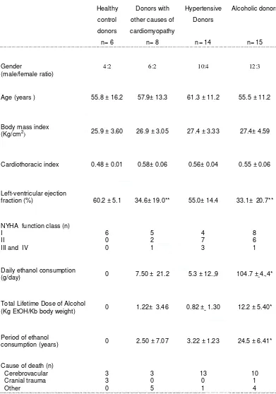
Table 1. Epidemiologic, clinical and heart function data of the different groups of donors.
Healthy control donors n= 6
Donors with other causes of cardiomyopathy n= 8 Hypertensive Donors n= 14 Alcoholic donors n= 15 Gender (male/female ratio)
4:2 6:2
10:4 12:3
Age (years ) 55.8 ± 16.2 57.9± 13.3 61.3 ± 11.2 55.5 ± 11.2
Body mass index
(Kg/cm2) 25.9 ± 3.60 26.9 ± 3.05 27.4 ± 3.33 27.4± 4.59
Cardiothoracic index 0.48 ± 0.01 0.58± 0.06 0.56± 0.04 0.55 ± 0.06
Left-ventricular ejection
fraction (%) 60.2 ± 5.1 34.6± 19.0** 55.0± 14.4 33.1± 20.7**
NYHA function class (n) I
II
III and IV
6 0 0 5 2 1 4 7 3 8 6 1
Daily ethanol consumption
(g/day) 0 7.50 ± 21.2 5.3 ± 12.,9 104.7 ± 4.,4*
Total Lifetime Dose of Alcohol
(Kg EtOH/Kb body weight) 0 1.22± 3.46 0.82 ± 1.30 12.2 ± 5.40*
Period of ethanol
consumption (years) 0 2.50 ± 7.07 3.22 ± 1.23 24.5 ± 6.41*
Cause of death (n) Cerebrovacular Cranial trauma Other 3 3 0 3 0 5 13 0 1 10 1 4
Results expressed as mean + SD
* p<0.01 compared to the other groups
** p<0.01 compared to controls
Page 24 of 29 Manuscripts submitted to Alcohol and Alcoholism
For Peer Review
Table 2. Cardiac myocyte Ki67 expression and TERT activity in different
subgroups of donors
Healthy control donors n=6 Hypertensive donors without CMP n=4 Hypertensive donors With CMP n=10 Alcoholic donors without CMP n= 8 Alcoholic donors with CMP n=7 Donors with other causes of CMP n=8 Ki67 (% of positive cells)
3.92 ± 0.99* 9.90 ± 5.80 13.5 ± 3.56** 9.92 ± 5.36 12.8 ± 4.99*** 15.9 ± 2.21
Increasing of ki67 expression with respect to healthy controls
x 2.5 x 3.4 x 2.5 x 3.2 x 4.0
TERT activity (% of positive cells)
0 0 0 0 0 0
Results expressed as mean + SD
CMP: Cardiomyopathy
* p <0.01 compared to the other groups with CMP
** p= 0.805 compared to hypertensive donors without CMP
*** p= 0.859 compared to alcoholic donors without CMP
Page 25 of 29 Manuscripts submitted to Alcohol and Alcoholism
For Peer Review
Page 26 of 29 Manuscripts submitted to Alcohol and Alcoholism
For Peer Review
[image:29.595.94.492.163.576.2]Figure 1. Cardiac Ki67 expression in the different groups of heart
donors
Figure 1a. Expression of Ki67 antigen in healthy control donors compared to donors with cardiomyopathy of alcoholic, hypertensive or other causes
CMP: cardiomyopathy
Results expressed as percentage of positive cells
Page 27 of 29 Manuscripts submitted to Alcohol and Alcoholism
For Peer Review
Figure 1b. Ki67 expression in healthy controls compared to all donors with cardiomyopathy
Statistically significant difference (p< 0.01)
Page 28 of 29 Manuscripts submitted to Alcohol and Alcoholism
For Peer Review
Figure 2. Ki67 h
eart immunohistochemical assay.
2.a. Control donor with low Ki-67 nuclear activity (magnification x 250)
Fig 2.b. Alcoholic donor with cardiomyopathy.
Increased Ki-67 activity is evident in some nuclei (arrows) (magnification x 250)
Page 29 of 29 Manuscripts submitted to Alcohol and Alcoholism
Estudi de la regeneració miocàrdica en la
miocardiopatia alcohòlica i la seva relació amb el dany
funcional i estructural miocàrdic, activació d’apoptosi
i activitat miostatina
Meritxell Lluís Padierna
ADVERTIMENT. La consulta d’aquesta tesi queda condicionada a l’acceptació de les següents condicions d'ús: La difusió d’aquesta tesi per mitjà del servei TDX (www.tdx.cat) ha estat autoritzada pels titulars dels drets de propietat intel·lectual únicament per a usos privats emmarcats en activitats d’investigació i docència. No s’autoritza la seva reproducció amb finalitats de lucre ni la seva difusió i posada a disposició des d’un lloc aliè al servei TDX. No s’autoritza la presentació del seu contingut en una finestra o marc aliè a TDX (framing). Aquesta reserva de drets afecta tant al resum de presentació de la tesi com als seus continguts. En la utilització o cita de parts de la tesi és obligat indicar el nom de la persona autora.
ADVERTENCIA. La consulta de esta tesis queda condicionada a la aceptación de las siguientes condiciones de uso: La difusión de esta tesis por medio del servicio TDR (www.tdx.cat) ha sido autorizada por los titulares de los derechos de propiedad intelectual únicamente para usos privados enmarcados en actividades de investigación y docencia. No se autoriza su reproducción con finalidades de lucro ni su difusión y puesta a disposición desde un sitio ajeno al servicio TDR. No se autoriza la presentación de su contenido en una ventana o marco ajeno a TDR (framing). Esta reserva de derechos afecta tanto al resumen de presentación de la tesis como a sus contenidos. En la utilización o cita de partes de la tesis es obligado indicar el nombre de la persona autora.
Editorial Manager(tm) for Alcoholism: Clinical and Experimental Research Manuscript Draft
Manuscript Number: ACER-D-10-2904R2
Title: INCREASED MYOSTATIN ACTIVITY AND DECREASED MYOCYTE PROLIFERATION IN CHRONIC ALCOHOLIC CARDIOMYOPATHY
Article Type: Original Research Article
Section/Category: Alcohol Effects on Other Organ Systems
Keywords: Alcohol; Myocardium; Apoptosis; Myostatin; Myocyte proliferation.
Corresponding Author: JOAQUIM FERNANDEZ-SOLA, MD, Ph.D.
Corresponding Author's Institution:
First Author: JOAQUIM FERNANDEZ-SOLA, MD, Ph.D.
Order of Authors: JOAQUIM FERNANDEZ-SOLA, MD, Ph.D.;MERITXELL LLUIS, M.D.;EMILIO SACANELLA, M.D., Ph.D.;RAMON ESTRUCH, M.D., Ph.D.;EMILIA ANTUNEZ, M.D., Ph.D.;ALVARO URBANO-MARQUEZ, Prof.
Manuscript Region of Origin: SPAIN
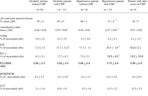
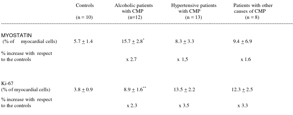
Abstract: Background: Apoptosis mediates in alcohol-induced heart damage leading to
cardiomyopathy (CMP). Myocyte proliferation may compensate for myocyte loss. Myostatin is up-regulated after cardiac damage and by alcohol consumption thereby decreasing myocyte renewal. We assess the potential role of alcohol in inducing myocyte apoptosis as well as in inhibiting myocyte proliferation.
Subjects and methods: Heart samples were obtained from organ donors, including 22 high alcohol consumers, 22 with hypertension, 8 with other causes of CMP, and 10 healthy donors. Evaluation included medical record with data on daily, recent and lifetime ethanol consumption, chest X ray, left ventricular (LV) function assessed by 2-D echocardiography and LV histology and
immunohistochemistry. Apoptosis was evaluated by TUNEL, BAX and BCL-2 assays. Myocyte proliferation was evaluated with Ki67 assay. Myostatin activity was measured with a specific immunohistochemical assay. CMP was assessed by functional and histological criteria.
Results: Alcoholic and hypertensive donors with CMP showed higher apoptotic indices than did their partners without CMP. Myostatin activity was higher in alcoholics than in controls, mainly in those with CMP. The increase in myostatin expression in alcoholic CMP was higher than in other groups. The Ki-67 proliferation index increased in all groups with CMP compared to those without CMP, with alcoholics showing a lower increase in this proliferation response.
INCREASED MYOSTATIN ACTIVITY AND DECREASED MYOCYTE PROLIFERATION IN CHRONIC ALCOHOLIC CARDIOMYOPATHY.
Joaquim Fernández-Solà1,2 M.D., Ph.D.; Meritxell Lluis M.D, Ph.D1.; Emilio
Sacanella1,2 M.D., Ph.D.; Ramón Estruch1,2 M.D., Ph.D.; Emilia Antúnez1,3 M.D, Ph.D,; Alvaro Urbano-Márquez1 M.D, Ph.D.
1Alcohol Research Unit,. Hospital Clinic. Institut d’Investigació August Pi i Sunyer
(IDIBAPS).Department of Medicine University of Barcelona. España
2CIBEROBN Fisiopatologia de la Obesidad y la Nutrición. Instituto de Salud Carlos
III, España
3Department of Medicine.University of Barcelona.Catalunya.España
ABSTRACT: 250 words FULL TEXT: 5,112 words TABLES: 4
FIGURES: 4
With support of SRG 2009-1158, Generalitat de Catalunya, Spain.
We thank Prof Se-Jin Lee ( Molecular Biology & Genetics, Johns Hopkins University School of Medicine. Baltimore, USA) for providing muscle of myostatin knockout mouse.
Corresponding author:
Joaquim Fernandez-Solà M.D.
Department of Internal Medicine. Alcohol Unit Villarroel 170. 08036. BARCELONA. ESPAÑA Phone and FAX: 00.34.93. 2279365
ABSTRACT
Background: Apoptosis mediates in alcohol-induced heart damage leading to cardiomyopathy (CMP). Myocyte proliferation may compensate for myocyte loss. Myostatin is up-regulated after cardiac damage and by alcohol consumption thereby decreasing myocyte renewal. We assess the potential role of alcohol in inducing myocyte apoptosis as well as in inhibiting myocyte proliferation.
Methods: Heart samples were obtained from organ donors, including 22 high alcohol consumers, 22 with hypertension, 8 with other causes of CMP, and 10 healthy donors. Evaluation included medical record with data on daily, recent and lifetime ethanol consumption, chest X ray, left ventricular (LV) function assessed by 2-D
echocardiography and LV histology and immunohistochemistry. Apoptosis was evaluated by TUNEL, BAX and BCL-2 assays. Myocyte proliferation was evaluated with Ki67 assay. Myostatin activity was measured with a specific immunohistochemical assay. CMP was assessed by functional and histological criteria.
Results: Alcoholic and hypertensive donors with CMP showed higher apoptotic indices than did their partners without CMP. Myostatin activity was higher in alcoholics than in controls, mainly in those with CMP. The increase in myostatin expression in alcoholic CMP was higher than in other groups. The Ki-67 proliferation index increased in all groups with CMP compared to those without CMP, with alcoholics showing a lower increase in this proliferation response.
Conclusions: Alcohol produces cardiac myocyte loss through apoptosis but also
partially inhibits myocyte proliferation through myostatin up-regulation. The final result may suppose an imbalance in myocyte homeostasis, with a net loss in total ventricular myocyte mass and progressive ventricular dysfunction.
INTRODUCTION
Alcoholic cardiomyopathy (CMP) is a recognised major complication of chronic alcoholism, (Moushmoush and Ali-Mansour, 1991; Urbano-Márquez and Fernández-Solà, 2004). The disorder is consequence of a common, dose-related, toxic effect of alcohol, affecting one third of long-term alcohol abusers (Urbano-Marquez et al, 1989). This distinct form of congestive heart failure is responsible for21–36% of all cases of non-ischaemic dilated CMP in Western society (Spies et al, 2001). The end-stage of this CMP is largely irreversible and is characterized by a significant loss of myocytes, hypertrophy of the remaining muscle cells and also development of progressive interstitial fibrosis (Fernández-Solà et al, 1994). Without complete abstinence, the 4-year mortality for alcoholic CMP is close to 50% (Laonigro et al, 2009). On the other hand, complete abstinence from alcohol may allow partial clinical and functional reversibility of alcoholic CMP (Guillo et al, 1997). In addition, Nicolás et al (2002) showed that controlled drinking (<60g/day) without complete abstinence was as effective as complete abstinence.
Mechanisms of alcohol-induced myocardial damage are multifactorial, with disruption of membrane composition, disturbances in calcium transients, excitation-contraction coupling mechanisms, energy and protein turnover, and changes in cell mechanisms of genetic control. All these factors are involved in the pathogenesis of this disease, often synchronously (Preedy et al, 1996; Molina et al, 2002; Urbano-Márquez and Fernández.Solà, 2004; Ren and World, 2008). As an end consequence of this process, ethanol induces myocyte damage that progress to myocyte death and cell loss, with replacement by interstitial fibrosis. This myocyte loss has long been attributed to non-inflammatory myocyte necrosis. However, as described in other CMP (Beltrami et al, 2001, 2003; Gürtl et al, 2009), myocyte apoptosis may play a role in the development of structural heart damage in this setting (Hajnóczky et al, 2005; Fernández-Solà et al, 2006).
are activated. When structural heart damage appears, the BCL2/BAX ratio decreases, and apoptosis is present in the heart muscle of high-dose alcohol consumers as occurs to a similar degree in other causes of structural heart damage such as longstanding hypertension (Fernández-Solà et al, 2006) or end-stage heart failure (Saraste et al, 1999; Williams, 1999).
Whereas cardiac myocytes have traditionally been considered to be terminally differentiated cells, the last few years have witnessed exciting new challenges to this dogma, including the discovery of a natural rate of apoptosis and renewal of cardiac myocytes via circulating bone marrow stem cells and resident cardiac progenitor cells (Anversa and Nadal-Ginard 2002; Bergmann et al 2009). The emerging concept of myocyte homeostasis supposes an equilibrium that is maintained with a balance between myocyte death and myocyte renewal (Hill and Olson, 2008; Buja and Vela, 2008). It is important to consider that myocyte death itself activates mechanisms of proliferation of the remaining cells and renewal from pluripotential local or systemic stem cells (Regula et al, 2004). In this scenario of heart remodelling and plasticity, there is place for physiological growth, for pathological growth, and also for antigrowth heart mechanisms (Hill and Olson, 2008). One of these antigrowth factors that control myocyte hypertrophy and proliferation is myostatin.
Myostatin is the growth differentiation factor-8 (GF-1), a potent inhibitor of skeletal muscle and heart growth (Joulia- Ekaza and Cabello, 2006). Myostatin controls cell cycle progression and inhibits skeletal myoblast proliferation and terminal differentiation. An increase in myostatin activity protects the cell from apoptosis. Its disruption causes increased skeletal mass with hypertrophy and hyperplasia of myocytes and increased myocyte proliferation (Joulia- Ekaza and Cabello, 2006; McKoy et al, 2007). Experimental studies suggest that myocyte myostatin increases with ethanol exposure (Lang et al, 2004). This effect of myostatin on skeletal myoblasts is not necessarily related to the same effects on the mature heart
tissue from human donors in which we can observe the effect of previous chronic alcohol consumption on the regulation of cardiac myostatin expression and also evaluate the induction of apoptosis and the degree of myocyte proliferation. We compared this effect of alcohol with that produced by longstanding hypertension and other causes of CMP, as well as in non-alcoholic, non-hypertensive controls.
MATERIAL AND METHODS
Patient and Control Selection
Over a four-year period (January 2003 to December 2006), we consecutively studied hearts from subjects who had brain death either of traumatic or cerebrovascular origin, and had been considered suitable as organ donors by the transplant team of the Hospital Clínic of Barcelona. Of 127 cadaveric donors younger than 70 years of age, 67 hearts were not suitable for transplantation. Of these latter organs we selected: 1) donors with chronic hypertension, 2) donors with a history of ethanol intake (≥ 60 g/day, longer than 10 years), 3) donors with other causes of heart disease, and 4) control hearts from healthy people who were not eligible for implantation because of a lack of matched receptor or size inadequacy.
All patients were white Caucasians of Spanish descent, who lived with their families in or around Barcelona and none was indigent. The study protocol was approved by the Ethics Committee of the Hospital Clínic and included informed consent from the families of the donors concerning the use of myocardium tissue for this research protocol study. One third of these subjects had been included in previous studies on heart antioxidant status (Fatjó et al, 2005) and cardiac apoptosis (Fernández-Solà et al, 2006).
All cases had been admitted to the Intensive Care Unit and ventilatory and haemodynamic parameters had been appropriately maintained at normal values
throughout hospitalisation: PaO2 greater than 60 mmHg, systolic blood pressure greater
A detailed history of ethanol intake was retrospectively obtained by consultation with family members using a structured questionnaire ("time-line follow-back method") (Sobel et al, 1979), as previously reported (Urbano-Márquez et al, 1989; Fernández-Solà et al, 1994). Duration of ethanol intake was calculated in each group as the total cumulated period of alcohol consumption in years, either recent or previous. The body mass index was determined as the actual body weight relative to the square of the body height (BMI, Kg/m2). Patients were considered to have caloric malnutrition if the BMI was less than 17 Kg/m2. Protein malnutrition was assessed by the following parameters obtained at hospital admission: haemoglobin, lymphocyte count, total protein, retinol-binding protein, pre-albumin and albumin.
Cardiac Studies
Past and present signs and symptoms of heart failure were evaluated in consultation with medical records and family members of the donors, and the New York Heart Association (NYHA) functional class was determined according to the Goldman activity scale (Goldman et al, 1981). Chest X-ray with measurement of cardiothoracic index and conventional electrocardiography were performed in all cases. Moreover, a bidimensional echocardiography was performed (Hewlet Packard Sonos 2500, USA) in 24 patients compared to none of the controls with a cardiothoracic index greater than or equal to 0.48 compared to none of the controls. End-diastolic and end-systolic
diameters, the shortening fraction, left ventricular (LV) mass, and the ejection fraction (EF) were measured according to the standards of the American Society of
Echocardiography (Gottdiener et al, 2004). Cardiomyopathy was defined in the presence of LVEF < 50% and LV enlargement. We observed a good correlation
between the cardiothoracic index and the left-ventricle end-diastolic diameter (r = 0.68, p < 0.01). The personnel who performed and evaluated these tests had no knowledge of the alcoholic history of the patients.
Myocardium histological studies
A distal 3 cm sample of the left-ventricle apex was surgically excised (total weight of 4-5 g) at the time the donor was under cold perfusion. The specimen was cut into