ESCUELA TÉCNICA SUPERIOR
DE INGENIERÍAS AGRARIAS
Máster en Investigación en Ingeniería para la Conservación y el Uso
Sostenible de los Sistemas Forestales
Effects of the mycorrhiza helper bacteria
Pseudomonas fluorescens Migula on the
mycorrhizal synthesis between Cistus ladanifer L.
and Boletus edulis Bull.
Alumna: Olaya Mediavilla Santos
Tutor: Pablo Martín Pinto
Director: Jaime Olaizola Suárez
Me gustaría agradecer en primer lugar a mi tutor, Pablo Martín Pinto, por
haberme abierto las puertas al mundo de la investigación y por haberme orientado.
Además me gustaría agradecer sus valiosas correcciones así como su disponibilidad
para adaptarse a mis complicados horarios.
En un segundo lugar pero con la misma relevancia, me gustaría expresar mi
agradecimiento a Jaime Olaizola por haberme dado la oportunidad de trabajar con él y
transmitirme sus conocimientos. También me gustaría agradecerle toda la ilusión,
esfuerzo y tiempo invertido en este trabajo. Sin su dedicación no hubiera sido posible.
A Valentín Pando por el asesoramiento y respaldo estadístico.
A Isbene Sánchez por contagiarme su pasión por los análisis moleculares y por
su atención y paciencia.
A Eneko Ochoa por acercarme al cultivo in vitro y por sus útiles explicaciones.
A Daniel L. Pérez Corona y Raquel Juan Ovejero por la revisión idiomática y
por ofrecerme su ayuda cada vez que lo necesito.
A las nenas, por todos los buenos ratos que pasamos juntas en los que hacéis
que desconecte de todo lo demás. También me gustaría agradecer todos los mensajes
de ánimo en los momentos difíciles.
A mi abuela por enseñarme las cosas importantes de la vida que no aparecen
en los libros, y por la ilusión y alegría que me aporta cada vez que estamos juntas.
A mis padres por acompañarme en cada uno de mis logros y fracasos y vivirlos
como si fuesen suyos.
A Nuria Mediavilla y Jaime Hermoso, por estar siempre ahí para todo, por
escucharme, comprenderme y por hacer que las cosas sean más fáciles a vuestro
Índice
0.- Abstract………..……….…………. 3
1.- Introduction..……….……….……….……...……. 4
2.- Material and methods………...………..….. 6
2.1.- Experimental design ………….…...…………... 6
2.2.- Mycorrhizal synthesis protocol...……….………. 6
2.2.1.- Fungal inoculum….. …….………. 6
2.2.2.- Bacterial inoculum………...………... 7
2.2.3.- Plant materials.………... 7
2.2.4.- Mycorrhizal and bacterial inoculation..……… 8
2.3.- Mycorrhizal colonization verification using molecular technique..……….. 8
2.4.- Data analysis……….. 9
3.- Results………..……...……… 9
3.1.- Mycorrhizal synthesis...…... 9
3.2.- Mycorrhizal infection presence………. 9
3.2.- Level of mycorrhization over the mycorrhized plants ……….. 10
4.- Discussion……….……...….. 13
5.- Conclusions……….………..…...….. 18
0. ABSTRACT
Boletus edulis (L.) is one of the most valuable fungi worldwide, both for its edibility
and economic importance. Sporocarps fruit associated symbiotically to different species of
trees with more than 40 years of age. However, natural productions of B. edulis have been
reported in 3 year old Cistus ladanifer shrubs. Several authors have found that the
ectomycorrhizal symbiosis can be significantly improved by mycorrhiza helper bacteria
(MHB). The aim of this work was to optimize the mycorrhizal protocol between B. edulis and
C. ladanifer as a tool for future reforestation with early productions of B. edulis in this host.
Effects of co-inoculating Pseudomonas fluorescens with B. edulis and mycelium culture time
on the presence of mycorrhizas and on the level of mycorrhization were assessed. The
results obtained were successful since it has was achieved the mycorrhizal synthesis at
medium scale between C. ladanifer and B. edulis from an own isolate of B. edulis.
Furthermore, results showed a significantly enhanced of the level of mycorrhization in the
co-inoculated plants although the bacteria did not improve the number of mycorrhized plants.
The same pattern followed the mycelium culture time. The obtained findings bring us closer
to the controlled B. edulis sporocarps productions in plantations.
Key words: MHB, Mycelium culture, Mycorrhizal plants, Sporocarps.
0. RESUMEN
Boletus edulis es uno de los hongos más valiosos en todo el mundo, tanto por su
comestibilidad como por su importancia económica. Fructifica asociado simbióticamente a
diferentes especies de árboles más de 40 años. Sin embargo, producciones naturales de B.
edulis se han observado en arbustos de C. ladanifer de 3 años de edad. Varios autores han
encontrado que la simbiosis micorrícica puede ser mejorada por bacterias facilitadoras. El
objetivo de este estudio fue optimizar el protocolo de micorrización entre C. ladanifer y B.
edulis como una herramienta para futuras reforestaciones de C. ladanifer con producciones
tempranas de B. edulis. Para ello evaluamos el efecto de la bacteria Pseudomonas
fluorescens y del tiempo de cultivo del micelio en la presencia de micorrizas y el nivel de
micorrización de la planta. Los resultados obtenidos fueron un éxito ya que se consiguió la
micorrización entre C. ladanifer y B. edulis a media escala y a partir de un aislamiento
propio. Además, los resultados mostraron una mejora del nivel de micorrización en las
plantas co-inoculadas, aunque la bacteria no aumentó el número de plantas micorrizadas. El
mismo patrón siguió el tiempo de cultivo de micelio. Los resultados obtenidos nos acercan a
las producciones controladas de cuerpos de fructificación de B. edulis.
1. INTRODUCTION
The species belonging to the Boletus edulis complex are currently among the most
appreciated fungi, being a major commercial mushroom consumed worldwide (Alonso-Ponce
et al., 2011). They are distributed in both hemispheres, from Scandinavia to Southern Africa
and Australia (Hall et al., 1998). These valued edible mushrooms reach an annual world
market around one billion Euros (Mello, 2012), harvesting in Spain an annual production of
Boletus between 2,000 and 20,000 Tm, depending on the year (Oria-de-Rueda et al., 2008).
But a drastic decrease in the presence and productivity of Boletus edulis has been reported
in several parts of Europe (Salerni and Perini, 2004). This phenomenon is accentuated by
the increase in the demand of edible fungi (Sitta and Floriani, 2008). The fact that for the
moment Boletus edulis is only collected from the wild (Cannon and Kirk, 2007), lead to
search alternatives which be feasible for controlling the production of these fungi, as it
happened with other mycorrhizal fungus like Tuber melanosporum (Bonet et al., 2009).
Most of the Boletes fructify associated to Pinus, Quercus and/ or Castanea (Olivier et
al., 1997). Moreover, the production of Boletus sporocarps is linked to relatively mature
forests, being observed mushroom productions in Pinus and Quercus stands with more than
40 years of age (Díaz-Balteiro et al., 2003). Boletus gr. edulis species have been also
reported under Cistus sp. shrubs in the northwest of Castile and Leon (Águeda et al., 2006;
Martín-Pinto et al., 2006). Early natural productions of B. edulis have been reported in 3 year
old Cistus ladanifer shrubs and high productions of these mushrooms in 8-year-old Cistus
plants (Oria-de-Rueda et al., 2008). The genus Cistus is widely distributed throughout the
Mediterranean region, forming vast scrublands (Comandini et al., 2006). Cistus ladanifer L.
is, within this genus, the most abundant species, present in areas affected by forest wildfires.
This natural association, along with sporocarps productions in early host ages allows being
closer of producing artificial mycorrhized plants which then, may yield earlier Boletus edulis
production.
In this sense, Águeda et al. (2008) synthesized ectomycorrhizas between Boletus
edulis and Cistus ladanifer under laboratory conditions using synthesis tubes and obtaining
plant material from seeds. After that, they identified and described the ectomycorrhizas
through visual analysis based on standard morphological and anatomical characters in four
replicates. The use of seeds in mycorrhizal synthesis causes some problems of
the use of vitro-plants may be an alternative method of commercial propagation, using it for
the propagation of plants at medium or large scale (George and Sherrington, 1984).
Mycorrhizal identification through traditional methods is usually confusing and time consuming and high experience is required. For this reason, the development of techniques
based on direct nucleic acid extraction coupled with polymerase chain reaction (PCR) amplification, as Real-time PCR, complement and confirm morphological studies, opening
new possibilities to explore the cryptic phases of the symbiosis (mycorrhizas) (Mello et al., 2006), resulting in prompt and reliable outcomes. The sensitivity of RT-PCR (greater than
conventional PCR) allows detecting the presence of B. edulis DNA when small quantities are present in the sample (De la Varga et al., 2011).
Also, it has been found that the formation of ectomycorrhizal symbiosis can be
improved by mycorrhizosphere bacteria (Deveau et al., 2007). The fungi-bacteria interaction
brings ecological consequences, such as stimulating mycorrhization process (Kurth et al.,
2013). The group of bacteria which promotes this effect is called mycorrhiza helper bacteria
(MHB) (Duponnois and Garbaye, 1991). For instance, they facilitate the mycorrhization from
stimulating fungal mycelial extension, increasing root–fungus contacts and colonization, and
reducing the impact of adverse environmental conditions on the mycelium of the mycorrhizal
fungi (Brulé et al., 2001; Frey-Klett et al., 2007).
Several works isolated the bacteria located into the ectomycorrhizae formed between
Rhizopogon luteolus- Pinus radiata and Laccaria bicolor- Douglas fir, and the most of the
isolated strains belonged to the group Pseudomonas fluorescens (Garbaye et al., 1989;
Garbaye et al., 1990). Various studies have confirmed that P. fluorescens improves the
symbiotic relationship with some ectomycorrhizae, stimulating the ectomycorrhiza fungal
growth (Frey-Klett et al., 2007), and increasing the ratio of mycorrhization (Duponnois and
Garbaye, 1991; Duponnois, 2006). Dominguez et al. (2012) reported the positive effect of
Pseudomonas fluorescens in the mycorrhizal symbiosis between Tuber melanosporum and Pinus halepensis, where mycorrhization was significantly improved with the co-inoculation of P. fluorescens.
The integration of the new technologies concerning fungi and plant production, DNA
detection applied to the mycology, as well as the positive results achieved with bacterial
Despite the results have been limited worldwide to the control of Boletus sp. production, the
possibility of obtaining productive plantations is a prospective aim.
Therefore, the general objective of this study was to optimize the mycorrhizal protocol
between B. edulis and C. ladanifer as a tool for future reforestation with early productions of
B. edulis in this host. In this sense, the following specific objectives have been addressed,
i) analyze the effect of P. fluorescens bacteria in the presence and level of mycorrhization, ii /
analyze the effect of the culture time of fungal inoculum in the presence and level of
mycorrhization, and iii) guarantee the presence of mycorrhizae by using molecular
techniques.
2. MATERIAL AND METHODS 2.1. Experimental design
The experiment included the following inoculation types: (1) Inoculation using Boletus
edulis (Be), (2) inoculation using B. edulis × Pseudomonas fluorescens CECT 844 (Be×Pf)
and (3) control without inoculation (C). Each of these inoculation types were established for
three different mycelium culture times (two, three and four months). Eighteen in vitro pots
were used per treatment and four plants were placed per pot. Thus, a total of 162 plants
were tested.
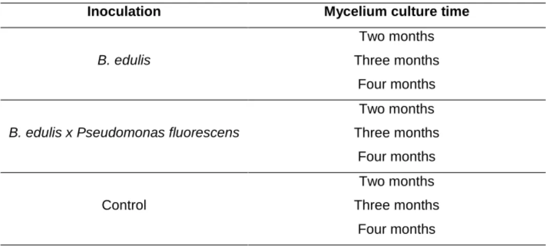
Table 1. Experimental design
Inoculation Mycelium culture time
B. edulis
Two months
Three months
Four months
B. edulis x Pseudomonas fluorescens
Two months
Three months
Four months
Control
Two months
Three months
Four months
2.2. Mycorrhizal syntensis protocol
2.2.1. Fungal inoculum
Sporocarps of Boletus edulis were collected from Cistus ladanifer shrubs located in
northwest Spain. They were stored in closed polyethylene bags until the isolation in
Isolation was carried out under aseptic conditions in order to avoid possible
contamination. A small piece of the fungus cap was placed on MMN nutritive medium plates
(Modified Melin-Norkrans) (Marx, 1969) at pH 5.5, growing at 22-24ºC in dark conditions
(Honrubia et al., 1994). Once the mycelium was large enough, it was transferred to fresh
MMN plates to ensure its vitality and growth. A molecular analysis was carried out in order to
verify the identification of the fungal species.
The solid expanding culture substrate was a mixture of vermiculite and peat (11/1;
v/v) moistened with liquid MMN nutritive medium at pH 5.5 and the glucose content reduced
at 2.5 g/l. The ratio of substrate to liquid was 2/1 (v/v). Substrate was transferred into glass
pots and sterilized at 121ºC for 20 min. Substrate was cooled down and the in vitro pots were
inoculated with B. edulis adding 20 plugs, 5 mm diameter, of active mycelium and cultured at
22ºC, for two, three or four months depending on the treatment.
2.2.2. Bacterial inoculum
The inoculum of Pseudomonas fluorescens CECT 844 was supplied by the CECT
(Spanish Type Culture Collection), University of Valencia. The liquid inoculum was prepared
by suspending P. fluorescens in a malt-glucose nutritive medium (3 g malt, 10 g glucose and
1l distilled water). Inoculated nutrient medium was cultured and shaken at 100 rpm and 22ºC.
After 48h, bacterial concentration was recorded through the Thoma camera and bacterial
inoculum was re-suspended in sterile water to get a concentration of 5×10⁸ spores per
milliliter.
2.2.3. Plant materials
Plant material was selected from Cistus ladanifer shrubs, which hosted Boletus edulis
fungi located in the Western of Zamora province.
Shoots tips from five years old Cistus ladanifer shrubs were selected in order to
establish in vitro cultures. Fifty shoots were introduced during 20 min in an antioxidant
solution (100 mg/l ascorbic acid and 150 mg/l citric acid) to avoid explants browning (M’Kada
et al., 1991). Then shoots were surface sterilized by introduction in 100% ethanol for 30 s,
rinsed with sterile distilled water and then further sterilized for 30 min in 1.5% sodium
hypochloride (NaClO) solution with one drop of Tween 20® detergent. Finally, they were
Proliferation was carried out culturing shoots tips on MS basal medium (Murashige
and Skoog, 1962) supplemented with 0.88 mg l-1 BAP, 30 g l-1 sucrose and 8 g l-1 agar. Shoot
tips were sub-cultured every four weeks. Rooting was performed transplanting microshoots
from the third subculture in MS basal media supplemented with 0.49 µm of IBA. Plants were
grown, until the inoculation, at 25 ± 1 ºC under a 16 h photoperiod with a light intensity of
2000 luxes provided by cool-white fluorescens lamps.
2.2.4. Mycorrhizal and bacterial inoculation
All of the inocula were applied at the same time (January 2014). Cistus ladanifer
seedlings were placed into the in vitro pots which contained the vermiculite-peat substrate
previously prepared. Depending on the treatment the plants were transferred into; substrate
inoculated with B. edulis (Be); substrate inoculated with B. edulis and injected with a bacterial
dose of 5×10⁸ spores/plant (Be×Pf); or substrate not inoculated (C). After performing
inoculations, plants were stored in a culture chamber under a 16 h photoperiod with a light
intensity of 2000 luxes and at 25 ± 1 ºC for five months.
2.3. Mycorrhizal colonization verification using molecular technique
After five months from inoculation, plants were carefully extracted from the pots and
roots were cleaning according to the methodology established by Fischer and Colinas,
(1997). All the plants inoculated were visualized by stereo microscope to analyze the
colonization of the mycorrhizal fungi in the roots by the morphological characterization and
identification of the mycorrhizas (Agerer, 1991). Description of the B. edulis ectomycorrhiza
performed by Águeda et al. (2008) was followed to ensure the identification.
The mycorrhization percentages were calculated. For that, roots were then chopped
into 1–2 cm pieces that were cleaned, rinsed in distilled water and placed into a Petri dish with water for analysis. In the mycorrhized plants, the total roots as well as the B. edulis
mycorrhized tips were counted.
The identification of B. edulis mycorrhiza was confirmed using molecular techniques.
DNA extractions for the roots were performed with the PowerSoilTM DNA Isolation Kit and the
presence by real-time polymerase chain reaction using specific primers and TaqMan® probe
(De la Varga et al., 2012). One plant were selected randomly and analyzed from each
2.4. Data analysis
Pearson Chi-square Test was used to compare frequency tables for mycorrhization
presence among treatments. For comparing the level of mycorrhization over the mycorrhized
plants, a factorial ANOVA analysis was performed for the data and means were compared
for the proposal factors by Least Significance Difference (LSD) Fisher Tests (P<0.05).
Statistical analysis was accomplished with STATISTICA ’08 Edition software (StatSoft Inc.,
1984–2008).
3. RESULTS
3.1. Mycorrhizal synthesis
The first noticeable result of this experiment is that ectomycorrhizas of Boletus edulis
in Cistus ladanifer plants were syntethized in vitro, furthermore is the first time that this was
possible at medium scale using vitro-plants. To get this result, in vitro propagation of plant
material was a relevant finding to optimize the inoculation protocol. The ectomycorrhizas
visualized had typical traits of Boletales, and followed the description realized by Águeda et
al. (2008) but with some differences in the color, showing brownish but with
plectenchymatous mantle and rhizomorphs with highly differentiated hyphae. The inflated,
smooth cystidia-like clavate end cells on the surface of the rhizomorphs and their slightly
twisted external hyphae are additional characterizing features.
In addition, this was the first time that B. edulis mycorrhizal synthesis was verified through
molecular analysis. Every plant selected randomly from each treatment was positive for B.
edulis mycorrhization in Real-Time PCR.
3.2. Mycorrhizal infection presence
Both Boletus edulis and B. edulis × Pseudomonas fluorescens formed
ectomycorrhizas with Cistus ladanifer. Among the total of 108 plants inoculated, 35 were
mycorrhized, which corresponds with a mycorrhizal infection ratio of 32.41% over the whole
experiment.
Among the 54 plants inoculated only with B. edulis, 18 were mycorrhized, which come
into a mycorrhizal infection ratio of 33.33%. From the plants inoculated with B. edulis ×
Pseudomonas fluorescens 17 were mycorrhized, reaching a mycorrhizal infection ratio of
Using co-inoculation with bacteria did not affect significantly the presence of
mycorrhized plants. In this sense, from the mycorrhized plants, 51.43% belonged to those
inoculated with B. edulis, whereas 48.57% were inoculated with B. edulis × Pseudomonas
fluorescens.
This same effect was observed analyzing the mycelium culture time. Thus, this
variable did not affect significantly the percentage of mycorrhized plants. With a mycelium
culture time of two months 27.78% of the inoculated plants were mycorrhized. The
mycorrhizal infection ratio was 36.11% at three and 33.33% at four months of mycelium
culture time.
Analyzing jointly the factors bacteria and mycelium culture time, no differences were
evidenced among treatments (Table 2).
Table 2. Presence of mycorrhizas (in percentage) according to inoculation and mycelium culture time
Inoculation Mycelium culture time Total
2 months 3 months 4 months
B.edulis 33.33aA 33.33aA 33.33aA 33.33A
B. edulis+bacteria 22.22aA 38.89aA 33.33aA 31.48A
Total 27.78a 36.11a 33.33a
Different minuscule letters in the same rows and capital letters in the same columns indicate significant differences according to Pearson Chi-square Test at P<0.05 levels.
3.3. Level of mycorrhization over the mycorrhized plants
Co-inoculation and mycelium culture time significantly affected the percentage of
mycorrhized plants. However, no interaction between these two factors was found, thus
bacteria affected increasing mycorrhization levels independently of the mycelium culture time
Table 3. Analysis of the varianza (ANOVA) of the level of mycorrhization over the
mycorrhized plants.
SS Degr. of
freedom MS F p
Intercept 6934,364 1 6934,364 119,8742 0,000000
Time 733,016 2 366,508 6,3358 0,005213
Bacteria 874,501 1 874,501 15,1175 0,000542
Time*Bacteria 74,915 2 37,457 0,6475 0,530740
Error 1677,563 29 57,847
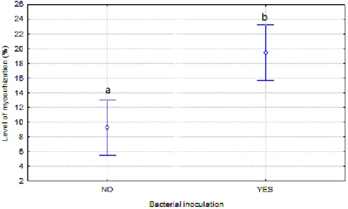
The most remarkable finding of this work was that co-inoculation of Boletus edulis
and bacteria Pseudomonas fluorescens had a positive effect on the level of mycorrhization,
being this the level significantly higher than in the case of plants mycorrhized exclusively with
Boletus edulis (P<0.001). Among the plants mycorrhized with B. edulis exclusively, the level
of mycorrhization was 9.25% whilst for the plants inoculated also with P. fluorescens, the
level reached 19.45% (Figure 1).
Figure 1. Effect of co-inoculation of B. edulis and P. flourescens on mycorrhization
level. Different letters indicate significant differences according to LSD at P<0.05
levels.
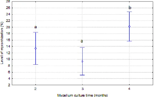
Mycelium culture time was favorable to the level of mycorrhization and, in this sense
the percentage of tips mycorrhized was higher in the plants inoculated with mycelium
cultured during four months, reaching 20.25% of tips mycorrhized. The level of
obtained at two (P=0.009) and three months of cultivation (P<0.001). However, differences
between the level of mycorrhization at two and three months of mycelium cultivation were not
significant (P=0.43).
Figure 2. Effect of the mycelium culture time on mycorrhization level. Different
letters indicate significant differences according to LSD at P<0.05 levels.
Analyzing together the use of bacteria and the mycelium culture time, some
interesting results were appreciated. Higher levels of mycorrhizas were achieved when using
the helper bacteria. For two and four months of mycelium culture time these differences were
significant. Although not significant, the trend was similar for three months of mycelium
cultivation. In all the cases, mycorrhization levels were duplicated when using co-inoculation
with B. edulis x P. fluorescens (Table 5).
Table 5. Effects of inoculation of B. edulis inoculated alone and together with
Pseudomonas fluorescens on Cistus ladanifer level of mycorrhization for the different
mycelium culture times.
Mycelium culture time
Inoculation
Be Be+bacteria
Two months 7.16 ± 2.70aA 19.69 ± 3.65bAB
Three months 6.32 ± 2.31aA 12.44 ± 2.81aA
Four months 14.28 ± 2.48aA 26.22 ± 2.58bB
Different minuscule letters in the same rows and capital letters in the same columns indicate significant differences according to LSD at P<0.05 levels.
Be is the plant inoculated with Boletus edulis. Be+bacteria is the plant inoculated with
The best results were achieved in the plants mycorrhized with B. edulis and bacteria,
with four months of mycelium cultivation; in this case the level of mycorrhization was
significantly higher 26.22% (Table 5).
If the mycelium culture time is analyzed separately for the two types of inoculation, a
similar trend was observed. Thus, the highest mycorrhization levels were found when the
culture time was four months in both cases, accordingly to the previous overall result. It was
observed that inoculation with B. edulis, mycorrhization percentage reached doubles the
ratios of two and three months. The higher value in the case of co-inoculation with the
bacteria was also achieved when the culture time was four months (Table 5).
4. DISCUSSION
The results obtained are successful since it has been achieved the mycorrhizal
synthesis between C. ladanifer and B. edulis, from own isolates of B. edulis at medium scale.
In order to develop the use of an ectomycorrhizal species in forestry, it is crucial to establish
an efficient protocol of controlled mycorrhization (Guerin-Laguette et al., 2000). In this sense,
an important improvement of the mycorrhizal protocol has been achieved with this work,
showing the effect of the co-inoculation with mycorrhizal helper bacteria on the
mycorrhization levels. Besides, different mycelium culture times have been analyzing to test
the effect over the presence of mycorrhizas and the level of mycorrhization.
Obtaining B. edulis mycorrhizas in C. ladanifer plants is a very relevant fact as it is the
first step which could enable to set future commercial plantations for early productions of
Boletus edulis sporocarps, whose wild productions may decline each year by over
exploitation and lack of regulation of this natural resource.
Mycorrhizas observed in plants were contrasted with those described by Águeda et
al. (2008), but differences were observed in the color of our mycorrhizas that were darker.
This difference in the color of the mycorrhizas may be due to the dryness of the substrate
shown at the end of the experiment. When the mycorrhiza loss water it tends to get obscure
because of the desiccation of the tissues. Furthermore, small differences evidenced in the
mycorrhizas could be due to the fact that the B. edulis mycorrhized by Águeda et al. (2008)
was associated to Cistus albidus. In spite of that, the rest of the traits were similar to those
characterized, plectenchymatous mantle, monopodial ectomycorrhizal system and frequent
In this point, molecular techniques play an important role when doubtful cases, as
happened in the comparison among our mycorrhizas with the previously described.
Molecular analysis will allow us to identify our sample quickly, accurate and reliable (Schena
et al., 2004). Our mycorrhizal identification was confirmed through molecular analysis, for
both, the inoculum used and the mycorrhizas obtained. Every plant selected randomly from
each treatment was positive for B. edulis mycorrhization. To our knowledge, this is the first
time that a molecular verification is performed for B. edulis after mycorrhization to check the
success of it. Some studies realized molecular analysis over the fungus previously to the
inoculation (Águeda et al. 2008). This subsequent test certifies with total guarantee that the
mycorrhiza obtained was caused by the isolated used and not by any contaminant.
Mycorrhizas were formed in the two treatments where there was B. edulis inoculation
(plants inoculated with B. edulis exclusively and plants inoculated with B. edulis and P.
fluorescens), whilst the control plants did not present any type of mycorrhizas, neither of B. edulis nor of possible contaminants. Despite the achievement in the mycorrhizal synthesis for
both B. edulis inoculated alone and also co-inoculated with P. fluorescens, the presence of
mycorrhizas was lower than expected. The relatively low percentages of B. edulis
mycorrhizas obtained may be due to an insufficient mycelium expansion, which
simultaneously may be promoted by various factors. An important factor to have into account
is the mycelium culture time. As it happens with other mycorrhizal fungi as Lactarius
deliciosus(Guerin-Laguette et al., 2000; Parladé et al., 2004), B. edulis grows slowly in pure culture and takes at least two months to complete the substrate colonization. In this sense,
an insufficient cultivation of the substrate may result in an uneven quality inoculum. The slow
colonization of B. edulis, as well as the uneven quality of the inoculums could be palliated by
adding a larger quantity of fungal mycelium at the inoculation time.
Another relevant factor may be the intraespecific variation of ectomycorrhizal fungi.
Thus, fungi of the same species may present different growing characteristics and also
different colonization ratio in pure cultures (Pera and Alvarez, 1995; Parladé et al., 2011).
Several authors found diversity in the responses within the same species, in this sense,
varying specificities among the fungal species has become apparent (Frey-Klett et al., 2007;
Aspray et al., 2013). A future possibility for optimizing the protocol would be to study several
A possible factor which may have affected negatively was the dryness that the
substrate presented at the moment of the roots evaluation. Despite the inoculum type utilized
is one of the most suitable for producing mycorrhizal seedlings (Marx and Kenney, 1982),
some disadvantages related to the desiccation are probable and difficult to manage (Parladé
et al., 2004). In this way, a control of the humidity of the culture chamber would be a requisite
for a successful mycelia expansion.
The relevance of the bacteria in the mycorrhiza formation was reported for first time
by Bowen and Theodorou (1979). They tested that some bacterial strains promoted and
others inhibited the colonization of Pinus radiata roots by Rhizopogon luteolus. Posterior
studies were conducted in order to get more knowledge. Thus, Garbaye and Bowen (1987);
de Oliveira and Garbaye (1989) confirmed that the co-inoculation with bacteria promoted the
mycorrhiza formation. More recently, satisfactory results gained in previous works with
Pseudomonas species. Duponnois and Plenchette (2003) co-inoculated Pseudomonas monteilii with several isolates of Pisolithus sp. and Scleroderma sp. in Australian Acacia
species. The ectomycorrhizal colonization for all the Acacia species was significantly
enhanced (from 45.8% to 70.3%). Deveau et al. (2007) tested the effect of various bacteria
(Collimonas fungivorans, Paenibacillus sp., Paenibacillus sp., Bacillus subtilis, Burkholderia
sp. and Pseudomonas fluorescens) through an in vitro confrontation with Laccaria bicolor.
Pseudomonas fluorescens was the unique bacteria which stimulated the growth of L. bicolor
mycelium, as well as the hypha density.Dominguez et al. (2012) stated the improvement of
P. fluorescens in the establishment and mycorrhizal symbiosis of Tuber melanosporum in Pinus halepensis. They obtained the bacteria inoculum from the CECT (Spanish Type
Culture Collection), University of Valencia. In this sense, regarding the good results obtained
by Dominguez et al. (2012) with P. fluorescens CECT 844, this isolated was selected for our
study.
According to the results acquired in our work when applying the bacteria, in the
co-inoculated treatment (B. edulis x P. fluorescens) the presence of plants mycorrhized did not
present differences respect to the inoculated with B. edulis exclusively. Specificity in
MHB-mycorrhizal fungus interactions was already indicated in early studies, which described
bacterial species that promote and others that were either neutral or inhibitory to mycorrhiza
The results obtained for the presence of mycorrhizas in the co-inoculation may also
be explained because of the competition of B. edulis with the bacteria during the
establishment into the substrate (Bowen and Theodorou, 1979). Various authors postulated
that some interactions between MHB and the fungus may be beneficial but some others may
be competitive to the mycorrhizal infection process (Garbaye, 1994; Frey-Klett et al., 2007;
Aspray et al., 2013). Bearing in mind that B. edulis is a sensitive species of slow growing in
saprophytic conditions (Olaizola, 2007), in a pre-symbiotic moment, i.e., before the
association between the plant and B. edulis, P. fluorescens and B. edulis may compete for
the nutrients and B. edulis may be particularly affected. This result agree with the
observations of Brulé et al. (2001), who affirmed a situation of dominance of the bacteria
over the fungus in rich growth media. Kurth et al. (2013) also suggested the inhibition of
Piloderma croceum because the competition for resources with bacteria. Thus, considering
the competition of P. fluorescens and B. edulis in the early stages, a reduction of the
inoculated bacterial dosage could be beneficial for the growth of B. edulis. The development
of methods for quantifying the abundance of bacteria and fungi in the presence of
one-another is essential (Kurth et al., 2013).
It was remarkable that our study showed that MHB increased the level of
mycorrhization. In this way, once B. edulis was associated with the plant, bacteria was
beneficial for its development which consequently was showed in a higher formation of
mycorrhizas. This result was in accordance with the obtained in previous studies. Dominguez
et al. (2012) co-inoculated P. fluorescens and Tuber melanosporum in Pinus halepensis
seedlings. They achieved to increment significantly the level of mycorrhization from 15% to
28%. Regarding the interaction between B. edulis and MHB, Wu et al. (2012) analyzed the
effects of co-inoculating the fungus and the bacteria Bacillus cereus in Pinus thunbergii. For
that, they isolated Bacillus cereus strains from B. edulis sporocarps and inoculated the plants
in nursery conditions. Interesting results were achieved since mycorrhizal infection ratio was
significantly increased in the plants co-inoculated with the fungus and bacteria. However,
they did not provide any molecular analysis to confirm the results which may lead to false
positives, i.e., a possible mycorrhization with other mycorrhizal fungi of similar morphological
appearance.
Although, the results obtained for the level of mycorrhization are apparently
contradictory with the obtained in the mycorrhizal infection, this fact has been explained by
samples. From them 12 stimulated the growth of two fungal isolates and the four best were
selected for further analysis. One of them increased the mycorrhizal presence significantly.
However, the other three isolates promoted fungal growth in vitro experiment but not
enhanced ectomycorrhiza formation. Zhao et al. (2013) suggested that the mechanism of
MHB effect may be diverse, and stimulation of fungi growth by MHB is not necessarily
correlated to effectiveness at promoting ectomycorrhiza formation. Brulé et al. (2001) studied
the effect of Pseudomonas fluorescens on the Douglas fir-Laccaria bicolor symbiosis. They
postulated that the success of the inoculation depends on the survival of the fungal
inoculums in the soil during pre-symbiotic life of the fungus. Furthermore, they found a
pattern of the bacteria in the fungus depending on the experimental conditions. Bacteria had
a negative effect on the symbiosis under favorable conditions (rich growth media) and
presented a positive effect under unfavorable conditions.
Considering the mycelium culture time, a positive overall correlation was showed for
the level of mycorrhizas over the mycorrhized plants, reaching the highest level of
mycorrhization at four months of mycelium cultivation. The maturity of the mycelium growing
for four months could cause higher mycorrhization levels. At four months of culture time, the
availability of nutrients is lower and the mycelium is becoming mature. Previous studies have
shown the ability of the ectomycorrhizal fungi to cope with stress situations such as nutrient
deficiency. Even in those poor conditions, fungi are able to increase their natural mycorrhizal
potential to guarantee plant mycorrhization (Requena et al., 1996).
However mycelium culture time did not affect mycorrhizal presence. Mycorrhization
percentages are directly linked to mycelium presence in the soil. In this sense, the result can
be explained because of the slow growth of B. edulis mycelium. Olaizola (2007) tested the
mycelium growth of twelve isolates of different mycorrhizal fungi under axenic conditions. In
that case, B. edulis was one of the isolates with lowest growth, with a growth significantly
lower than that for Lactarius deliciosus which get the whole colonization of the substrate by
the mycelium after two months as minimum under optimum conditions (Parladé et al., 2004).
For explaining our results regarding to fungal culture time, we hypothesize the
possibility that the quantity of mycelium achieved at two months was similar than that present
in the soil after four months. However, the maturity of the mycelium growing for 4 months
Furthermore, we have to highlight the achievement in the proliferation of Cistus
ladanifer plants through in vitro culture. To date only few studies on the micropropagation of Cistaceae have been conducted (M’Kada et al., 1991; Morte and Honrubia, 1992; Iriondo et al., 1995; Pela et al., 2000; Madesis et al., 2011), and C. ladanifer had not been cultured
using in vitro propagation previously due to its scarce economic value and its great
colonizing ability. Seeds of Cistus species have a form of dormancy due to their impermeable
seed coat, which is broken when the seeds are exposed to heat (Madesis et al., 2011).
Furthermore, sometimes it is difficult obtaining axenic seedlings proceeding from seeds
because of the contamination problems. When stronger disinfection treatments are
performed, germination and seed viability are drastically reduced. De la Varga et al. (2011)
confirmed the necessity of improving the production of C. ladanifer plants under axenic
conditions pointing the difficulty of producing a large quantity of plants mycorrhized because
of the high economic and time costs. Thus, plant tissue culture is an alternative method of
commercial propagation, used widely for the propagation of a large number of plant species
(George and Sherrington, 1984). Taking into account that our prospective objective is to
place plantations of C. ladanider mycorrhized with B. edulis, our work has gone a step further
on the idea of making B. edulis fruiting plants a commercial product. This method would be
more feasible for plant proliferation at medium and large scale.
5. CONCLUSIONS
Mycorrhizal synthesis between C. ladanifer and B. edulis was achieved from an own
B. edulis isolate. Consecutive identification of B. edulis mycorrhizas was verified through
molecular techniques. The obtained results confirmed the beneficial effects of P. fluorescens
in an increment of the level of mycorrhization respect to the inoculation with B. edulis alone.
A higher mycelium culture time also enhanced the level mycorrhization. Furthermore, the
achievement in the production of Cistus ladanifer vitro-plants enable faster production,
avoiding the high problems of contamination observed from seeds and facilitating the plant
production at large scale. The accomplished results bring us closer to form controlled
plantations of C. ladanifer producing B. edulis, where it will be possible to collect fruiting
bodies. This will enable return a valuable resource to the rural areas.
6. REFERENCES
Agerer R. 1991. Characterization of ectomycorrhiza, in: Norris JR, Read DJ, Varma AK.
Águeda B, Parladé J, de Miguel, AM, Martínez-Peña F. 2006. Characterization and
identification of field ectomycorrhizae of Boletus edulis and Cistus ladanifer. Mycologia
98, 23–30.
Águeda B, Parladé J, Fernández-Toirán, LM, Cisneros O, de Miguel AM, Modrego MP,
Martínez-Peña F, Pera J. 2008. Mycorrhizal synthesis between Boletus edulis species
complex and rockroses (Cistus sp.). Mycorrhiza 18, 443–9.
Alonso-Ponce R, Águeda B, Modrego MP, Aldea J, Fernández-Toirán LM, Martínez-Peña F.
2011. Rockroses and Boletus edulis ectomycorrhizal association: realized niche and
climatic suitability in Spain. Fungal Ecol. 4, 224–232.
Aspray TJ, Jones EE, Davies MW, Shipman M. 2013. Increased hyphal branching and
growth of ectomycorrhizal fungus Lactarius rufus by the helper bacterium Paenibacillus
sp. Mycorrhiza 23, 403–410.
Bonet JA, Oliach D, Fischer CR, Martínez de Aragón J, Colinas C. 2009. Cultivation methods
of the black truffle, the most profitable mediterranean non-wood forest product; a state
of the art review, in: Palahí M, Birot Y, Bravo F, Gorriz E. (Eds.), Modeling, Valuing and
Managing Mediterraneam Forests Ecosystems for Non-Timber Goods and Services.
EFI Procedings, 57–71.
Bowen GD and Theodorou CU. 1979. Interactions between bacteria and ectomycorrhizal
fungi. Soil Biol. Biochem. 11, 119–126.
Brulé C, Frey-Klett P, Pierrat JC, Courrier S, Gérard F, Lemoine MC, Rousselet JL, Sommer
G, Garbaye J. 2001. Survival in the soil of the ectomycorrhizal fungus Laccaria bicolor
and the effects of a mycorrhiza helper Pseudomonas fluorescens. Soil Biol. Biochem.
33, 1683–1694.
Cannon PF and Kirk PM. 2007. Fungal Families of the World. CAB International, Wallingford,
Oxfordshire, UK.
Comandini O, Contu M, Rinaldi AC. 2006. An overview of Cistus ectomycorrhizal fungi.
De la Varga H, Águeda B, Martínez-Peña F, Parladé J, Pera J. 2011. Quantification of
extraradical soil mycelium and ectomycorrhizas of Boletus edulis in a Scots pine forest
with variable sporocarp productivity. Mycorrhiza 22, 59–68.
De Oliveira VL and Garbaye J. 1989. Les microorganismes auxiliaires de retabhssemeni des
symbioses ectomycorrbiziennes. European Journal of Forest Pathology 19, 54–64,
Deveau A, Palin B, Delaruelle C, Peter M, Kohler A, Pierrat, JC, Sarniguet A, Garbaye J,
Martin F, Frey-Klett P. 2007. The mycorrhiza helper Pseudomonas fluorescens BBc6R8
has a specific priming effect on the growth, morphology and gene expression of the
ectomycorrhizal fungus Laccaria bicolor S238N. New Phytol. 175, 743–55.
Díaz-Balteiro L, Álvarez-Nieto A, Oria-de-Rueda JA. 2003. Integración de la Producción
Fúngica en la Gestión Forestal. Aplicación al Monte “Urcido” (Zamora). Investigaciones
Agrarias: Sistemas y Recursos Forestales 121:5–19.
Dominguez JA, Martin A, Anriquez A, Albanesi A. 2012. The combined effects of
Pseudomonas fluorescens and Tuber melanosporum on the quality of Pinus halepensis
seedlings. Mycorrhiza 22, 429–36.
Duponnois R. 2006. Bacteria Helping Mycorrhiza Development. In: Mukerji KG,
Manoharachary C, Singh J, eds. Microbial activity in the rhizosphere. Berlin, Germany:
Springer-Verlag, 297–310.
Duponnois R and Garbaye J. 1991. Mycorrhization helper bacteria associated with the
Douglas fir-Laccaria laccata symbiosis : effects in aseptic and in glasshouse conditions
Correspondence and reprints 48, 239–251.
Duponnois R and Plenchette C. 2003. A mycorrhiza helper bacterium enhances
ectomycorrhizal and endomycorrhizal symbiosis of Australian Acacia species.
Mycorrhiza 13, 85–91.
Fischer C and Colinas C. 1997. Propuesta de metodología para la certificación de planta de
Quercus ilex inoculada con Tuber melanosporum para la aplicación comercial. Soria.
Frey-Klett P, Garbaye J, Tarkka M. 2007. The mycorrhiza helper bacteria revisited. New
Garbaye J. 1994. Tansley Review No. 76 Helper bacteria: a new dimension to the
mycorrhizal symbiosis. New Phytol. 128, 197–210.
Garbaye J and Bowen GD. 1987. Effect of different microflora on the success of mycorrhizal
inoculation of Pinus radiata. Can. J. For. Researcl 17, 941–943.
Garbaye J and Bowen GD. 1989. Stimulation of ectomycorrhizal infection of Pinus radiata by
some microorganisms associated with the mantle of ectomycorrhizas. New Phytol. 112,
383–388.
Garbaye J, Duponnois R, Wahl JL. 1990. The bacteria associated with Laccaria laccata
ectomycorrhizas or sporocarps: effect on symbiosis establishment on Douglas-fir,
Symbiosis 9, 267-273.
George EF and Sherrington PD. 1984. In Vitro: Plant Propagation by Tissue Culture.
Exegetics Ltd, Eversley, England.
Guerin-Laguette A, Plassard C, Mousain D. 2000. Effects of experimental conditions on
mycorrhizal relationships between Pinus sylvestris and Lactarius deliciosus and
unprecedent fruit-body formation of the Saffron milk cap under controlled soilless
conditions. Can. J. Microbiol. 46, 790–799.
Hall IR, Lyon AJE, Sinclair L. 1998. Ectomycorrhizal fungi with edible fruiring bodies 2.
Boletus edulis. Econ. Bot. 52, 44–56.
Honrubia M, Torres P, Diaz G, Morte A. 1994. Biotecnología forestal: Técnicas de
micorrización y micropropagación de plantas.
Iriondo JM, Moreno C, Pérez C. 1995. Micropropagation of Six Rockrose (Cistus) Species.
Hortscience 30, 1080–1081.
Kurth F, Zeitler K, Feldhahn L, Neu TR, Weber T, Kri V. 2013. Detection and quantification of
a mycorrhization helper bacterium and a mycorrhizal fungus in plant-soil microcosms at
different levels of complexity Detection and quantification of a mycorrhization helper
bacterium and a mycorrhizal fungus in plant-soil. BMC Microbiol. 13, 205.
M’Kada J, Dorion N, Bigot C. 1991. In vitro propagation of Cistus × purpureus Lam. Sci.
Madesis P, Konstantinidou E, Tsaftaris A, Nianiou-Obeidat I. 2011. Micropropagation and
shoot regeneration of Cistus creticus ssp. Creticus 01, 54–58.
Martín-Pinto P, Vaquerizo H, Peñalver F, Olaizola J, Oria-de-Rueda JA. 2006. Early effects
of a wildfire on the diversity and production of fungal communities in Mediterranean
vegetation types dominated by Cistus ladanifer and Pinus pinaster in Spain. For. Ecol.
Manage. 225, 296–305.
Marx D. 1969. The influence of ectotrophic mycorrhizal fungi on the resistance of pine roots
to pathogenic infections. I. Antagonism of mycorrhizal fungi to root pathogenic fungi and
soil bacteria. Phytopathology 59, 153–163.
Marx DH and Kenney DS. 1982. Production of ectomycorrhizal fungus inoculum, in: Schenck
NC. (Ed.), Methods and Principles of Mycorrhizal Research. American
Phytopathological Society, St Paul, Minn., 131–146.
Mello A. 2012. State of the Art of the Research on Boletus edulis, in: Zambonelli A, Bonito
GM (Eds.), Edible Ectomycorrhizal Mushrooms. Springer Berlin Heidelberg, 73–81.
Mello A, Ghignone S, Vizzini A, Sechi C, Ruiu P, Bonfante P. 2006. ITS primers for the
identification of marketable Boletus. J. Biotechnol. 121, 318–329.
Morte A and Honrubia M. 1992. In vitro propagation of Helianhthemum almeriense Pau
(Cistaceae). Agronomie 12, 807–809.
Murashige T and Skoog F. 1962. A revised medium for rapid growth and bioassays with
tobacco tissue cultures. Physiol Plant. 15, 473–479.
Olaizola J. 2007. Selección de hongos ectomicorrícicos comestibles para su utilización en el
control biológico del Damping-off causado por Fusarium oxysporum Schlecht y
Fusarium verticillioides (Sacc.) Nirenberg. Universidad de Valladolid.
Olivier JM, Guinberteau J, Rondet J, Mamoun M. 1997. Vers l’inoculation contrôlée des
cèpes et bolets comestibles? Rev Fr 49, 222–234.
Oria-de-Rueda JA, Martín-Pinto P, Olaizola J. 2008. Bolete Productivity of Cistaceous
Parladé J, Hortal S, Varga H, Pera J. 2011. Intraspecific variability of Lactarius deliciosus
isolates : colonization ability and survival after cold storage. Mycorrhiza 393–401.
Parladé J, Pera J, Luque J. 2004. Evaluation of mycelial inocula of edible Lactarius species
for the production of Pinus pinaster and P. sylvestris mycorrhizal seedlings under
greenhouse conditions. Mycorrhiza 171–176.
Pela Z, Pencheva M, Gerasopoulos D, Maloupa E. 2000. In vitro induction of adventitious
roots and proliferation of Cistus creticous creticous plants. Acta Hortic. 541, 518–524.
Pera J and Alvarez, IF. 1995. Ectomycorrhizal fungi of Pinus pinaster. Mycorrhiza 5, 193–
200.
Requena N, Jeffries P, Barea, JM. 1996. Assessment of natural mycorrhizal potential in a
desertified semi-arid ecosystem. Appl. Environ. Microbiol. 62, 842-847.
Salerni E and Perini C. 2004. Experimental study for increasing productivity of Boletus edulis
s.l. in Italy. For. Ecol. Manage. 201, 161–170.
Schena L, Nigro F, Ippolito A, Gallitelli D. 2004. Real-time quantitative PCR: a new
technology to detect and study phytopathogenic and antagonistic fungi. Eur. J. Plant
Pathol. 110, 893–908.
Sitta N and Floriani M. 2008. Nationalization and globalization trends in the wild mushroom
commerce of Italy with emphasis on Porcini (Boletus edulis and allied species). Econ.
Bot. 62, 307–322.
Wu XQ, Hou LL, Sheng JM, Ren JH, Zheng L, Chen D, Ye JR. 2012. Effects of
ectomycorrhizal fungus Boletus edulis and mycorrhiza helper Bacillus cereus on the
growth and nutrient uptake by Pinus thunbergii. Biol. Fertil. Soils 48, 385–391.
Zhao L, Wu XQ, Ye JR, Li H, Li GE. 2013. Isolation and characterization of a mycorrhiza