Primary hepatic neuroendocrine
tumour requiring live donor liver transplantation:
case report and concise review
Ananta Gurung,* Eric M. Yoshida,** Charles H. Scudamore,*** Almoutaz Hashim,** Siegfried R. Erb,** Douglas L. Webber*
* Department of Pathology and Laboratory Medicine,** Division of Gastroenterology, Department of Medicine,*** Department of Surgery, Faculty of Medicine, Vancouver General Hospital, Vancouver, BC, Canada.
ABSTRACT
Primary hepatic neuroendocrine tumours are rare tumours effecting relatively young patients. As metasta-tic neuroendocrine tumours to the liver are much more common, extensive investigations are crucial to exclude a primary tumour elsewhere. We report a case of a 27 year old woman who presented with fati-gue, increased abdominal girth and feeling of early satiety and bloating. Extensive work up failed to show tumour at another primary site. Hepatic artery embolization showed no effect, so the patient underwent total hepatectomy and live-donor liver transplant. Grossly the tumour measured 27 cm. Microscopic exami-nation showed bland, monomorphic cells growing in tubuloglandular and trabecular growth patterns. Cells were positive for neuroendocrine (synaptophysin, chromogranin, CD56) and epithelial markers (MOC31, CK7, CK19). Cytoplasmic dense neurosecretory vesicles were seen on ultrastructural examination. Based on the Ki-67 rate, mitotic count, lack of marked nuclear atypia and absence of necrosis, a diagnosis of primary neuroendocrine grade 2 was conferred.
Key words. Neuroendocrine tumour. Liver transplantation. Live donor.
Correspondence and reprint request: Ananta Gurung, M.D.
Department of Pathology and Laboratory Medicine, Vancouver General Hospital, 899 West 12th Avenue, Vancouver, B.C., V5Z 1M9, Canada. Fax: 604-875-4797. Ph.: 778-858-9755
E-mail: [email protected]
Manuscript received: February 22, 2012. Manuscript accepted: March 06, 2012.
INTRODUCTION
Neuroendocrine tumours have an annual inciden-ce of 1 to 2 per 100,000, and represent 2% of all tu-mours in the gastrointestinal tract. The most frequent site of neuroendocrine tumours is in the gastrointestinal tract (followed by the bronchopul-monary system), accounting for approximately 70% of the total neuroendocrine tumours in the body. The most common sites are the appendix, ileum and rectum, followed by the colon, stomach, duodenum and jejunum.1-3 Neuroendocrine tumours frequently
metastasize to the liver, but the liver itself seldom is the site of a primary tumour. A review of the En-glish literature on neuroendocrine tumours in 2005
showed only 95 cases of primary hepatic neuroendo-crine tumours.3 Since then, 5 other cases have been
reported.4-8
Clinicopathological findings as summarized by previous reviews1-4 articles include:
• Patients are relatively young (average 45 years of age, range of 8-83), with no gender predomi-nance.
• Clinical presentation varies considerably ranging from no symptomatology, to symptoms seconda-ry to mass effect of the tumour on the hepatic pa-renchyma and adjacent structures (abdominal pain, weight loss, palpable mass, gastric outlet obstruction). The pathognomonic features of car-cinoid syndrome which occur from metastatic neuroendocrine tumours to the liver, occur infre-quently (despite evidence showing the presence of bioactive amines) with primary hepatic neuroen-docrine tumours (5-10%).
• Pathologic diagnosis requires routine hema-toxylin and eosin (for general morphology and grading) with ancillary studies demonstrating evidence of neuroendocrine differentiation. • A variety of therapeutic approaches have been
used (hepatic lobectomy, systemic chemotherapy, hepatic artery chemoembolization, octreotide alo-ne or with surgery), with surgical removal regar-ded as the most effective.
• 5 year survival rates are approximately 10-20%, but more favorable outcomes have been docu-mented in aggressive treatment with liver resec-tion and orthotopic liver transplant.10
CASE REPORT
A healthy 27 year old woman presented with fati-gue, increased abdominal girth and feeling of early satiety and bloating in April 2010. On examination her right hemidiaphragm was raised with decreased air entry to the right base and right middle lobe, the liver edge was palpable at the level of the umbili-cus in the mid-clavicular line. There were no stig-mata of chronic liver disease. Clinical examination and an extensive work up (which included PET scan) failed to show tumour at another primary site. A CT scan performed in June 2010 (Figure 1) showed multiple cystic lesions with the overall mass measuring 24 x 14 cm which was involving primari-ly the caudate and right lobe of liver with sparing of segment 2/4, parts of segments 4, 5 and 6. She was
scheduled for drainage of these cysts as the etiology was believed to be Echinococcus infection. Intraope-ratively however, extensive tumour mass was seen and a biopsy was performed, with a subsequent diagnosis of gland forming intrahepatic cholangio-carcinoma.
Pertinent investigators include negative Echino-coccus and hepatitis serology, normal serum levels of chromogranin A, alpha-fetoprotein, and carcinoe-mbryonic antigen. CA19-9B was elevated to 69 kU/L (normal < 37 kU/L). A repeat CT scan in October 2010 and MRI performed in November 2010 demons-trated no interval change in the large heterogenous liver mass, the radiographic impression was that the enhancement characteristics and the appearances were highly suggestive of a hepatocellular carcino-ma. Cholangiocarcinoma was considered less likely due to the relative lack of hepatic duct dilatation and capsular retraction. The patient underwent a hepatic artery embolization procedure in December 2010; however, a follow up post embolization CT scan performed in January of 2011 demonstrated no effect.
She underwent total hepatectomy and live-donor liver transplant. Immunosuppression con-sisted of tapering corticosteroids, mycophenolate mofetil and tacrolimus. Intraoperatively, frozen sections performed on multiple enlarged nodes and diaphragmatic nodules failed to show tumour im-plants or extrahepatic disease. She was transfe-rred to the ICU post-operatively and did well. A day after a re-look laparotomy (performed on post-operative day 2), she was extubated and transferred to the ward. Post-operatively she had minor complications but with quick resolution.
Figure 1. Intravenous contrast enhanced CT scan of the
abdomen demonstrates a very large 23.6 cm (transverse) x 15.7 cm (AP) mass involving most of the right liver lobe with extension in to the medial segment of the left liver lobe.
On follow up 6 months post-transplant, she was doing well with no evidence primary tumour recu-rrence or metastases.
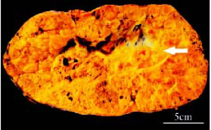
Gross examination (Figure 2) of the massively larged explanted liver (5,470 g) showed that the en-tire right lobe and one half of the left lobe replaced by a large multinodular tumour measuring 27 cm (transverse) x 22 cm (anterior to posterior) x 12 cm (superior to inferior). The nodules were white, ye-llow with focal hemorrhages, central scarring and were separated by thin fibrous septae. A large 8 cm
cyst was seen in the centre of the lesion. The majori-ty of the tumour was grossly viable. There was no rupture of the tumour capsule and there was no gross invasion into the porta hepatis, region of the bile duct and portal vein, and hepatic vein.
Microscopic examination showed a hepatic neo-plasm with an unusual growth pattern with two se-parate histological patterns that blended into one another arising in a background of non-cirrhotic li-ver. The first pattern had well-developed tubuloglan-dular architecture with blood and eosinophilic
secretions filling tubular spaces (Figures 3A and 3B). Tubules were lined by neoplastic cuboidal cells with large vesicular nuclei with prominent nucleoli and a moderate amount of eosinophilic granular cytoplasm. This component of the tumour had scan-ty mitoses (maximum of 3 per 10 high power fields) and the tubuloglandular structures were arranged in a back-to-back pattern with minimal or no inter-vening stroma or desmoplastic reaction. The second pattern observed was a solid sheet-like pattern punc-tuated by delicate fibrovascular cores which impar-ted focally a papillary appearance. In this solid portion of the tumour, there were some areas with a trabecular architecture. The sheets of cells in the so-lid component had identical morphology to the cells in the tubuloglandular area. There was no tumour necrosis and no vascular invasion. Scanty possible mucin in the lumina of some tubules as well as in-tracytoplasmic globules was demonstrated by PAS diastase stain. The mucicarmine stain and immuno-histochemical stains for mucin (MUC-1, MUC-2, MUC-5AC) however failed to show positivity for mu-cin. In both components, neuroendocrine markers were positive (synaptophysin strongly positive, chromogranin focally positive, CD56 weakly focally positive) as was MOC-31 (in membrane fashion), CK7, CK-19, but the tumour was negative for CEA, Hepar-1, CK20, TTF-1, thyroglobulin and CD10 (Fi-gures 3C-3H). The Ki-67 rate on average was 15% (focally up to 25%).
Electron microscopy was performed and in both the solid sheet-like as well as tubuloglandular por-tions of tumour, electron dense neurosecretory vesi-cles were demonstrated (Figure 4). Intriguingly, in
both areas, the neoplastic cells showed similar ultrastructural features of microvilli formation and cells were attached to one another through tight junctions in their cell membranes.
Extensive workup to exclude a primary tumour elsewhere was negative. Given the lack of marked nuclear atypia and necrosis, and an average of 3 mi-tosis per 10 high power fields and 15% Ki-67 index, the patient was diagnosed with a primary hepatic neuroendocrine tumour, grade 2 (see later for gra-ding criteria).
DISCUSSION
Primary neuroendocrine tumours of the liver are extremely rare. The diagnosis of this tumour poses a challenge to clinicians, radiologists, and patholo-gists. Not only is it extremely rare with non-specific clinical and radiographic presentation, but it also is a heterogeneous entity histologically. In this pa-tient, the radiographic diagnosis of Echinococcus in-fection followed by hepatocellular carcinoma, and the diagnosis of cholangiocarcinoma based on initial biopsies highlights this diagnostic challenge.
pancreatic or adrenal origin, neuroendocrine cells from within the intrahepatic biliary tree, or from neuroendocrine-programmed ectoblasts.11,12
Microscopically neuroendocrine tumours show polygonal, monomorphic cells with finely granular nuclear chromatic with small or not visible nucleoli, and bland cytological features. However, some tu-mours can show relative pleomorphism. The type of growth pattern that is seen (A-insular/nested, B-tra-becular, C-glandular/acinar, D-poorly differentiated) may be characteristic for neuroendocrine tumours for specific sites. In our case the tumour showed trabecular as well as glandular/acinar patterns, sug-gesting that the neoplastic neuroendocrine cells are either of hindgut or foregut derivation.
Given the lack of universally accepted nomencla-ture of neuroendocrine tumours of the gastrointesti-nal tract, a new classification scheme was introduced by the WHO in 2010. The term carcinoid in the 1980 WHO, is now divided into 3 separate tiers: neuroendocrine tumour grade 1 (NET G1), NET G2 and neuroendocrine carcinoma (NEC). A NET is a well-differentiated neoplasm that is sepa-rated into G1 (< 2 mitosis per 10 high power fields, and/or ≤ 2% Ki-67 index) or G2 (2-20 mitosis per 10 high power fields, and/or 3-20% Ki-67 index) and en-compasses neoplasms previously termed ‘carcinoid tumour’. The high grade (G3) neuroendocrine neo-plasm which shows greater nuclear atypia, multifo-cal necrosis and greater mitoses (> 20 mitosis per 10 high power fields, and/or > 20% Ki-67 index) is now termed ‘neuroendocrine carcinoma’ and refers to neoplasms previously classified as small cell car-cinoma, large cell neuroendocrine carcar-cinoma, and poorly differentiated neuroendocrine carcinoma.13 In
our case, neoplastic cells showed mitotic count and Ki-67 index, consistent with a grade 2 NET.
The immunoprofile in this case was consistent with cells of a neuroendocrine origin (positive sy-naptophysin, chromogranin and CD56); however, ex-pression of markers of epithelial differentiation (MOC31, CK7 and CK19) were also observed. Given the rarity of primary hepatic neuroendocrine tu-mours, there is no literature available regarding ex-pression of epithelial markers in these tumours. However, expression of CK7 and CK19 in conjunction with neuroendocrine cell markers has been previous-ly reported in extrahepatic neuroendocrine gastroin-testinal tumours as well as neuroendocrine lung tumours. CK7 expression for example, has been shown in approximately 10% of GI (stomach and lar-ge intestine), 50% of pancreatic carcinoid tumours,14
and in the lung 5% of carcinoid tumours, 40% of
small cell carcinomas and 70% of large cell neuroen-docrine carcinomas.15 CK19 expression has also been
shown in 80% of carcinoids from the appendix16 and
in the lung 70% of carcinoid tumours and 80% of both small cell carcinomas and large cell neuroendo-crine carcinomas show CK19 expression.15
Additional evidence of neuroendocrine differentia-tion was provided through electron microscopic exa-mination. This method has been used in the past by other investigators to show uniform electron dense neurosecretory granules in the cytoplasm of neo-plastic cells.17-19 In our case, we also observed these
granules and also discovered that cells lining glan-dular cystic spaces had microvilli projecting in to lu-mens and these cells were attached to each other with tight junctions, a phenomenon which has been previously described.17
An important differential is cholangiocarcinoma, an entity which would require a much different che-motherapeutic approach should the lesion grow or metastasize. Typically, cholangiocarcinoma develops in a non-cirrhotic liver. Macroscopically it is classi-fied into three types (mass-forming, periductal infil-tration, and intraductal-growth type). Histologically cholangiocarcinoma are adenocarcinomas with tubu-lar growth being the most common pattern. Micro-papillary and acinar (or cord-like) growth patterns also occur. Tumour cells, which may be pleomor-phic, are usually small to medium sized, cuboidal or columnar, with small nuclei and nucleoli and pale eosinophilic cytoplasm. An important characteristic is the presence of fibrous stroma with the central portion of the tumour being sclerotic and hyalini-zed. Histochemical stains such as mucicarmine, PASD, Alcian blue, and immunohistochemical stains for mucin core (MUC) proteins are positive. Tumour cells are also immunoreactive for cytokera-tins, such as CK7 and CK19.
In summary, primary hepatic neuroendocrine tu-mours are rare tutu-mours that effect relatively young patients and typically do not present with the carci-noid syndrome. They typically arise in a background of non-cirrhotic liver with diagnosis requiring routi-ne hematoxylin and eosin as well as ancillary studies (primarily immunohistochemistry) demonstrating evidence of neuroendocrine differentiation. The exact etiology remains elusive; our knowledge on the range of morphology is still gaining experience and warrants further investigation.
REFERENCES
2. Modlin IM, Sandor A. An analysis of 8305 cases of carcinoid tumors. Cancer 1997; 79: 813-29.
3. Modlin IM, Shapiro MD, Kidd M. An analysis of rare carci-noid tumors: clarifying these clinical conundrums. World J Surg 2005; 29: 92-101.
4. Fenoglio LM, Severini S, Ferrigno D, Gollè G, Serraino C, Bracco C, Castagna E, et al. Primary hepatic carcinoid: a case report and literature review. World J Gastroente-rol 2009; 15: 2418-22.
5. Lin CW, Lai CH, Hsu CC, Hsu CT, Hsieh PM, Hung KC, Chen YS. Primary hepatic carcinoid tumor: a case report and review of the literature. Cases J 2009; 2: 90.
6. Schwartz G, Colanta A, Gaetz H, Olichney J, Attiyeh F. Primary carcinoid tumors of the liver. World J Surg Oncol
2008; 6: 91.
7. Zhu H, Sun K, Ward SC, Schwartz M, Thung SN, Qin L. Pri-mary hepatic signet ring cell neuroendocrine tumor: a case report with literature review. Semin Liver Dis 2010; 30: 422-8 [Epub 2010 Oct 19].
8. Mima K, Beppu T, Murata A, Otao R, Miyake K, Okabe H, Masuda T, et al. Primary neuroendocrine tumor in the li-ver treated by hepatectomy: report of a case. Surg Today
2011; 4: 1655-60 [Epub 2011 Oct 4].
9. Pilichowska M, Kimura N, Ouchi A, Lin H, Mizuno Y, Nagura H. Primary hepatic carcinoid and neuroendocrine carcino-ma: clinicopathological and immunohistochemical study of five cases. Pathol Int 1999; 49: 318-24.
10. Fenwick SW, Wyatt JI, Toogood GJ, Lodge JP. Hepatic re-section and transplantation for primary carcinoid tumors of the liver. Ann Surg 2004; 239: 210-9.
11. Gravante G, De Liguori Carino N, Overton J, Manzia TM, Orlando G. Primary carcinoids of the liver: a review of
symptoms, diagnosis and treatments. Dig Surg 2008; 25: 364-8 [Epub 2008 Nov 4].
12. Nikfarjam M, Muralidharan V, Christophi C. Primary hepa-tic carcinoid tumours. HPB (Oxford) 2004; 6: 13-7. 13. Bosman FT, Carneiro F, Hruban RH, Theise ND. WHO
Classification of Tumours of the Digestive System. 4th. Ed. Lyon: International Agency for Research on Cancer; 2010.
14. Cai YC, Banner B, Glickman J, Odze RD. Cytokeratin 7 and 20 and thyroid transcription factor 1 can help distinguish pulmonary from gastrointestinal carcinoid and pancreatic endocrine tumors. Hum Pathol 2001; 32: 1087-93. 15. Jerome Marson V, Mazieres J, Groussard O, Garcia O,
Ber-jaud J, Dahan M, Carles P. Expression of TTF-1 and cytokeratins in primary and secondary epithelial lung tu-mours: correlation with histological type and grade.
Histopathology 2004; 45: 125-34.
16. Alsaad KO, Serra S, Perren A, Hsieh E, Chetty R. CK19 and CD99 immunoexpression profile in goblet cell (mucin-produ-cing neuroendocrine tumors) and classical carcinoids of the vermiform appendix. Int J Surg Pathol 2007; 15: 252-7. 17. Andreola S, Lombardi L, Audisio RA, Mazzaferro V, Koukouras D, Doci R, Gennari L, et al. A clinicopathologic study of primary hepatic carcinoid tumors. Cancer 1990; 65: 1211-8.
18. Oh YH, Kang GH, Kim OJ. Primary hepatic carcinoid tumor with a paranuclear clear zone: a case report. J Korean Med Sci 1998; 13: 317-20.