Response to Loco-Regional Therapy Predicts
Outcomes After Liver Transplantation for Combined
Hepatocellular-Cholangiocarcinoma
Samuel O. Antwi,**,♦ Yacob Y. Habboush,*,♦ Lori A. Chase,* David D. Lee,* Tushar Patel* * Department of Transplantation, Mayo Clinic, Jacksonville, FL, USA. ** Department of Health Sciences Research, Mayo Clinic, Jacksonville, FL, USA. ♦ These individuals contributed equally. November-December, Vol. 17 No. 6, 2018: 969-979
The Official Journal of the Mexican Association of Hepatology, the Latin-American Association for Study of the Liver and
the Canadian Association for the Study of the Liver
Manuscript received: Manuscript received:Manuscript received:
Manuscript received:Manuscript received: November 13, 2017. Manuscript accepted:Manuscript accepted:Manuscript accepted:Manuscript accepted:Manuscript accepted: January 25, 2018. DOI:10.5604/01.3001.0012.2232
A B S T R A C T A B S T R A C T A B S T R A C T A B S T R A C T A B S T R A C T
Introduction and aim. Introduction and aim.Introduction and aim. Introduction and aim.
Introduction and aim. Combined hepatocellular-cholangiocarcinoma (HCC-CCA) is a rare liver malignancy distinct from either hepatocellular carcinoma (HCC) or cholangiocarcinoma. Liver transplantation (LT) is not recommended for HCC-CCA because of suboptimal outcomes. Non-invasive diagnosis of HCC-CCA is extremely challenging; thus, some HCC-CCAs are presumed as HCC on imaging and listed for LT with the correct diagnosis ultimately made on explant pathology. We compared HCC-CCA with HCC to determine the utility of response to pre-transplant loco-regional therapy (LRT) in predicting outcomes for HCC-CCA after LT as a potential means of identifying appropriate HCC-CCA patients for LT. Material and methods.Material and methods.Material and methods.Material and methods.Material and methods. Retrospective review of 19 pa-tients with pathologically confirmed HCC-CCA were individually matched to 38 HCC papa-tients (1:2) based on age, sex, and Milan cri-teria at listing was performed. The modified response evaluation cricri-teria in solid tumors was used to categorize patients as responders or non-responders to ptransplant LRT based on imaging performed before and after LRT. Overall survival (OS) and re-currence-free survival (RFS) were examined. Results.Results.Results.Results.Results. OS at 3 years post-transplant was 74% for HCC-CCA and 87% for HCC. RFS at 3 years was 74% for HCC-CCA, and 87% for HCC. Among responders to LRT, the 3-year OS was 92% for HCC-CCA and 88% for HCC; among non-responders, 3-year OS was 43% for HCC-CCA and 83% for HCC. Higher 3-year OS was observed among HCC-CCA responders (77%) compared with HCC-CCA non-responders (23%). Conclusions.Conclusions.Conclusions.Conclusions.Conclusions. OS was similarly high among responders to pre-transplant LRT irrespective of tumor type. Radiologic response to LRT could potentially be used to select appropri-ate HCC-CCA patients for LT if the findings are validappropri-ated in independent studies.
Key words. Key words.Key words. Key words.
Key words. Loco-regional therapy. LRT. combined hepatocellular and cholangiocarcinoma. HCC. Overall survival. Recurrence-free survival.
INTRODUCTION
Combined hepatocellular-cholangiocarcinoma (HCC-CCA) is a rare primary liver cancer, with histopathologi-cal features that overlap both hepatocellular carcinoma (HCC) and intrahepatic cholangiocarcinoma (iCCA).1 HCC-CCA accounts for some 1-5% of primary liver can-cers and is distinct from HCC and iCCA, in that, it is more aggressive and associated with worse prognosis.1-6 The optimal management of HCC-CCA is not well estab-lished, due to the lack of specific and definitive non-inva-sive diagnostic criteria, and the rarity of this malignancy. There are limited data on the benefits of liver transplanta-tion (LT) in HCC-CCA patients. At present, HCC-CCA
is not an indication for LT due, in part, to the absence of data supporting improved prognosis following LT and aggressive disease course observed in practice.7,8
liv-er. Such incidentally identified HCC-CCA may occur in 1-2% of transplants for HCC.13,14 The pathogenesis of HCC-CCA is not completely understood, and tumors could arise from either dual differentiation along hepato-cellular and cholangiohepato-cellular lineages or from malignant transformation of hepatic progenitor cells (HPC).15 The World Health Organization (WHO) classification divides HCC-CCA into two histologically distinguished groups, classical type and subtypes with stem cell features.16,17 A recent molecular profiling of HCC-CCA by Moeini, et al. showed that HCC-CCA is a heterogeneous disease with subtypes that include cholangiocellular carcinoma and a stem cell-derived subtype that is more aggressive and asso-ciated with worse prognosis.18
Co-presentation of HCC with iCCA on explant pa-thology can be categorized into three distinct types. Type I, or collision tumors, consist of contiguous or separate HCC and CCA that exist coincidentally. These tumors have mucin production and lack transitional zone be-tween different tumor types. Type II tumors have a transi-tional area between HCC and CCA where a mixture of cells with hepatocellular and cholangiocellular differenti-ation are present. These tumors have bile and/or mucin se-cretion. Type III, or fibrolamellar tumors, have a combination and mingling of cellular features of hepato-cellular and cholangiohepato-cellular differentiation. These tu-mors have mucin-producing pseudoglands and larger cells with eosinophilic granular cytoplasm. Compared to the other types, type III is more prevalent in younger patients but it has a longer survival after diagnosis if left untreat-ed.19-22 Other histological classifications have been pro-posed but have not been shown to provide prognostic insight.23
Management of HCC-CCA patients include the use of Loco-regional therapy (LRT) with or without surgical re-section.7,22,24 Patients with HCC awaiting LT may undergo LRT, such as trans-arterial chemoembolization and radi-ofrequency ablation to control tumor growth while
await-ing a replacement organ.25 Pre-transplant response to LRT has been associated with a post-transplant survival benefit for HCC, and proposed as a proxy for tumor biology.26 Al-though response to LRT could be useful for identifying a subset of HCC-CCA patients who will have favorable out-comes from LT, the prognostic value of response to LRT for HCC-CCA is currently unknown.27 The purpose of this study was to investigate whether radiologic response to pre-transplant LRT could predict overall survival (OS) and recurrence-free survival (RFS) after LT for HCC-CCA by comparing HCC-HCC-CCA patients with HCC pa-tients.
MATERIAL AND METHODS
Study subjects
Following approval by our Institutional Review Board, we identified patients who underwent orthotopic LT for presumed HCC at our institution, between January 01, 2001 and October 31, 2016, from a prospective database and reviewed their medical records. Patients were eligible for the study if they received neoadjuvant LRT and had magnetic resonance imaging (MRI) scan performed be-fore and after LRT to assess treatment response. Pre-trans-plant diagnosis of HCC was determined by a dedicated team of oncologists, hepathologist, radiologists and liver transplant surgeons, following the American Association for the Study of Liver Diseases practice guidelines.8 Based on the medical records review, 19 incidentally transplant-ed HCC-CCA cases were identifitransplant-ed from explant pathol-ogy reports. Each HCC-CCA case was matched to 2 pathologically confirmed HCC cases diagnosed at the same institution and over the same period, based on age (± 5 years), sex, and the Milan criteria used for selecting patients for orthotopic LT (i.e., 1 lesion ≤ 5 cm, or 2-3 le-sions each ≤ 3 cm and no vascular invasion).28 Patients who did not have an imaging prior to and post LRT, did
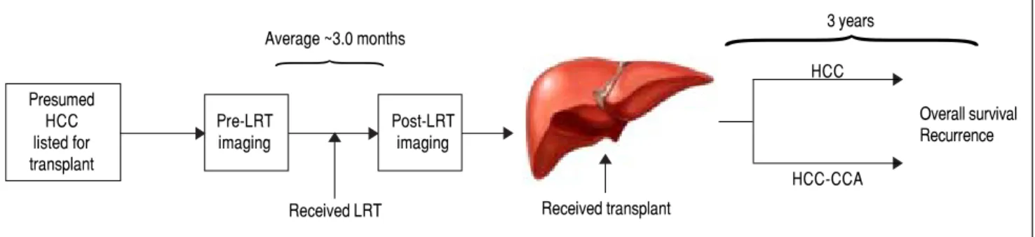
Figure 1. Figure 1. Figure 1. Figure 1.
Figure 1. Study overview. Time intervals are reported as the average months among hepatocellular carcinoma (HCC) and combined hepatocellular-cholangi-ocarcinoma (HCC-CCA) patients. All patients were presumed to have hepatocellular carcinoma (HCC) at diagnosis, received loco-regional therapy (LRT) with pre- and post-treatment magnetic resonance imaging (MRI) scan to access response to treatment, followed by liver transplantation. Patients were followed for up to 3 years.
Presumed
HCC Pre-LRT Post-LRT
listed for imaging imaging
transplant
Average ~3.0 months
Received LRT Received transplant
3 years
HCC
HCC-CCA
Overall survival Recurrence
not undergo LT, or did not have a diagnosed hepatic mass were excludid from this study. The study population in-cluded 19 HCC-CCA cases and 38 HCC control patients. This study was approved by the Mayo Clinic Institutional Review Board (IRB 12-004111).
An overview of the study design is provided in figure 1. In brief, all patients had presumed HCC and were listed for LT. All patients received neoadjuvant LRT with an av-erage 3-month interval between pre- and post-imaging LRT for assessment of treatment response. The patients then underwent LT and histopathological evaluation was performed on the explanted liver for definitive diagnosis. Although some patients had evidence of complete radio-logical response to pre-transplant LRT, none had a com-plete pathological response of target lesions where histological confirmation of tumor type could not be made. All patients were followed for up to 3 years for as-sessment of recurrence and vital status.
Data collection
Data abstracted from patients’ medical records includ-ed demographics (e.g., age and sex), MELD (model for end-stage liver disease) score at listing for LT including exception points, native MELD score without exception points, number and size of tumor lesions identified on imaging, tumor vascular invasion, type of LRT received, dates of pre- and post-LRT imaging used to assess treat-ment response, dates of LT, time between post-LRT im-aging and LT, and vital and recurrence status at 1 and 3 years following LT. The number and size of hepatic le-sions and evidence of tumor invasion or extrahepatic spread were used to determine whether or not a patient met the Milan criteria. Response to LRT was determined by hepatic MRI performed before and after LRT, but prior to LT. The modified response evaluation criteria in solid tumors (mRECIST) were used to assess treatment sponse and categorized as complete response, partial re-sponse, stable disease, or progressive disease.29 In mRECIST, complete response is defined as disappearance of all intratumoral arterial enhancements in all target le-sions, partial response as at least a 30% decrease in the sum of the diameters of viable target lesions, stable disease as neither growing nor shrinking lesions, and progressive disease as an increase of 20% or more in diameter of target viable lesions.29,30 Multiple tumors were present in 26 pa-tients (8 in HCC-CCA group, and 18 in the HCC groups). In such instances, the sum of the diameter of viable le-sions assessed before and after LRT was averaged to one response category for each patient and classified as a com-plete radiologic response, partial radiologic response, sta-ble disease, or progressive disease. To ensure more meaningful interpretation of the data given the small
num-bers, patients with complete or partial response to LRT were combined into a “responders” group, while patients with stable or progressive disease after LRT were com-bined into a “non-responders” group.
Data analysis
Means and proportions were used to compare demo-graphic and clinical attributes between the HCC-CCA and the HCC only patients, using Students’ t-tests for continuous variable and chi-square tests for categorical variables. OS and RFS rates were assessed at 1-year and at 3-years following LT using Fisher’s exact tests to compare the HCC-CCA patients with the HCC patients. Separate analyses were performed among responders to pre-trans-plant LRT and non-responders assessing both OS and RFS by comparing rates between HCC-CCA and HCC pa-tients in each of the treatment response groups. For the as-sessment of RFS, patients who died over the 3-year study period were censored at the time of death. Kaplan-Meier survival curves were used to describe OS and RFS pat-terns over the 3-year follow-up period, using log-rank tests to examine differences in survival patterns between the HCC-CCA and HCC patients, and stratified by the treatment response groups. We also compared OS and RFS between HCC-CCA responders versus HCC-CCA non-responders. In examining the survival rates and pat-terns, OS was defined by the length of time (months) from the date of LT to date of death or date of last follow-up, while RFS was defined by time from transplant date to date of recurrence, date of death, or date of the last follow-up, whichever occurred first. All statistical tests were two-sided, and a p-value < 0.05 was considered statistically significant. Given the rarity of HCC-CCA,1 reflected by our small sample size, we considered p-values lower than 0.15 as marginally significant. All analyses were performed in SAS® version 9.4 (SAS Institute, Cary, NC, USA).
RESULTS
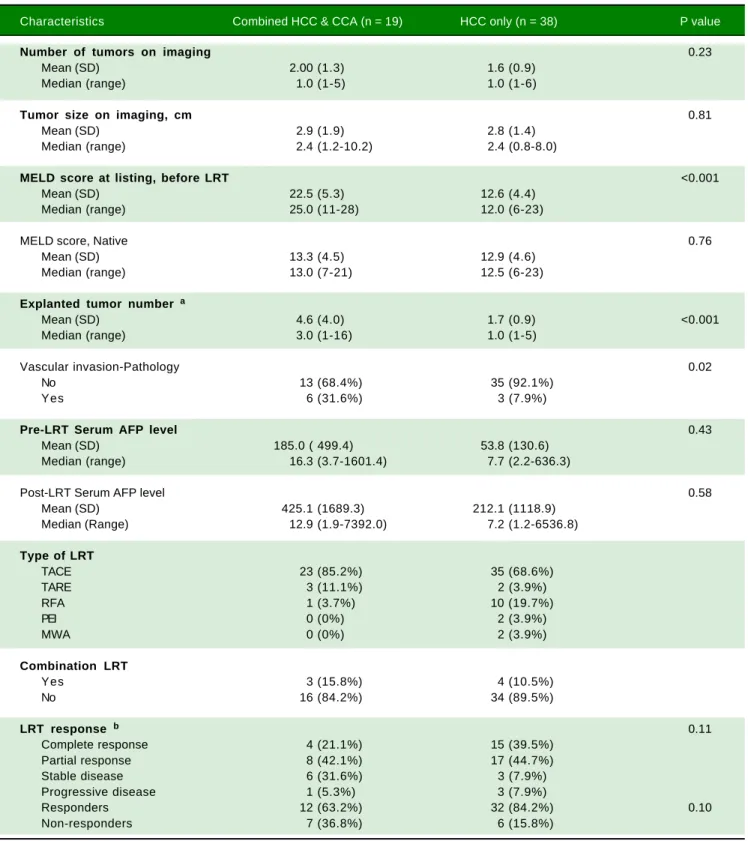
Because of the individual matching design, the HCC-CCA and HCC patients did not differ in age, sex, mean tu-mor number from pre-LT imaging, mean tutu-mor size from pre-LT imaging, or the proportion that were within the Milan criteria. Five HCC-CCA and 10 HCC patients were downstaged to within Milan criteria following LRT and before LT. Only one HCC-CCA patient received post-LT chemotherapy and none of the HCC patients re-ceived post-LT chemotherapy. The types of LRT rere-ceived by the patients were transcatheter arterial chemoemboli-zation (TACE), transarterial radioembolichemoemboli-zation (TARE), radiofrequency ablation (RFA), percutaneous ethanol abla-tion (PEI), and microicrowave ablaabla-tion (MWA). Three HCC-CCA patients had a combination of TACE with TARE, while four HCC patients had combination LRT of TACE with RFA (two), TARE (one) or MWA (one) (Ta-ble 2). Among the 19 CCA patients, five had HCC-CCA and concurrent HCC, six had HCC-CCA and HCC, seven had HCC-CCA only, and one had HCC-CCA and con-current CCA pathologically identified in the explanted liver.
On average, patients with HCC-CCA had 2 lesions with a mean diameter of 2.9 cm, while those with HCC had 2 lesions with a mean diameter of 2.8 cm. Compared with the HCC patients, the HCC-CCA patients had
high-er MELD score at listing, before LRT (22 vs. 13), a highhigh-er number of tumors on explanted liver (5 vs. 2), and were more likely to have a vascular invasion on histopathologi-cal report (32 vs. 8%) (Table 2). However, they did not dif-fer on native MELD score (without exception points) or serum alpha-fetoprotein level post-LRT. There was a higher proportion of pre-transplant LRT responders among HCC patients (84%) than among HCC-CCA pa-tients (63%), but the two groups were similar in terms of the time between pre- and post-LRT imaging, the number of LRT performed, or the time between post-LRT ing and LT. The average interval between post-LRT imag-ing and transplantation was 3.2 months for HCC-CCA patients and 2.4 months for HCC, with a combined aver-age of 3 months. The HCC-CCA patients underwent an average of 1.4 LRT treatments, whereas those with HCC underwent an average of 1.3 LRT treatments prior to LT.
The OS and RFS rates among the HCC-CCA and the HCC patients were examined after 1 and 3 years of fol-low-up (Table 3). At 1 year of folfol-low-up, OS was 84% among patients with HCC-CCA and 95% among patients with HCC (p-value = 0.18). At 3 years, the OS rates were 74% for HCC-CCA and 87% for HCC (p-value = 0.22). RFS rates at 1 and 3 years were 79% and 74%, respectively, for HCC-CCA and 92% and 87%, respectively, for the
Table 1. Descriptive characteristics of study participants (n = 57).
Characteristics HCC-CCA (n = 19) HCC only (n = 38) P value
Age group, years 1.00
< 50 2 (10.5%) 4 (10.5%)
50-54 1 (5.3%) 2 (5.3%)
55-59 3 (15.8%) 6 (15.8%)
60-64 5 (26.3%) 10 (26.3%)
65-69 5 (26.3%) 10 (26.3%)
70+ 3 (15.8%) 6 (15.8%)
Mean (SD) 61.5 (10.5) 61.9 (7.5) 0.86
Median (range) 64.0 (30.0-76.0) 63.5 (46.0-75.0)
Sex 1.00
Female 4 (21.1%) 8 (21.1%)
Male 15 (78.9%) 30 (78.9%)
Within Milan criteria at listing 1.00
Yes 14 (73.7%) 28 (73.7%)
No 5 (26.3%) 10 (26.3%)
Etiology
Hepatitis C 12 (63.1%) 22 (57.9%) 0.65
Hepatitis B 0 (0%) 3 (7.9%)
Alcohol 2 (10.5%) 4 (10.5%)
NASH 1 (5.3%) 1 (2.6)
Primary sclerosing cholangitis 1 (5.3%) 0 (0%)
Primary biliary cirrhosis 0 (0%) 2 (5.3%)
Cryptogenic cirrhosis 3 (15.8%) 6 (15.8%)
Table 2. Tumor features and treatment response among study participants (n = 57).
Characteristics Combined HCC & CCA (n = 19) HCC only (n = 38) P value
Number of tumors on imaging 0.23
Mean (SD) 2.00 (1.3) 1.6 (0.9)
Median (range) 1.0 (1-5) 1.0 (1-6)
Tumor size on imaging, cm 0.81
Mean (SD) 2.9 (1.9) 2.8 (1.4)
Median (range) 2.4 (1.2-10.2) 2.4 (0.8-8.0)
MELD score at listing, before LRT <0.001
Mean (SD) 22.5 (5.3) 12.6 (4.4)
Median (range) 25.0 (11-28) 12.0 (6-23)
MELD score, Native 0.76
Mean (SD) 13.3 (4.5) 12.9 (4.6)
Median (range) 13.0 (7-21) 12.5 (6-23)
Explanted tumor number a
Mean (SD) 4.6 (4.0) 1.7 (0.9) <0.001
Median (range) 3.0 (1-16) 1.0 (1-5)
Vascular invasion-Pathology 0.02
No 13 (68.4%) 35 (92.1%)
Yes 6 (31.6%) 3 (7.9%)
Pre-LRT Serum AFP level 0.43
Mean (SD) 185.0 ( 499.4) 53.8 (130.6)
Median (range) 16.3 (3.7-1601.4) 7.7 (2.2-636.3)
Post-LRT Serum AFP level 0.58
Mean (SD) 425.1 (1689.3) 212.1 (1118.9)
Median (Range) 12.9 (1.9-7392.0) 7.2 (1.2-6536.8)
Type of LRT
TACE 23 (85.2%) 35 (68.6%)
TARE 3 (11.1%) 2 (3.9%)
RFA 1 (3.7%) 10 (19.7%)
PEI 0 (0%) 2 (3.9%)
MWA 0 (0%) 2 (3.9%)
Combination LRT
Yes 3 (15.8%) 4 (10.5%)
No 16 (84.2%) 34 (89.5%)
LRT response b 0.11
Complete response 4 (21.1%) 15 (39.5%)
Partial response 8 (42.1%) 17 (44.7%)
Stable disease 6 (31.6%) 3 (7.9%)
Progressive disease 1 (5.3%) 3 (7.9%)
Responders 12 (63.2%) 32 (84.2%) 0.10
Non-responders 7 (36.8%) 6 (15.8%)
HCC patients (p-values ≥ 0.15). As shown in figure 2, OS and RFS were better across follow-up for HCC than for HCC-CCA patients (log-rank p-values for OS and RFS were each = 0.04).
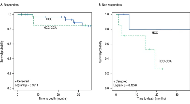
The results for overall and RFS rates were stratified by response to ptransplant LRT (Table 4). Among re-sponders, the OS rate at 1-year was 92% for HCC-CCA and 97% for HCC (p-value = 0.46). In comparison, 1-year OS rates were poorer among non-responders regardless of tumor type, with a 71% OS rate for HCC-CCA and 83% for HCC. The difference between HCC and HCC-CCA
survival was not statistically significant (p-value = 0.61). Three-year OS was also inferior among non-responders compared to responders, and was much worse for non-responders with HCC-CCA, such that while the 3-year OS among responders was 92% for HCC-CCA and 88% for HCC (p-value = 0.70), among non-responders, the 3-year OS rates were 43% for HCC-CCA and 83% for HCC (p-value = 0.13). Figure 3 demonstrates compara-ble OS pattern between HCC-CCA and HCC with no statistically significant difference between patients who responded to LRT with either HCC or HCC-CCA
Table 3. Overall survival and recurrence rates among patients with combined hepatocellular-cholangiocarcinoma (HCC-CCA), and pa-tients with hepatocellular carcinoma (HCC) only.
Tumor type Overall Survival (n = 57)
1-year 3-year
Dead (n = 5) Alive (n = 52) P-value Dead (n = 10) Alive (n = 47) P-value
HCC-CCA 3 (16%) 16 (84%) 0.18 5 (26%) 14 (74%) 0.22
HCC only 2 (5%) 36 (95%) 5 (13%) 33 (87%)
Recurrence (n = 57)
Recurrence Recurrence-free Recurrence Recurrence-free
HCC-CCA 4 (21%) 15 (79%) 0.15 5 (26%) 14 (74%) 0.21
HCC only 3 (8%) 35 (92%) 5 (13%) 33 (87%)
Percentages are presented as row percent because of the 1:2 matching of HCC-CCA and HCC patients, respectively.
Figure 2. Figure 2. Figure 2. Figure 2.
Figure 2. Kaplan-Meier survival curves for (AAAAA) overall survival and (BBBBB) recurrence-free survival by tumor type. Solid blue line: hepatocellular carcinoma (HCC); Dashed green line: combined hepatocellular-cholangiocarcinoma (HCC-CCA).
A. A. A. A.
A. Overall survival. B.B.B.B.B. Recurrence-free survival.
+ Censored Logrank p = 0.0455 1.0
0.8
0.6
0.4
0.2
0.0
Survival probability
0 10 20 30
Time to death (months)
1.0
0.8
0.6
0.4
0.2
0.0
Survival probability
0 10 20 30
Time to recurrence (months) HCC
HCC-CCA
+ Censored Logrank p = 0.0446
HCC
(log-rank p-value = 0.99). Among non-responders; how-ever, the curves show a trend towards worse survival in the HCC-CCA group as compared with the HCC group (log-rank p-value = 0.13).
Among responders to pre-transplant LRT, the 1- and 3-year RFS rates were respectively 83% and 75% for patients with HCC-CCA, and 97% and 91% for HCC patients. Non-responders with HCC-CCA and HCC had equally poor RFS, with 1- and 3-year recurrence-free rates of 71% and 57%, respectively, for HCC-CCA and corresponding rates of 67% and 50% for HCC patients. In line with these results, the RFS curves (Figure 4) show a slightly better RFS across follow-up for responders with HCC than for responders with HCC-CCA (log-rank p-value = 0.08). In
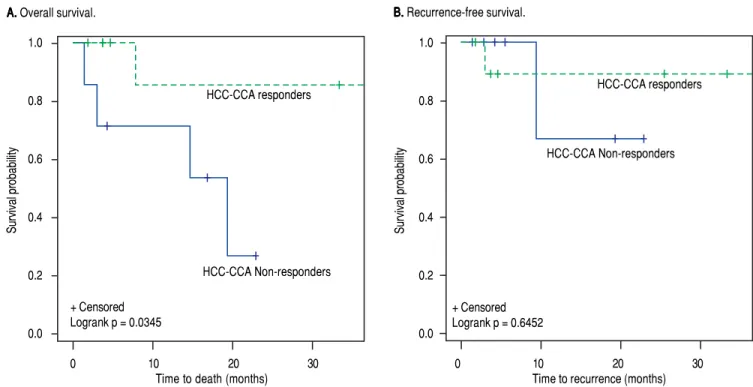
contrast, the RFS curves show similarly dismal survival across follow-up among non-responders with HCC-CCA and HCC (log-rank p-value = 0.94). Further analyses showed a generally higher survival rates among HCC-CCA responders compared to HCC-HCC-CCA non-respond-ers. Three-year overall survival was particularly higher among HCC-CCA responders (77%) compared with the HCC-CCA non-responders (23%) (p-value = 0.07). We also found a significantly better survival across follow-up among HCC-CCA responders than among HCC-CCA non-responders, with log-rank p-value = 0.03 (Figure 5). However, RFS did not differ significantly between the HCC-CCA responders and HCC-CCA non-responders (Table 4 and Figure 5).
Table 4. Rates of survival and recurrence among patients with combined hepatocellular-cholangiocarcinoma (HCC-CCA), and patients with hepatocellular carcinoma (HCC) only, stratified by response to loco-regional therapy (LRT).
LRT Response Groups * Overall survival (n = 57)
1-year 3-year
Dead Alive P-value Dead Alive P-value
Responders (n = 44) Tumor type
HCC-CCA 1 (8%) 11 (92%) 0.46 1 (8%) 11 (92%) 0.70
HCC 1 (3%) 32(97%) 4 (12%) 28 (88%)
Non-responders (n = 13) Tumor type
HCC-CA 2 (29%) 5 (71%) 0.61 4 (57%) 3 (43%) 0.13
HCC 1 (17%) 5 (83%) 1 (17%) 5 (83%)
Recurrence (n = 57)
Recurrence Recurrence-free Recurrence Recurrence-free
Responders (n = 44) Tumor type
HCC-CCA 2 (17%) 10 (83%) 0.11 3 (25%) 9 (75%) 0.18
HCC 1 (3%) 31 (97%) 3 (9%) 29 (91%)
Non-responders (n = 13) Tumor type
HCC-CCA 2 (29%) 5 (71%) 0.85 3 (43%) 4 (57%) 0.71
HCC 2 (33%) 4 (67%) 3(50%) 3 (50%)
Overall survival (n = 19)
Dead Alive Dead Alive
HCC-CCA only
Responders (n = 12) 1 (33%) 11 (69%) 0.24 2 (33%) 10 (77%) 0.07
Non-responders (n = 7) 2 (67%) 5 (31%) 4 (67%) 3 (23%)
Recurrence (n = 19)
Recurrence Recurrence-free Recurrence Recurrence-free
HCC-CCA only
Responders (n = 12) 1 (50%) 11 (65%) 0.68 1 (33%) 11 (69%) 0.43
Non-responders (n = 7) 1 (50%) 6 (35%) 2 (67%) 5 (31%)
Figure 4. Figure 4. Figure 4. Figure 4.
Figure 4. Recurrence-free survival curves among (AAAAA) responders and (BBBBB) non-responders to loco-regional therapy. Solid blue line: hepatocellular carcinoma (HCC). Dashed green line: combined hepatocellular-cholangiocarcinoma (HCC-CCA).
A. A. A. A.
A. Responders. B.B.B.B.B. Non-responders.
Figure 3. Figure 3. Figure 3. Figure 3.
Figure 3. Overall survival curves among (AAAAA) responders and (BBBBB) non-responders to loco-regional therapy. Solid blue line: hepatocellular carcinoma (HCC). Dashed green line: combined hepatocellular-cholangiocarcinoma (HCC-CCA).
A. A. A. A.
A. Responders. B.B.B.B.B. Non-responders.
1.0
0.8
0.6
0.4
0.2
0.0
Survival probability
0 10 20 30
Time to death (months) HCC
HCC-CCA
+ Censored Logrank p = 0.9911
1.0
0.8
0.6
0.4
0.2
0.0
Survival probability
0 10 20 30
Time to death (months) HCC
HCC-CCA
+ Censored Logrank p = 0.1270
1.0
0.8
0.6
0.4
0.2
0.0
Survival probability
0 10 20 30
Time to recurrence (months)
HCC
HCC-CCA
+ Censored Logrank p = 0.0791
1.0
0.8
0.6
0.4
0.2
0.0
Survival probability
0 10 20 30
Time to recurrence (months) HCC
HCC-CCA
DISCUSSION
Patients with HCC-CCA are usually excluded from list-ing for LT due to the generally poor outcomes. To our knowledge, this is the largest study of treatment response on survival outcomes among HCC-CCA patients conduct-ed to date. Our findings are consistent with the reportconduct-ed poor outcomes of HCC-CCA, in that, patients who were incidentally found to have HCC-CCA post-LT had worse OS and RFS as compared with individually matched HCC patients. These may be due, in part, to greater vascular inva-sion among the HCC-CCA patients. We found also that among patients who had a complete or partial response to neoadjuvant LRT, OS rates were equally favorable for HCC and HCC-CCA patients. These findings suggest that re-sponse to LRT could predict OS and RFS following LT, pending verification in independent samples, leading us to propose that a favorable response to LRT could potentially be used to identify those HCC-CCA patients who may ben-efit from LT.
LT provides the most favorable survival outcome for patients with primary liver cancer and even for patients with HCC-CCA, survival after LT is better than with any other therapeutic modality. Unfortunately, the scarcity of donor liver allografts and the high mortality in patients with primary liver failure have put restrictions on which patients can be considered for expedited access to donor
organs in the setting of cancer.31 As a benchmark, LT of-fers a 4-year survival of ~85% for HCC within the Milan criteria. Many centers have modified these criteria by ex-panding the tumor number and size criteria for HCC or by including patients with other types of primary liver can-cers (e.g., CCA) and have demonstrated similarly favora-ble outcomes in carefully selected patients.32,33 Response to LRT has been proposed by a number of groups to be used as a selective tool for patients with HCC because it appears to act as a surrogate for “favorable tumor biolo-gy”.27 A recent meta-analysis of incidental HCC-CCA upon explant, showed a wide 3- year RFS of 33-86% and OS of 22-70%, in comparison, the current study shows 3-year OS and RFS of 74% each.34 The heterogeneity of tu-mor behavior supports the use of criteria based on tutu-mor biology rather than arbitrary size and number to identify candidates who will equally benefit from the life-saving intervention of LT and have excellent outcomes to justify expedited access to the scarce resource of a liver trans-plant. Our findings show that the response to LRT could identify suitable candidates with HCC-CCA who may have favorable OS after LT. To our knowledge, this is the first such report to suggest these findings.
The greatest challenge to any potential prospective trial investigating the benefits of LT for patients with HCC-CCA is the limited ability to accurately diagnose these can-cers in the pre-operative setting. The consistency between
Figure 5. Figure 5.Figure 5.
Figure 5.Figure 5. Kaplan-Meier survival curves for (AAAA) overall survival and (BA BBB) recurrence-free survival among combined hepatocellular-cholangiocarcinoma (HCC-B CCA) patients who responded to loco-regional therapy (LRT) vs. HCC-CCA patients who did not respond to LRT. Solid blue line: HCC-CCA responders to LRT. Dashed green line: HCC-CCA non-responders to LRT.
A. A.A.
A.A. Overall survival. B.B.B. Recurrence-free survival.B.B. 1.0
0.8
0.6
0.4
0.2
0.0
Survival probability
0 10 20 30
Time to death (months) + Censored
Logrank p = 0.0345
1.0
0.8
0.6
0.4
0.2
0.0
Survival probability
0 10 20 30
Time to recurrence (months) + Censored
Logrank p = 0.6452 HCC-CCA Non-responders
HCC-CCA responders
radiological and histopathological reports for HCC-CCA tumors is not high.15 Almost every reported experience with HCC-CCA and LT has been based upon incidental, post-LT diagnosis of HCC-CCA in the explant. Reports from some centers have suggested that incidental HCC-CCA may be identified in 1-3% of all LTs performed. The accuracy of imaging-based diagnosis for HCC-CCA is low, even with large tumors, and the potential for misdiagnosis as HCC is well recognized.12,35 The imaging features of HCC-CCA are heterogeneous and overlap with those of HCC and CCA with the more dominant histopathological component determining the predominant radiographic fea-tures.36 The use of ancillary features within the LI-RADS algorithm such as rim enhancement and liver surface retrac-tion can improve the ability to detect non-HCC tumors even when all major imaging features of HCC such as arte-rial phase enhancement, washout, and capsule appearance are present.35 Developing refined and specific radiological criteria of HCC-CCA with pathological validation, as well as a determination of their specificity in lesions within Mi-lan criteria will be essential in order to enable diagnosis of HCC-CCA in the absence of pathological data. However, this goal has been stymied by the rarity of these tumors.2,12,35 The limitations of the present study include its retro-spective and non-randomized nature, which precludes de-finitive causal inferences. The small sample size of the HCC-CCA group impeded reliable estimation of hazard ratios since such small sample sizes generally result in over-inflated risk estimates and wide confidence intervals. Major strengths of the study include the use of a standard-ized assessment of radiographic response to LRT, and consistent protocol based approach to patient selection for transplantation, which makes the results generalizable to HCC-CCA patients across centers and can be used to guide the management of HCC-CCA patients in different centers. However, our study is limited in generalizability because of the small sample size and the single center ap-proach. The possibility exists that HCC-CCA patients with diagnosis established prior to transplant may have a different prognosis. This should be considered in the in-terpretation of results. The ability to individually match the HCC-CCA cases with HCC cases on relevant demo-graphic and clinical factors add to the study strengths by reducing the potential impact of confounding factors. Moreover, HCC-CCA is an extremely rare cancer and this is the largest study conducted to date on treatment re-sponse among HCC-CCA patients.
In summary, this study shows that HCC-CCA has poorer post-transplant survival than HCC. However, among responders to pre-transplant LRT, HCC-CCA and HCC both had a similarly favorable OS after transplant. In contrast, OS is dismal for responders, particular non-responders with HCC-CCA. The findings, therefore,
in-dicate that a radiological response to LRT predicts favora-ble OS and RFS in both HCC-CCA and HCC. Response to pre-transplant LRT could potentially be useful for identifying HCC-CCA patients who will receive the greatest benefit from LT; however, the findings need veri-fication in independent cohorts. If the findings are con-firmed by other studies, response to LRT may be useful also for guiding post-transplant management of immuno-suppression, surveillance protocols for recurrence of the tumor, and the use of post-transplant cancer preventive strategies. Although, this is the largest study of its kind conducted to date, prospective validation in larger cohorts is warranted.
CONFLICTS OF INTEREST
The authors declares that there is no conflict of interest regarding the publication of this article.
FUNDING
Mayo Clinic.
ABBREVIATIONS
• CI: confidence interval.
• CT: computed tomography scan. • HCC: hepatocellular carcinoma.
• HCC-CCA: combined hepatocellular and
cholangi-ocarcinoma. • HR: hazard ratio.
• iCCA: intrahepatic cholangiocarcinoma.
• LRT: loco-regional therapy.
• MELD: Model for end-stage liver disease.
• mRECIST: modified response evaluation criteria in
solid tumors.
• MRI: magnetic resonance imaging. • OS: overall survival.
• RFS: recurrence-free survival.
REFERENCES
1. Jarnagin WR, Weber S, Tickoo SK, Koea JB, Obiekwe S,
Fong Y, DeMatteo RP, et al. Combined hepatocellular and cholangiocarcinoma: demographic, clinical, and prognostic factors. Cancer 2002; 94: 2040-6.
2. Maximin S, Ganeshan DM, Shanbhogue AK, Dighe MK, Yeh
MM, Kolokythas O, Bhargava P, et al. Current update on
com-bined hepatocellular-cholangiocarcinoma. Eur J Radiol Open
2014; 1: 40-8.
3. Yeh MM. Pathology of combined
hepatocellular-cholangi-ocarcinoma. J Gastroenterol Hepatol 2010; 25: 1485-92.
4. Liu CL, Fan ST, Lo CM, Ng IO, Lam CM, Poon RT, Wong J.
He-patic resection for combined hepatocellular and cholangi-ocarcinoma. Arch Surg 2003; 138: 86-90.
5. Chi M, Mikhitarian K, Shi C, Goff LW. Management of
literature review. Gastrointest Cancer Res 2012; 5: 199-202. 6. Allen RA, Lisa JR. Combined liver cell and bile duct
carcino-ma. Am J Pathol 1949; 25: 647-55.
7. Panjala C, Senecal DL, Bridges MD, Kim GP, Nakhleh RE,
Nguyen JH, Harnois DM. The diagnostic conundrum and liver transplantation outcome for combined hepatocellular-cholan-giocarcinoma. Am J Transplant 2010; 10: 1263-7.
8. Bruix J, Sherman M. Management of hepatocellular
carcino-ma: an update. Hepatology 2011; 53: 1020-2.
9. Fowler KJ, Sheybani A, Parker RA 3rd, Doherty S, M Brunt E,
Chapman WC, Menias CO. Combined hepatocellular and cholangiocarcinoma (biphenotypic) tumors: imaging features and diagnostic accuracy of contrast-enhanced CT and MRI.
AJR 2013; 201: 332-9.
10. Jung DH, Hwang S, Kim KH, Hong SM, Lee YJ, Ahn CS, Moon DB, et al. Clinicopathological Features and Post-resection Prognosis of Double Primary Hepatocellular Carcinoma and
Intrahepatic Cholangiocarcinoma. World J Surg 2017; 41:
825-34.
11. Li R, Yang D, Tang CL, Cai P, Ma KS, Ding SY, Zhang XH, et al. Combined hepatocellular carcinoma and cholangiocarci-noma (biphenotypic) tumors: clinical characteristics, imaging features of contrast-enhanced ultrasound and computed
to-mography. BMC Cancer 2016; 16: 158.
12. Shetty AS, Fowler KJ, Brunt EM, Agarwal S, Narra VR, Meni-as CO. Combined hepatocellular-cholangiocarcinoma: what the radiologist needs to know about biphenotypic liver
carci-noma. Abdom Imaging 2014; 39: 310-22.
13. Sapisochin G, de Lope CR, Gastaca M, de Urbina JO, López-Andujar R, Palacios F, Ramos E, et al. Intrahepatic cholangi-ocarcinoma or mixed hepatocellular-cholangicholangi-ocarcinoma in patients undergoing liver transplantation: a Spanish matched cohort multicenter study. Ann Surg 2014; 259: 944-52. 14. Takahashi K, Obeid J, Burmeister CS, Bruno DA, Kazimi MM,
Yoshida A, Abouljoud MS, et al. Intrahepatic Cholangiocarci-noma in the Liver Explant After Liver Transplantation: Histo-logical Differentiation and Prognosis. Ann Transplant 2016; 21: 208-15.
15. Mao Y, Xu S, Hu W, Huang J, Wang J, Zhang R, Li S. Imaging features predict prognosis of patients with combined hepa-tocellular-cholangiocarcinoma. Clin Radiol 2017; 72: 129-35. 16. Akiba J, Nakashima O, Hattori S, Tanikawa K, Takenaka M, Nakayama M, Kondo R, et al. Clinicopathologic analysis of combined hepatocellular-cholangiocarcinoma according to
the latest WHO classification. Am J Surg Pathol 2013; 37:
496-505.
17. Jung DH, Hwang S, Hong SM, Chung YK, Song GW, Lee YJ, Kim KH, et al. Post-resection Prognosis of Combined Hepato-cellular Carcinoma-Cholangiocarcinoma According to the
2010 WHO Classification. World J Surg 2017; 41: 1347-57.
18. Moeini A, Sia D, Zhang Z, Camprecios G, Stueck A, Dong H, Montal R, et al. Mixed hepatocellular cholangiocarcinoma tu-mors: Cholangiolocellular carcinoma is a distinct molecular entity. J Hepatol 2017; 66: 952-61.
19. Goodman ZD, Ishak KG, Langloss JM, Sesterhenn IA, Rabin L. Combined hepatocellular-cholangiocarcinoma. A histologic
and immunohistochemical study. Cancer 1985; 55: 124-35.
20. Wachtel MS, Zhang Y, Xu T, Chiriva-Internati M, Frezza EE. Combined hepatocellular cholangiocarcinomas; analysis of a large database. Clin Med Pathol 2008; 1: 43-7.
21. Liver Cancer Study Group of J. Primary liver cancer in Ja-pan. Clinicopathologic features and results of surgical treat-ment. Ann Surg 1990; 211: 277-87.
22. Wu CH, Yong CC, Liew EH, Tsang LL, Lazo M, Hsu HW, Ou HY, et al. Combined Hepatocellular Carcinoma and
Cholangi-ocarcinoma: Diagnosis and Prognosis After Resection or Transplantation. Transplant Proc 2016; 48: 1100-4.
23. Sasaki M, Sato H, Kakuda Y, Sato Y, Choi JH, Nakanuma Y. Clinicopathological significance of ‘subtypes with stem-cell feature’ in combined hepatocellular-cholangiocarcinoma.
Liv-er Int 2015; 35: 1024-35.
24. Groeschl RT, Turaga KK, Gamblin TC. Transplantation ver-sus resection for patients with combined hepatocellular
car-cinoma-cholangiocarcinoma. J Surg Oncol 2013; 107:
608-12.
25. Theodoropoulos J, Brooks A. Inconsistency in the manage-ment of patients with hepatocellular carcinoma: the need for a strict protocol. Am Surg 2011; 77: 207-14.
26. Yao FY, Kinkhabwala M, LaBerge JM, Bass NM, Brown R Jr., Kerlan R, Venook A, et al. The impact of pre-operative loco-regional therapy on outcome after liver transplantation for hepatocellular carcinoma. Am J Transplant 2005; 5: 795-804.
27. Morris PD, Laurence JM, Yeo D, Crawford M, Strasser SI, McCaughan GW, Sandroussi C, et al. Can response to loco-regional therapy help predict long-term survival after liver transplantation for hepatocellular carcinoma? A systematic review. Liver Transpl 2017; 23: 375-85.
28. Mazzaferro V, Regalia E, Doci R, Andreola S, Pulvirenti A, Bozzetti F, Montalto F, et al. Liver Transplantation for the Treatment of Small Hepatocellular Carcinomas in Patients with Cirrhosis. N Engl J Med 1996; 334: 693-9.
29. Lencioni R, Llovet JM. Modified RECIST (mRECIST)
assess-ment for hepatocellular carcinoma. Semin Liver Dis 2010;
30: 52-60.
30. Patel T, Harnois D. Assessment of response to therapy in hepatocellular carcinoma. Ann Med 2014; 46: 130-7. 31. Magistri P, Tarantino G, Serra V, Guidetti C, Ballarin R, Di
Benedetto F. Liver transplantation and combined hepatocel-lular-cholangiocarcinoma: Feasibility and outcomes. Dig
Liv-er Dis 2017; 49: 467-70.
32. Sapisochin G, Facciuto M, Rubbia-Brandt L, Marti J, Mehta N, Yao FY, Vibert E, et al. Liver transplantation for “very early” intrahepatic cholangiocarcinoma: International retrospective
study supporting a prospective assessment. Hepatology
2016; 64: 1178-88.
33. Jung DH, Hwang S, Song GW, Ahn CS, Moon DB, Kim KH, Ha TY, et al. Long term prognosis of combined hepatocellular carcinoma-cholangiocarcinoma following liver transplanta-tion and resectransplanta-tion. Liver Transpl 2017; 23: 330-41.
34. Gupta R, Togashi J, Akamatsu N, Sakamoto Y, Kokudo N. Im-pact of incidental/misdiagnosed intrahepatic cholangiocarci-noma and combined hepatocellular cholangiocarcicholangiocarci-noma on the outcomes of liver transplantation: an institutional case series and literature review. Surg Today 2017; 47: 908-17. 35. Potretzke TA, Tan BR, Doyle MB, Brunt EM, Heiken JP,
Fowl-er KJ. Imaging Features of Biphenotypic Primary LivFowl-er Carci-noma (HepatocholangiocarciCarci-noma) and the Potential to Mimic Hepatocellular Carcinoma: LI-RADS Analysis of CT and MRI Features in 61 Cases. AJR 2016; 207: 25-31.
36. Gera S, Ettel M, Acosta-Gonzalez G, Xu R. Clinical features, histology, and histogenesis of combined hepatocellular-cholangiocarcinoma. World J Hepatol 2017; 9: 300-9.
Correspondence and reprint request:
Tushar Patel, M.B.Ch.B, F.A.A.S.L.D.
Mayo Clinic, 4500 San Pablo Road S, Jacksonville, FL, 32224, USA. Tel.: 1-904-953-6454. Fax: 1- 904-953-0575