Noninvasive markers of fibrosis:
key concepts for improving accuracy in daily clinical practice
Andrés Duarte-Rojo,* José Trinidad Altamirano,**Jordan J. Feld**** Division of Gastroenterology and Hepatology, University of Arkansas for Medical Sciences, Little Rock, USA. ** Liver Unit; Hospital Clinic, Institut d’Investigacions Biomediques August Pi I Sunyer, Barcelona, Spain. *** Toronto Western Hospital Francis Family Liver Clinic, University Health Network, University of Toronto, Canada.
ABSTRACT
Noninvasive markers of fibrosis have emerged as an alternative to the staging of fibrosis by means of liver biopsy. Apart from being noninvasive and thus lacking the adverse effects of liver biopsy, they offer some advantages such as reduced risk of sampling error, objectiveness in the interpretation of the result, appropriateness for repeated measurements and lower cost. Many studies have validated different panels of blood markers and imaging/transient elastography for the estimation of fibrosis with acceptable accuracy. Clinical scenarios leading to inacurate or failed estimation must be acknowledged, as well as the fact that performance of blood markers and transient elastography, and their diagnostic cut-off values vary among specific liver diseases. The combination of two blood markers or of a blood marker and transient elastography has been shown to increase accuracy of the estimation. Further, unlike liver biopsy the noninvasive markers of fibrosis are not associated with a ceiling effect after cirrhosis is identified, but can discriminate early from advanced stages of cirrhosis. Longitudinal studies have shown their utility as pre-dictors of complications from portal hypertension and mortality, outperforming liver biopsy. In conclusion, noninvasive markers of fibrosis provide major advantages over liver biopsy. The reported performance of some of the available tests -particularly when used in combination- make them a reliable tool, very attrac-tive for daily clinical practice.
Key words. APRI. Fibrotest. Transient elastography. Mortality.
Correspondence and reprint request: Andres Duarte-Rojo Division of Gastroenterology and Hepatology,
University of Arkansas for Medical Sciences
4301 W. Markham St. #567, Shorey S8/68, Little Rock, AR, USA Phone: (501) 686-5126. Fax: (501) 405-8125
E-mail: [email protected]
Manuscript received: January 27, 2012. Manuscript accepted: February 14, 2012.
INTRODUCTION
Although liver biopsy continues to be an invalua-ble diagnostic method in hepatology as well as the gold standard to evaluate fibrosis, it is an invasive procedure with the potential for side effects: pain in 84% of cases, bleeding in l 1/500, and death in l 1/ 10,000.1 Furthermore, liver biopsy is an expensive procedure when one considers physician, nursing, pathology and facility costs. Because fibrogenesis is a dynamic process, under specific clinical situations the ongoing surveillance of fibrosis progression is necessary, hence mandating repeated evaluations.
This multiplies anxiety and the economic burden, making liver biopsy a less than ideal method to eva-luate fibrosis. For these reasons, noninvasive mar-kers for the estimation of liver fibrosis have been developed, most of them based on combinations of blood parameters or using transient elastography (TE). This review will describe the fundamentals be-hind noninvasive markers of fibrosis, as well as some of the relevant validation studies with their re-ported diagnostic accuracies. The aim is to reveal useful information that can help the clinician in dai-ly practice, including the variations in tests accor-ding to the liver disease and the known limitations under specific clinical situations. Other noninvasive markers addressing hepatic inflammatory activity or steatosis will not be discussed.
Liver biopsy
427
Noninvasive markers of fibrosis. , 2012; 11 (4): 426-439
yields a sample of l1/50,000 times the size of the li-ver used for the semiquantitative assessment of fi-brosis. Diverse scales have been developed for the staging of fibrosis, with METAVIR and Ishak as the most popular systems for viral hepatitis, and the Brunt score for nonalcoholic fatty liver disease (NAFLD)2 (Table 1). In spite of the standardization of fibrosis staging, the main limiting factor conti-nues to be sampling variability, which can lead to an inaccurate assessment of fibrosis in up to 33% of ca-ses, and disagreement between pathologists
regar-ding staging in about 30% of biopsies.3
Furthermore, it has been shown that the smaller the liver biopsy, the lower the stage of fibrosis ob-served,4 suggesting that by increasing the size of the liver biopsy sample, the accuracy of fibrosis assess-ment is improved. It is thus recommended that the optimal liver biopsy specimen should exceed 20-25 mm long, and/or include at least 11 portal tracts.3 However this goal is a very difficult to achieve and less than half of liver biopsies fulfill this require-ment. In their systematic review Cholongitas, et al.
reviewed 32 studies reporting the length of liver biopsy (12 reporting the number of portal tracts) and could determine that the mean ± SD length was 17.7 ± 5.8 mm, whereas the mean ± SD number of portal tracts was 7.5 ± 3.4.3
Another important issue is the use of semiquanti-tative scales to stage fibrosis. The distance between adjacent stages, as well as the clinical implications of progressing one stage (i.e. F0 to F1 = F2 to F3), is certainly not equal because despite the use of the numbers, the scales are not linear. There is also the
issue of a ceiling effect to all of the fibrosis scoring systems. Once the diagnosis of cirrhosis (F4) is esta-blished, it is not possible to determine more advan-ced stages (stages 2 to 4 of cirrhosis) with higher risk of complications on the basis of liver biopsy alone, and other diagnostic methods have to be considered (e.g. hepatic venous pressure gradient, HVPG).5 These issues highlight the fact that liver biopsy is a very imperfect gold standard for assessing fibrosis, greatly limiting its utility in validation studies of noninvasive markers. Ideally, validation studies for noninvasive markers of fibrosis should include liver biopsies with a length ≥ 2 cm (or ≥ 11 portal tracts) and perhaps base the analysis in more objectively quantitative methods with a continuous scale such as the collagen proportionate area.6 Unfortunately most of the validation studies have based their con-clusions on biopies ≥ 1.0-1.5 cm long with METAVIR as the gold standard.
NONINVASIVE MARKERS: GENERAL CONCEPTS
Noninvasive markers of fibrosis offer several ad-vantages over liver biopsy: near absence of adverse effects (only those associated with phleobotomy, when applicable) and reduced risk of sampling error, objectiveness in the interpretation of the result, lack of a ceiling effect, appropriateness for repeated as-sessment, and lower cost. Nevertheless, as with any diagnostic test, studies show that there is a variable degree of overlap among different stages of fibrosis score (0-4) and most of the tests are not applicable
Table 1. Semiquantitative methods for staging fibrosis in liver biopsy.
Chronic hepatitis B and C NAFLD
METAVIR Ishak Brunt
F0-No fibrosis. F0-No fibrosis. F0-No fibrosis.
F1-Fibrous F1-Fibrous expansion of some portal areas with F1A-Mild perisinusoidal fibrosis. portal expansion. or without short fibrous septa.
F2-Periportal fibrosis F2-Fibrous expansion of most portal areas with F1B-Moderate perisinusoidal fibrosis. with rare septa formation. or without short fibrous septa.
F3-Abundant F3-Fibrous expansion of most portal areas F1C-Only portal/periportal fibrosis. bridging fibrosis. with occasional portal to portal bridging.
F4-Cirrhosis. F4-Fibrous expansion of portal areas with marked F2-Both perisinusoidal portal-portal and portal-central bridging. and portal/periportal fibrosis. F5-Marked bridging with occasional nodules F3-Bridging fibrosis.
(incomplete cirrhosis).
to all liver diseases or clinical scenarios. It it likely that future advances in the ‘omics’ (genomics, meta-bolomics and proteomics) may be useful in the futu-re yielding an accurate pfutu-rediction of fibrosis progression even at the very early stages of fibroge-nesis (Figure 1).
Remarkably, the gold standard in the validation studies for noninvasive markers of fibrosis has been the liver biopsy, despite the fact that the gold stan-dard will yield equivocal results in about a third of cases. Consequently, noninvasive markers will ap-pear to be particularly inferior to liver biopsy whe-never the latter misclassifies the stages of fibrosis, even when the former are more accurate. Thus, with the current validation methodology, noninvasive markers by definition will never outperform liver biopsy even if they are in truth better assessors of liver fibrosis. Interestingly, it has been shown that the larger the size of the biopsy specimen, the better the accuracy of noninvasive markers of fibrosis sug-gesting that it is the accuracy of biopsy that limits the correlation with the new systems.7,8 Clearly, cross-sectional comparisons with liver biopsy are not enough to determine the real usefulness of no-ninvasive markers, but rather longitudinal studies assessing their predictive value for clinical outcomes must be analyzed. These should include not only
death and complications from portal hypertension, but regression of fibrosis with resolution of the un-derlying liver disease (eg. sustained virological res-ponse in viral hepatitis, alcohol abstinence in alcoholic liver disease, and improvement in metabo-lic conditions in non-alcohometabo-lic steatohepatitis [NASH]).
Because of their dynamic nature, chronic viral hepatitis C and B (CHC, CHB) are perhaps the two liver diseases that benefit the most from noninvasive assessment of fibrosis. Determining disease progres-sion to clinically relevant fibrosis (CRF or F ≥ 2) or early stages of cirrhosis (F4) cannot be performed with routine tests and they are of great importance in daily clinical practice: CRF determines disease progression and the need for antiviral treatment, whereas cirrhosis has prognostic implications both for antiviral response and survival, and dictates the need to start screening tests for esophageal varices and hepatocellular carcinoma. Thus, both positive and negative predictive values (PPV, NPV) for F ≥ 2 and F4 are the most important parameters to consi-der. Because of the variation in the PPV and NPV according to prevalence of these stages among stu-dies, a direct application of results from published studies is limited. In the ideal world, each liver center would know their F ≥ 2 and F4 prevalences Figure 1. Noninvasive assessment of liver fibrosis. Most of the non-invasive methods currently used have a good accuracy to identify clinically relevant/advanced fibrosis (F ≥ 2/3). It is possible that in the near future the incorporation of genetic, meta-bolic and proteomic profiles could be a feasible strategy to allow the identification of liver fibrosis at its early stages. ARFI, acoustic radiation force impulse (photographs courtesy of Rosa Miquel and Cristina Millán, Hospital Clinic, Barcelona, Spain).
Hepatic stellate Mild fibrosis Clinically relevant Advanced fibrosis Cirrhosis
Cell activation (F1) Fibrosis (F2) (F3) (F4)
Predictomics? (Genomics, Metabolomics,
Proteomics)
Direct blood markers
429
Noninvasive markers of fibrosis
.
, 2012; 11 (4): 426-439
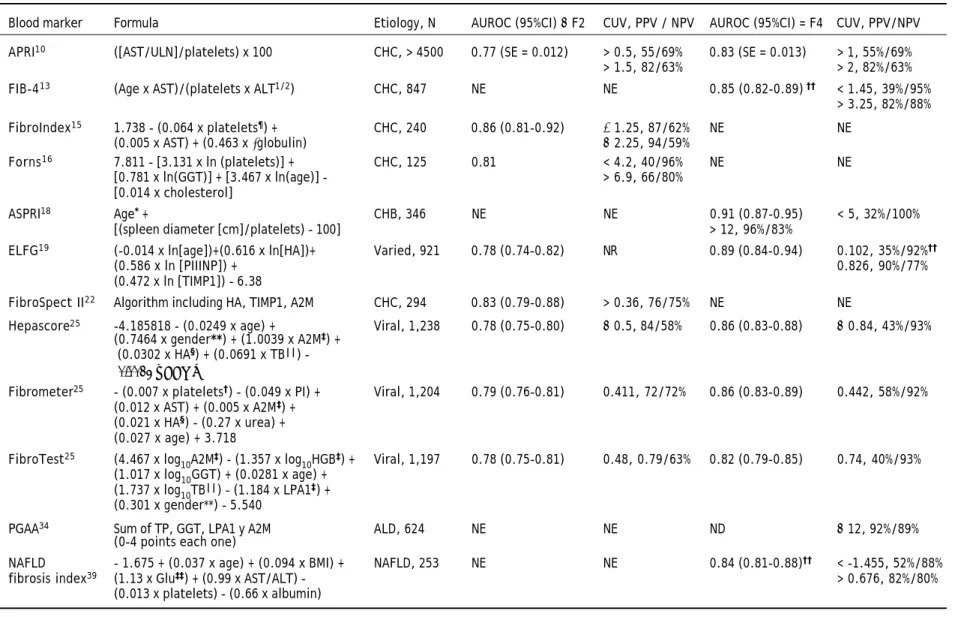
Table 2. Formulas and performance of blood markers for the estimation of clinically relevant fibrosis and cirrhosis.
Blood marker Formula Etiology, N AUROC (95%CI) ≥ F2 CUV, PPV / NPV AUROC (95%CI) = F4 CUV, PPV/NPV
APRI10 ([AST/ULN]/platelets) x 100 CHC, > 4500 0.77 (SE = 0.012) > 0.5, 55/69% 0.83 (SE = 0.013) > 1, 55%/69%
> 1.5, 82/63% > 2, 82%/63%
FIB-413 (Age x AST)/(platelets x ALT1/2) CHC, 847 NE NE 0.85 (0.82-0.89) †† < 1.45, 39%/95%
> 3.25, 82%/88%
FibroIndex15 1.738 - (0.064 x platelets¶) + CHC, 240 0.86 (0.81-0.92) ≤ 1.25, 87/62% NE NE
(0.005 x AST) + (0.463 x γglobulin) ≥ 2.25, 94/59%
Forns16 7.811 - [3.131 x ln (platelets)] + CHC, 125 0.81 < 4.2, 40/96% NE NE
[0.781 x ln(GGT)] + [3.467 x ln(age)] - > 6.9, 66/80%
[0.014 x cholesterol]
ASPRI18 Age* + CHB, 346 NE NE 0.91 (0.87-0.95) < 5, 32%/100%
[(spleen diameter [cm]/platelets) - 100] > 12, 96%/83%
ELFG19 (-0.014 x ln[age])+(0.616 x ln[HA])+ Varied, 921 0.78 (0.74-0.82) NR 0.89 (0.84-0.94) 0.102, 35%/92%††
(0.586 x ln [PIIINP]) + 0.826, 90%/77%
(0.472 x ln [TIMP1]) - 6.38
FibroSpect II22 Algorithm including HA, TIMP1, A2M CHC, 294 0.83 (0.79-0.88) > 0.36, 76/75% NE NE
Hepascore25 -4.185818 - (0.0249 x age) + Viral, 1,238 0.78 (0.75-0.80) ≥ 0.5, 84/58% 0.86 (0.83-0.88) ≥ 0.84, 43%/93%
(0.7464 x gender**) + (1.0039 x A2M‡) +
(0.0302 x HA§) + (0.0691 x TB||) -(0.0012 x GGT)]
Fibrometer25 - (0.007 x platelets†) - (0.049 x PI) + Viral, 1,204 0.79 (0.76-0.81) 0.411, 72/72% 0.86 (0.83-0.89) 0.442, 58%/92%
(0.012 x AST) + (0.005 x A2M‡) +
(0.021 x HA§) - (0.27 x urea) +
(0.027 x age) + 3.718 FibroTest25 (4.467 x log
10A2M‡) - (1.357 x log10HGB‡) + Viral, 1,197 0.78 (0.75-0.81) 0.48, 0.79/63% 0.82 (0.79-0.85) 0.74, 40%/93%
(1.017 x log10GGT) + (0.0281 x age) + (1.737 x log10TB||) – (1.184 x LPA1‡) +
(0.301 x gender**) - 5.540
PGAA34 Sum of TP, GGT, LPA1 y A2M ALD, 624 NE NE ND ≥ 12, 92%/89%
(0-4 points each one)
NAFLD - 1.675 + (0.037 x age) + (0.094 x BMI) + NAFLD, 253 NE NE 0.84 (0.81-0.88)†† < -1.455, 52%/88%
fibrosis index39 (1.13 x Glu‡‡) + (0.99 x AST/ALT) - > 0.676, 82%/80%
(0.013 x platelets) - (0.66 x albumin)
Age is expressed in years, albumin in g/dL, ALT, AST and GGT in UI/L, cholesterol in mg/dL, γglobulin in g/dL, BMI in kg/m2, platelets in 109/L, PI protrombin index in percentage, urea in mmol/L; unless otherwise stated. * Sum 1 point for every each 10 years starting at 30 years, until reaching 5 points in patients ≥ 70 years (< 30 years = 0). ††††† In g/L. ‡‡‡‡‡In mg/dL. §§§§§ In mcg/L. |||||||||| In mcmol/L. ¶ Expressed in 104/mm3. **Gender: male = 1, female = 0. ††In this study comparison was between F0-2 and F3-4. ††††††††††Glucose > 110 mg/dL or diabetes gives 1 point, whereas their absence equals 0.
2M
A2M: alpha-2-macroglobulin. HA: hyaluronic acid. APRI: AST/plaquetas ratio index. P ELFG: European Liver Fibrosis Group. : HHGB: haptoglobin. LPA1: apolipoprotein-A1. NE NE: not evaluated.
NR: not reported. PIIINP: amino-terminal propeptide of type III collagen. I SPRI: spleen/platelets ratio index. P I TB: total bilirubin. TIMP: tissue inhibitor of matrix metalloproteinase 1.I
(pre-test probability), which would allow for more accurate use of the noninvasive markers of fibrosis (higher prevalence increases PPV but decreases NPV, and viceversa).
BLOOD MARKERS
Blood markers used in the estimation of liver fi-brosis can be classified as direct, when they measure components derived from the extracellular matrix or hepatic stellate cell (e.g. hyaluronic acid, α -2-macro-globulin), or indirect, when they are molecules released by hepatic parenchyma after fibrosis injury (e.g. cellular damage [GGT, AST], are indicative of compromised hepatic synthesis [bilirubin, INR], or are markers of portal hypertension [platelets, gam-ma globulin]). Even though direct gam-markers allow an earlier assessment of liver fibrosis, they lack specifi-city as non-hepatic scarring processes can yield posi-tive results. It is generally considered that the combination of direct and indirect markers can pro-vide a better estimation of fibrosis, as they combine the need for both sensitive and specific parameters. Given that direct markers sometimes require sophis-ticated laboratory techniques, it is advisable to re-quest them at reference laboratories with good quality control and standardization processes; otherwise, proposed cut-off points may inadvertently be modified.
None of the existing blood markers fulfills crite-ria as an ideal noninvasive marker of fibrosis: sim-ple, accesible, low cost, accurate and reliable. In spite of this, available methods offer many advanta-ges. Many panels have been described to date but only those considered most relevant due to either popularity or good performance characteristics, will be described (Table 2). Also relevant are the clinical situations for which noninvasive markers are less valid, or possible pitfalls with suggestions to impro-ve accuracy.9 Some of these are shown in table 3.
Viral hepatitis
The APRI (AST to platelet ratio index) combines two biological phenomena that occur during progre-ssion to cirrhosis and portal hypertension: the in-crease in AST and dein-crease in platelet count, respectively. Given its simplicity it continues to be very useful in clinical practice, even though a recent meta-analysis including 40 studies showed some li-mitations according to the reported PPV and NPV.10 APRI has been validated for CHC and CHB, as well as for coinfection with HCV/HIV.10,11 FIB-4 is ano-ther simple method which makes use of the biologi-cal phenomena of APRI, considering the concomitant decrease in ALT, while AST increases. This was ori-ginally designed to be used in HCV/HIV coinfection and was validated against the Ishak fibrosis scale,12
Table 3. Possible caveats of noninvasive markers for the estimation of fibrosis and their effects on interpretation.
Blood markers
Limiting factor Effect
Hemolysis Reduces haptoglobin → Fibrotest ↑
Gilbert syndrome Increases bilirubin → Fibrotest ↑
Inflammatory condition Increases α2−macroglobulin → Fibrotest ↑
Increases γglobulin → Fibroindex ↑
Postprandium, gastrectomy9 Increases hyaluronic acid → Fibrometer ↑
Active alcoholism Increases GGT → Fibrotest ↑
Statin use, HCV genotype 3 Reduces cholesterol → Forns ↑
Transient elastography
Limiting factor Effect − Possible solution
BMI ≥ 28 - 30 kg/m2* Failure - Use XL -probe
Narrow intercostal space Failure or TE ↑ - Patient reposition
Ascites Failure - Assess need for TE, diuretics
Elevated ALT TE ↑- Repeat after acute event improves
Hepatic infiltration TE ↑- Assess need for TE, alternative method Retrogade vascular congestion TE ↑- Consider alternative method
Extrahepatic cholestasis TE ↑- Consider alternative method
431
Noninvasive markers of fibrosis. , 2012; 11 (4): 426-439
but has now been validated for CHC and CHB with METAVIR as well.13,14 The limitation of APRI or FIB-4 in CHC is the lack of ability to differentiate early stages of fibrosis (F0-2), however they are use-ful to detect advanced fibrosis (F3-4) with high va-lues in the area under the receiver operator characteristic curve (AUROC) reported.13 For CHB, a PPV of 91% and NPV of 93% for the diagnosis of cirrhosis was reported.14 Fibroindex adds to AST and platelet count the determination of gamma glo-bulin under the assumption that this increases in the presence of portosystemic shunting. One might expect that the test is not appropriate to evaluate early stages of fibrosis, however it showed a high AUROC with a PPV of 94% for the detection of CRF in CHC.15 Forns’ index is based on patient age, which is associated with more advanced fibrosis particular-ly in CHC; GGT as a marker of biliary damage, and thus commonly elevated in advanced fibrosis; and cholesterol, which decreases due to impaired synthetic capacity or as a direct cytopathic viral effect (more evident with genotype 3).16,17 The major utility of the Forns index is to rule out CRF with an NPV of 96%. It was been validated for both CHC and CHB. ASPRI (Age-Spleen-Platelet Radio Index), validated in CHB, uses patient’s age and an ultraso-nographic parameter –spleen diameter– in combina-tion with platelet count (from its predecesor SPRI).18 This system is based on clear pathophysio-logic principles of portal hypertension and may be very useful in ruling in/out cirrhosis in CHC and CHB. However, two possible caveats are the opera-tor-dependent reliability of ultrasonographic spleen measurement, as well as variations in body compo-sition that may affect measurement or the normal relation with platelet count.
Unlike the previously described estimators that were based only on indirect markers, there are two that exclusively combine direct markers. The ELFG (European Liver Fibrosis Group) measures hyaluro-nic acid (HA), amino-terminal propeptide of type III collagen (PIIINP), the tissue inhibitor of matrix me-talloproteinase 1 (TIMP1), as well as age. The first two biomarkers are components of the extracellular matrix and the third one is a fibrogenesis regulator that inhibits collagen degradation and hepatic stella-te cell apoptosis. ELFG has shown a good AUROC for the identification of cirrhosis in CHC, presuma-bly with a high NPV given the reported sensitivity of 91%.19 A more recent study demonstrated that eli-minating age from the formula does not change the usefulness of the test, and now this is known as ELF (Enhanced Liver Fibrosis).20 Fibrospect II
(Prometheus Labs., San Diego, CA) uses an algorithm including HA, TIMP1 and α-2-macroglobulin (A2M). The latter is a protease inhibitor expressed by the hepatic stellate cell upon activation.21 The valida-tion study of Fibrospect II in CHC found a fair AUROC but reported PPV and NPV that are difficult to use in clinical practice.22 Notably, a sub-sequent study using quantitive measurements of fibrosis did not show improved accuracy for this tool.23 The major issue with either ELF or Fibros-pect II is the relatively limited accessibility of the tests required in general clinical practice.
caused underestimation of the PPV and overestima-tion of the NPV.25 Similar results have been repor-ted in other studies and meta-analyses (including a recent one for CHB).26-29 At least part of the discor-dance in AUROC results between studies can be ex-plained by ‘spectrum bias’: variations in the representation of the different stages of fibrosis (e.g.
prevalence of F4 oscilates between 10% and 25% among studies), and their transformation from an ordinal to a binary scale (F0-4) by clustering adja-cent stages (F0-1 vs F2-4 or F0-3 vs. F4). When sta-tistical methods adjusting for spectrum bias are used (DANA, Obuchowski),30 a modest improvement in the ‘real accuracy’ of blood markers has been ob-served for the 3 tests with AUROC for the diagnosis of CRF increasing from 0.80-0.82 to 0.83-0.85 after adjustment.31 Accumulation of future studies asses-sing blood markers for the noninvasive estimation of fibrosis with ajustment for spectrum bias may provi-de a better unprovi-derstanding on their accuracy.
Other liver disease
In both alcoholic liver disease (ALD) and NAFLD fibrosis deposition differs from viral hepatitis in that it starts with a perisinusoidal and perivenular dis-tribution instead of the classical periportal fibrosis32 (Table 1). This phenomenon may cause blood mar-kers designed for viral hepatitis to have different performance characteristics when applied to ALD and NAFLD. Not only may the sensitivity, specifici-ty, PPV and NPV change but the thresholds used to rule in or out various degrees of fibrosis may also differ between diseases. These differences may be im-portant clinically to ensure that the decisions are made with the specific test characteristics for a gi-ven disease in mind. Arguably, there may be some advantage to developing specific markers for disea-ses with different patterns of fibrogenesis.
Several panels have been specifically developed or validated for alcoholic liver disease (ALD). The PGA index, named after its components: protrombin, GGT and LPA1, was initially developed for ALD. The diagnostic accuracy was improved with the addition of A2M, hence becoming PGAA. Because of the methodology employed in these studies it is diffi-cult to ascertain the accuracy for the diagnosis of CRF, but PGAA is a good discriminator for cirrhosis with PPV and NPV close to 90%.33,34 Remarkably, Fibrometer, ELFG and Fibrotest have all been vali-dated in ALD, showing better performance than what has been observed in CHC. The reported AU-ROC for ALD and CHC, respectively were:
Fibrome-ter 0.92 (SE = 0.03) and 0.83 (SE = 0.02); ELFG 0.94 (SE = 0.06) and 0.77 (SE = 0.04); and Fibro-test (meta-analysis) 0.88 (0.81-0.84) and 0.79 (0.76-0.82).7,19,27 Recently, one study compared the accuracy of Fibrotest, Fibrometer and Hepascore and did not find differences among them for the diagnosis of alcoholic cirrhosis: Fibrotest and Fibrometer AUROC = 0.94 ± 0.02, Hepascore AUROC = 0.92 ± 0.02, and were significantly grea-ter than those of nonpatented biomarkers (APRI, Forns, FIB4; P < 0.01).35 Another recent study showed relatively limited usefulness of the Forns in-dex in ALD.36
HA by itself has also shown adequate diagnostic usefulness and it outperformed Fibrotest for the diagnosis of CRF (AUROC 0.94 and 0.83, respective-ly) but not for cirrhosis (AUROC 0.93 and 0.95, res-pectively), according to one study.37 Finally, serum markers of hepatocyte death and apoptosis have re-cently been evaluated in fibrosis progression among alcoholic patients. Lavallard, et al. found that ci-tokeratin 18 (CK18) and its fragments showed good diagnostic accuracy (AUROC 0.84, 95% CI 0.76-0.90 and 0.76, 95% CI 0.66-0.83; respectively) for predic-ting advanced fibrosis among heavy drinkers.38
yiel-433
Noninvasive markers of fibrosis. , 2012; 11 (4): 426-439
ded better results;44 and another study showed Hepascore, Fibrotest and FIB-4 to have better accu-racy than both BARD index and APRI.45
In autoimmune and other liver diseases there is a paucity of studies of noninvasive estimation of liver fibrosis through blood tests. APRI, FIB-4, Fibroindex and Fibrotest have shown to have limited usefulness in autoimmune hepatitis and primary biliary cirrhosis, although the Forns index looked to be promising for the identification of cirrhosis.46-48
TRANSIENT
ELASTOGRAPHY OF THE LIVER
TE measures the stiffnes or ability of a tissue not to undergo deformation when mechanical stress is applied to it, by measuring the propagation velocity of a shear wave within the tissue: the stiffer the tis-sue the faster the shear wave propagates. The prin-ciple is similar to percusion during clinical exam in that we can identify the content of a tissue (i.e. so-lid, liquid, gas) according to the audible feedback we generate. With the Fibroscan (FS) (Echosens, Paris, France), a vibrating probe with low frequency and amplitude (50 MHz) is mounted to an ultrasound transducer (3.5 MHz). The probe generates a shear wave or elastic wave that propagates through the hepatic tissue and the ultrasound transducer captu-res the wave. The data are processed to expcaptu-ress the elastic wave as a function of time represented in an elastogram, and stiffness is provided as a numeric output expressed in kilopascals (kPa). The results range from 2.5 to 75 kPa. The probe assesses a total hepatic volume of 3 cm3, greater than 100 times the size of a liver biopsy, thus theoretically reducing sampling error. With the standard probe, measure-ment starts at a depth of 2.5 cm from the skin (up to 6.5 cm in depth) and hence the evaluation in pa-tients with an excess amount of fat over the right hypochondrium may be limited.49
TE offers some advantages over other noninvasi-ve markers of fibrosis: the procedure is easy to per-form, is relatively operator-independent, is easy to learn (> 100 cases), and provides immediate results.49 Moreover, it is a reliable method with an intra-class correlation coefficient of 0.98, particular-ly in stages F ≥ 2.50 However, caveats remain, some of which are shown in table 3.
TE determinations should be considered valid when there are 10 succesful measurements with a success rate > 60%, and when the interquartile range (IQR) of all measurements is ≤ 30% of the value of the median (IQR/M < 30%); although more recent data suggested that an IQR/M < 21% would increase the accuracy of the test.51 According to a study inclu-ding 7,261 patients, it was not possible to obtain at least one measurement in 4% of individuals, and in 17% measurements were unreliable, mostly in rela-tion to an IQR/M > 30%. The main limiting factors leading to a failed study were the lack of experience of the operator and a BMI > 28 kg/m2.52 For heavier patients a new probe (XL) allowing examinations at a depth from 3.5 to 7.5 cm below the skin has been de-signed, increasing the rate of succesful measure-ments. The validation study showed that the rate of failed measurements could be decreased from 16% with the standard probe to 1.1% with the XL probe, in patients with a BMI ≥ 28 kg/m2.53 However, even with the XL-probe BMI continues to be the main factor favoring discordance with biopsy results, particularly in patients with a BMI ≥ 40 kg/m2.54
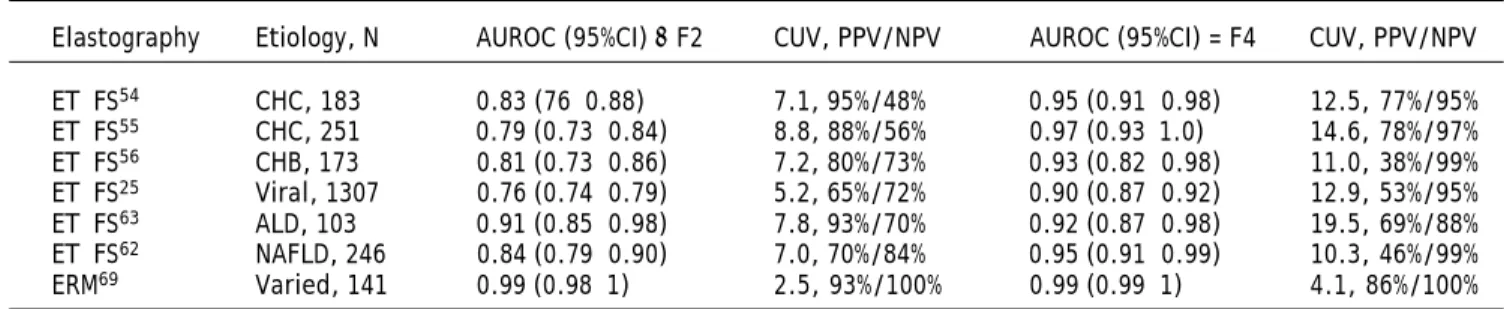
Table 4 summarizes some of the published studies regarding the noninvasive estimation of liver fibro-sis with TE. As can be observed the cut-off points used for CRF and cirrosis vary among studies. A re-cent meta-analysis evaluated the diagnostic accura-cy of TE showing that the sentitivity and specificity for the diagnosis of CRF (31 studies) were 79% (95%CI: 74-82%) and 78% (95%CI: 72-83%), respec-tively, and the median of reported cut-off values was 7.2 kPa (range of 4 to 10.1 kPa). For the diagnosis
Table 4. Performance of studies using elastography for the estimation of clinically relevant fibrosis and cirrhosis.
Elastography Etiology, N AUROC (95%CI) ≥ F2 CUV, PPV/NPV AUROC (95%CI) = F4 CUV, PPV/NPV
ET−FS54 CHC, 183 0.83 (76−0.88) 7.1, 95%/48% 0.95 (0.91−0.98) 12.5, 77%/95%
ET−FS55 CHC, 251 0.79 (0.73−0.84) 8.8, 88%/56% 0.97 (0.93−1.0) 14.6, 78%/97% ET−FS56 CHB, 173 0.81 (0.73−0.86) 7.2, 80%/73% 0.93 (0.82−0.98) 11.0, 38%/99% ET−FS25 Viral, 1307 0.76 (0.74−0.79) 5.2, 65%/72% 0.90 (0.87−0.92) 12.9, 53%/95% ET−FS63 ALD, 103 0.91 (0.85−0.98) 7.8, 93%/70% 0.92 (0.87−0.98) 19.5, 69%/88% ET−FS62 NAFLD, 246 0.84 (0.79−0.90) 7.0, 70%/84% 0.95 (0.91−0.99) 10.3, 46%/99%
of cirrhosis (30 studies) sensitivity was 83% (95%CI: 79-86%) and specificity 89% (95%CI: 87-91%), with a median cut-off of 14.5 kPa (range 9 to 26.5 kPa).55
Viral hepatitis
TE by means of FS has proved to be very useful for both CHC and CHB. As shown in table 4, the AUROC are particularly high for cirrhosis but less accurate for the diagnosis of CRF. This is related to some overlap among adjacent stages that is more evident in the low spectrum of fibrosis. Although the clinical application for CRF seems to be somewhat limited, in cirrhosis the NPV are all ≥ 95%.56-58 Whe-ther statistical methods adjusting for the spectrum bias (DANA, Obuchowski) will be useful for TE is unknown, but such an approach seems rational and a recent study also showed a modest improvement when standard AUC were compared to Obuchowski (0.82 vs. 0.84, respectively).31 Although the afore-mentioned meta-analysis showed that the cut-off va-lues to discriminate CRF or cirrhosis were slightly higher for CHC than for CHB, it must be considered that TE readings are elevated during ALT flares in CHB and they return to baseline after normaliza-tion of ALT.59,60 Some authors have proposed to use a cut-off point of 10.1-12 kPa for F4 when ALT is normal, and another of 13.4-15.5 kPa when ALT is above the upper limit of normal.61,62
Other liver diseases
Despite some controversies about the effect of steatosis on TE results,57,63,64 this technique has been used for fibrosis evaluation among patients with ALD and NAFLD showing good diagnostic accuracy, comparable to that of viral hepatitis64,65 (Table 4). Other studies have shown similar results36,53,66,67 and support the utility of TE in the-se patients. Different cut-off values for cirrhosis have been suggested for ALD/NAFLD than for viral hepatitis, which may reflect differences in the fibro-genic process between diseases. Similar to CHB fla-res, severe steatohepatitis, particularly in ALD, may also affect the TE results.
Recently, variation in the accuracy of TE as a function of AST levels was evaluated in patients with alcoholic steatohepatitis. By performing se-quential TE before and after of alcohol detoxifica-tion, Mueller, et al. could observe a parallel decrease of both TE and AST, with the former decreasing more than 3.5 kPa in patients arriving with an AST
≥ 100 U/L, after a fall in AST. In a second cohort of patients it was confirmed that the lower the AST at the time of assessment the more accurate the TE re-sults.68 This study provides evidence that, in pa-tients with ALD, a more accurate noninvasive assessment of fibrosis stage by TE can be achieved if the degree of steatohepatitis is considered. However, validation in large and independent series is still needed before making specific recommendations in routine clinical practice.
As with blood markers, there is a lack of valida-tion studies for TE in autoimmune and other liver diseases. Nonetheless, and in spite of being diseases of heterogeneous involvement within the liver, TE has shown to be useful in a limited sample of pa-tients with either primary biliary cirrhosis or pri-mary sclerosing cholangitis.69
Magnetic resonance
elastography and other imaging modalities
Magnetic resonance elastography (MRE) uses the same principles as TE, differences being that the low amplitude (60 MHz) vibrating signal is provided by a pneumatic transducer positioned over the right hypochondrium (or last ribs at the back) and the elastic waves are captured by the magnetic resonan-ce machine. With this method the elastogram can in-volve the whole liver instead of only the 3 cm3 of FS, and it is not limited by a narrow intercostal spa-ce, also allowing the evaluation of obese patients and those with ascites.70 Nevertheless, the techni-que suffers from the same drawbacks of all magnetic resonance approaches, such as the longer-time for acquisition and interpretation of results, and eleva-ted cost. In addition, it cannot be used to assess pa-tients with significant iron overload because of signal-to-noise limitations. Table 4 shows the per-formance characteristics of a representative study using MRE. This study also reported on the superio-rity of MRE over FS-TE, particulary at early stages of fibrosis (AUROC for CRF with MRE was 0.99 vs.
0.84 with FS-TE; for cirrhosis with MRE 0.99 vs.
0.93 with FS-TE), due to better discrimination of ad-jacent stages; and it also showed improved reliabili-ty.71 Given the very high predictive values reported by this study for both CRF and cirrhosis, MRE see-ms to be a very promising tool for noninvasive esti-mation of fibrosis and confirmatory large studies are awaited. However, availability and cost may be im-portant limiting factors even in specialized centers.
435
Noninvasive markers of fibrosis. , 2012; 11 (4): 426-439
through magnetic resonance; and acoustic radiation force impulse (ARFI) and real-time elastography, which make use of ultrasound technology. A study with diffusion-weighted imaging showed it to be infe-rior to MRE,72 and in the case of ARFI73-75 most of the available data does not support that it outperfor-ms TE, although it is more accurate than APRI.76 However, a recent study used ARFI to evaluate liver stiffness in 20 different spots of the right lobe (seg-ments V to VIII) in patients with CHC, providing a larger and more varied sample than TE. With this novel approach authors could demonstrate superio-rity of ARFI over TE, particularly for the diagnosis of CRF and advanced fibrosis.77 This may be espe-cially useful for liver diseases with a heterogenous distribution such as primary biliary cirrhosis and primary sclerosing cholangitis. It will be interesting to follow the performance of ARFI in future studies, as access to this technology may be favored over FS-TE and MRE in some centers.
COMBINING BLOOD MARKERS AND TRANSIENT ELASTOGRAPHY
Tables 2 and 3 clearly demonstrate improved performance of noninvasive tests for the diagnosis cirrhosis over CRF. The main issue for all tests is discrimination between adjacent stages of fibrosis. Fibrotest is more accurate in the lower range of fi-brosis stages (F0 vs. F1), while TE performs better with more advanced fibrosis (F3 vs. F4).26 Mo-reover, some of the predictive values seem to com-plement each other supporting the concept that the combination of a blood marker and TE would yield greater diagnostic accuracy. This has been investi-gated for CHC78 and CHB.61,62 In patients with CHC, a report showed that an algorithm combining APRI and Fibrotest correctly classified 97% of pa-tients with CRF (PPV 96%, NPV 100%), and would have avoided 48% of biopsies; whereas an algori-thm combining Fibrotest and FS correctly classi-fied 96% of patients with cirrhosis (PPV 95%, NPV 96%) and would have avoided 79% of biopsies.79 These results make the combination of noninvasive markers of fibrosis look very attractive for daily clinical practice. Moreover this has been confirmed by a more recent publication with a larger sample, also proposing a new noninvasive algorithm combi-ning Fibrometer and FS-TE with very promising results.80 It is worth mentioning that the new cli-nical practice guidelines from the European Asso-ciation for the Study of Liver on CHC have endorsed the use of noninvasive markers of
fibro-sis, and recommend to combine them to increase diagnostic accuracy.81
Longitudinal studies with noninvasive markers of fibrosis
Sustained virological response (SVR) after treat-ment with interferon and ribavirin in CHC patients is associated with regression of fibrosis after long-term follow up. A few studies have evaluated the change in noninvasive markers of fibrosis after anti-viral treatment, noting that in patients who achieve an SVR there is a significant decrease (APRI, FIB-4, Forns, ELF, Fibrotest, Fibrospect II) not observed in those without an SVR.82-85 The improvement in Fibrospect II and ELF as early as 24 weeks after the end of treatment suggests reversion of the profibro-genic environment in the liver, as these panels in-clude molecules involved in matrix remodelling and stellate cell activation.83,85 Perhaps not surprisingly, it has been shown that low scores from blood mar-kers before the start of antiviral treatment are pre-dictive of treatment response, similar to what has been described for early stages of fibrosis on liver biopsy.83,85,86
degree of change in noninvasive markers will allow identifying what the clinically important change is. This is particularly relevant as it is conceivable thinking that the initial improvement in noninvasi-ve markers of fibrosis after viral eradication/control is to some extent related to resolved inflammatory activity, and not to regressed fibrosis. Long-term fo-llow up studies may clarify things and set the stage for accurate prognostication with noninvasive mar-kers after regressed fibrosis.
The association between noninvasive markers of fibrosis and portal hypertension or robust outcomes (i.e. death and complications from portal hyperten-sion) has also been assessed. There is evidence showing that TE correlates with HVPG (ρ = 0.858, p = 0.001), and a TE measurement of > 21 kPa had a PPV of 93% and NPV of 91% for predicting an HVPG ≥ 10 mmHg, according to one study inclu-ding 150 patients with varied etiologies of liver di-sease.91 Follow up of these patients showed that TE (same cut-off value of > 21 kPa) was also useful to predict complications from portal hypertension such as development of ascites, hepatic encephalopathy, variceal bleeding, hepatocellular carcinoma and death.92 Although indirectly assessing liver fibrosis, spleen stiffness measured by elastography better correlates with HVPG when compared to liver stiff-ness,93 and it is another promising method for iden-tifying clinically relevant portal hypertension in the form of gastroesophageal varices.94-95 The associa-tion with hepatocellular carcinoma and death was also recently shown for TE readings > 10kPa in pa-tients with CHB.96
The prognostic significance of the blood panels has also been evaluated. The correlation between Fi-brotest and HVPG is moderate (ρ = 0.58, p = 0.001),97 but a Fibrotest determination ≥ 0.58 is as-sociated with the development of complications from portal hypertension and increased mortality in CHC patients.98 Similar findings have been reported for
Fibrotest in CHB,99 and for ELF in primary biliary cirrhosis.100 In ALD a Fibrotest value > 0.58 is also associated with higher mortality, and this effect is independent of liver biopsy stage, whereas Fibrome-ter A and Hepascore did not show any independent effect on mortality.35
A recent study evaluated liver biopsy, blood mar-kers and TE as predictors of 5-year mortality in pa-tients with CHC. As seen in table 5, both Fibrotest and TE were associated with mortality, outperfor-ming the prognostic value of fibrosis stage by liver biopsy, and independently of antiviral treatment res-ponse.101 Thus, noninvasive markers of fibrosis can predict both complications from portal hypertension and mortality even once cirrhosis is identified, a ma-jor benefit over liver biopsy. This can be explained by the fact that they can differentiate early from ad-vanced cirrhosis (i.e. stage 1 vs. stages 2-4), due to the lack of a ceiling effect as observed with liver biopsy after reaching F4.
CONCLUSIONS
Noninvasive markers of fibrosis are making their way into daily clinical practice, predominantly for the identification of CRF and/or cirrhosis. Their ac-curacy and reliability are good enough to be used in the clinical field and may be underestimated due to imprecision of the ‘gold standard’ to which they are compared (i.e. liver biopsy). Use of non-invasive tests provides conclusive enough results to avoid half of the liver biopsies performed for staging of fi-brosis. It is critical to remember that non-invasive markers perform differently in distinct clinical sce-narios and therefore it is necessary to consider diffe-rent cut-off values with their associated PPV and NPV for each clinical scenario, and in some instan-ces for each specific liver disease. Although most of the approaches have fairly good test characteristics, the greatest accumulated experience exists with APRI, Fibrotest, Fibrometer, TE and MRE. Results may be improved with all tests with knowledge of clinical situations that may lead to test failure as well as possible solutions. The combination of noninvasive markers increases their diagnostic performance and therefore it is useful to consider more than one test, either in sequential or parallel algorithms, according to the information they provide (complementary tests with either high sensitivity or high specificity, or strengthening of results when two tests with similar performance coincide). Noninvasive markers provide a major advance over liver biopsy in terms of the ability to do longitudinal
Table 5. Multivariate model for the association between nonin-vasive markers of fibrosis and 5−year mortality in chronic hepa-titis C.
Pronostic marker RR (95%CI) P
TE 2.9 (2.0−4.3) < 0.0001
Fibrotest 60 (14−255) < 0.0001
Actitest 0.19 (0.07−0.53) 0.002
Antiviral treatment 0.28 (0.19−0.42) < 0.0001
Age 1.03 (1.01−1.04) 0.002
437
Noninvasive markers of fibrosis. , 2012; 11 (4): 426-439
evalutions, particularly given their utility in predic-ting clinical events and even mortality. It has taken a remarkable international effort to develop the va-rious noninvasive approaches available today and the future holds promise of improving current tools as well as the development of novel approaches. Liver biopsy still has a major diagnostic role for the hepa-tologist, but its days as the gold standard for fibrosis evaluation and prognostication may be numbered.
REFERENCES
1. Rockey DC, Caldwell SH, Goodman ZD, Nelson RC, Smith AD, American Association for the Study of Liver Diseases. Li-ver biopsy. Hepatology 2009; 49: 1017-44.
2. Goodman ZD. Grading and staging systems for inflamma-tion and fibrosis in chronic liver diseases. J Hepatol 2007; 47: 598-607.
3. Cholongitas E, Senzolo M, Standish R, et al. A systematic review of the quality of liver biopsy specimens. Am J Clin Pathol 2006; 125: 710-21.
4. Colloredo G, Guido M, Sonzogni A, Leandro G. Impact of li-ver biopsy size on histological evaluation of chronic viral hepatitis: the smaller the sample, the milder the disease. J Hepatol 2003; 39: 239-44.
5. Garcia-Tsao G, Friedman S, Iredale J, Pinzani M. Now the-re athe-re many (stages) whethe-re befothe-re thethe-re was one: In search of a pathophysiological classification of cirrhosis.
Hepatology 2010; 51: 1445-9.
6. Germani G, Hytiroglou P, Fotiadu A, Burroughs AK, Dhillon AP. Assessment of fibrosis and cirrhosis in liver biopsies: an update. Semin Liver Dis 2011; 31: 82-90.
7. Calès P, Oberti F, Michalak S, et al. A novel panel of blood markers to assess the degree of liver fibrosis. Hepatology
2005; 42: 1373-81.
8. Mallet V, Dhalluin-Venier V, Roussin C, et al. The accuracy of the FIB-4 index for the diagnosis of mild fibrosis in chronic hepatitis B. Aliment Pharmacol Ther 2009; 29: 409-15.
9. Idobe Y, Murawaki Y, Ikuta Y, Koda M, Kawasaki H. Post-prandial serum hyaluronan concentration in pa-tients with chronic liver disease. Intern Med 1998; 37: 568-75.
10. Lin ZH, Xin YN, Dong QJ, et al. Performance of the aspar-tate aminotransferase-to-platelet ratio index for the sta-ging of hepatitis C-related fibrosis: an updated meta-analysis. Hepatology 2011; 53: 726-36.
11. Stibbe KJ, Verveer C, Francke J, et al. Comparison of non-invasive assessment to diagnose liver fibrosis in chronic hepatitis B and C patients. Scand J Gastroenterol 2011; 46: 962-72.
12. Sterling RK, Lissen E, Clumeck N, et al. Development of a simple noninvasive index to predict significant fibrosis in patients with HIV/HCV coinfection. Hepatology 2006; 43: 1317-25.
13. Vallet-Pichard A, Mallet V, Nalpas B, et al. FIB-4: an inex-pensive and accurate marker of fibrosis in HCV infection. Comparison with liver biopsy and fibrotest. Hepatology
2007; 46: 32-6.
14. Kim BK, Kim DY, Park JY, et al. Validation of FIB-4 and comparison with other simple noninvasive indices for pre-dicting liver fibrosis and cirrhosis in hepatitis B virus-in-fected patients. Liver Int 2010; 30: 546-53.
15. Koda M, Matunaga Y, Kawakami M, Kishimoto Y, Suou T, Murawaki Y. FibroIndex, a practical index for predicting significant fibrosis in patients with chronic hepatitis C.
Hepatology 2007; 45: 297-306.
16. Forns X, Ampurdanès S, Llovet JM, et al. Identification of chronic hepatitis C patients without hepatic fibrosis by a simple predictive model. Hepatology 2002; 36: 986-92. 17. Siagris D, Christofidou M, Theocharis GJ, et al. Serum lipid
pattern in chronic hepatitis C: histological and virological correlations. J Viral Hepat 2006; 13: 56-61.
18. Kim BK, Kim SA, Park YN, et al. Noninvasive models to pre-dict liver cirrhosis in patients with chronic hepatitis B. Li-ver Int 2007; 27: 969-76.
19. Rosenberg WM, Voelker M, Thiel R, et al. Serum markers detect the presence of liver fibrosis: a cohort study. Gas-troenterology 2004; 127: 1704-13.
20. Parkes J, Guha IN, Roderick P, et al. Enhanced Liver Fibro-sis (ELF) test accurately identifies liver fibroFibro-sis in patients with chronic hepatitis C. J Viral Hepat 2011; 18: 23-31. 21. Kawser CA, Iredale JP, Winwood PJ, Arthur MJ. Rat hepatic
stellate cell expression of alpha2-macroglobulin is a feature of cellular activation: implications for matrix remodelling in hepatic fibrosis. Clin Sci (Lond) 1998; 95: 179-86.
22. Patel K, Gordon SC, Jacobson I, et al. Evaluation of a panel of non-invasive serum markers to differentiate mild from moderate-to-advanced liver fibrosis in chronic hepatitis C patients. J Hepatol 2004; 41: 935-42.
23. Patel K, Nelson DR, Rockey DC, et al. Correlation of FI-BROSpect II with histologic and morphometric evaluation of liver fibrosis in chronic hepatitis C. Clin Gastroenterol Hepatol 2008; 6: 242-7.
24. Adams LA, Bulsara M, Rossi E, et al. Hepascore: an accura-te validaaccura-ted predictor of liver fibrosis in chronic hepatitis C infection. Clin Chem 2005; 51: 1867-73.
25. Degos F, Perez P, Roche B, et al. Diagnostic accuracy of FibroScan and comparison to liver fibrosis biomarkers in chronic viral hepatitis: a multicenter prospective study (the FIBROSTIC study). J Hepatol 2010; 53: 1013-21. 26. Poynard T, de Ledinghen V, Zarski JP, et al. FibroTest(®)
and Fibroscan(®) performances revisited in patients with chronic hepatitis C. Impact of the spectrum effect and the applicability rate. Clin Res Hepatol Gastroenterol
2011 [epub ahead of print].
27. Halfon P, Munteanu M, Poynard T. FibroTest-ActiTest as a non-invasive marker of liver fibrosis. Gastroenterol Clin Biol 2008; 32: 22-39.
28. Leroy V, Halfon P, Bacq Y, et al. Diagnostic accuracy, re-producibility and robustness of fibrosis blood tests in chronic hepatitis C: a meta-analysis with individual data.
Clin Biochem 2008; 41: 1368-76.
29. Poynard T, Ngo Y, Munteanu M, Thabut D, Ratziu V. Nonin-vasive Markers of Hepatic Fibrosis in Chronic Hepatitis B.
Curr Hepat Rep 2011; 10: 87-97.
30. Guha IN, Myers RP, Patel K, Talwalkar JA. Biomarkers of li-ver fibrosis: What lies beneath the receili-ver operating characteristic curve? Hepatology 2011; 54: 1454-62. 31. Zarski JP, Sturm N, Guechot J, et al. Comparison of nine
blood tests and transient elastography for liver fibrosis in chronic hepatitis C: The ANRS HCEP-23 study. J Hepatol
2011 [epub ahead of print].
32. Bataller R, Rombouts K, Altamirano J, Marra F. Fibrosis in alcoholic and nonalcoholic steatohepatitis. Best Pract Res Clin Gastroenterol 2011; 25: 231-44.
33. Poynard T, Aubert A, Bedossa P, et al. A simple biological index for detection of alcoholic liver disease in drinkers.
34. Naveau S, Poynard T, Benattar C, Bedossa P, Chaput JC. Alpha-2-macroglobulin and hepatic fibrosis. Diagnostic in-terest. Dig Dis Sci 1994; 39: 2426-32.
35. Naveau S, Gaudé G, Asnacios A, et al. Diagnostic and prog-nostic values of noninvasive biomarkers of fibrosis in pa-tients with alcoholic liver disease. Hepatology 2009; 49: 97-105.
36. Janssens F, de Suray N, Piessevaux H, Horsmans Y, de Ti-mary P, Stärkel P. Can transient elastography replace li-ver histology for determination of advanced fibrosis in alcoholic patients: a real-life study. J Clin Gastroenterol
2010; 44: 575-82.
37. Naveau S, Raynard B, Ratziu V, et al. Biomarkers for the prediction of liver fibrosis in patients with chronic alcoho-lic liver disease. Clin Gastroenterol Hepatol 2005; 3: 167-74.
38. Lavallard VJ, Bonnafous S, Patouraux S, et al. Serum mar-kers of hepatocyte death and apoptosis are non invasive biomarkers of severe fibrosis in patients with alcoholic li-ver disease. PLoS One 2011; 6: 17599.
39. Angulo P, Hui JM, Marchesini G, et al. The NAFLD fibrosis score: a noninvasive system that identifies liver fibrosis in patients with NAFLD. Hepatology 2007; 45: 846-54. 40. Harrison SA, Oliver D, Arnold HL, Gogia S,
Neuschwander-Tetri BA. Development and validation of a simple NAFLD cli-nical scoring system for identifying patients without advanced disease. Gut 2008; 57: 1441-7.
41. Ratziu V, Massard J, Charlotte F, et al. Diagnostic value of biochemical markers (FibroTest-FibroSURE) for the predic-tion of liver fibrosis in patients with non-alcoholic fatty li-ver disease. BMC Gastroenterol 2006; 6: 6.
42. Calès P, Lainé F, Boursier J, et al. Comparison of blood tests for liver fibrosis specific or not to NAFLD. J Hepatol
2009; 50: 165-73.
43. Guha IN, Parkes J, Roderick P, et al. Noninvasive markers of fibrosis in nonalcoholic fatty liver disease: Validating the European Liver Fibrosis Panel and exploring simple markers. Hepatology 2008; 47: 455-60.
44. Shah AG, Lydecker A, Murray K, et al. Comparison of no-ninvasive markers of fibrosis in patients with nonalcoholic fatty liver disease. Clin Gastroenterol Hepatol 2009; 7: 1104-12.
45. Adams LA, George J, Bugianesi E, et al. Complex non-inva-sive fibrosis models are more accurate than simple models in non-alcoholic fatty liver disease. J Gastroenterol Hepa-tol 2011; 26: 1536-43.
46. Loaeza-del-Castillo A, Paz-Pineda F, Oviedo-Cardenas E, et al. AST to platelet ratio index (APRI) for the noninva-sive evaluation of liver fibrosis. Ann Hepatol 2008; 7: 350-7.
47. Floreani A, Cazzagon N, Martines D, Cavalletto L, Baldo V, Chemello L. Performance and utility of transient elasto-graphy and noninvasive markers of liver fibrosis in prima-ry biliaprima-ry cirrhosis. Dig Liver Dis 2011; 43: 887-92. 48. Friedrich-Rust M, Müller C, Winckler A, et al. Assessment
of liver fibrosis and steatosis in PBC with FibroScan, MRI, MR-spectroscopy, and serum markers. J Clin Gastroente-rol 2010; 44: 58-65.
49. Castera L, Forns X, Alberti A. Non-invasive evaluation of liver fibrosis using transient elastography. J Hepatol
2008; 48: 835-47.
50. Fraquelli M, Rigamonti C, Casazza G, et al. Reproducibili-ty of transient elastography in the evaluation of liver fi-brosis in patients with chronic liver disease. Gut 2007; 56: 968-73.
51. Lucidarme D, Foucher J, Le Bail B, et al. Factors of accu-racy of transient elastography (fibroscan) for the diagno-sis of liver fibrodiagno-sis in chronic hepatitis C. Hepatology
2009; 49: 1083-9.
52. Castéra L, Foucher J, Bernard PH, et al. Pitfalls of liver stiffness measurement: a 5-year prospective study of 13,369 examinations. Hepatology 2010; 51: 828-35. 53. Myers RP, Pomier-Layrargues G, Kirsch R, et al. Feasibility
and diagnostic performance of the fibroscan xl probe for liver stiffness measurement in overweight and obese pa-tients. Hepatology 2012; 54: 199-208.
54. Myers RP, Pomier-Layrargues G, Kirsch R, et al. Discor-dance in fibrosis staging between liver biopsy and tran-sient elastography using the fibroscan XL probe. J Hepatol 2012 [epub ahead of print].
55. Tsochatzis EA, Gurusamy KS, Ntaoula S, Cholongitas E, Da-vidson BR, Burroughs AK. Elastography for the diagnosis of severity of fibrosis in chronic liver disease: a meta-analy-sis of diagnostic accuracy. J Hepatol 2011; 54: 650-9. 56. Castéra L, Vergniol J, Foucher J, et al. Prospective
com-parison of transient elastography, fibrotest, APRI, and li-ver biopsy for the assessment of fibrosis in chronic hepatitis C. Gastroenterol 2005; 128: 343-50.
57. Ziol M, Handra-Luca A, Kettaneh A, et al. Noninvasive as-sessment of liver fibrosis by measurement of stiffness in patients with chronic hepatitis C. Hepatology 2005; 41: 48-54.
58. Marcellin P, Ziol M, Bedossa P, et al. Non-invasive assess-ment of liver fibrosis by stiffness measureassess-ment in patients with chronic hepatitis B. Liver Int 2009; 29: 242-7. 59. Coco B, Oliveri F, Maina AM, et al. Transient elastography:
a new surrogate marker of liver fibrosis influenced by ma-jor changes of transaminases. J Viral Hepat 2007; 14: 360-9.
60. Arena U, Vizzutti F, Corti G, et al. Acute viral hepatitis in-creases liver stiffness values measured by transient elas-tography. Hepatology 2008; 47: 380-4.
61. Kim SU, Kim do Y, Park JY, et al. How can we enhance the performance of liver stiffness measurement using FibroScan in diagnosing liver cirrhosis in patients with chronic hepatitis B? J Clin Gastroenterol 2010; 44: 66-71. 62. Chan HL, Wong GL, Choi PC, et al. Alanine
aminotransfera-se-based algorithms of liver stiffness measurement by transient elastography (Fibroscan) for liver fibrosis in chronic hepatitis B. J Viral Hepat 2009; 16: 36-44. 63. Lupsor M, Badea R, Stefãnescu H, et al. Analysis of
histo-pathological changes that influence liver stiffness in chro-nic hepatitis C. Results from a cohort of 324 patients. J Gastrointestin Liver Dis 2008; 17: 155-63.
64. Wong VW, Vergniol J, Wong GL, et al. Diagnosis of fibrosis and cirrhosis using liver stiffness measurement in nonalco-holic fatty liver disease. Hepatology 2010; 51: 454-62. 65. Nguyen-Khac E, Chatelain D, Tramier B, et al. Assessment of
asymptomatic liver fibrosis in alcoholic patients using fibros-can: prospective comparison with seven non-invasive labo-ratory tests. Aliment Pharmacol Ther 2008; 28: 1188-98. 66. Nahon P, Kettaneh A, Tengher-Barna I, et al. Assessment
of liver fibrosis using transient elastography in patients with alcoholic liver disease. J Hepatol 2008; 49: 1062-8. 67. Yoneda M, Mawatari H, Fujita K, et al. Noninvasive
assess-ment of liver fibrosis by measureassess-ment of stiffness in pa-tients with nonalcoholic fatty liver disease (NAFLD). Dig Liver Dis 2008; 40: 371-8.
68. Mueller S, Millonig G, Sarovska L, et al. Increased liver stiff-ness in alcoholic liver disease: differentiating fibrosis from steatohepatitis. World J Gastroenterol 2010; 16: 966-72.
439
Noninvasive markers of fibrosis. , 2012; 11 (4): 426-439
69. Corpechot C, El Naggar A, Poujol-Robert A, et al. As-sessment of biliary fibrosis by transient elastography in patients with PBC and PSC. Hepatology 2006; 43: 1118-24.
70. Yin M, Talwalkar JA, Glaser KJ, et al. Assessment of hepa-tic fibrosis with magnehepa-tic resonance elastography. Clin Gastroenterol Hepatol 2007; 5: 1207-13.
71. Huwart L, Sempoux C, Vicaut E, et al. Magnetic resonance elastography for the noninvasive staging of liver fibrosis.
Gastroenterology 2008; 135: 32-40.
72. Wang Y, Ganger DR, Levitsky J, et al. Assessment of chro-nic hepatitis and fibrosis: comparison of MR elastography and diffusion-weighted imaging. AJR Am J Roentgenol
2011; 196: 553-61.
73. Friedrich-Rust M, Romen D, Vermehren J, et al. Acoustic radiation force impulse-imaging and transient elastogra-phy for non-invasive assessment of liver fibrosis and stea-tosis in NAFLD. Eur J Radiol 2011 [in press].
74. Friedrich-Rust M, Wunder K, Friener S, et al. Liver fibrosis in viral hepatitis: noninvasive assessment with acoustic radiation force impulse imaging versus transient elasto-graphy. Radiology 2009; 252: 595-604.
75. Ebinuma H, Saito H, Komuta M, et al. Evaluation of liver fi-brosis by transient elastography using acoustic radiation force impulse: comparison with Fibroscan(®). J Gastroen-terol 2011; 46: 1238-48.
76. Palmeri ML, Wang MH, Rouze NC, et al. Noninvasive eva-luation of hepatic fibrosis using acoustic radiation force-based shear stiffness in patients with nonalcoholic fatty liver disease. J Hepatol 2011; 55: 666-72.
77. Rizzo L, Calvaruso V, Cacopardo B, et al. Comparison of transient elastography and acoustic radiation force impulse for non-invasive staging of liver fibrosis in patients with chronic hepatitis c. Am J Gastroenterol 2011; 106: 2112-20. 78. Bourlière M, Pénaranda G, Adhoute X, Oules V, Castellani P. Combining non-invasive methods for assessment of liver fibrosis. Gastroenterol Clin Biol 2008; 32: 73-9.
79. Castéra L, Sebastiani G, Le Bail B, de Lédinghen V, Couzi-gou P, Alberti A. Prospective comparison of two algorithms combining non-invasive methods for staging liver fibrosis in chronic hepatitis C. J Hepatol 2010; 52: 191-8.
80. Boursier J, de Ledinghen V, Zarski JP, et al. Comparison of eight diagnostic algorithms for liver fibrosis in hepatitis C: new algorithms are more precise and entirely noninva-sive. Hepatology 2011; 55: 58-67.
81. European Association for the Study of the Liver. EASL Cli-nical Practice Guidelines: management of hepatitis C virus infection. J Hepatol 2011s; 55: 245-64.
82. Poynard T, Imbert-Bismut F, Ratziu V, et al. Biochemical markers of liver fibrosis in patients infected by hepatitis C virus: longitudinal validation in a randomized trial. J Vi-ral Hepat 2002; 9: 128-33.
83. Patel K, Benhamou Y, Yoshida EM, et al. An independent and prospective comparison of two commercial fibrosis marker panels (HCV FibroSURE and FIBROSpect II) during albinterferon alfa-2b combination therapy for chronic he-patitis C. J Viral Hepat 2009; 16: 178-86.
84. Vergniol J, Foucher J, Castéra L, et al. Changes of non-in-vasive markers and FibroScan values during HCV treat-ment. J Viral Hepat 2009; 16: 132-40.
85. Martinez SM, Fernández-Varo G, González P, et al. As-sessment of liver fibrosis before and after antiviral the-rapy by different serum marker panels in patients with chronic hepatitis C. Aliment Pharmacol Ther 2011; 33: 138-48.
86. Poynard T, Munteanu M, Colombo M, et al. FibroTest is an independent predictor of virologic response in chronic he-patitis C patients retreated with pegylated interferon alfa-2b and ribavirin in the EPIC3 program. J Hepatol 2011; 54: 227-35.
87. Hézode C, Castéra L, Roudot-Thoraval F, et al. Liver stiff-ness diminishes with antiviral response in chronic hepati-tis C. Aliment Pharmacol Ther 2011; 34: 656-63.
88. Ogawa E, Furusyo N, Toyoda K, Takeoka H, Maeda S, Ha-yashi J. The longitudinal quantitative assessment by tran-sient elastography of chronic hepatitis C patients treated with pegylated interferon alpha-2b and ribavirin. Antivi-ral Res 2009; 83: 127-34.
89. van der Meer, Veldt BJ, Feld JJ, et al. Improved platelet count and smaller spleen size long after sustained virologi-cal response in chronic hepatitis c patients with advan-ced fibrosis. Hepatology 2011; 54(Suppl.): 820A.
90. Enomoto M, Mori M, Ogawa T, et al. Usefulness of tran-sient elastography for assessment of liver fibrosis in chro-nic hepatitis B: Regression of liver stiffness during entecavir therapy. Hepatol Res 2010; 40: 853-61. 91. Bureau C, Metivier S, Peron JM, et al. Transient
elasto-graphy accurately predicts presence of significant portal hypertension in patients with chronic liver disease. Ali-ment Pharmacol Ther 2008; 27: 1261-8.
92. Robic MA, Procopet B, Métivier S, et al. Liver stiffness ac-curately predicts portal hypertension related complications in patients with chronic liver disease: A prospective stu-dy. J Hepatol 2011; 55: 1017-25.
93. Hirooka M, Ochi H, Koizumi Y, et al. Splenic elasticity mea-sured with real-time tissue elastography is a marker of portal hypertension. Radiology 2011; 261: 960-8.
94. Talwalkar JA, Yin M, Venkatesh S, et al. Feasibility of in vivo MR elastographic splenic stiffness measurements in the assessment of portal hypertension. AJR Am J Roentgenol 2009; 193: 122-7.
95. Stefanescu H, Grigorescu M, Lupsor M, Procopet B, Maniu A, Badea R. Spleen stiffness measurement using fibroscan for the noninvasive assessment of esophageal varices in liver cirrhosis patients. J Gastroenterol Hepatol 2011; 26: 164-70.
96. Fung J, Lai CL, Seto WK, Wong DK, Yuen MF. Prognostic significance of liver stiffness for hepatocellular carcinoma and mortality in HBeAg-negative chronic hepatitis B. J Vi-ral Hepat 2011; 18: 738-44.
97. Thabut D, Imbert-Bismut F, Cazals-Hatem D, et al. Relation-ship between the Fibrotest and portal hypertension in patients with liver disease. Aliment Pharmacol Ther 2007; 26: 359-68.
98. Ngo Y, Munteanu M, Messous D, et al. A prospective analy-sis of the prognostic value of biomarkers (FibroTest) in patients with chronic hepatitis C. Clin Chem 2006; 52: 1887-96.
99. Ngo Y, Benhamou Y, Thibault V, et al. An accurate defini-tion of the status of inactive hepatitis B virus carrier by a combination of biomarkers (FibroTest-ActiTest) and viral load. PLoS One 2008; 3: 2573.
100.Mayo MJ, Parkes J, Adams-Huet B, et al. Prediction of cli-nical outcomes in primary biliary cirrhosis by serum en-hanced liver fibrosis assay. Hepatology 2008; 48: 1549-57.