Transient elastography in non-alcoholic fatty liver disease
Ludovico Abenavoli,*,** Michel Beaugrand*** Department of Health Sciences, University “Magna Græcia” of Catanzaro, Italy. ** Service d’Hépato-Gastroentérologie, Hôpital Jean Verdier, Bondy Cedex, France.
ABSTRACT
Non-alcoholic fatty liver disease (NAFLD) is a relevant issue in public health owing to its epidemiological burden. It represents the most common chronic liver disease in the general population and is expected to increase in future as a result of an ageing population. Liver biopsy is still considered the “gold standard” for distinguishing the broad range of NAFLD. However, liver biopsy is often not recommended in the NAFLD patients, because of its cost, the potential risk of bleeding and the absence of consensus regarding the histopathological criteria that firmly differentiate between the NAFLD entities. Due to the remarkable in-crease in the prevalence of NAFLD and the concomitant efforts in developing novel therapies, a non-invasi-ve, simple and reproducible technique is needed in the clinical practice. Transient elastography is a non-invasive technique for liver stiffness measurement (LSM) as a function of the extent of hepatic fibro-sis. This review focuses on practical issues in the use of LSM in the NAFLD patients and suggests areas for further research and development.
Key words. Liver. Stiffness. Elastic wave. Steatosis. Obesity.
Correspondence and reprint request: Ludovico Abenavoli M.D. Ph. D. Department of Health Sciences
University “Magna Græcia”
Viale Europa, 88100, Catanzaro, Italy. Tel.: +39 0961 3697113. Fax: +39 0961 754220 E mail: [email protected]
Manuscript received: September 19, 2011. Manuscript accepted: November 28, 2011.
INTRODUCTION
Non-alcoholic fatty liver disease (NAFLD) repre-sents the most common chronic liver disease in the general population and is expected to increase in fu-ture as a result of an ageing population, the impro-ving control of other major causes of the chronic liver disease and the epidemics of obesity and diabe-tes.1,2
Liver steatosis consists of an accumulation of fat in the liver cells and it is not a harmful disease it-self. It is an aggravating factor in certain pathologi-cal situations of varying degrees of gravity and becomes dangerous when it is associated with infla-mmation or fibrosis. However, it is reversible, through diet and exercise. In fact, NAFLD may pre-sent a spectrum ranging from asymptomatic steato-sis with elevated or normal aminotransferases to
steatohepatitis [non-alcoholic steatohepatitis (NASH)], all the way to cirrhosis with the complica-tions of liver function to hepatocellular carcinoma. NAFLD occurs in 60-95% patients with obesity, in 28-55% patients with type 2 diabetes mellitus (T2DM) and in 27-92% patients with dyslipidemia. Insulin resistance with compensatory hyperinsuline-mia is a common denominator of obesity, T2DM and dyslipidemia, and it can play a pathogenetic role in NAFLD. Accordingly, it has been reported that the insulin resistance is a single laboratory finding most closely associated with NAFLD in a great number of patients, irrespective of the body mass in-dex (BMI), fat distribution or glucose tolerance. Therefore, NAFLD has recently been proposed as an additional feature of the metabolic syndrome.3
Liver biopsy (LB) is still considered the gold stan-dard for distinguishing between the broad range of NAFLD. However, LB is often not recommended in the NAFLD patients because of its cost, the potential risk of bleeding, and the absence of consensus regar-ding the histopathological criteria that firmly define NASH and differentiate between NAFLD entities.4
Currently, many techniques exist to detect steato-sis, although ultrasound (US) is perhaps the easiest and most common approach in the clinical practice and for epidemiological studies. In particular, US is the least expensive modality for detecting the chan-ges associated with NAFLD. However, echography-based US techniques become sensitive at moderately high levels of steatosis (33% or more) and suffer from low intra- and inter-operator repeatability.6
In the last years, a new technique called vibra-tion-controlled transient elastography ([VCTE], Fi-broscan®, Echosens, Paris, France) has been proposed for liver stiffness measurement (LSM). It is a technique using elastic waves of low-audio-fre-quency (50 Hz), whose propagation velocity depends on tissue elasticity, indicative of liver stiffness, con-sidered to be one of the direct consequences of the fi-brotic evolution of chronic liver disease.7
A few meta-analyses have suggested liver stiffness measurement (LSM) by VCTE to be a reliable tool to detect advanced liver fibrosis and early liver cirrho-sis.8,9 In the NAFLD patients, various degrees of
he-patic steatosis may attenuate the elastic shear wave, but does not change its underlying speed which is the parameter used for the stiffness measurement. Clearly positive correlation between LSM and the severity of liver fibrosis in the patients with NAFLD has been reported in the literature. Recently, a new measurement technique, the controlled attenuation parameter (CAP®, EchoSens, Paris, France), has been introduced which provides good accuracy even with the low levels of steatosis in the liver. Owing to its sensitivity at early phases of steatosis, CAP could provide a means to monitor the progression or regression of steatosis. A combination of the measu-rement by VCTE and CAP could provide the means to follow the NAFLD patients, during the different phases of the disease history.
This review focuses on practical issues in the use of LSM in NAFLD patients and suggests areas for further research and development.
Elastography assessment of NAFLD
The development of non-invasive methods to eva-luate liver disease stages is a clinical and research priority. NAFLD is strongly associated with meta-bolic syndrome and obesity. NASH may progress to cirrhosis and hepatocellular carcinoma. The progno-sis depends heavily on histological severity. Several questions concerning the management of the pa-tients with NAFLD and, in particular, how to diag-nose NAFLD and its type, how to select the patient
candidates for the treatment and how to treat these patients have been debated in the literature.
The limitations of LB have stimulated the search for non-invasive approaches for the assessment of steatosis and liver fibrosis in the patients with NA-FLD. A variety of new methods, including serum markers, imaging techniques such as US, computed tomography, magnetic resonance imaging, and more recently LSM, have been proposed. In particular, LSM has shown excellent results for the diagnosis of severe fibrosis and cirrhosis and moderate results for the diagnosis of significant fibrosis in the pa-tients with NAFLD/NASH.
A prospective study aimed to assess the ability of VCTE to identify histological parameters, including steatosis, in asymptomatic healthy individuals such as the potential liver donors, and to compare these findings with the results in the patients with liver disease.10 Forty-seven patients with abnormal liver
function and/or hepatitis symptoms and eighty li-ving related potential liver donors were consecutive-ly enrolled. LB and LSM were performed in each subject. Histological parameters were evaluated ac-cording to Metavir score by a single pathologist. In the liver disease patients, stiffness was significantly correlated with fibrosis stage (P < 0.001), and the optimal stiffness cut-off values for F ≥ 2, F ≥ 3, and F = 4 were 7.35, 8.85, and 15.1 kPa respectively. In the potential liver donors, stiffness was not correla-ted with fibrosis (P = 0.851). In the latter group, the area under the receiver-operating characteristics curve (AUROC) was 0.70 (95%, confidence interval [CI], 0.58-0.81), and the optimal stiffness cut-off va-lue was 4.00 kPa for F ≥ 2, which was lower than that in the liver disease patients. Steatosis was not correlated with stiffness (P = 0.463) in the potential liver donors. The authors concluded that VCTE pre-set limited the value for detecting steatosis in the asymptomatic healthy subjects.
Nobili, et al. assessed the value of VCTE in a co-hort of the paediatric patients with NASH.11 LSM
va-lues between 7 and 9 kPa predicted fibrosis stages 1 or 2, but could not discriminate between these two stages. The VCTE values of at least 9 kPa were associated with the presence of advanced fi-brosis. In summary this study reported the reliabi-lity of VCTE to predict a normal liver or the presence of significant fibrosis and advanced fibro-sis in these special populations.
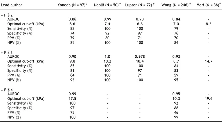
Yoneda, et al. investigated the usefulness of LSM in the assessment of liver fibrosis in the NAFLD pa-tients.12 VCTE was performed for LSM in 97
NA-FLD patients. The authors also investigated the relationship between LSM and the serum levels of hyaluronic acid and type IV collagen 7s domain. Li-ver stiffness was well correlated with the different stages of Metavir score (P < 0.0001). The AUROC were (Table 1):
• 0.927 for ≥ F1. • 0.865 for ≥ F2. • 0.904 for ≥ F3. • 0.991 for ≥ F4.
Only the fibrosis stage was strictly correlated with LSM by multiple regression analysis. Liver sti-ffness was also strongly correlated with the serum levels of type IV collagen 7s domain (r = 0.525,
P < 0.0001) and hyaluronic acid (r = 0.457, P < 0.0001). These data showed the significant correla-tion between LSM and the fibrosis stage in the NA-FLD patients, as confirmed by the results of LB.
Lupsor, et al. assessed VCTE performance in the NASH patients, as well as the factors determining the discordance between the VCTE-predicted and the fibrosis stage evaluated by the Brunt system.13
LB and VCTE were performed on 72 consecutive NASH patients. LSM ranged from 2.80 to 16.90 kPa. In the univariate analysis, LSM was correla-ted with fibrosis (P < 0.0001), steatosis (P < 0.0001), ballooning (P = 0.001) and lobular inflam-mation (P = 0.002). In multivariate analysis, only fibrosis significantly correlated with LSM (P < 0.0001). The median LSM values according to the fibrosis stages were:
• 4.90 kPa (range: 2.80-7.30) for F0. • 6.15 kPa (4.80-12.50) for F1. • 6.90 kPa (3.30-16.90) for F2, and
• 14.00 kPa (10.70-14.10) for F3, with significant difference between stages, except for F1-F2 (P = 0.249).
The cut-off values were calculated for predicting each fibrosis stage (Table 1):
Table 1. Analysis of VCTE cut-off for the diagnosis of NAFLD in adult patients. AUROC: area under receiver operative curve. PPV: positive predictive value. NPV: negative predictive value. Histological evaluation by *Metavir and †Brunt score.
Lead author Yoneda (N = 97)* Nobili (N = 50) † Lupsor (N = 72) † Wong (N = 246) † Mori (N = 36)†
• F ≥ 2
AUROC 0.86 0.99 0.78 0.84
-Optimal cut-off (kPa) 6.6 7.4 6.8 7.0 8.3
Sensitivity (%) 88 100 100 79
-Specificity (%) 74 92 97 76
-PPV (%) 79 80 71 70
-NPV (%) 85 100 100 84
-• F ≥ 3
AUROC 0.90 1.0 0.978 0.93
-Optimal cut-off (kPa) 9.8 10.2 10.4 8.7 14.7
Sensitivity (%) 85 100 100 84
-Specificity (%) 81 100 97 83
-PPV (%) 64 100 71 59
-NPV (%) 93 100 100 95
-• F ≥ 4
AUROC 0.99 - - 0.95
-Optimal cut-off (kPa) 17.5 - - 10.3 19.6
Sensitivity (%) 100 - - 92
-Specificity (%) 97 - - 88
-PPV (%) 75 - - 46
-• 5.3kPa (AUROC = 0.879) for F1. • 6.8kPa (AUROC = 0.789) for F2, and • 10.4kPa (AUROC = 0.978) for F3.
The patients with false positive results had a sig-nificantly higher alanine aminotransferase (ALT) le-vel than those with concordant results (P = 0.039). These data show that steatosis degree, ballooning and inflaammation do not influuence LSM.
In the NAFLD patients, various degrees of hepa-tic steatosis may attenuate the elashepa-tic shear wave, possibly leading again to an underestimation of liver damage. Wong, et al. aimed to evaluate the accuracy of VCTE in the diagnosis of fibrosis and cirrhosis in the patients with NAFLD and to study the factors associated with discordance between LSM and histo-logical data.14 Two hundred forty-six consecutive
patients from two ethnic groups had successful LSM and satisfactory liver biopsy specimens. The AUROC of VCTE for F3 or higher and F4 disease was 0.93 and 0.95, respectively, and was significantly higher than that of other biochemical scores for fibrosis prediction. AUROC ranged from 0.62 to 0.81, P < 0.05 for all comparisons. At a cut-off value of 7.9 kPa, the sensitivity, specificity and positive and ne-gative predictive values for F3 or greater disease were 91, 75, 52 and 97%, respectively (Table 1). LSM was not affected by hepatic steatosis, necro-in-flammation, or BMI. Discordance of at least two sta-ges between VCTE and histology was observed in 33 (13.4%) patients. By multivariate analysis, LB leng-th less leng-than 20 mm and F0-F2 disease were associa-ted with discordance. Unsatisfactory LB specimens rather than VCTE accounted for most cases of dis-cordance. However, when the diagnostic characteris-tics were compared with the inclusion of the subjects who had unsuccessful LSM, the sensitivity and specificity values were not dissimilar from the clinical/biochemical models.
A prospective study assessed the accuracy and the efficacy of VCTE for the detection of fibrosis in the patients with the liver disease of different aetiology (chronic hepatitis B - CHB, C- CHC and NAFLD) and evaluated the effect of steatosis on LSM.15
VCTE was performed in 219 consecutive patients with the chronic liver disease (35% CHC, 32% CHB, and 33% NAFLD) within 6 months of the liver biop-sy. LSM was related to the fibrosis stage in each group:
• CHC: P = 0.596, P < 0.001. • CHB: P = 0.418, P < 0.001. • NAFLD: P = 0.573, P < 0.001.
But the correlation was less strong in the CHB and NAFLD than in CHC patients. The median LSM values were:
• 7 kPa (3.2-26) in the HCV patients (n = 77). • 7.6 kPa (3.7-30.7) in the HBV patients (n = 70). • 6.6 kPa (3.0-44.3) in the NAFLD patients (n =
72) compared to 4.35 kPa (range 2.6-7) in the controls (n = 40), (P < 0.001).
The median values of LSM according to the fibro-sis stage evaluated by Metavir were in the 72 NA-FLD patients:
• F0: 5.3 kPa (3.0-9.7). • F1: 6.15 kPa (3.2-12.1). • F2: 7.75 kPa (4.3-13.9). • F3: 6.5 kPa (4.3-10.3).
• F4: 11.9 kPa (7.9-44.3), (P = 0.001).
VCTE underestimated the stage of fibrosis in 75% of patients with F3 and steatosis > 33%. At the mul-tiple logistic regression analysis, in the CHC and CHB patients, LSM was the only predictive variable of severe fibrosis/cirrhosis (odds ratio [OR] = 1.42, P = 0.003 and OR = 1.354, P = 0.003, respectively), while in the NAFLD subjects the BMI and transa-minase levels (OR = 1.433, P = 0.002 and OR = 1.053, P = 0.020, respectively), but not LSM, were independently related with advanced fibrosis and ci-rrhosis. This study confirmed that VCTE could be considered a valid support to detect fibrosis in chro-nic liver disease related to HCV, but it should be in-terpreted cautiously in the CHB and NAFLD patients.
In a recent study, Mori, et al. confirmed the rela-tionship between LSM and the Brunt score (P = 0.000149) in 36 NAFLD biopsy (Table 1).16 In
addi-tion, the authors showed that liver stiffness signifi-cantly correlated with the stage of fibrosis determined by the Sirius red-positive fibrotic area (r = 0.390, P = 0.0184) and α-smooth muscle actin-po-sitive area (r = 0.333, P = 0.0471), respectively, the markers of collagen deposit and myofibroblasts pre-sence.
Elastography assessment of NAFLD in obese patients
failure, as the results may provide guidance to refe-rral physicians regarding the optimal candidates for LSM. The knowledge would also assist clinicians to counsel patients before arranging LSM. The mea-surement is normally difficult for the obese pa-tients because of the thick subcutaneous and perihepatic fat.
Studies have shown that BMI > 28 is an indepen-dent risk factor for a LSM failure.17 A prospective
pi-lot study evaluated the new XL-probe in 99 obese patients (mean BMI 40.5 kg/m2).18 LSM was
succes-sful in 45% of the cases with M-probe, versus 76% of the cases with XL-probe (P < 0.001). 59% of those who could not be measured (< 10 valid measure-ments) using M-probe could successfully be measu-red using XL-probe. Both probes were successfully measured in 44 patients. LSM was correlated with the platelet count, prothrombin time, gamma-gluta-myltransferase (GGT), aspartate aminotransferase (AST), fasting glucose, AST/platelet ratio index, Forns (combines age, GGT, cholesterol, and platelet count) and FIB-4 (combines age, AST, platelet count, and ALT) scores. XL-probe provided a higher rate of LSM than M-probe in the patients with an increased BMI and showed the promising results for the evaluation of liver fibrosis.
Wong, et al. have recently investigated the rates of unreliable LSM and LSM failure in the patients suffering from chronic liver diseases using the same procedure.19 They also aimed to evaluate the factors
including BMI and central obesity associated with unreliable LSM and LSM failure. Among 3205 Chi-nese patients with LSM, 370 (12%) with liver steato-sis, 371 (11.6%) and 88 (2.7%) had unreliable LSM and LSM failure, respectively. The rates started to increase when BMI ≥ 28.0 kg/m2. Comparing the
pa-tients with BMI ≥ 28.0-29.9 kg/m2 with those with
BMI ≥ 30.0 kg/m2 the rates of unreliable LSM (16.4
vs. 18.9%; P = 0.62) and LSM failure (11.8 vs. 17.8%; P = 0.16) were similar. BMI ≥ 28.0 kg/m2
was the most important factor associated with unre-liable LSM (OR = 2.9, 95% CI = 2.1-3.9, P < 0.0001) and LSM failure (OR = 10.1, 95% CI = 6.4-14.2, P < 0.0001). Central obesity, defined as a waist circumference > 80 cm in women and > 90 cm in men, was another independent risk factor of un-reliable LSM (OR 1 3, 95% CI = 1.0-1.6, P = 0.04) and LSM failure (OR = 5.8, 95% CI = 2.9–11.5, P < 0.0001).
Finally, Petta, et al. evaluated the reliability of LSM in 169 NAFLD patients and the role of BMI.20
Twenty-three patients (14%) failed to obtain 10 va-lid LSM acquisitions due to the higher BMI (P <
0.001), and showed a not-significantly different pre-valence of significant (F2-F4) and severe (F3-F4) fi-brosis (P = 0.20), evaluated by Kleiner score. A LSM value > 7.25 kPa was the best cut-off for pre-dicting significant fibrosis at biopsy (AUC 0.794); however, this cut-off still failed to rule out F2-F4 fi-brosis in 31% (false-negative rate) or rule in F3-F4 in 29% (false-positive rate). Similarly, a LSM value > 8.75 kPa was the best cut-off for severe fibrosis (F3-F4) (AUC 0.870) with a rate of false-negatives 24% and of false-positives 2%. The authors found that BMI values reduced the performance of LSM in evaluating the stage of fibrosis.
CAP approach to liver steatosis
Fat affects US propagation. US is the most com-mon liver imaging technique used to detect the pre-sence of steatosis. In B-mode images, steatosis appears as an increased parenchymal echogenicity caused by the reflectivity induced by fatty accumula-tion. Many studies have reported averaged specifici-ty and sensitivispecifici-ty of this technique as between 60 and 95%. However, US can only detect steatosis from around 30% of fatty infiltration. Furthermore, it is, therefore, impossible to distinguish between in-creased echogenicity caused by an extensive fibrosis from increased echogenicity caused by fatty infiltra-tion. However, a quantitative attenuation parameter can overcome this limitation.
Recently a new parameter has been developed to detect and quantify the degree of liver steatosis. This parameter is based on the US properties of the radiofrequency back propagated signals acquired by VCTE. It is called CAP because it is devised to tar-get the liver specifically.21 This coefficient is an
esti-mate of the total US (go-and-return path) at 3.5 MHz and is expressed in decibel per meter (dB.m-1).
CAP is evaluated using the same radio-frequency data and in the same region of interest used for LSM and is only appraised if the acquisition is va-lid. CAP is, therefore, VCTE guided, ensuring the operator obtains an ultrasonic attenuation value of the liver only. Therefore, CAP can be assessed by an operator who does not have any ultrasound imaging skills. Furthermore, CAP is designed to be an imme-diate, reproducible operator and machine-indepen-dent. The performance of CAP was evaluated by Sasso, et al. on 115 patients, taking the histological grade of steatosis as reference.21 Liver conditions
to steatosis (P = 10-16). AUROC was equal to 0.91
and 0.95 for the detection of more than 10 and 33% of steatosis, respectively. These data showed that CAP can efficiently separate several steatosis gra-des. CAP appears to be a promising diagnostic tool for non-invasive assessment and quantification of steatosis, enhancing the spectrum of the non-invasi-ve method for the exploration and follow-up of the patients with fatty liver. However, the validation of this novel indicator is ongoing in a large multi-ae-tiology cohort study.
CONCLUSION
LB will still be a part of the clinical practice in the coming years, but the progress in medicine will challenge previously entrenched assumptions and will change our current approach to liver diseases in future. VCTE is a simple, non-invasive and inexpen-sive technique to assess LS, in particular, in the pa-tients with CHC.22 In other highly prevalent
diseases, such as NAFLD, there is still room for im-provement.23,24
A new statistical approach is needed in order to obtain reliable and clinically useful information from LSM.25 In fact, most of the published studies on
the NAFLD patients reported the cut-off values se-lected by binary measures, like sensitivity and speci-ficity. This seems insufficiently informative to discriminate the optimal cut-off value in a wide ran-ge of values, especially in the context of liver steato-sis. However, the data have shown that the degree of hepatic steatosis does not appear to affect liver stiff-ness, which is an important consideration with the adoption of transient elastography as a tool for fi-brosis assessment in NAFLD. In addition, the re-cent studies on patients with chronic viral hepatitis have reported that the necro-inflammatory hepatic activity or transaminase levels contribute signifi-cantly to hepatic stiffness, interfering with the LSM.11-15 To the contrary, in the NAFLD patients,
this relation is not reported, probably due to the lo-wer inflammatory grade observed in NAFLD than in viral hepatitis.19 In this setting, a high BMI
repre-sents the major determinant of diagnostic errors in predicting significant and severe fibrosis.
A new parameter, named CAP, has been developed to process the raw ultrasonic signals stored in the VCTE examination file. Performance of CAP has to be validated into specific etiologic groups. In addition, the influence of the steatosis topography and other histological features specific to each aetiology has to be considered. Large cohorts are required to determine
the appropriate threshold to detect and quantify stea-tosis. Furthermore, the development of new probes, as well as that for the obese patients, can broaden the utility of LSM in the clinical practice.
ABBREVIATIONS
• NAFLD: Non-alcoholic fatty liver disease.A D: • NASH: Non-alcoholic steatohepatitis.A : • T2DM: Type 2 diabetes mellitus.2 • BMI: Body mass index.M
• LB: Liver biopsy. • US: Ultrasound.
• VCTE: Vibration-controlled transient elastography. • LSM: Liver stiffness measurement.:
• CAP: Controlled attenuation parameter.P:
• AUROC: Area under the receiver-operating cha-U racteristics curve.
• CI: Confidence interval.:
• ALT: Alanine aminotransferase.L • CHB: Chronic hepatitis B. • CHC: Chronic hepatitis C.H
• GGT: Gamma-glutamyltransferase. • AST: Aspartate aminotransferase.ST • OR: Odds ratio.:
• PPV: Positive predictive value. • NPV: Negative predictive value.PV
ACKNOWLEDGMENT
We would like to thank Natasa Milic for the lin-guistic revision.
REFERENCES
1. Vernon G, Baranova A, Younossi ZM. Systematic review: the epidemiology and natural history of non-alcoholic fatty liver disease and non-alcoholic steatohepatitis in adults. Aliment Pharmacol Ther 2011; 34: 274-85
2. Bellentani S, Marino M. Epidemiology and natural history of non-alcoholic fatty liver disease (NAFLD). Ann Hepatol
2009; 8(Suppl. 1): S4-S8.
3. Krawczyk M, Bonfrate L, Portincasa P. Nonalcoholic fatty liver disease. Best Pract Res Clin Gastroenterol 2010; 24: 695-708.
4. Brunt EM, Tiniakos DG. Histopathology of nonalcoholic fatty liver disease. World J Gastroenterol 2010; 16: 5286-96. 5. Adams LA, Feldstein AE. Non-invasive diagnosis of
nonalco-holic fatty liver and non-alcononalco-holic steatohepatitis. J Dig Dis 2011; 12: 10-6.
6. Schwenzer NF, Springer F, Schraml C, Stefan N, Machann J, Schick F. Non-invasive assessment and quantification of liver steatosis by ultrasound, computed tomography and magnetic resonance. J Hepatol 2009; 51: 433-45.
7. Sandrin L, Fourquet B, Hasquenoph JM, Yon S, Fournier C, Mal F, Christidis C, et al. Transient elastography: a new non-invasive method for assessment of hepatic fibrosis.
8. Friedrich-Rust M, Ong MF, Martens S, Sarrazin C, Bojunga J, Zeuzem S, et al. Performance of transient elastography for the staging of liver fibrosis: a meta-analysis. Gas-troenterology 2008; 134: 960-74.
9. Tsochatzis EA, Gurusamy KS, Ntaoula S, Cholongitas E, Da-vidson BR, Burroughs AK. Elastography for the diagnosis of severity of fibrosis in chronic liver disease: a meta-analy-sis of diagnostic accuracy. J Hepatol 2011; 54: 650-9. 10. Kim KM, Choi WB, Park SH, Yu E, Lee SG, Lim YS, et al.
Diagnosis of hepatic steatosis and fibrosis by transient elastography in asymptomatic healthy individuals: a pros-pective study of living related potential liver donors. J Gastroenterol 2007; 42: 382-8.
11. Nobili V, Vizzutti F, Arena U, Abraldes JG, Marra F, Pietro-battista A, et al. Accuracy and reproducibility of transient elastography for the diagnosis of fibrosis in pediatric no-nalcoholic steatohepatitis. Hepatology 2008; 48: 442-8. 12. Yoneda M, Yoneda M, Mawatari H, Fujita K, Endo H, Iida
H, et al. Noninvasive assessment of liver fibrosis by mea-surement of stiffness in patients with nonalcoholic fatty li-ver disease (NAFLD). Dig Liver Dis 2008; 40: 371-8. 13. Lupsor M, Badea R, Stefanescu H, Grigorescu M, Serban A,
Radu C, et al. Performance of unidimensional transient elastography in staging non-alcoholic steatohepatitis. J Gastrointestin Liver Dis 2010; 19: 53-60.
14. Wong VW, Vergniol J, Wong GL, Foucher J, Chan HL, Le Bail B, et al. Diagnosis of fibrosis and cirrhosis using liver stiffness measurement in nonalcoholic fatty liver disease.
Hepatology 2010; 51: 454-62.
15. Gaia S, Carenzi S, Barilli AL, Bugianesi E, Smedile A, Brune-llo F, Marzano A, et al. Reliability of transient elastogra-phy for the detection of fibrosis in non-alcoholic fatty liver disease and chronic viral hepatitis. J Hepatol 2011; 54: 64-71.
16. Mori M, Fujii H, Ogawa T, Kobayashi S, Iwai S, Morikawa H, et al. Close correlation of liver stiffness with collagen de-position and presence of myofibroblasts in non-alcoholic fatty liver disease. Hepatol Res 2011; 41: 897-903.
17. Foucher J, Castéra L, Bernard PH, Adhoute X, Laharie D, Bertet J, Couzigou P, et al. Prevalence and factors asso-ciated with failure of liver stiffness measurement using Fi-broScan in a prospective study of 2114 examinations. Eur J Gastroenterol Hepatol 2006; 18: 411-2.
18. de Lédinghen V, Vergniol J, Foucher J, El-Hajbi F, Me-rrouche W, Rigalleau V. Feasibility of liver transient elas-tography with FibroScan using a new probe for obese patients. Liver Int 2010; 30: 1043-8.
19. Wong GL, Wong VW, Chim AM, Yiu KK, Chu SH, Li MK, Chan HL. Factors associated with unreliable liver stiffness mea-surement and its failure with transient elastography in the Chinese population. J Gastroenterol Hepatol 2011; 26: 300-5.
20. Petta S, Di Marco V, Cammà C, Butera G, Cabibi D, Craxì A. Reliability of liver stiffness measurement in non-alcoho-lic fatty liver disease: the effects of body mass index. Ali-ment Pharmacol Ther 2011; 33: 1350-60.
21. Sasso M, Beaugrand M, de Ledinghen V, Douvin C, Marcellin P, Poupon R, Sandrin L, et al. Controlled attenuation parameter (CAP): a novel VCTE™ guided ultrasonic attenuation measurement for the evalua-tion of hepatic steatosis: preliminary study and vali-dation in a cohort of patients with chronic liver disease from various causes. Ultrasound Med Biol
2010; 36: 1825-35.
22. Abenavoli L, Corpechot C, Poupon R. Elastography in hepa-tology. Can J Gastroenterol 2007; 21: 839-42.
23. Abenavoli L, Addolorato G, Riccardi L, Gasbarrini A, Gas-barrini G, Rapaccini GL. Elastography assessment in pa-tients with chronic HCV infection. Int J Clin Pract 2008; 62: 1108-12.
24. Vizzutti F, Arena U, Nobili V, Tarquini R, Trappoliere M, Laffi G, Marra F, et al. Non-invasive assessment of fibrosis in non-alcoholic fatty liver disease. Ann Hepatol 2009; 8: 89-94.