AF Hofmann. Biliary secretion and excretion in health and disease 15
edigraphic.com
SUSTRAÍDODE-M.E.D.I.G.R.A.P.H.I.C:ROP ODAROBALE FDP VC ED AS, CIDEMIHPARG ARAP
ACIDÉMOIB ARUTARETIL :CIHPARGIDEM
Annals of Hepatology 2007; 6(1): January-March: 15-27
Annals of Hepatology
Concise Review
Biliary secretion and excretion in health and
disease: Current concepts
Alan F Hofmann1
Abstract
Biliary secretion in health and disease is reviewed. The powerful techniques of molecular biology have enabled cloning of the transporters involved in bil-iary secretion and the enterohepatic circulation of bile acids. This, in turn has permitted elucidation of their function as well as their regulation by nuclear receptors. Bile acid secretion is required for efficient lipid absorption, and bile acids also possess powerful direct and indirect antimicrobial functions in the small intestine. The enterohepatic circulation results from efficient ileal absorption, and is highly regulat-ed at two sites. In the hepatocyte, biosynthesis of bile acids is regulated in negative feedback manner by the nuclear receptor FXR as well as by cytokines and by a peptide (FGF-19) liberated by bile acids from the il-eal enterocyte. In the ilil-eal enterocyte, bile acid recla-mation is regulated in negative feedback manner by FXR and other nuclear receptors. The bile salt export pump (BSEP) mediates uphill canalicular bile acid se-cretion. Inborn defects in its function cause intrahe-patic cholestasis in infants; inhibition of its function by drugs causes hepatotoxicity. Bile acid therapy is based on correction of bile acid deficiency by supple-mental bile acids or displacement in which a noncyto-toxic bile acid (ursodeoxycholic acid, ursodiol, UDCA) is administered and dilutes out the endogenous cytotoxic bile acids. Administration of primary bile
1Division of Gastroenterology. Department of Medicine. University of California, San Diego.
The author’s work is supported in part by a grant from the National Institutes of Health DK 64891.
Address for correspondence: Alan F. Hofmann, M.D.
Department of Medicine, MC0813 University of California, San Diego La Jolla, California 92093-0813 Telephone: (858)459-1754 Fax: (619)543-2770 E-mail: [email protected]
Manuscript received and accepted: 3 December, 2006
acids may be lifesaving in inborn defects of bile acid biosynthesis. A synthetic bile acid, norUDCA is ab-sorbed by the biliary ductules after secretion and cures the peribiliary fibrosis occurring in the MDR2 -/-mouse which lacks biliary phospholipid.
Key words: Bile acids, bile salts, bile acid metabolism, bile acid transport, biliary disease
In this brief review, emphasis will be on current con-cepts of biliary secretion and excretion as perceived by the author. Alterations in disease and therapeutic ap-proaches to correcting defects will be summarized. The emphasis will be on human physiology and pathophysiol-ogy, but studies in experimental animals will be consid-ered when they elucidate events in man. Two review arti-cles in the Handbook of Physiology, the first1 being an
overview of bile secretion and the second2 being an
over-view of the enterohepatic circulation of bile acids serve as a summary of understanding about twenty years ago. More recent reviews of the enterohepatic circulation of bile acids are available.3,4 Other reviews deal with nuclear receptors
for which bile acids are the ligands;5 reviews of the
canali-cular transporter for bile acids6,7 and overall hepatocyte
transport of organic anions are also available.8-11
Bile as a digestive secretion: Bile, a detergent-rich fluid secreted by the liver into the intestinal tract is present in all vertebrates, so far as is known. The gallbladder is a reservoir between the liver and the small intestine, in which bile is stored, concentrated, and discharged during digestion. The gallbladder is present in most vertebrates, including early vertebrates such as the coelacanth. [The gallbladder is lack-ing in ancient mammals (manatee, hyrax, elephant, rhinoc-eros, and horse), in some pigeon species, and in a few mam-mals such as certain even-toed ungulates (musk and other deer, the giraffe, some rodents, as well as whales and por-poises]. I have argued that the presence of a gallbladder in-dicates a recycling pool of bile salt, if this view is correct, then the genes for development of the gallbladder arose at about the same time as the genes for bile salt synthesis. Nonetheless, a recycling pool of bile acids occurs in species that lack a gallbladder as well as in the cholecystectomized person. Thus the presence of a gallbladder indicates the presence of a bile acid pool but the absence of a gallbladder does not exclude a recycling bile acid pool.
Artemisa
edigraphic.com
Powerful new tools of molecular and cell biology
Great advances in understanding canalicular transport have been made in the past two decades. The most impor-tant advance has been the identification (cloning) of the genes encoding the proteins mediating transport by the sinusoidal canalicular membrane of the hepatocyte.8-12
Cloning of a gene in turn permits studies of its regulation by defining the elements of its promoter site (reviewed in 13). In addition, knowledge of gene structure permits the development of animals in which the gene is ablated (knocked out) or over-expressed (so called transgenic an-imals). It also permits tissue specific expansion or dele-tion of a gene. This technique is valuable when the ge-netic knockout is fatal in the embryonic stage. One can also perform site directed mutagenesis to find which parts of the gene are essential for transcribing a functional molecule.
In the past few years, the presence of small, «interfer-ing» RNA molecules that destroy complementary mes-senger RNA and thereby prevent protein translation has been identified.14 Thus, it is possible to knock down a
specific gene product in a cell using synthetic interfer-ence RNA. All of these tools have led to an enormous un-derstanding of the transport proteins of the hepatocyte.
Another conceptual advance was the recognition that hepatocytes are polarized, and this led in time, to meth-ods15 permitting separation of basolateral domains (the
membrane of the hepatocyte facing the sinusoid) and of apical domains (canalicular membranes).
The ability to transfect cells has led to the creation of what may be considered artificial hepatocytes in which either a basolateral(sinusoidal) or apical (canalicular) transporter or both are transfected. Such cells mediate vectorial transport and are invaluable to define unequiv-ocally which substrates are actually transported by indi-vidual transporters.16,17
Two new techniques are rapidly changing the whole concept of genetic expression. The first is microarray analysis (c.f. 18). In this technique changes in RNA are measured by hybridizing a cell’s RNA (converted to DNA for the experiment) to a library of DNA molecules. The technique can be used to measure which mRNA mole-cules are upregulated or downregulated, in response to a given perturbation of a cell in culture. For example, the effect of adding a bile acid at several concentrations can be assessed. This technique permits changes in thousands of RNA molecules to be assessed in a relatively short time. Of course, results have to be confirmed by the poly-merase chain reaction, a technique that is routine in every laboratory today.
Microarray analysis only provides information on changes in RNA molecules and such do not always corre-late with protein content as proteins may be degraded at differing rates once synthesized. To assess the protein content of the cell, specific antibodies are required.
Alter-natively, the technique of proteome analysis19 is an
alter-native; this new method is undergoing rapid develop-ment. In this technique proteins are fractionated by charge and/or molecular weight and then quantified by the combination of enzymatic cleavage, mass spectrome-try and complex computer programs. Examples of teome analysis are an early study of cholangiocyte pro-teins20 and a more recent study of the proteome of the
outer membrane of yeast mitochondria21 It is not
impossi-ble to envision a complete proteomic analysis of the canalicular membrane in the next decade.
The last technique that has proved to be extremely powerful for understanding events in biliary secretion is improved imaging using confocal laser microscopy. An-tibodies (either polyclonal or monoclonal) are generated to a given transport protein and tagged with a fluorescent dye. The protein can then now be visualized together with an antibody for a protein or organelle whose loca-tion is known. One then visualizes the first fluorescent dye, then the second, then both (so called merge) and thereby identifies the precise cellular localization of the protein in question.
Two other imaging techniques should be noted briefly. The first are dyes (usually fluorescent) that are used as in-dicators to measure the intracellular concentrations of sig-naling molecules such as Ca 2+ and ATP. The second is the
generation of DNA constructs in which a gene for a fluores-cent protein or luciferase is added to the gene for a desired protein. When such fusion genes are transfected into cells, it is possible to visualize the protein in question by its flu-orescence or luminescence, enabling its movement in the cell to be traced using videomicroscopy. Using videomi-croscopy, one can watch the actual movement of such tagged proteins in real time, for example, as they move from the Golgi to the canalicular membrane.22
Using these techniques, there has been a great expan-sion in our knowledge of cholangiocytes, the epithelial cells of the biliary ductules. As noted above, the proteins of these cells have been determined by 2 dimensional electrophoresis and mass spectrometry,20 and differences
in function between small and large cholangiocytes have been elucidated23 Indeed, it is fair to say that the last
de-cades have witnessed an explosion in cholangiocyte bi-ology and pathobibi-ology.
Thus the evolution of these enormously powerful technique in genetics, and cell and molecular biology have resulted in a vast increase in our understanding of the events of biliary secretion.
Bile as a digestive secretion
edigraphic.com
SUSTRAÍDODE-M.E.D.I.G.R.A.P.H.I.C:ROP ODAROBALE FDP VC ED AS, CIDEMIHPARG ARAP
ACIDÉMOIB ARUTARETIL :CIHPARGIDEM salts. Bile salts have multiple functions in the organism, and current concepts of bile acid (salt) function are sum-marized in Table I.
Bile salts may be defined as water soluble, amphipath-ic end products of cholesterol metabolism formed by conjugating a bile acid to taurine or glycine (or a bile al-cohol to sulfate). The anions of such conjugated bile salts are impermeable to cell membranes and have a re-markable ability to dissolve biliary phosphatidylcho-line, as well as the products generated by the action of pancreatic lipases and esterases on dietary esterified lipid [fatty acids (partly ionized) and 2-monoacyl glycerol (monoglyceride)]. Esterified lipid in the diet of carni-vores and fish consists mostly of triglyceride although a small proportion of waxes (long chain fatty acids esteri-fied to long chain alcohols) may also be present in di-etary lipids. Herbivores must be able to hydrolyze the great variety of plant lipids by their pancreatic lipases and esterases.
The final product of such lipolysis is mostly fatty acid. Long chain fatty acids are poorly soluble at small intesti-nal pH because of the formation of acid soaps (one mole-cule of ionized fatty acid and one molemole-cule of protonat-ed (non-ionizprotonat-ed fatty acid) that have extremely low
solu-bility at the slightly acidic pH of jejunal content.24
How-ever, the partly ionized fatty acids readily form mixed micelles with bile salts. Such micellar solubilization in-creases the amount of fatty acid present in the aqueous phase by about a thousand fold. The micelle diffuses more slowly than single molecules because of its size. Because diffusion rates are related to volume, which is a cube root of the molecular weight, the mixed micelle, de-spite its having a molecular weight at least 200 times that of a fatty acid monomer, nonetheless diffuses at about one seventh the rate of individual molecules. Therefore micellar solubilization increases the rate of dif-fusion to the cell membrane by a factor of at least a hun-dred. Fatty acids that are water soluble do not require sol-ubilization by bile acids for absorption. This includes fatty acids with a chain length ≤ 14 carbon atoms as well as most unsaturated fatty acids.
The mixed micelle has a hydrocarbon core composed of the hydrocarbon chains of the fatty acids. This hydro-carbon can in turn dissolve other dietary lipids such as fat soluble vitamins. Solubilization of fat soluble vita-mins thereby enabling their efficient intestinal absorp-tion is probably the most important funcabsorp-tion of mixed micelles. However, little is known about the role of mixed micelles in fat soluble vitamin absorption in non-mammalian species such as reptiles.
To form mixed micelles, bile salts must be present at a concentration at which they form mixed micelles. High conjugated bile acid concentrations are present in the small intestine for several reasons. First, because of con-centration in the gallbladder, bile salts enter the small in-testine at very high concentrations. Second, because they are present solely as anions at small intestinal pH (be-cause they are strong acids), they are membrane imper-meable. Finally, in humans and presumably in other mammals dilution of small intestinal content during di-gestion is not great.
How does one check for the presence of micelles? One can add to a sample of any body fluid a dye that is lipid soluble but water-insoluble.25 The solubilization of such
a dye (Orange OT is used by chemists), but the principle is not different than staining fat droplets in a histologi-cal section with Sudan III. The current view is that mi-celles are present in bile and small intestinal content and nowhere else in the body.
The micelle is believed to be spherical based on bio-physical studies. The bile salts are wedge shaped mole-cules. They put their hydrophobic back between the heads of the fatty acids and push them apart, converting a bilayer arrangement to a spherical arrangement. The mo-lecular arrangement in the mixed micelle was proposed on the basis of complex small angle neutron scattering techniques led by Rex Hjelm and his colleagues at the Los Alamos National Laboratory.26 The spherical micelle
differs from the original drum shaped micelle proposed by Small in which the a bilayer of fatty acid was
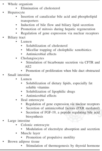
sur-Table I. Functions of bile acids currently recognized
• Whole organism
• Elimination of cholesterol • Hepatocyte
• Insertion of canalicular bile acid and phospholipid transporters
• Induction of bile flow and biliary lipid secretion • Promotion of mitosis during hepatic regeneration • Regulation of gene expression via nuclear receptors • Biliary tract
• Lumen
• Solubilization of cholesterol
• Micellar trapping of cholephilic xenobiotics • Antimicrobial effects
• Cholangiocytes
• Stimulation of bicarbonate secretion via CFTR and AE2
• Promotion of proliferation when bile duct obstructed • Small intestine
• Lumen
• Solubilization of dietary lipids, especially fat soluble vitamins
• Solubilization of lipophilic drugs • Antimicrobial effects
• Ileal enterocyte
• Regulation of gene expression via nuclear receptors • Secretion of antimicrobial factors (FXR mediated) • Secretion of FGF-19, a peptide regulating bile acid biosynthesis
• Large intestine
• Colonic enterocyte
• Modulation of electrolyte absorption and secretion • Muscle layer
• Promotion of propulsive motility • Brown adipose tissue
edigraphic.com
rounded by a one molecule thick bilayer of bile acid molecules.
Both the bile salts and the fatty acids remain in any given micelle for only some milliseconds. They move out from the micelle and exchange with fatty acids and bile salt molecules present as monomers in the surrounding aqueous phase. The mixed micelle is a flickering cluster. Uptake at the cell membrane is believed to be both pas-sive (high capacity, low affinity) and carrier-mediated (low capacity, high affinity). Passive flipflop of fatty ac-ids (in protonated, uncharged form) across a lipid bilay-ers is bidirectional, the direction being determined by the concentration gradient. At least one fatty acid trans-porter is also a coenzyme A synthetase.27 Linking a fatty
acid to Coenzyme A in thio-ester linkage prevents back diffusion.
The presence of a micellar phase during digestion in man is well established by sampling small intestinal content and isolating the micellar phase by ultracentrif-ugation28 or ultrafiltration,29 and similar studies have
been performed in large animals such as the dog and the cow. However, as noted, experimental isolation of a mi-cellar phase has been performed in relatively few other vertebrates.
Bile salts might also play a role in protein digestion. Bile salts will adsorb to any hydrophobic domains of di-etary proteins, and this might in turn promote protein de-naturation, rendering proteins more susceptible to diges-tion by the proteolytic enzymes.
Bile salts solubilize biliary lipids (phosphatidyl-choline and cholesterol) as well as dietary lipids. Bil-iary phospholipid – about 6 grams/day in the adult – serves to emulsify dietary triglyceride, but is rapidly hydrolyzed to lysophosphatidylcholine and fatty acid. Lysophosphatidylcholine is water soluble and presum-ably partitions between the aqueous phase and the mixed micelles. It is rapidly absorbed. Biliary choles-terol is solubilized in mixed micelles and mixes with dietary cholesterol.
Cholesterol absorption varies between individuals and ranges widely. In the past, changing cholesterol ab-sorption was not considered very important as increasing dietary cholesterol (c.f. 30) or blocking cholesterol ab-sorption by feeding high dose plant sterols31 had only
small effects on levels of plasma or biliary cholesterol. The reason for the lack of influence of dietary cholester-ol on biliary or plasma lipids was related to homeostatic control by negative feedback of cholesterol synthesis by the hepatocyte. When less cholesterol reached the hepa-tocyte, cholesterol biosynthesis increased; when more cholesterol reached the hepatocyte, cholesterol biosyn-thesis decreased.
However, in the past few years, cholesterol absorption has become a topic of intense scrutiny. This view has been modified strikingly in the past few years, as NPC1L1, a cholesterol transport protein present in the
apical membrane of the enterocyte was identified, and as a new drug, ezetimibe, was shown to block cholesterol uptake by this transporter.32 Moreover, combining
ezetimibe with a statin (which inhibits cholesterol syn-thesis) was shown to greatly enhance the hypocholester-olemic effect of the statin.33
The mixed micelles also solubilize plant sterols such as sitosterol. In the past, these molecules were considered to be poorly absorbed because of their different molecular shape. With the identification of an ATP-energized choles-terol (and plant scholes-terol) efflux pump34 formed by two half
transporters (ABC5 and ABC8), it became clear that plant sterols were absorbed (presumably by the cholesterol im-porter). However, as plant sterols in contrast to cholesterol underwent little esterification, they are not incorporated into chylomicrons and effluxed back into the lumen via ABC5/ABC8. In the past, it had been assumed erroneously that plant sterols did not enter the enterocyte.
An extremely rare disease, sitosterolemia, was shown to be caused by defects in ABC5 and/or ABC8,35,36 and is
characterized by the accumulation of plant sterols in the body evidenced by sterol-rich xanthomata. The laborato-ry group of Gerald Salen has just reported that feeding ezetimibe to such a patient caused a dramatic fall in plas-ma levels of plant sterols and regression of xanthoplas-mata.37
The ultimate fate of fatty acid in the enterocyte is re-esterification to form triglyceride, and packaging of the triglyceride droplets into chylomicrons. Details of intrac-ellular processing is beyond the scope of this review, but reviews are available.
It is reasonable to speculate that bile salts play a gen-eral role in keeping the absorptive surface of the small in-testine clean. Bile salts should adsorb to food residues, giving them a negative charge, thereby precluding their aggregation. However, this has not been tested experi-mentally. Cells at the tip of the villus are undergoing continuous apoptosis followed by shedding. Cellular lip-ids will be solubilized by bile salts and delivered to more caudal enterocytes.
Besides forming mixed micelles with dietary lipids and their lipolysis products, and cleaning the small intes-tinal surface, conjugated bile acids also have potent anti-microbial effects in the small intestine.38 Conjugated bile
acids thus join defensins and IgA as luminal molecules inhibiting the growth of bacteria.
The evidence for a potent antimicrobial effect of con-jugated bile acids is based on a number of reports, mostly in the surgical literature, showing that bile duct ligation leads to bacterial proliferation in the small intestine. Ex-tending this work was a study by Lorenzo-Zuniga et al39
edigraphic.com
SUSTRAÍDODE-M.E.D.I.G.R.A.P.H.I.C:ROP ODAROBALE FDP VC ED AS, CIDEMIHPARG ARAP
ACIDÉMOIB ARUTARETIL :CIHPARGIDEM two conjugated bile acids (cholylglycine or cholylsar-cosine) abolished bacterial overgrowth, decreased endot-oxemia, and increased survival. Inagaki et al40 extended
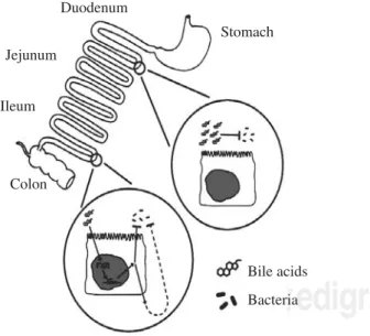
this work by testing whether administration of an agonist of the nuclear receptor FXR (known to have bile acids as its major ligand) would have an effect on the bacterial overgrowth occurring in the bile duct ligated mouse. They found that administration of GW4064, an FXR ago-nist synthesized by Glaxo Smith Kline, led to a remark-able fall in bacterial proliferation in the small intestine. As these animals had bile duct ligation, the effect of this FXR agonist must indicate a second indirect effect by which bile acids exert an antimicrobial effect. Figure 1
summarizes the antimicrobial effects of bile acids in the small intestine.
Intraluminal deficiency of bile acids. A conjugated bile acid deficiency occurs when the enterohepatic circu-lation is obstructed, diverted, or when intestinal conser-vation of bile acids is impaired because of ileal dysfunc-tion. In patients with a short bowel syndrome, severe bile acid malabsorption occurs because most patients with this condition have lost their ileum. The therapy current-ly practiced is to enrich dietary trigcurrent-lyceride in medium chain triglyceride, as medium chain fatty acids are water soluble and do not require micellar solubilization for ab-sorption. Moreover they are absorbed extremely rapidly as they are absorbed both transcellularly and paracellu-larly. Fat soluble vitamins are given parenterally.
Patients with short bowel syndrome have both a loss of intestinal absorptive surface as well as defective micel-lar solubilization. The feeding of conjugated bile acids
can correct the defect in fat digestion. A conjugated bile acid analogue, cholylsarcosine, was synthesized, found the physicochemical properties of the natural conjugates of cholic acid41 and shown to be resistant to bacterial
degradation (deconjugation and dehydroxylation) in ani-mals42 and man.43 Addition of cholylsarcosine to the diet
increased triglyceride absorption in dogs with bile acid malabsorption induced by ileal resection.44 In patients
with short bowel syndrome cholylsarcosine administra-tion increased triglyceride absorpadministra-tion and induced weight gain.44,46 However, cholylsarcosine is
investiga-tional and few patients have been treated to date. Phar-maceutical companies have shown little interest in cholylsarcosine because there is no patent protection and the perceived market is small. Cholylsarcosine can cause gastric irritation, but if an enteric coating is used, the compound must be rapidly released in the duodenum, as this is a major site of fat absorption, and small intestinal transit is often very rapid in patients with short bowel syndrome. Such a formulation of cholylsarcosine has been reported.47
Bile acid recycling: the enterohepatic circulation
In health, daily bile acid secretion when measured by an indicator dilution technique is 30-50 mmoles day.48
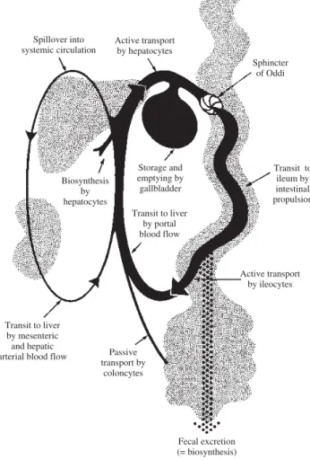
Daily synthesis averages about 1 mmole/day. The ability to secrete more bile acid than is synthesized results from a recycling bile acid pool. Development of a bile acid pool results from efficient intestinal conservation mediat-ed by in large part by the ileal conjugatmediat-ed bile acid trans-port system. Schematic views of the enterohepatic circu-lation of bile acids are shown in Figures 2 and Figure 3.
The enterohepatic circulation is regulated at two sites. The first is regulation of biosynthesis from cholesterol, which is mediated in negative feedback manner by sever-al mechanisms. First, bile acids in the hepatocyte activate a heterodimeric nuclear receptor (RXR-FXR) whose acti-vation induce the synthesis of a protein named shp.5Shp
together with activators binds to the promoter site of cyp7A1, (cholesterol 7α-hydroxylase) which is the rate limiting enzyme in bile acid biosynthesis. Binding of shp
down regulates bile acid biosynthesis. Second, there is a
shp independent pathway for down regulation, activated by inflammatory cytokines.49 Finally, FGF-19, a newly
identified protein that is released from the ileal entero-cyte by bile acids, also down regulates bile acid biosyn-thesis.50,51 Ileal absorption of bile acids is also regulated
in a negative feedback manner by bile acids, as dis-cussed below.
Bile acid uptake across the apical membrane of the il-eal enterocyte is mediated by a sodium dependent conju-gated bile acid cotransporter [apical sodium dependent bile acid transporter (ASBT) that has been found in every vertebrate in which it is sought.3 In humans, it is
ex-pressed weakly in cholangiocytes, in the gallbladder, in
Figure 1. Schematic depiction of the antimicrobial effects of bile acids in the small intestinal lumen. Bile acids, possibly aided by fatty acids, have a direct antimicrobial effect on luminal bacteria. Bile acids also have an indirect effect on luminal bacteria, media-ted by the nuclear receptor FXR; the mechanism of this effect has not been clarified. Taken from reference 38.
Stomach Duodenum
Jejunum
Ileum
Colon
Bile acids
edigraphic.com
the renal tubule and in the placenta.3 A sodium
indepen-dent bile salt transporter (oatp3) is present throughout the small intestine and may promote limited bile salt ab-sorption.52 There is little information on the contribution
of this transporter to bile salt conservation in vertebrates. Paracellular absorption of bile acids is believed to be negligible as the bile salt molecule is too large to pass via the tight junctions between small intestinal epithe-lial cells. Whether subjects with increased intestinal per-meability absorb bile acids via a paracellular route is not known. Were this to occur, the intraluminal concentra-tion of bile acids might fall, leading to bacterial prolifer-ation. Were bacterial proliferation to damage the paracel-lular junctions, a vicious cycle might ensue.
A bile acid binding protein in the ileal enterocyte plays an as yet undefined role in promoting vectorial transport. Exit from the ileal enterocyte is mediated by a heterodimeric bile salt transporter composed of two sub-units OSTalpha and OSTbeta.53,54Figure 4 shows a
sche-matic view of vectorial transport of bile acids through the ileal enterocyte.
Ileal transport in man and the mouse appears to be also regulated in a negative feedback manner – thus bile acid feeding down regulates bile acid transport, and bile acid sequestrant feeding upregulates bile acid transport.3
Up-regulation may involve recruitment of more orad epithe-lial cells rather than enhanced transport by individual il-eal enterocytes. Details of regulation of the ilil-eal apical sodium dependent transporter are being clarified.
Bile acids are transported to the liver in portal venous blood are efficiently extracted despite being highly albu-min-bound. The extent to which bile acids are bound to albumin depends on bile acid structure: trihydroxy bile acids are bound much less completely (70%) than dihy-droxy bile acids (99%). Uptake is dependent on bile acid structure and is greater for trihydroxy bile acids than
di-Figure 2. Schematic illustration of the enterohepatic circulation of bile acids. Conjugated bile acid absorption mediated by OATP3 may also occur throughout the small intestine, but the magnitude of this flux in man is not known.
Sphincter of Oddi Active transport
by hepatocytes Spillover into
systemic circulation
Transit to ileum by intestinal propulsion Storage and
emptying by gallbladder Transit to liver
by portal blood flow Biosynthesis
by hepatocytes
Active transport by ileocytes
Transit to liver by mesenteric and hepatic
arterial blood flow Passive transport by
coloncytes
Fecal excretion (= biosynthesis)
Figure 3. Schematic depiction of the en-terohepatic circulation and metabolism of bile acids. Normally, bile acids are efficiently conjugated (amidated) with glycine or taurine, and cholehepatic shunting is small. The ileal apical bile acid transporter is present on the apical membrane of cholangiocytes, so a mo-dest amount of cholehepatic cycling of conjugated bile acids does occur. Bile acid synthesis is balanced by fecal loss.
Input of “new” (unconjugated) 7-deoxy BA
(damaged)
Unconjugated) BA (damaged)
Input of “new” BA biosynthesized from cholesterol (pericentral
hepatocytes)
Transport and repair of “old” BA (periportal
hepatocytes)
Conjugated BA (undamaged)
Biliary ductules
Cholehepatic Cycling
(Unconjugated lipophilic BA)
Conjugated BA (undamaged and repaired) Duodenum
Jejunum Ileum
edigraphic.com
SUSTRAÍDODE-M.E.D.I.G.R.A.P.H.I.C:ROP ODAROBALE FDP VC ED AS, CIDEMIHPARG ARAP
ACIDÉMOIB ARUTARETIL :CIHPARGIDEM hydroxy bile acids, and for a given steroid moiety, is greater for conjugated bile acids than unconjugated bile acids. Fractional extraction of bile acids remains constant despite varying bile acid loads to the liver. Therefore bile acid extraction is said to be «blood flow limited».
Vectorial transport by the hepatocyte involves uptake at the basolateral membrane and active secretion across the canalicular membrane. Basolateral uptake is mediat-ed by both sodium dependent and sodium independent transporters. Although there are continuing efforts to de-fine the substrate specificity and role of the many sinuso-idal membrane uptake proteins,8-11 It is still not clear
which transporters other than the sodium dependent transporter are involved in bile acid uptake. Figure 5 il-lustrates schematically the major transporters involved in vectorial transport of bile acids through the hepatocyte.
Secretion across the canalicular membrane involves predominantly the canalicular bile salt export pump (BSEP), which is energized by hydrolysis of ATP. Since its cloning in 1998,55 BSEP has been studied in
consider-able detail.6,7 The human BSEP transports both
conjugat-ed and unconjugatconjugat-ed bile acids as well as sulfatconjugat-ed litho-cholyl conjugates. About 50 mutations have been identi-fied in infants born with «primary familial intrahepatic cholestasis» (type 2).56 Defects in BSEP may involve
fail-ure of mRNA to be formed, biosynthesis of a non-func-tional transporter, or biosynthesis of a transporter that is formed but not delivered to the canalicular membrane. When infants are born with non-functioning BSEP, they develop hepatocyte necrosis and liver fibrosis, leading ultimately to liver failure. Liver transplantation is re-quired and is life-saving.
BSEP transports not only bile acids, but also a variety of drugs, including several statins.57,58 Inhibition of this
transporter by drugs can cause hepatotoxicity,57,59 and in
vitro screening techniques are being developed that will permit elucidation of interaction of drugs (and their me-tabolites) with BSEP.60
The major canalicular transporter for organic anions other than bile acids is MRP2. It transports many drug
Figure 4. Schematic illustration of the bile acid transport by the ileal entero-cyte. During active absorption, the ileal bile acid binding protein (IBA-BP) may translocate to the nucleus. When IBAPB is ablated, bile acid ab-sorption still occurs, so the protein is not required for active bile acid ab-sorption. Abbreviations of the protein and gene are given in the insert.
Basolateral membrane
BA
OST / OST
a
b ASBT
X
IBABP
Apical membrane
BA
Na
Abbreviation: X , unidentified anion (s)
Protein ASBT OST /OST IBABP (FABP6)
a b
Gene SLC10A2 OST /OST FABP6
a b
- +
-Figure 5. Schematic illustration of bile acid transport by the hepatocyte. The substrate specificity of the three canali-cular transporters is not yet perfectly defined and varies between species. ABCG2 is also known as the breast can-cer related gene. Normally, the propor-tion of bile acid sulfates or bile acid glu-curonosides (glucuronides) in bile is quite low. MRP4 promotes the cotrans-port of conjugated bile acids and redu-ced glutathione. MRP3 is thought to be involved in efflux of glucuronosides. OSTα/OSTβ upregulate markedly in cholestatic liver disease and pump bile acids out of the hepatocyte into sinusoi-dal blood. Abbreviations of the protein and gene are given in the insert.
Protein NTCP OATP´S MRP3 MRP4 BSEP MRP2 ABCG2 OST /OSTa b
Gene SLC10A1 SLC21.. (many) ABCC3 ABCC4 ABCB11 ABCC2 ABCG2 OST /OSTa b BA
MRP4
GSH BA X
-OST / OST
a b Basolateral
membrana
Canalicular membrane
BA
BA
X Na
NTCP
OATP´S
MRP3
BSEP
ABCG2
MRP2
Glucuronosides
BA sulfates
BA glucuronosides BA amidates +
-edigraphic.com
metabolites and bilirubin glucuronides. In animals, MRP2 mediates canalicular secretion of bile acid sul-fates. However, in man, BSEP appears to mediate the canalicular secretion of sulfated (and amidated) deriva-tives of lithocholic acid.61 Such transport should be
im-portant in patients with cholestatic liver disease who in-gest ursodiol, as ursodiol is converted to lithocholic acid by colonic bacteria.
Bile as an excretory fluid
Overview: The major excretory constituents of bile are bile acids – end products of cholesterol metabolism – and bile pigments (conjugated bilirubin and/or biliver-din – end products of heme metabolism, and polyvalent metal cations. A survey of biliary lipid composition in over 100 vertebrates showed that in many vertebrates, only bile acids are present in appreciable proportions, i.e. in many vertebrates the phospholipid/bile acid and cho-lesterol/bile acid ratio is extremely low.62
Human bile is lipid rich containing a high ratio of phospholipid to bile acid (0.3) and a high ratio of choles-terol to bile acid (0.07). Man appears to differ from all other animals characterized to date in eliminating choles-terol from the body to a greater extent as cholescholes-terol than by conversion to bile acids. Balance studies suggest that perhaps 60% of cholesterol is eliminated from the body as cholesterol, the remainder being eliminated as bile ac-ids.63 In contrast, in most animals, the virtual absence of
cholesterol in bile suggests that most cholesterol is elimi-nated from the body by biotransformation to bile acids.62
Biliary excretion permits the organism to eliminate substrates that cannot be eliminated via renal excretion. Besides bile acids, biliary excretion involves the excre-tion of organic molecules and heavy metal caexcre-tions that are highly protein bound.64,65 Thus bile serves as the
elim-ination route for plant sterols, lipophilic drug metabo-lites, and heavy metals. Because none of such molecules have an appreciable enterohepatic circulation, their con-centration in bile is quite low.
The digestive function of bile acids has already been summarized, and bile acid transport into bile by BSEP was discussed above.
Biliary phospholipid secretion. The major phospholip-id of mammalian bile is phosphatphospholip-idylcholine (PC) which is not believed to have any important digestive function. This is because phospholipids are present in dietary lipids, and in addition, the enterocyte can biosynthesize PC which is a key part of the lipid surface layer of chylomicrons. Phos-pholipid serves to solubilize cholesterol in bile, as it forms the lipid core of the mixed micelles present in bile that can in turn solubilize cholesterol and other biliary lipids. The secretion of PC into bile is mediated by a phospholipid flip-pase termed MDR2 in mice and MDR3 in man.66
To date, the only canalicular protein involved in bil-iary phospholipid secretion is MDR2.66 This transporter
can be shown in vitro to flip PC molecules across mem-branes, but it has been proposed that the flippase also contributes to the generation of hemivesicles containing mostly PC, as these are present by electron microscopy in wild type mice, but not in MDR2 knockout mice.67 Bile
acids convert the hemivesicles into mixed micelles by ad-sorbing to the lipid bilayer and at a sufficiently high bile acid/phospholipid ratio destroying the bilayer stability.
The luminal side of the canalicular membrane must be highly resistant to the solubilizing action of bile acids. Sphingomyelin which is a membrane stabilizer is present in the luminal face of the canalicular membrane but is present in mammalian bile in only trace amounts. Phos-phatidylserine is believed to be flipped back from the lu-minal face to the cellular face by FIC1. Primary familial intrahepatic cholestasis type I involves mutations in a gene called FIC1 . One view of the protein encoded for by this gene is that it promotes the flipflop of phosphati-dylserine from the biliary face of the canalicular mem-brane to the cytosolic face. Defective removal of this phospholipid from the biliary face of the canalicular membrane results canalicular membrane fragility. As a re-sult phosphatidylserine as well as canalicular proteins are released into bile when bile flow is induced by infus-ing bile acids.68
It is generally assumed that the PC molecules that en-ter bile at the canaliculus remain in the mixed micelle and are not absorbed by the biliary ductules. The validi-ty of this assumption is not known. In the gallbladder, the phospholipid/bile salt ratio decreases as bile is con-centrated, suggesting that some phospholipid is ab-sorbed by the gallbladder epithelium.69
Knockout of this gene in mice results in the absence of biliary phospholipid and causes the development of a peribiliary fibrosis.66 Thus biliary PC serves to protect
the biliary ductules from the membrane solubilizing ac-tion of bile acids. The monomeric activity of bile acids is higher in the absence of phospholipids, and the in-creased monomeric activity of bile acids has been con-sidered the causal agent for biliary ductule injury, None-theless, the increase in the monomeric bile acid concen-tration is modest, if findings in model systems apply to the in vivo situation.70 When phospholipid is absent from
bile, the cholesterol concentration is also greatly re-duced. Some work on other organs suggests71 that the
combination of PC and cholesterol in bile may render membranes resistant to bile acids. If this speculation is correct, the bile duct injury observed in the MDR-/- mouse
might result from the decreased concentration of both phospholipid and cholesterol.
The phenotype of decreased MDR function in man has been reported to be calculous biliary disease,72
pre-sumably because of defective micellar solubilization of cholesterol; mutations in MDR have also been reported in cholestasis of pregnancy.73 Biliary lipid analyses have
pa-edigraphic.com
SUSTRAÍDODE-M.E.D.I.G.R.A.P.H.I.C:ROP ODAROBALE FDP VC ED AS, CIDEMIHPARG ARAP
ACIDÉMOIB ARUTARETIL :CIHPARGIDEM tients with gallbladder inflammation despite the absence of gallstones, a disease named chronic acalculous chole-cystitis.74 Biliary lipid analyses showed dilute bile with a
low PC/bile acid ratio. This observation has not been confirmed. In these patients, the decreased phospholipid could result from rather than cause the mucosal inflam-mation. There would appear need for additional work to characterize the phenotype of MDR3 deficiency in man.
In the National Cooperative Gallstone study per-formed in the United States, phospholipid/bile acid ratios showed a normal distribution, and patients with low phospholipid/bile acid ratios were not identified as hav-ing a distinct phenotype.75 It is rational to treat MDR3
deficiency patients with ursodeoxycholic acid (UDCA), but no controlled studies have examined efficacy.
The MDR2 knockout mouse develops a striking peribiliary fibrosis. This can be treated successfully by administering norursodeoxycholic acid, the C23 (C24-nor) homologue of UDCA;76 such a molecule has an
isobu-tanoic acid side chain and thereby differs from UDCA which has an isopentanoic acid side chain. NorUDCA is secreted intact into bile and reabsorbed in the biliary ductules to undergo cholehepatic shunting in animals77
and apparently in man.78 In MDR2 knockout mice, there
is a striking improvement in fibrosis, a decrease in leuko-cyte infiltration and ductular proliferation, and a marked increase in the activity of detoxifying enzymes such as sulfotransferase. UDCA has a much weaker effect. Wheth-er norUDCA will prove to have any clinical value in man is quite uncertain. In contrast to other natural bile acids, it has considerable renal excretion, especially of its ester glucuronide, its major metabolite. Although the peribil-iary fibrosis of the MDR2 knockout mouse resembles pri-mary sclerosing cholangitis, biliary phospholipid secre-tion is quite normal in this disease, so it remains quite uncertain that norUDCA will have a therapeutic effect in sclerosing cholangitis.
Biliary cholesterol secretion: Cholesterol is secreted into bile largely by the heterodimeric transporter ABC5/ ABC8.79,80 The protein is considered to be a cholesterol
flippase, flipping cholesterol molecules from the cytoso-lic face of the canacytoso-licular membrane to the luminal face.81
Over expression of this transporter results in increased biliary cholesterol secretion.82 Knockout of this
trans-porter does not fully eliminate biliary cholesterol secre-tion, indicating that either other cholesterol transporters are present in the canaliculus, or that postcanalicular mechanisms for cholesterol entry into bile exist, or both.
Cholesterol is absorbed in the biliary tract in some species, based on the observation that at least in the dog, the cholesterol/bile acid ratio increases markedly when ezetimibe is given. Ezetimibe is secreted into bile as a glucuronide that maintains pharmacodynamic activity.83
Cholesterol absorption in the biliary tract is inhibited and biliary cholesterol increases markedly. As yet, whether such vigorous cholesterol absorption by
cholan-giocytes that occurs in the dog, is also present in other species is not known. Cholesterol can exit the cholangio-cyte by basolateral ATP5/ATP8. During gallbladder stor-age, the proportion of cholesterol in bile falls, indicating that cholesterol is absorbed by the healthy gallbladder.84
Chronic absorption of cholesterol from supersaturated bile leads to oxidative stress that in turn causes impaired gallbladder motility, and this promotes cholesterol gall-stone formation.85,86
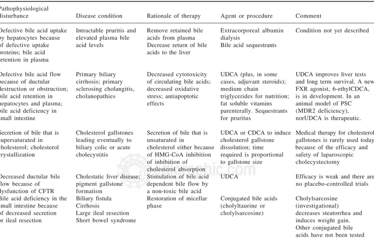
Bile acid therapy: Bile acid therapy is still used infre-quently in liver and intestinal disease, with the single exception of ursodiol that is used by most physicians for a variety of uncommon hepatobiliary diseases in which cholestasis occurs. An overview of the rationale of bile acid therapy is given in Table II and Table III.
At present, there are four rationales for bile acid thera-py. Bile acid replacement is when bile acids are adminis-tered to replace a deficiency state. Such occurs in inborn errors of bile acid biosynthesis or bile acid conjugation, as well as in conditions when there is a deficiency in the small intestine because of ileal dysfunction. This occurs in short bowel syndrome, as the ileum has usually been resected in such patients. Bile acid deficiency in the small intestine also occurs in cholestatic liver disease, but usually bile acid secretion into the small intestine is sufficient to permit adequate lipid absorption in the adult, and it has been considered dangerous to administer exogenous bile acids other than ursodiol when endoge-nous bile acids are already.
Bile acid displacement occurs when the composition of the circulating bile acids is changed by exogenous bile acid administration, but there is relatively little change in bile acid secretion. Bile acid displacement is the rationale when ursodiol is used in cholestatic liver diseases. The mechanism of action of ursodiol is com-plex and involves replacement of endogenous cytotoxic bile acids [chenodeoxycholic (CDCA) and DCA) by UDCA a non-cytotoxic bile acid; the mechanism is like-ly to be competition for active ileal transport. UDCA also has antiapoptotic effects, and anti-oxidative injury ef-fects. It may also reduce endoplasmic reticulum «stress» and appears to also have anti-inflammatory effects. UDCA is also used to lower the cholesterol proportion in bile and thereby induce gallstone dissolution. UDCA is amidated with glycine or taurine in the liver, and the re-sulting UDCA conjugates, as any natural conjugated bile acid, induce bile acid dependent bile flow. In cystic fi-brosis, ductular bile flow is decreased because of non-functioning of the CFTR chloride channel. Increased canalicular bile flow induced by UDCA administration is thought to benefit children with cystic fibrosis by reduc-ing the likelihood of developreduc-ing chronic liver disease.
A third rationale for bile acid therapy may be termed
edigraphic.com
Table II. Rationale of bile acid therapy: liver defects.Pathophysiological
disturbance Disease condition Rationale of therapy Agent or procedure Comment
Defective biosynthesis of bile Cholestatic liver disease in Suppression of biosynthesis Primary bile acids Disease conditions are acids because of defects in infants; cerebrotendinous of toxic precursors (cholic acid and/or extremely rare; therapy enzymes involved in bile xanthomatosis (CTX) Restoration of enterohepatic chenodeoxycholic may be life saving for acid biosynthesis; exuberant circulation of bile acids acid (CDCA) inborn errors of bile
bile acid biosynthesis because acid biosynthesis; therapy
of lack of feedback inhibition with CDCA in CTX
slows disease progression Defective bile acid N-acyl Malabsorption of fat Restore enterohepatic In principle: ox bile No successful therapy amidation with taurine or soluble vitamins; circulation of conjugated salts, or synthetic reported as yet. Use of glycine because of defective possibly cholestatic bile acids; restore mixed bile acid N-acyl conjugated bile acids CoA formation or defective liver disease micelles in small intestinal amidates, e.g. would be investigational. bile acid amino acid lumen taurocholate or Ox bile salts are available
transferase cholylsarcosine as nutritional supplements
Defective canalicular transport Primary familial Reduce toxicity of Ursodiol Some 50 defects in because of deficiency in bile intrahepatic cholestasis, retained bile acids BSEP have been identified salt export pump (BSEP) or type II (neonatal Remove retained bile acids Extracorporeal
canalicular dysfunction; bile hepatitis on biopsy) Prevent inappropriate albumin dialysis Generally, infants acid retention in hepatocytes ileal absorption of any progress to liver failure and plasma; bile acid secreted bile acids Bile acid sequestrants and require liver deficiency in small intestinal Enrich diet in triglycerides transplantation
lumen whose fatty acids are Add medium chain
water soluble triglycerides to diet Replace defective
hepatocytes or entire Hepatocyte infusion or liver liver transplantation
Table III. Rationale of bile acid therapy: liver disease (continued) and intestinal disease.
Pathophysiological
disturbance Disease condition Rationale of therapy Agent or procedure Comment
Defective bile acid uptake Intractable pruritis and Remove retained bile Extracorporeal albumin Condition not yet described by hepatocytes because elevated plasma bile acids from plasma dialysis
of defective uptake acid levels Decrease return of bile Bile acid sequestrants proteins; bile acid acids to the liver
retention in plasma
Defective bile acid flow Primary biliary Decreased cytotoxicity UDCA (plus, in some UDCA improves liver tests because of ductular cirrhosis; primary of circulating bile acids; cases, adjuvant steroids); and long term survival. A new destruction or obstruction; sclerosing cholangitis, decreased oxidative medium chain FXR agonist, 6-ethylCDCA, bile acid retention in cholanopathies stress; antiapoptotic triglycerides for nutrition; is in development. In an hepatocytes and plasma; effects fat soluble vitamins animal model of PSC bile acid deficiency in parenterally. Sequestrants (MDR2 deficiency),
small intestine for pruritus norUDCA is therapeutic.
Secretion of bile that is Cholesterol gallstones Secretion of bile that is UDCA or CDCA to induce Medical therapy for cholesterol supersaturated in leading eventually to unsaturated in cholesterol gallstone gallstones is rarely used today cholesterol; cholesterol biliary colic or acute cholesterol either because dissolution; time because of the efficacy and crystallization cholecystitis of HMG-CoA inhibition required is proportional safety of laparoscopic
of inhibition of to gallstone size cholecystectomy cholesterol absorption
Decreased ductular bile Cholestatic liver disease; Stimulation of bile acid UDCA Efficacy is weak and there are flow because of pigment gallstone dependent bile flow by no placebo-controlled trials dysfunction of CFTR formation a non-toxic bile acid
Bile acid deficiency in the Biliary fistula Restoration of micellar Conjugated bile acids Cholylsarcosine small intestine because Cirrhosis phase (cholyltaurine or (investigational) of decreased secretion Large ileal resection cholylsarcosine) decreases steatorrhea and or ileal resection Short bowel syndrome induces weight gain.
edigraphic.com
SUSTRAÍDODE-M.E.D.I.G.R.A.P.H.I.C:ROP ODAROBALE FDP VC ED AS, CIDEMIHPARG ARAP
ACIDÉMOIB ARUTARETIL :CIHPARGIDEM and shown to have antifibrotic effects and anticholestatic effects in experimental models in the mouse. The effect of this FXR agonist could involve not only FXR activa-tion in hepatocytes but also. The compound is undergo-ing early clinical studies with hopes of eventual market-ing.
A fourth rationale for bile acid therapy is ductular tar-geting, using a bile acid that is secreted in part in uncon-jugated form by BSEP and whose unconuncon-jugated form is membrane permeable, resulting in absorption in the bil-iary ductules. As noted above, an example of such a bile acid is norUDCA, which causes a marked diminution in the peribiliary fibrosis occurring in the MDR-/- knockout
mouse that is unable to secrete phosphatidylcholine into bile. NorUDCA is an investigational compound, and as yet there is no proof of principle that the compound will have useful efficacy in man.
Epilogue: The inability to sample canalicular bile means that details of biliary lipid secretion are often de-duced or inferred; nonetheless, great progress has been made in understanding canalicular bile secretion. The explanation for the increased cholesterol/phospholipid ratio (or cholesterol/bile acid ratio) present in cholesterol gallstone patients is being actively pursued at a genetic level.87-89 Much needs to be done to clarify the clinical
phenotype of decreased MDR3 function in man. None-theless, identification of the major biliary lipid transport-ers and elucidation of their regulation has been a remark-able achievement. Progress in this area is likely to be great in the coming decades.
References
1. Hofmann AF. Overview of bile secretion. In: Schultz SG, ed. Handbook of Physiology. Section on the Gastrointestinal Sys-tem. American Physiological Society, Bethesda, 1989: 549-566. 2. Hofmann AF. Enterohepatic circulation of bile acids. In: Handbook of Physiology. The Gastrointestinal System. Volume III. Salivary, gastric, pancreatic, and hepatobiliary secretion. Schultz SG, editor. American Physiological Society, Bethesda, 1989: 567-596. 3. Dawson PA, Shneider BL, Hofmann AF. Bile formation and the
enterohepatic circulation. In: Barrett KE, Ghishan FK, Merchant JL, Said HM, Wood JD, Johnson LR, eds. Physiology of the Gastrointestinal tract. Elsevier Academic Press, San Diego 2006: 1437-62.
4. Pauli-Magnus C, Stieger B, Meier Y, Kullak-Ublick GA, Meier PJ. Enterohepatic transport of bile salts and genetics of cholestasis.
J Hepatol 2005; 43: 342-57.
5. Chiang JY. Nuclear receptor regulation of lipid metabolism: potential therapeutics for dyslipidemia, diabetes, and chronic heart and liver diseases. Curr Opin Investig Drugs 2005; 6: 994-1001.
6. Stieger B, Meier Y, Meier PJ. The bile salt export pump. Pflugers Arch 2006 (in press).
7. Suchy FJ, Ananthanarayanan M. Bile salt excretory pump: biol-ogy and pathobiolbiol-ogy. J Pediatr Gastroenterol Nutr 2006; 43 Suppl 1: S10-6.
8. Faber KN, Muller M, Jansen PL. Drug transport proteins in the liver. Adv Drug Deliv Rev 2003; 55: 107-24.
9. Keppler D. Uptake and efflux transporters for conjugates in human hepatocytes. Methods Enzymol 2005; 400: 531-42.
10. Suzuki H, Sugiyama Y. Transporters for bile acids and organic anions. Pharm Biotechnol 1999; 12: 387-439.
11. Kullak-Ublick GA, Stieger B, Meier PJ. Enterohepatic bile salt transporters in normal physiology and liver disease. Gastroen-terology 2004; 126: 322-42.
12. Oude Elferink RP, Paulusma CC, Groen AK. Hepatocanalicular transport defects: pathophysiologic mechanisms of rare diseases.
Gastroenterology 2006; 130: 908-25.
13. Eloranta JJ, Meier PJ, Kullak-Ublick GA. Coordinate transcrip-tional regulation of transport and metabolism. Methods Enzymol
2005; 400: 511-30.
14. Engels BM, Hutvagner G. Principles and effects of microRNA-mediated post-transcriptional gene regulation. Oncogene 2006; 25: 6163-9.
15. Meier PJ, Boyer JL. Preparation of basolateral (sinusoidal) and canalicular plasma membrane vesicles for the study of hepatic transport processes. Methods Enzymol 1990; 192: 534-45. 16. Cui Y, Konig J, Keppler D. Vectorial transport by
double-trans-fected cells expressing the human uptake transporter SLC21A8 and the apical export pump ABCC2. Mol Pharmacol 2001; 60: 934-43. 17. Mita S, Suzuki H, Akita H, Hayashi H, Onuki R, Hofmann AF, Sugiyama Y. Vectorial transport of unconjugated and conju-gated bile salts by monolayers of LLC-PK1 cells doubly trans-fected with human NTCP and BSEP or with rat Ntcp and Bsep.
Am J Physiol Gastrointest Liver Physiol 2006; 290: G550-6. 18. Abdullah-Sayani A, Bueno-de-Mesquita JM, van de Vijver MJ.
Technology Insight: tuning into the genetic orchestra using microarrays—limitations of DNA microarrays in clinical prac-tice. Nat Clin Pract Oncol 2006; 3: 501-16.
19. Hu S, Loo JA, Wong DT. Human body fluid proteome analysis.
Proteomics 2006.
20. Tietz P, de Groen PC, Anderson NL, Sims C, Esquer-Blasco R, Meheus L, Raymackers J, et al. Cholangiocyte-specific rat liver proteins identified by establishment of a two-dimensional gel protein database. Electrophoresis 1998; 19: 3207-12. 21. Schmitt S, Prokisch H, Schlunck T, Camp DG, 2nd, Ahting U,
Waizenegger T, Scharfe C, et al. Proteome analysis of mitochon-drial outer membrane from Neurospora crassa. Proteomics 2006; 6: 72-80.
22. Sai Y, Nies AT, Arias IM. Bile acid secretion and direct targeting of mdr1-green fluorescent protein from Golgi to the canalicular membrane in polarized WIF-B cells. J Cell Sci 1999; 112 (Pt 24): 4535-45.
23. Marzioni M, Glaser SS, Francis H, Phinizy JL, LeSage G, Alpini G. Functional heterogeneity of cholangiocytes. Semin Liver Dis
2002; 22: 227-40.
24. Lucassen J. Hydrolysis and precipitates in carboxylate soap so-lutions. J Phys Chem 1966; 70: 1824-30.
25. Roda A, Hofmann AF, Mysels KJ. The influence of bile salt structure on self-association in aqueous solutions. J Biol Chem
1983; 258: 6362-70.
26. Hjelm RP, Schteingart CD, Hofmann AF, Thiyagarajan P. The structure of conjugated bile salt-fatty acid monoglyceride mixed colloids: studies by small-angle neutron scattering. J Phys Chem
2000; 523: 299-307.
27. Doege H, Baillie RA, Ortegon AM, Tsang B, Wu Q, Punreddy S, Hirsch D, Watson N, et al. Targeted deletion of FATP5 reveals multiple functions in liver metabolism: alterations in hepatic lipid homeostasis. Gastroenterology 2006; 130: 1245-58. 28. Hofmann AF, Borgstrom B. The intraluminal phase of fat
diges-tion in man: the lipid content of the micellar and oil phases of intestinal content obtained during fat digestion and absorption.
J Clin Invest 1964; 43: 247-57.
29. Mansbach CM, 2nd, Cohen RS, Leff PB. Isolation and proper-ties of the mixed lipid micelles present in intestinal content dur-ing fat digestion in man. J Clin Invest 1975; 56: 781-91. 30. Dam H, Prange I, Jensen MK, Kallehauge HE, Fenger HJ.
Stud-ies on human bile. IV. Influence of ingestion of cholesterol in the form of eggs on the composition of bile in healthy subjects.
edigraphic.com
31. Tangedahl TN, Thistle JL, Hofmann AF, Matseshe JW. Effect of beta-sitosterol alone or in combination with chenic acid on cho-lesterol saturation of bile and chocho-lesterol absorption in gallstone patients. Gastroenterology 1979; 76: 1341-6.
32. Altmann SW, Davis HR, Jr., Zhu LJ, Yao X, Hoos LM, Tetzloff G, Iyer SP, et al. Niemann-Pick C1 Like 1 protein is critical for intestinal cholesterol absorption. Science 2004; 303: 1201-4. 33. Feldman T, Davidson M, Shah A, Maccubbin D, Meehan A,
Zakson M, Tribble D, et al. Comparison of the lipid-modifying efficacy and safety profiles of ezetimibe coadministered with simvastatin in older versus younger patients with primary hy-percholesterolemia: a post Hoc analysis of subpopulations from three pooled clinical trials. Clin Ther 2006; 28: 849-59. 34. Graf GA, Yu L, Li WP, Gerard R, Tuma PL, Cohen JC, Hobbs
HH. ABCG5 and ABCG8 are obligate heterodimers for protein trafficking and biliary cholesterol excretion. J Biol Chem 2003; 278: 48275-82.
35. Lee MH, Lu K, Patel SB. Genetic basis of sitosterolemia. Curr Opin Lipidol 2001; 12: 141-9.
36. Yu L, von Bergmann K, Lutjohann D, Hobbs HH, Cohen JC. Selective sterol accumulation in ABCG5/ABCG8-deficient mice.
J Lipid Res 2004; 45: 301-7.
37. Salen G, Starc T, Sisk CM, Patel SB. Intestinal cholesterol ab-sorption inhibitor ezetimibe added to cholestyramine for sitosterolemia and xanthomatosis. Gastroenterology 2006; 130: 1853-7.
38. Hofmann AF, Eckmann L. How bile acids confer gut mucosal protection against bacteria. Proc Natl Acad Sci USA 2006; 103: 4333-4.
39. Lorenzo-Zuniga V, Bartoli R, Planas R, Hofmann AF, Vinado B, Hagey LR, Hernandez JM, et al. Oral bile acids reduce bacterial overgrowth, bacterial translocation, and endotoxemia in cir-rhotic rats. Hepatology 2003; 37: 551-7.
40. Inagaki T, Moschetta A, Lee YK, Peng L, Zhao G, Downes M, Yu RT, et al. Regulation of antibacterial defense in the small intestine by the nuclear bile acid receptor. Proc Natl Acad Sci
USA 2006; 103: 3920-5.
41. Lillienau J, Schteingart CD, Hofmann AF. Physicochemical and physiological properties of cholylsarcosine. A potential replace-ment detergent for bile acid deficiency states in the small intes-tine. J Clin Invest 1992; 89: 420-31.
42. Schmassmann A, Angellotti MA, Ton-Nu HT, Schteingart CD, Marcus SN, Rossi SS, Hofmann AF. Transport, metabolism, and effect of chronic feeding of cholylsarcosine, a conjugated bile acid resistant to deconjugation and dehydroxylation. Gastroen-terology 1990; 98: 163-74.
43. Schmassmann A, Fehr HF, Locher J, Lillienau J, Schteingart CD, Rossi SS, Hofmann AF. Cholylsarcosine, a new bile acid ana-logue: metabolism and effect on biliary secretion in humans.
Gastroenterology 1993; 104: 1171-81.
44. Longmire-Cook SJ, Lillienau J, Kim YS, Schteingart CD, Danzinger RG, Esch O, Hofmann AF. Effect of replacement therapy with cholylsarcosine on fat malabsorption associated with severe bile acid malabsorption. Studies in dogs with ileal resection. Dig Dis Sci 1992; 37: 1217-27.
45. Gruy-Kapral C, Little KH, Fordtran JS, Meziere TL, Hagey LR, Hofmann AF. Conjugated bile acid replacement therapy for short-bowel syndrome. Gastroenterology 1999; 116: 15-21. 46. Kapral C, Wewalka F, Praxmarer V, Lenz K, Hofmann AF.
Con-jugated bile acid replacement therapy in short bowel syndrome patients with a residual colon. Z Gastroenterol 2004;42:583-9. 47. Furst T, Bott C, Stein J, Dressman JB. Enteric-coated cholylsarcosine microgranules for the treatment of short bowel syndrome. J Pharm Pharmacol 2005; 57: 53-60.
48. van Berge Henegouwen GP, Hofmann AF. Nocturnal gallblad-der storage and emptying in gallstone patients and healthy sub-jects. Gastroenterology 1978; 75: 879-85.
49. Li T, Jahan A, Chiang JY. Bile acids and cytokines inhibit the human cholesterol 7 alpha-hydroxylase gene via the JNK/c-jun pathway in human liver cells. Hepatology 2006; 43: 1202-10.
50. Holt JA, Luo G, Billin AN, Bisi J, McNeill YY, Kozarsky KF, Donahee M, et al. Definition of a novel growth factor-depen-dent signal cascade for the suppression of bile acid biosynthesis.
Genes Dev 2003; 17: 1581-91.
51. Lundasen T, Galman C, Angelin B, Rudling M. Circulating in-testinal fibroblast growth factor 19 has a pronounced diurnal variation and modulates hepatic bile acid synthesis in man. J Intern Med 2006; 260: 530-6.
52. Walters HC, Craddock AL, Fusegawa H, Willingham MC, Dawson PA. Expression, transport properties, and chromosomal location of organic anion transporter subtype 3. Am J Physiol Gastrointest Liver Physiol 2000; 279: G1188-200.
53. Dawson PA, Hubbert M, Haywood J, Craddock AL, Zerangue N, Christian WV, Ballatori N. The heteromeric organic solute transporter alpha-beta, Ostalpha-Ostbeta, is an ileal basolateral bile acid transporter. J Biol Chem 2005; 280: 6960-8. 54. Ballatori N, Christian WV, Lee JY, Dawson PA, Soroka CJ, Boyer
JL, Madejczyk MS, et al. OSTalpha-OSTbeta: a major basolateral bile acid and steroid transporter in human intestinal, renal, and biliary epithelia. Hepatology 2005; 42: 1270-9.
55. Gerloff T, Stieger B, Hagenbuch B, Madon J, Landmann L, Roth J, Hofmann AF, et al. The sister of P-glycoprotein repre-sents the canalicular bile salt export pump of mammalian liver. J Biol Chem 1998; 273: 10046-50.
56. Thompson RJ, Azevedo RA, Galoppo C, Lewindon P, McKiernan P. Cholestatic and metabolic liver diseases: Working Group re-port of the second World Congress of Pediatric Gastroenterol-ogy, HepatolGastroenterol-ogy, and Nutrition. J Pediatr Gastroenterol Nutr
2004; 39 Suppl 2: S611-5.
57. Pauli-Magnus C, Meier PJ. Hepatobiliary transporters and drug-induced cholestasis. Hepatology 2006; 44: 778-87.
58. Hirano M, Maeda K, Hayashi H, Kusuhara H, Sugiyama Y. Bile salt export pump (BSEP/ABCB11) can transport a nonbile acid substrate, pravastatin. J Pharmacol Exp Ther 2005; 314: 876-82.
59. Mita S, Suzuki H, Akita H, Hayashi H, Onuki R, Hofmann AF, Sugiyama Y. Inhibition of bile acid transport across Na+/ taurocholate cotransporting polypeptide (SLC10A1) and bile salt export pump (ABCB 11)-coexpressing LLC-PK1 cells by cholestasis-inducing drugs. Drug Metab Dispos 2006; 34: 1575-81.
60. Hirano H, Kurata A, Onishi Y, Sakurai A, Saito H, Nakagawa H, Nagakura M, et al. High-speed screening and QSAR analysis of human ATP-binding cassette transporter ABCB11 (bile salt ex-port pump) to predict drug-induced intrahepatic cholestasis. Mol Pharm 2006; 3: 252-65.
61. Hayashi H, Takada T, Suzuki H, Onuki R, Hofmann AF, Sugiyama Y. Transport by vesicles of glycine- and taurine-conjugated bile salts and taurolithocholate 3-sulfate: a comparison of human BSEP with rat Bsep. Biochim Biophys Acta 2005; 1738: 54-62. 62. Moschetta A, Xu F, Hagey LR, van Berge-Henegouwen GP, van Erpecum KJ, Brouwers JF, Cohen JC, et al. A phylogenetic survey of biliary lipids in vertebrates. J Lipid Res 2005; 46: 2221-32. 63. Quintao E, Grundy SM, Ahrens EH, Jr. Effects of dietary
choles-terol on the regulation of total body cholescholes-terol in man. J Lipid Res 1971; 12: 233-47.
64. Gregus Z, Klaassen CD. Disposition of metals in rats: a compara-tive study of fecal, urinary, and biliary excretion and tissue distribution of eighteen metals. Toxicol Appl Pharmacol 1986; 85: 24-38.
65. Klaassen CD. Biliary excretion of metals. Drug Metab Rev 1976; 5: 165-96.
66. Oude Elferink RP, Paulusma CC. Function and pathophysiological importance of ABCB4 (MDR3 P-glycoprotein). Pflugers Arch
2006.
67. Crawford AR, Smith AJ, Hatch VC, Oude Elferink RP, Borst P, Crawford JM. Hepatic secretion of phospholipid vesicles in the mouse critically depends on MDR2 or MDR3 P-glycoprotein expression. Visualization by electron microscopy. J Clin Invest
edigraphic.com
SUSTRAÍDODE-M.E.D.I.G.R.A.P.H.I.C:ROP ODAROBALE FDP VC ED AS, CIDEMIHPARG ARAP
ACIDÉMOIB ARUTARETIL :CIHPARGIDEM 68. Paulusma CC, Groen A, Kunne C, Ho-Mok KS, Spijkerboer AL,
Rudi de Waart D, Hoek, et al. Atp8b1 deficiency in mice reduces resistance of the canalicular membrane to hydrophobic bile salts and impairs bile salt transport. Hepatology 2006; 44: 195-204. 69. Shiffman ML, Sugerman HJ, Kellum JM, Moore EW. Changes in gallbladder bile composition following gallstone formation and weight reduction. Gastroenterology 1992; 103: 214-21. 70. Higuchi WI, Liu CL, Adachi Y, Mazer NA, Lee PH. Equilibrium
dialysis studies on aqueous taurocholate-lecithin solutions: fur-ther validation of the method. Hepatology 1990; 12: 45S-49S; discussion 49S-50S.
71. Duane WC, Wiegand DM. Mechanism by which bile salt dis-rupts the gastric mucosal barrier in the dog. J Clin Invest 1980; 66: 1044-9.
72. Rosmorduc O, Hermelin B, Boelle PY, Parc R, Taboury J, Poupon R. ABCB4 gene mutation-associated cholelithiasis in adults.
Gastroenterology 2003; 125: 452-9.
73. Wasmuth HE, Glantz A, Keppeler H, Simon E, Bartz C, Rath W, Mattsson LA, et al. Intrahepatic cholestasis of pregnancy: the severe form is associated with common variants of the hepatobiliary phospholipid transporter gene ABCB4. Gut 2006. 74. Venkataramani A, Strong RM, Anderson DS, Gilmore IT, Stokes K, Hofmann AF. Abnormal duodenal bile composition in pa-tients with acalculous chronic cholecystitis. Am J Gastroenterol
1998; 93: 434-41.
75. Hofmann AF, Grundy SM, Lachin JM, Lan SP, Baum RA, Hanson RF, Hersh T, et al. Pretreatment biliary lipid composition in white patients with radiolucent gallstones in the National Coop-erative Gallstone Study. Gastroenterology 1982; 83: 738-52. 76. Fickert P, Wagner M, Marschall HU, Fuchsbichler A, Zollner G,
Tsybrovskyy O, Zatloukal K, et al. 24-norursodeoxycholic acid is superior to ursodeoxycholic acid in the treatment of scleros-ing cholangitis in MDR2 (Abcb4) knockout mice. Gastroenter-ology 2006; 130: 465-81.
77. Yoon YB, Hagey LR, Hofmann AF, Gurantz D, Michelotti EL, Steinbach JH. Effect of side-chain shortening on the physiologic properties of bile acids: hepatic transport and effect on biliary secretion of 23-nor-ursodeoxycholate in rodents. Gastroenter-ology 1986; 90: 837-52.
78. Hofmann AF, Zakko SF, Lira M, Clerici C, Hagey LR, Lambert KK, Steinbach JH, et al. Novel biotransformation and
physi-ological properties of norursodeoxycholic acid in humans.
Hepatology 2005; 42: 1391-8.
79. Yu L, Gupta S, Xu F, Liverman AD, Moschetta A, Mangelsdorf DJ, Repa JJ, et al. Expression of ABCG5 and ABCG8 is required for regulation of biliary cholesterol secretion. J Biol Chem 2005; 280: 8742-7.
80. Kosters A, Frijters RJ, Schaap FG, Vink E, Plosch T, Ottenhoff R, Jirsa M, et al. Relation between hepatic expression of ATP-bind-ing cassette transporters G5 and G8 and biliary cholesterol se-cretion in mice. J Hepatol 2003; 38: 710-6.
81. Kosters A, Kunne C, Looije N, Patel SB, Oude Elferink RP, Groen AK. The mechanism of ABCG5/ABCG8 in biliary choles-terol secretion in mice. J Lipid Res 2006;47:1959-66. 82. Yu L, Li-Hawkins J, Hammer RE, Berge KE, Horton JD, Cohen
JC, Hobbs HH. Overexpression of ABCG5 and ABCG8 pro-motes biliary cholesterol secretion and reduces fractional ab-sorption of cholesterol. J Clin Invest 2002; 110: 671-80. 83. Kosoglou T, Statkevich P, Johnson-Levonas AO, Paolini JF,
Bergman AJ, Alton KB. Ezetimibe: a review of its metabolism, pharmacokinetics and drug interactions. Clin Pharmacokinet
2005; 44: 467-94.
84. Duane WC, Ginsberg RL, Bennion LJ. Effects of fasting on bile acid metabolism and biliary lipid composition in man. J Lipid Res 1976; 17: 211-9.
85. Behar J, Lee KY, Thompson WR, Biancani P. Gallbladder con-traction in patients with pigment and cholesterol stones. Gastro-enterology 1989; 97: 1479-84.
86. Xiao ZL, Amaral J, Biancani P, Behar J. Impaired cytoprotective function of muscle in human gallbladders with cholesterol stones.
Am J Physiol Gastrointest Liver Physiol 2005; 288: G525-32. 87. Lyons MA, Wittenburg H. Cholesterol Gallstone Susceptibility
Loci: A Mouse Map, Candidate Gene Evaluation, and Guide to Human LITH Genes. Gastroenterology 2006; 131: 1943-70. 88. Katsika D, Grjibovski A, Einarsson C, Lammert F, Lichtenstein
P, Marschall HU. Genetic and environmental influences on symp-tomatic gallstone disease: a Swedish study of 43,141 twin pairs.
Hepatology 2005; 41: 1138-43.