Review
Nuclear envelope defects in muscular dystrophy
Kyle J. Roux, Brian Burke
⁎
Department of Anatomy and Cell Biology, The University of Florida College of Medicine, 1600 SW Archer Road, Gainesville, FL 32606, USA
Received 16 May 2006; accepted 3 June 2006 Available online 7 June 2006
Abstract
Muscular dystrophies are a heterogeneous group of disorders linked to defects in 20–30 different genes. Mutations in the genes encoding a pair of nuclear envelope proteins, emerin and lamin A/C, have been shown to cause the X-linked and autosomal forms respectively of Emery–Dreifuss muscular dystrophy. A third form of muscular dystrophy, limb girdle muscular dystrophy 1b, has also been linked to mutations in the lamin A/C gene. Given that these two genes are ubiquitously expressed, a major goal is to determine how they can be associated with tissue specific diseases. Recent results suggest that lamin A/C and emerin contribute to the maintenance of nuclear envelope structure and at the same time may modulate the expression patterns of certain mechanosensitive and stress induced genes. Both emerin and lamin A/C may play an important role in the response of cells to mechanical stress and in this way may help to maintain muscle cell integrity.
© 2006 Elsevier B.V. All rights reserved.
Keywords:Muscular dystrophy; Nuclear envelope; Nuclear lamia; Laminopathy; Emerin
1. Introduction
Muscular dystrophies (MDs) represent a diverse group of several dozen inherited disorders [1]. While their common feature is always progressive weakness and degeneration of skeletal muscle, these various disorders may differ, quite considerably, with respect to location of affected tissues, disease progression and severity. Disparity in affected muscles can easily be appreciated when comparing facioscapulohumeral MD 1A (FSHMD1A OMIM #158900) to limb girdle MD 1A (LGMD1A OMIM #159000). In the former, the muscle groups affected are in the face, shoulder girdle and lower legs. In the latter, proximal weakness of the hip girdle is observed which only later progresses to the shoulder girdle. Other forms of muscular dystrophy, for example Emery–Dreifuss MD (EDMD OMIM #310300), may feature degeneration of cardiac muscle in addition to skeletal muscle. Finally, certain forms of muscular dystrophy such as EDMD may appear early in life whereas others such as LGMD1A display an adult onset. Mutations in at least 20–30 genes[2,3]have been associated with MD. Proteins encoded by these genes can be grouped according to their subcellular localization. While this review will focus primarily
on MD-linked nuclear proteins, functional parallels between protein groups will be explored.
2. Cytoskeletal and extracellular matrix related muscular dystrophies
The most common form of MD is Duchenne MD (DMD OMIM #310200)[1]. This is an X-linked disorder with an early onset of about 3–5 years of age. The affected gene in DMD encodes dystrophin, an extremely large (∼400 kDa) protein related to alpha actinin and spectrin. In muscle cells, dystrophin functions to link the actin cytoskeleton to the plasma membrane and extracellular matrix (ECM) [2,3]. The N-terminus of dystrophin interacts directly with cytoskeletal actin filaments, but not actin filaments of the contractile apparatus. Distal regions of the molecule bind a complex of plasma membrane proteins containing, among others, members of the dystrogly-can and sarcoglydystrogly-can families of glycoproteins. Alpha-dystro-glycan in turn binds to alpha2-laminin on the extracellular face of the plasma membrane providing a link to the ECM. Perhaps not surprisingly, mutations in the genes encoding dystroglycans, sarcoglycans and laminin have all been linked to various forms of muscular dystrophy[2,3]. In addition, forms of MD such as Fukuyama congenital muscular dystrophy appear to involve proteins that are implicated in the intracellular processing of
⁎ Corresponding author. Tel.: +1 352 392 0040; fax: +1 352 392 3305. E-mail addresses:bburke@ufl.edu,kroux@ufl.edu(B. Burke).
newly synthesized dystroglycans and sarcoglycans[4,5]. What all of these proteins have in common is their contribution to the integrity of a structural network, with signaling properties, that connects the muscle cell cytoskeleton to the ECM through the plasma membrane. Other MD-associated genes encode cyto-solic proteins like calpain-3 and sarcomeric proteins such as titin. The latter functions both as a molecular ruler in sarcomere assembly as well as an elastic component of the contractile apparatus. In this way titin makes a direct contribution to muscle cell functionality.
3. Nuclear envelope related muscular dystrophies
In recent years an additional group of MDs have been linked to defects in nuclear envelope proteins [1]. The prototype of these is Emery–Dreifuss MD. EDMD displays two inheritance patterns, X-linked (EDMD OMIM #310300) and autosomal (EDMD2 OMIM #181350). Both forms of the disease display similar physical symptoms featuring degeneration of muscles of the upper arms, shoulder girdle and lower legs, and contractures of the Achilles tendons as well as of tendons of the elbows and neck. These contractures have a childhood onset and are one of the early signs of the disease. EDMD also features a very significant cardiac involvement with both cardiac muscle degeneration and associated conduction system block. The latter frequently requires the implantation of a pacemaker in early adulthood and may ultimately necessitate a heart transplant.
In 1994, the X-linked form of EDMD was mapped to a gene encoding emerin, a 29 kDa membrane protein (named after Professor Alan Emery, who originally described the disease[6])
[7]. Emerin immediately provided two surprises. First, it turned out to be a nuclear envelope membrane protein and second it was not specific to muscle [8,9]. Instead it is expressed in virtually all human-cell types. Subsequent discussion will delve further into the etiology of both X-linked and autosomal EDMD, as well as other associated disorders, in an attempt to elucidate how defects in ubiquitously expressed proteins might give rise to tissue specific diseases. Finally, recent findings will be examined which might functionally connect nuclear envelope components with dystrophin and dystrophin asso-ciated proteins (dytroglycans and sarcoglycans etc.) that are linked to Duchenne, Becker and related forms of MD.
4. The nuclear envelope
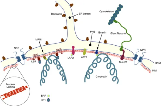
The nuclear envelope (NE) is a selective barrier that forms the interface between the nucleus and the cytoplasm, and as such plays a central role in defining the biochemical identities of each compartment[10,11]. In addition to its barrier function, it is becoming increasingly clear that the NE is a key determinant of nuclear architecture and may strongly influence cytoplasmic organization. The NE contains several discrete structural elements, the most prominent of which are the inner and outer nuclear membranes (Fig. 1). In mammalian somatic cells these two membranes are separated by a uniform gap of about 30– 50 nm referred to as the perinuclear space (PNS). The INM and ONM are connected at annular junctions which create aqueous
channels between the nucleoplasm and cytoplasm. These channels are occupied by nuclear pore complexes (NPCs), massive multi-protein assemblies that regulate the trafficking of macromolecules across the NE. A mammalian somatic cell nucleus typically contains several thousand NPCs.
In addition to its continuities with the INM at the periphery of each NPC, the ONM also displays multiple connections to the peripheral endoplasmic reticulum (ER) to which it is functionally related. Evidently the INM, ONM and ER form a single continuous membrane system. Similarly, the PNS constitutes a perinuclear extension of the ER lumen, and contains both secretory proteins and soluble ER resident proteins, including ER chaperones.
The final major structural feature of the NE is the nuclear lamina [12]. This is a relatively thin (20–50 nm) protein meshwork that is closely associated with both the nuclear face of the INM and the underlying chromatin. The key components of the nuclear lamina are a group of proteins known as A- and B-type lamins. The lamin proteins are members of the more extensive cytoplasmic intermediate filament (IF) family and like all IF proteins contain a central coiled-coil domain flanked by non-helical head and tail domains. In contrast to their cytoplasmic counterparts, each of the lamins contains a nuclear localization sequence (NLS) within the C-terminal non-helical domain required for efficient nuclear import of newly synthesized lamin proteins. Both A- and B-type lamins are known to interact with membrane proteins of the INM[12]as well as with chromatin[13,14]. In this way, the nuclear lamina may provide anchoring sites at the nuclear periphery for higher order chromatin domains in addition to stabilizing and organizing the NE. While the bulk of the lamins appear to reside at the nuclear periphery, nucleoplasmic lamins have also been observed[15–17]with proposed roles in several aspects of nuclear metabolism, including DNA replication[18–21].
In mammalian cells there are two major A-type lamins, A and C, encoded by a single gene, LMNA [22]. These two proteins are identical for the first 566 amino acid residues. Both proteins possess unique C-terminal extensions. In the case of lamin C this consists of a sequence of six amino acids. The unique region of lamin A is considerably larger at 98 amino acids. Two other A-type lamins have been described. The first of these, lamin AΔ10 [23], lacks a 30 amino acid sequence within the unique lamin A specific region that is encoded by exon 10 (LMNAcontains 12 exons). While it is found in somatic cells, its abundance and distribution has yet to be well defined. Lamin C2 [24] is male germ cell-specific and essentially consists of a truncated form of lamin C that contains an alternative N-terminus modified by myristoylation.
Mammalian somatic cells also contain two B-type lamins, lamins B1 and B2[25], encoded by separate genes (LMNB1and
cell populations[31,32]. In the mouse, A-type lamin expression commences only midway through gestation at embryonic day 8 or day 9, initially in cells of the trophoblast and visceral endoderm[29].
The ubiquitous expression of B-type lamins led to the conclusion early on that these were essential proteins. This has certainly turned out to be true of lamin B1, at least at the organismal level. Gene targeting experiments in mice have revealed that lamin B1 is required for the development of viable embryos[33]. However, mouse embryonic fibroblasts derived from Lmnb1-null embryos can nevertheless be maintained in culture. Evidently this protein is dispensable in certain cell types. This observation is supported by findings that HeLa cells depleted of lamin B1 [34] and/or lamin B2 using RNA interference continue to proliferate in culture, at least in the short term (Kyle Roux, Melissa Crisp and Brian Burke, unpublished observations).
Lamin A and lamins B1 and B2, feature C-terminal CaaX motifs (whereCis cysteine,ais an aliphatic amino acid andXis usually a hydrophobic residue). The CaaX motif was originally described in small Ras-related GTPases and represents a site of farnesylation[35]. This modification is mediated by a protein farnesyl transferase and occurs on the CaaX cysteine residue soon after completion of lamin synthesis[36–39]. Farnesylation is followed by C-terminal proteolysis to remove the aaX residues[40]. Processing of the CaaX motif is then completed by carboxy methylation of the newly exposed C-terminal cysteine residue [40]. Farnesylation of the CaaX cysteine residue is a prerequisite for the efficient assembly of newly synthesized lamins into the interphase nuclear lamina[41–43].
While the B-type lamins remain permanently farnesylated, lamin A is unique in that this modification is lost following proteolytic cleavage 14 residues upstream from the farnesy-lated cysteine [44]. This cleavage event is catalyzed by ZmpSte24, a membrane associated proteinase [45,46], and occurs soon after incorporation into the nuclear lamina, typically within 30–60 min of synthesis [47]. In this way, full length, or pre-lamin A exists only transiently in normal cells.
While individual lamin monomers are known to assemble to form coiled-coil homodimers, the higher order organization of the lamina is still a topic of considerable debate. As members of the IF family, the lamins are thought to be organized in the form of filaments. Certainly this has been borne out in ultrastructural studies ofXenopusoocyte nuclear envelopes where the lamina appears as an oftentimes orthogonal meshwork of 10 nm filaments [48]. However, the oocyte lamina is composed primarily of a single lamin isoform (lamin L3) [49]. The organization of the more complex mammalian somatic cell lamina containing lamins A, C, B1, B2 and perhaps AΔ10, has yet to be satisfactorily addressed.
5. Nuclear membrane proteins
Despite their numerous connections at the periphery of each NPC, the INM and ONM are biochemically quite distinct. This could be surmised even from early ultrastructural studies since the ONM, but not the INM, contains numerous bound ribosomes. Recent proteomic studies have revealed the existence of as many as 67 integral membrane proteins that
are enriched in the NE. The bulk of these appear to reside within the INM [50]. The mechanism by which proteins become localized to the INM has been a topic of some debate. The consensus that has emerged is that it involves, at least in part, a process of selective retention[51–53]. In this model, proteins that are synthesized on the peripheral ER or ONM gain access to the INM by lateral diffusion via the membrane continuities surrounding each NPC. Only proteins that can interact with nuclear, other INM or lamina components will be retained and concentrated. However, recent findings that movement of membrane proteins between the ONM and INM involves an energy dependent mechanism[54]and which appears to operate at the level of the NPC, suggest that we have not yet heard the final word on INM protein sorting.
The recent identification of a number of ONM-specific integral membrane proteins has raised additional questions[55–
57]. In particular, what prevents ONM proteins from simply drifting away into the peripheral ER? The issue of ONM protein localization was originally addressed inC. elegans. Starr and Han[58]demonstrated that the appropriate localization of Anc-1, a very large type II ONM protein involved in actin-dependent nuclear positioning, was dependent upon Unc-84, an INM protein [58]. Localization of Unc-84 itself was found to be dependent upon the singleC. eleganslamin[59]. Based upon these and similar findings, both Lee et al. and Starr and Han
[59,60]proposed a model in which Unc-84 and Anc-1 would interact across the PNS via their respective lumenal domains. In this way, Unc-84 would act as a trans-lumenal tether for Anc-1 in the ONM.
In mammalian cells two giant (800–1000 kDa!) actin binding proteins have been identified (variously termed NUANCE, nesprin 2 Giant, nesprin 1, enaptin, Syne 1, syne 2, myne 1) as integral proteins of the ONM[55–57,61,62]. Due to a complex array of alternatively spliced isoforms a very large family of proteins are encoded by thenesprin 1and nesprin 2
genes[56]. Nesprins are related to Anc-1, as well as to a Dro-sophila ONM protein known as Klarsicht [63–66]. All three proteins contain an∼60 amino acid C-terminal KASH domain (Klarsicht, Anc-1, Syne Homology) that is comprised of a single transmembrane anchor and a short segment of about 40 residues that resides within the PNS.
A third nesprin family member, nesprin 3, has also been described[67]. Like nesprins 1 and 2, nesprin 3 possesses a C-terminal KASH domain. However, its distinct N-C-terminal cytoplasmic domain features a binding site for plectin, a very large (466 kDa) intermediate filament-associated protein. Thus, whereas nesprins 1 and 2 connect the NE to microfilaments, nesprin 3 may function as a link between the NE and the cytoplasmic intermediate filament network.
One of the defining features of theC. elegansUnc-84 protein is a 200 amino acid region of homology with Sad1p, an S. pombepolypeptide that is associated with the spindle pole body
[68]. This region of homology is known as the SUN domain (for
Sad1p, UNc-84) and extends into the PNS. Mammalian cells contain several SUN domain proteins. Indeed there are five that are encoded within the human genome. Two of these, Sun1 and Sun2, are INM proteins and have a topology similar to that of
Unc-84 with a nucleoplasmic N-terminal domain and a C-terminal SUN domain in the PNS[69–71]. At least in the case of Sun1, its nucleoplasmic domain interacts with farnesylated pre-lamin A raising the possibility that this protein could function in lamin A targeting and/or assembly[69].
Recent reports indicate that both Sun1 and Sun2 cooperate in tethering nesprin 2 Giant (nesp2G) within the ONM [69,72]. This tethering involves the establishment of molecular interac-tions that span the PNS[73]similar to that suggested for Anc-1 and Unc84 in C. elegans (Fig. 2). Circumstantial evidence based upon competition between nesprin 1 and nesprin 2 KASH domains indicates that nesprin 1 Giant (nesp1G, enaptin) is similarly tethered by Sun1 and Sun2. It follows, therefore, that Sun1 and Sun2 function as links in a molecular chain that connects the actin cytoskeleton, via giant nesprin proteins, to nuclear lamins and other components of the nuclear interior. We now refer to this assembly as the LINC complex (forLInker of
Nucleoskeleton andCytoskeleton)[69]. The recent discovery of nesprin 3 as a link to the IF system [67], suggests that there maybe multiple functionally distinct isoforms of the LINC complex that are responsible for integrating the nucleus with different components of the cytoskeleton. The implication of the
existence of these linkages across the NE is that the nucleus and cytoplasm may display interdependent mechanical properties. As will be discussed below, this has recently been shown to be the case.
6. The nuclear envelope and muscular dystrophy
X-linked EDMD was the first human disorder to be linked to defects in a component (emerin) of the NE. Emerin is a type II transmembrane protein that localizes exclusively to the INM
[7–9]. The bulk of its mass resides within its 220 amino acid N-terminal nucleoplasmic domain. The majority of emerin mutations, either point or nonsense mutations, that are associated with EDMD lead to complete loss of the emerin protein or to its mislocalization[74–77]. It would appear that EDMD must be caused by loss of some essential emerin function. The nature of this function, however, is still a matter of debate. Although emerin is expressed in the majority of adult cell types, only skeletal and cardiac muscle seem to be adversely affected by its loss.
Detailed analyses of the emerin protein both in vivo and in vitro have revealed that the nucleoplasmic domain of emerin interacts with multiple nuclear proteins [78–82]. The N-terminus of emerin shares a sequence of about 40 amino acids with several other proteins including the INM proteins lamina associated polypeptide 2 (LAP2) and MAN1. This region of homology, known as the LEM domain (forLAP2,emerin and
MAN1) functions as a binding site for BAF (barrier to autointegration factor) a small DNA binding protein [83,84]. In this way BAF functions as a link between emerin and chromatin. Emerin also binds to several transcriptional regulators, germ cell less (GCL)[80]and Btf[78]. Binding of GCL and BAF to emerin are mutually exclusive[80]. Btf when overexpressed induces apoptosis. Thus sequestration of Btf by emerin could potentially have an anti-apoptotic function. In terms of regulatory activities, emerin might also modulate pre-mRNA processing through interactions with YT521-B, a factor involved in splice site selection[82].
In addition to these regulatory molecules, emerin interacts with a number of structural proteins including actin and A-type lamins. Emerin has been shown to promote the polymerization of actin and to cap the pointed end of actin filaments in vitro. It likely binds nuclear actin in vivo. As will be described further below, the interaction with A-type lamins contributes to the appropriate localization of emerin to the INM.
Studies on the role of emerin in vivo have so far shed only a little light on the etiology of X-linked EDMD. In C. elegans, depletion of the emerin orthologue by RNA intereference yields no detectable phenotype [85]. Emerin depletion is however, synthetic lethal with depletion of MAN1, which like its mammalian orthologue, is also a LEM domain protein [85]. Mice harboring a deletion of the emerin gene have no overt symptoms of muscular dystrophy and display no obvious skeletal or cardiac muscle pathology[86]. However, in common with human EDMD patients, fibroblasts derived from emerin deficient mice often have irregularly shaped nuclei featuring blebbing of the nuclear membranes [87]. Furthermore, emerin
deficient fibroblasts exhibit impaired signaling responses to mechanical stress. This may be observed in terms of reduced induction ofiex-1andegr-1, a pair of mechanosensitive genes
[87]. The overall effect is that emerin-null cells, when compared with wild type cells, display increased rates of apoptosis when subjected to mechanical strain[87].
Several years after the identification of the emerin gene as the site of mutations causing X-linked EDMD, the autosomal dominant form of the disease was mapped to theLMNAgene
[88]. Soon thereafter, limb girdle MD 1B (LGMD1B) and dilated cardiomyopathy (DCM) were also linked to mutations in LMNA[89,90]. In total more than 70 distinct mutations within
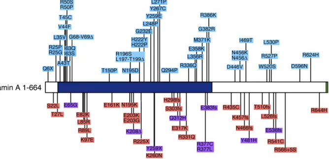
LMNAhave now been linked to skeletal and cardiac muscle diseases (Fig. 3). A few rare mutations which cause autosomal recessive EDMD have also been mapped toLMNA.
Muscular dystrophy (EDMD and LGMD1B) and cardio-myopathy are only three of at least 11 (depending upon definition) other disorders linked to mutations in the LMNA
gene[91]. These diseases, which are commonly referred to as
“laminopathies”, include Dunnigan type familial partial lipo-dystrophy (FPLD) [92,93], Charcot–Marie–Tooth disease (CMT2)[94], mandibuloacral dysplasia (MAD)[95], restrictive dermopathy (RD) [96] and two premature aging syndromes, Hutchinson–Gilford progeria (HGPS) [97–99] and atypical Werner's syndrome (aWRN)[100,101]. A major goal now is to determine how mutations in the widely expressedLMNAgene can give rise to such a bewildering array of tissue specific disorders. What is becoming increasingly clear is that multiple disease mechanisms must be at work[12,102,103]. A complete discussion of the laminopathies is beyond the scope of this review. therefore the remainder of the article will focus primarily on the molecular causes of EDMD, LGMD1B and DCM.
Dilated cardiomyopathy is a feature of both EDMD and LGMD1B. These two disorders differ only in terms of the affected muscle groups (e.g. distal versus proximal leg muscles). Evidently, certainLMNAmutations may cause heart disease (DCM) but spare skeletal muscle. However, either additional genetic or environmental factors may tip the balance towards skeletal muscle involvement [104]. This view is supported by observations of Brodski et al. who have identified a family carrying a single lamin A mutation (a frameshift caused by a single nucleotide deletion at position 959) where different members have been diagnosed with EDMD, LGMD or with DCM [105]. The bulk of LMNA mutations associated with muscle disease involve single amino acid changes, although deletions, frameshifts (above) and nonsense mutations are well represented (Fig. 3). Indeed the earliest report linkingLMNAto autosomal dominant EDMD highlighted a nonsense mutation at codon number six [88]. This would in effect represent a functional gene deletion, thus LMNA haploinsufficiency is sufficient to cause muscle disease.
It is certainly true that some (but by no means all) muscle disease-linked lamin mutations cause abnormalities in A-type lamin assembly at the nuclear periphery [106–108]. The non-helical lamin A tail is known to be comprised in part by an immunoglobulin type fold consisting of apposed beta sheets
[109,110]. Those EDMD-associated mutations found in this region of the lamin A molecule are predicted to significantly disrupt the 3D organization of the Ig domain. Taken together, these observations are consistent with the notion that muscle pathologies are linked to loss of structurally functional lamin proteins.
Homologous recombination has been used to eliminate the
Lmnagene in mice[111]. Animals homozygous for the deletion are born without any striking abnormalities, indicating that A-type lamin expression is not a prerequisite for normal embryonic development. However, newborn Lmna-null mice fail to thrive and display clear evidence of muscle weakness. Histological analyses reveal extensive muscular dystrophy and cardiomyopathy. These animals, which never survive beyond eight weeks of age, display a syndrome that is very similar to human EDMD. There are, however, two important differences. The first is that Lmna-null mice display a peripheral axonal neuropathy similar to CMT2 (in humans this is caused by an R298C mutation) [112]. There is no evidence of any other laminopathy-related pathology in these knock out animals. Secondly, heterozygous animals (Lmna+/−) are asymptomatic
[111]. As described above, loss of just one LMNA allele in humans is associated with EDMD. Regardless of these differences, however, the mouse studies lend considerable support for the view that it is loss of A-type lamin function that underlies the skeletal and cardiac muscle pathologies.
At the cellular level, loss of A-type lamin expression is associated with prominent changes in nuclear morphology
[111]. In both fibroblasts fromLmna-null mice as well as cells
from laminopathy patients[113], nuclei are frequently observed to have highly irregular shapes. This is usually associated with the appearance of NE “herniations” in which B-type lamins, INM proteins, NPCs and chromatin are withdrawn from one pole of the nucleus. This is accompanied by localized dilation of the ONM in the NPC free regions. Changes in heterochromatin organization, particularly loss of peripheral heterochromatin, are also observed[114]. Given that heterochromatin is generally transcriptionally silent, loss of A-type lamins could be associated with changes in gene expression patterns.
A final conspicuous feature of fibroblasts derived from
Lmna-null mice is the frequent mislocalization of emerin from the INM to the peripheral ER [111]. Introduction of human lamin A in to these cells by transfection will restore the normal localization of emerin to the INM. Similar mislocalization of emerin can be observed in HeLa cells following depletion of A-type lamins by RNA interference [34]. These observations clearly demonstrate that A-type lamins contribute to the normal localization of emerin and provide a molecular relationship between X-linked and autosomal EDMD.
Mutations of titin are associated with multiple MD disorders where conventional thought suggests its role as a structural protein of the muscle sarcomere is presumably disrupted[115]. However, a recent report by Zastrow et al. identified nuclear titin as a binding partner for A- and B-type lamins [116], making titin the third NE protein mutated in MD. It remains to be seen if perturbation of titin function at the NE might underlie some aspects of disease pathology within muscle cells.
At present there are two views of how defects in NE proteins might give rise to skeletal and cardiac muscle disease, and involve either mechanical stress or gene expression based models[102,117]. As will be seen, however, these views are not mutually exclusive. The mechanical stress model proposes that muscle cell nuclei lacking functional A-type lamins or emerin
may be excessively prone to mechanical damage caused by repeated cycles of muscle contraction. This notion has some merit. Firstly, we know that nuclei containing either defective A-type lamins or depleted of A-type lamins exhibit structural abnormalities. Furthermore, nuclei isolated from Lmna-null mouse livers are far more prone to fragmentation than their wild-type derived counterparts[111]. Recently Lammerding et al.[118]and Broers et al.[119]have demonstrated exactly such nuclear fragility in Lmna-null fibroblasts in vivo. Basically these investigators used direct mechanical methods to deform nuclei in cells in culture. They were able to show that the nuclei in Lmna-null fibroblasts were more deformable and ruptured under lower applied forces than nuclei in wild-type fibroblasts. Lammerding et al. also demonstrated that the cytoplasm of
Lmna-null cells was mechanically less resilient than that of wild-type cells[87]. This begins to make some sense given that we now know that nuclei are coupled to both the actin and IF cytoskeleton via the LINC complex of SUN proteins and nesprin proteins [56,57,67,69,72]. In muscle cells actin filaments and desmin IF filaments are linked to dystroglycans and sarcoglycans at the cell surface via contacts with dystrophin and other dystrophin interacting proteins. This raises the possibility that in EDMD and related myopathies, mechanical stress might have detrimental effects on both nuclear and cytoplasmic (and perhaps plasma membrane) organization. In this respect, the etiology of EDMD could have more in common with that of Duchenne and related MDs than immediately meets the eye.
The alternative view of EDMD, LGMD1B and DCM pathology proposes that changes in NE organization due to emerin or lamin mutations might lead to changes in muscle cell gene expression patterns. Both emerin and A-type lamins are known to interact with a number of transcriptional regulators including GCL and Btf (in the case of emerin) [78,80] and SREBP [120], Rb[121] and MOK2 (in the case of lamin A)
[122]. Furthermore, the well documented rearrangements in heterochromatin organization would be entirely consistent with this idea. Compelling evidence in favor of the gene expression model comes from several quarters. As described above, emerin deficient mice exhibit no overt pathology. However, muscle regeneration in these mice show clear abnormalities. In particular, myogenic differentiation is delayed, a phenomenon that is associated with perturbations in transcriptional pathways that are regulated by Rb and MyoD [86]. Similarly, lamin A mutants have been found to interfere with the differentiation program of C2C12 myoblasts[123,124].
Both mechanical stress and gene expression defects may be integrated in other observations. Lmna-null fibroblasts exhibit grossly impaired mechanotransduction and decreased viability under mechanical strain [118,119]. Induction of the mechan-osensitive genes, iex-1 and egr-1 is strongly attenuated[118]. Similar but milder effects are observed inemerin-null cells[87].
Lmna-null cells (but notemerin-null cells) also exhibit reduced NF-κB-regulated transcription in response to either cytokine or mechanical stimulation [118]. Taken together, all of these observations suggest that emerin-and Lmna-null cells exhibit reduced viability when subjected to mechanical stress. This
reduced viability may be due to direct mechanical effects (i.e., physical damage), inability to induce mechanosensitive genes or indeed to both. In this way both the mechanical stress and gene expression models may accurately describe different aspects of the molecular basis of EDMD and related myopathies.
Clearly we have come a considerable way in improving our understanding of NE biology since the original realization that X-linked EDMD was caused by defects in an INM protein. However, the recognition that at least nine or ten more human diseases are linked to defects in A-type lamins has raised puzzling questions. How for instance can defects in a near ubiquitously expressed gene give rise to such an array of tissue specific phenotypes? The answer may well lie with other proteins that are themselves expressed in a tissue specific fashion but which interact with A-type lamins. Some such proteins may be represented among the sixty or more nuclear membrane proteins identified using proteomics approaches. Given the role of the LINC complex in integrating the nucleus with the cytoskeleton a better understanding of some lamino-pathies may even arise through studies of NE-associated cytoplasmic proteins. What is certain is that this area of research at the interface of medicine and cell biology will continue to present us with surprises that redefine our under-standing of cellular structure and function while hopefully revealing novel avenues for disease therapy.
References
[1] A.E. Emery, The muscular dystrophies, Lancet 359 (2002) 687–695. [2] R.D. Cohn, K.P. Campbell, Molecular basis of muscular dystrophies,
Muscle Nerve 23 (2000) 1456–1471.
[3] I. Dalkilic, L.M. Kunkel, Muscular dystrophies: genes to pathogenesis, Curr. Opin. Genet. Dev. 13 (2003) 231–238.
[4] K. Kobayashi, Y. Nakahori, M. Miyake, K. Matsumura, E. Kondo-Iida, Y. Nomura, M. Segawa, M. Yoshioka, K. Saito, M. Osawa, K. Hamano, Y. Sakakihara, I. Nonaka, Y. Nakagome, I. Kanazawa, Y. Nakamura, K. Tokunaga, T. Toda, An ancient retrotransposal insertion causes Fukuyama-type congenital muscular dystrophy, Nature 394 (1998) 388–392. [5] Y.K. Hayashi, M. Ogawa, K. Tagawa, S. Noguchi, T. Ishihara, I. Nonaka,
K. Arahata, Selective deficiency of alpha-dystroglycan in Fukuyama-type congenital muscular dystrophy, Neurology 57 (2001) 115–121. [6] A.E. Emery, F.E. Dreifuss, Unusual type of benign x-linked muscular
dystrophy, J. Neurol. Neurosurg. Psychiatry 29 (1966) 338–342. [7] S. Bione, E. Maestrini, S. Rivella, M. Mancini, S. Regis, G. Romeo, D.
Toniolo, Identification of a novel X-linked gene responsible for Emery– Dreifuss muscular dystrophy, Nat. Genet. 8 (1994) 323–327.
[8] A. Nagano, R. Koga, M. Ogawa, Y. Kurano, J. Kawada, R. Okada, Y.K. Hayashi, T. Tsukahara, K. Arahata, Emerin deficiency at the nuclear membrane in patients with Emery–Dreifuss muscular dystrophy, Nat. Genet. 12 (1996) 254–259.
[9] S. Manilal, T.M. Nguyen, C.A. Sewry, G.E. Morris, The Emery–Dreifuss muscular dystrophy protein, emerin, is a nuclear membrane protein Hum, Mol. Genet. 5 (1996) 801–808.
[10] B. Burke, C.L. Stewart, Life at the edge: the nuclear envelope and human disease, Nat. Rev., Mol. Cell Biol. 3 (2002) 575–585.
[11] M.W. Hetzer, T.C. Walther, I.W. Mattaj, Pushing the envelope: structure, function, and dynamics of the nuclear periphery, Annu. Rev. Cell Dev. Biol. 21 (2005) 347–380.
[12] Y. Gruenbaum, A. Margalit, R.D. Goldman, D.K. Shumaker, K.L. Wilson, The nuclear lamina comes of age, Nat. Rev., Mol. Cell Biol. 6 (2005) 21–31.
The alpha-helical rod domain of human lamins A and C contains a chromatin binding site, EMBO J. 12 (1993) 4413–4424.
[14] M.E.E. Ludérus, A. de Graf, E. Mattia, J.L. den Blaauwen, M.A. Grande, L. de Jong, R. van Driel, Binding of matrix attachment regions to lamin B1, Cell 70 (1992) 949–959.
[15] J.M. Bridger, I.R. Kill, M. O'Farrell, C.J. Hutchison, Internal lamin structures within G1 nuclei of human dermal fibroblasts, J. Cell Sci. 104 (Pt 2) (1993) 297–306.
[16] A.E. Goldman, R.D. Moir, M. Montag-Lowy, M. Stewart, R.D. Goldman, Pathway of incorporation of microinjected lamin A into the nuclear envelope, J. Cell Biol. 119 (1992) 725–735.
[17] J.L. Broers, B.M. Machiels, G.J. van Eys, H.J. Kuijpers, E.M. Manders, R. van Driel, F.C. Ramaekers, Dynamics of the nuclear lamina as monitored by GFP-tagged A-type lamins, J. Cell Sci. 112 (Pt. 20) (1999) 3463–3475.
[18] R.D. Moir, T.P. Spann, H. Herrmann, R.D. Goldman, Disruption of nuclear lamin organization blocks the elongation phase of DNA replication, J. Cell Biol. 149 (2000) 1179–1192.
[19] T.P. Spann, R.D. Moir, A.E. Goldman, R. Stick, R.D. Goldman, Disruption of nuclear lamin organization alters the distribution of replication factors and inhibits DNA synthesis, J. Cell Biol. 136 (1997) 1201–1212.
[20] D.A. Barbie, B.A. Kudlow, R. Frock, J. Zhao, B.R. Johnson, N. Dyson, E. Harlow, B.K. Kennedy, Nuclear reorganization of mamma-lian DNA synthesis prior to cell cycle exit, Mol. Cell. Biol. 24 (2004) 595–607.
[21] B.K. Kennedy, D.A. Barbie, M. Classon, N. Dyson, E. Harlow, Nuclear organization of DNA replication in primary mammalian cells, Genes Dev. 14 (2000) 2855–2868.
[22] F. Lin, H.J. Worman, Structural organization of the human gene encoding nuclear lamin A and nuclear lamin C, J. Biol. Chem. 268 (1993) 16321–16326.
[23] B.M. Machiels, A.H. Zorenc, J.M. Endert, H.J. Kuijpers, G.J. van Eys, F.C. Ramaekers, J.L. Broers, An alternative splicing product of the lamin A/C gene lacks exon 10, J. Biol. Chem. 271 (1996) 9249–9253.
[24] K. Furukawa, H. Inagaki, Y. Hotta, Identification and cloning of an mRNA coding for a germ cell-specific A-type lamin in mice, Exp. Cell Res. 212 (1994) 426–430.
[25] C.F. Lehner, V. Kurer, H.M. Eppenberger, E.A. Nigg, The nuclear lamin protein family in higher vertebrates: identification of quantitatively minor lamin proteins by monoclonal antibodies, J. Biol. Chem. 261 (1986) 13293–13301.
[26] T.H. Hoeger, G. Krohne, W.W. Franke, Amino acid sequence and molecular charactarization of murine lamin B as deduced from cDNA clones, Eur. J. Cell Biol. 47 (1988) 283–290.
[27] T.H. Hoeger, K. Zatloukal, I. Waizenegger, G. Krohne, Characterization of a second highly conserved B-type lamin present in cells previously thought to contain only a single B-type lamin, Chromosoma 99 (1990) 379–390.
[28] K. Furukawa, Y. Hotta, cDNA cloning of a germ cell-specific lamin B3 from mouse spermatocytes and analysis of its ectopic expression in somatic cells, EMBO J. 12 (1993) 97–106.
[29] C. Stewart, B. Burke, Teratocarcinoma stem cells and early mouse embryos contain only a single major lamin polypeptide closely resembling lamin B, Cell 51 (1987) 383–392.
[30] S. Lebel, C. Lampron, A. Royal, Y. Raymond, Lamins A and C appear during retinoic acid-induced differentiation of mouse embryonal carcinoma cells, J. Cell Biol. 105 (1987) 1099–1104.
[31] R.-A. Roeber, H. Sauter, K. Weber, M. Osborn, Cells of the cellular immune and hemopoietic system of the mouse lack lamins A/C: distinction versus other somatic cells, J. Cell Sci. 95 (1990) 587–598. [32] R.-A. Roeber, K. Weber, M. Osborn, Differential timing of lamin A/C
expression in the various organs of the mouse embryo and the young animal: a developmental study, Development 105 (1989) 365–378. [33] L. Vergnes, M. Peterfy, M.O. Bergo, S.G. Young, K. Reue, Lamin B1 is
required for mouse development and nuclear integrity, Proc. Natl. Acad. Sci. U. S. A. 101 (2004) 10428–10433.
[34] J. Harborth, S.M. Elbashir, K. Bechert, T. Tuschl, K. Weber,
Identifica-tion of essential genes in cultured mammalian cells using small interfering RNAs, J. Cell Sci. 114 (2001) 4557–4565.
[35] A.I. Magee, M. Hanley, Sticky fingers and CAAX boxes, Nature 335 (1988) 114–115.
[36] L.A. Beck, T.J. Hosick, M. Sinensky, Incorporation of a product of mevalonic acid metabolism into proteins of Chinese hamster ovary cell nuclei, J. Cell Biol. 107 (1988) 1307–1316.
[37] S.L. Wolda, J.A. Glomset, Evidence for modification of lamin B by a product of mevalonic acid, J. Biol. Chem. 263 (1988) 5997–6000. [38] C.C. Farnsworth, S.L. Wolda, M.H. Gelb, J.A. Glomset, Human lamin B
contains a farnesylated cysteine residue, J. Biol. Chem. 264 (1989) 20422–20429.
[39] K. Vorburger, G.T. Kitten, E.A. Nigg, Modification of nuclear lamin proteins by a mevalonic acid derivative occurs in reticulocyte lysates and requires the cysteine residue of the C-terminal CXXM motif, EMBO J. 8 (1989) 4007–4013.
[40] M. Sinensky, K. Fantle, M. Trujillo, T. McLain, A. Kupfer, M. Dalton, The processing pathway of prelamin A, J. Cell Sci. 107 (1994) 61–67.
[41] D. Holtz, R.A. Tanaka, J. Hartwig, F. McKeon, The CaaX Motif of lamin A functions in conjunction with the nuclear localization signal to target assembly to the nuclear envelope, Cell 59 (1989) 969–977.
[42] G.T. Kitten, E.A. Nigg, The CaaX motif is required for isoprenylation, carboxy methylation and nuclear membrane association of lamin B2, J. Cell Biol. 113 (1991) 13–24.
[43] G. Krohne, I. Waizenegger, T.H. Hoeger, The conserved carboxy-terminal cysteine of nuclear lamins is essential for association with the nuclear envelope, J. Cell Biol. 109 (1989) 2003–2011.
[44] K. Weber, U. Plessmann, P. Traub, Maturation of nuclear lamin A involves a specific carboxy-terminal trimming, which removes the polyisoprenylation site from the precursor; implications for the structure of the nuclear lamina, FEBS Lett. 257 (1989) 411–414.
[45] D.P. Corrigan, D. Kuszczak, A.E. Rusinol, D.P. Thewke, C.A. Hrycyna, S. Michaelis, M.S. Sinensky, Prelamin A endoproteolytic processing in vitro by recombinant Zmpste24, Biochem. J. 387 (2005) 129–138. [46] A.M. Pendas, Z. Zhou, J. Cadinanos, J.M. Freije, J. Wang, K. Hultenby,
A. Astudillo, A. Wernerson, F. Rodriguez, K. Tryggvason, C. Lopez-Otin, Defective prelamin A processing and muscular and adipocyte alterations in Zmpste24 metalloproteinase-deficient mice, Nat. Genet. 31 (2002) 94–99.
[47] L. Gerace, C. Comeau, M. Benson, Organization and modulation of nuclear lamina structure, J. Cell Sci. (Suppl. 1) (1984) 137–160. [48] U. Aebi, J.B. Cohn, L. Buhle, L. Gerace, The nuclear lamina is a
meshwork of intermediate type filaments, Nature 323 (1986) 560–564. [49] R. Stick, P. Hausen, Changes in the nuclear lamina composition during
early development ofXenopus laevis, Cell 41 (1985) 191–200. [50] E.C. Schirmer, L. Florens, T. Guan, J.R. Yates III, L. Gerace, Nuclear
membrane proteins with potential disease links found by subtractive proteomics, Science 301 (2003) 1380–1382.
[51] J. Ellenberg, E.D. Siggia, J.E. Moreira, C.L. Smith, J.F. Presley, H.J. Worman, J. Lippincott-Schwartz, Nuclear membrane dynamics and reassembly in living cells: targeting of an inner nuclear membrane protein in interphase and mitosis, J. Cell Biol. 138 (1997) 1193–1206. [52] L. Powell, B. Burke, Internuclear exhchange of an inner nuclear
membrane protein (p55) in heterokaryons: in vivo evidence for the association of p55 with the nuclear lamina, J. Cell Biol. 111 (1990) 2225–2234.
[53] B. Soullam, H.J. Worman, Signals and structural features involved in integral membrane protein targeting to the inner nuclear membrane, J. Cell Biol. 130 (1995) 15–27.
[54] T. Ohba, E.C. Schirmer, T. Nishimoto, L. Gerace, Energy- and temperature-dependent transport of integral proteins to the inner nuclear membrane via the nuclear pore, J. Cell Biol. 167 (2004) 1051–1062. [55] V.C. Padmakumar, S. Abraham, S. Braune, A.A. Noegel, B. Tunggal, I.
Karakesisoglou, E. Korenbaum, Enaptin, a giant actin-binding protein, is an element of the nuclear membrane and the actin cytoskeleton, Exp. Cell Res. 295 (2004) 330–339.
P.L. Weissberg, J.A. Ellis, C.M. Shanahan, Nesprins: a novel family of spectrin-repeat-containing proteins that localize to the nuclear mem-brane in multiple tissues, J. Cell Sci. 114 (2001) 4485–4498. [57] Y.Y. Zhen, T. Libotte, M. Munck, A.A. Noegel, E. Korenbaum,
NUANCE, a giant protein connecting the nucleus and actin cytoskeleton, J. Cell Sci. 115 (2002) 3207–3222.
[58] D.A. Starr, M. Han, Role of ANC-1 in tethering nuclei to the actin cytoskeleton, Science 298 (2002) 406–409.
[59] K.K. Lee, D. Starr, M. Cohen, J. Liu, M. Han, K.L. Wilson, Y. Gruenbaum, Lamin-dependent localization of UNC-84, a protein required for nuclear migration inCaenorhabditis elegans, Mol. Biol. Cell 13 (2002) 892–901.
[60] D.A. Starr, M. Han, ANChors away: an actin based mechanism of nuclear positioning, J. Cell Sci. 116 (2003) 211–216.
[61] E.D. Apel, R.M. Lewis, R.M. Grady, J.R. Sanes, Syne-1, a dystrophin-and Klarsicht-related protein associated with synaptic nuclei at the neuromuscular junction, J. Biol. Chem. 275 (2000) 31986–31995. [62] J.M. Mislow, M.S. Kim, D.B. Davis, E.M. McNally, Myne-1, a spectrin
repeat transmembrane protein of the myocyte inner nuclear membrane interacts with lamin A/C, J. Cell Sci. 115 (2002) 61–70.
[63] J.A. Fischer, S. Acosta, A. Kenny, C. Cater, C. Robinson, J. Hook, Drosophilaklarsicht has distinct subcellular localization domains for nuclear envelope and microtubule localization in the eye, Genetics 168 (2004) 1385–1393.
[64] K.L. Mosley-Bishop, Q. Li, L. Patterson, J.A. Fischer, Molecular analysis of the klarsicht gene and its role in nuclear migration within differentiating cells of theDrosophilaeye, Curr. Biol. 9 (1999) 1211–1220.
[65] K. Patterson, A.B. Molofsky, C. Robinson, S. Acosta, C. Cater, J.A. Fischer, The functions of Klarsicht and nuclear lamin in developmentally regulated nuclear migrations of photoreceptor cells in theDrosophila eye, Mol. Biol. Cell 15 (2004) 600–610.
[66] M.A. Welte, S.P. Gross, M. Postner, S.M. Block, E.F. Wieschaus, Developmental regulation of vesicle transport inDrosophilaembryos: forces and kinetics, Cell 92 (1998) 547–557.
[67] K. Wilhelmsen, S.H. Litjens, I. Kuikman, N. Tshimbalanga, H. Janssen, I. van den Bout, K. Raymond, A. Sonnenberg, Nesprin-3, a novel outer nuclear membrane protein, associates with the cytoskeletal linker protein plectin, J. Cell Biol. 171 (2005) 799–810.
[68] I. Hagan, M. Yanagida, The product of the spindle formation gene sad1+ associates with the fission yeast spindle pole body and is essential for viability, J. Cell Biol. 129 (1995) 1033–1047.
[69] M. Crisp, Q. Liu, K. Roux, J.B. Rattner, C. Shanahan, B. Burke, P.D. Stahl, D. Hodzic, Coupling of the nucleus and cytoplasm: role of the LINC complex, J. Cell Biol. 172 (2006) 41–53.
[70] S. Hasan, S. Guttinger, P. Muhlhausser, F. Anderegg, S. Burgler, U. Kutay, Nuclear envelope localization of human UNC84A does not require nuclear lamins, FEBS Lett. 580 (2006) 1263–1268.
[71] D.M. Hodzic, D.B. Yeater, L. Bengtsson, H. Otto, P.D. Stahl, Sun2 is a novel mammalian inner nuclear membrane protein, J. Biol. Chem. 279 (2004) 25805–25812.
[72] V.C. Padmakumar, T. Libotte, W. Lu, H. Zaim, S. Abraham, A.A. Noegel, J. Gotzmann, R. Foisner, I. Karakesisoglou, The inner nuclear membrane protein Sun1 mediates the anchorage of Nesprin-2 to the nuclear envelope, J. Cell Sci. 118 (2005) 3419–3430.
[73] M.D. McGee, R. Rillo, A.S. Anderson, D.A. Starr, UNC-83 IS a KASH protein required for nuclear migration and is recruited to the outer nuclear membrane by a physical interaction with the SUN protein UNC-84, Mol. Biol. Cell 17 (2006) 1790–1801.
[74] J.A. Ellis, C.A. Brown, L.D. Tilley, J. Kendrick-Jones, J.E. Spence, J.R. Yates, Two distal mutations in the gene encoding emerin have profoundly different effects on emerin protein expression, Neuromuscul. Disord. 10 (2000) 24–30.
[75] J.A. Ellis, M. Craxton, J.R. Yates, J. Kendrick-Jones, Aberrant intracellular targeting and cell cycle-dependent phosphorylation of emerin contribute to the Emery–Dreifuss muscular dystrophy phenotype, J. Cell Sci. 111 (1998) 781–792.
[76] J.A. Ellis, J.R. Yates, J. Kendrick-Jones, C.A. Brown, Changes at P183 of emerin weaken its protein-protein interactions resulting in X-linked
Emery–Dreifuss muscular dystrophy, Hum. Genet. 104 (1999) 262–268.
[77] E.A. Fairley, J. Kendrick-Jones, J.A. Ellis, The Emery–Dreifuss muscular dystrophy phenotype arises from aberrant targeting and binding of emerin at the inner nuclear membrane, J. Cell Sci. 112 (1999) 2571–2582. [78] T. Haraguchi, J.M. Holaska, M. Yamane, T. Koujin, N. Hashiguchi, C.
Mori, K.L. Wilson, Y. Hiraoka, Emerin binding to Btf, a death-promoting transcriptional repressor, is disrupted by a missense mutation that causes Emery–Dreifuss muscular dystrophy, Eur. J. Biochem. 271 (2004) 1035–1045.
[79] J.M. Holaska, A.K. Kowalski, K.L. Wilson, Emerin caps the pointed end of actin filaments: evidence for an actin cortical network at the nuclear inner membrane, PLoS Biol. 2 (2004) E231.
[80] J.M. Holaska, K.K. Lee, A.K. Kowalski, K.L. Wilson, Transcriptional repressor germ cell-less (GCL) and barrier to autointegration factor (BAF) compete for binding to emerin in vitro, J. Biol. Chem. 278 (2003) 6969–6975.
[81] K.K. Lee, T. Haraguchi, R.S. Lee, T. Koujin, Y. Hiraoka, K.L. Wilson, Distinct functional domains in emerin bind lamin A and DNA-bridging protein BAF, J. Cell Sci. 114 (2001) 4567–4573.
[82] F.L. Wilkinson, J.M. Holaska, Z. Zhang, A. Sharma, S. Manilal, I. Holt, S. Stamm, K.L. Wilson, G.E. Morris, Emerin interacts in vitro with the splicing-associated factor, YT521-B, Eur. J. Biochem. 270 (2003) 2459–2466.
[83] F. Lin, D.L. Blake, I. Callebaut, I.S. Skerjanc, L. Holmer, M.W. McBurney, M. Paulin-Levasseur, H.J. Worman, MAN1, an inner nuclear membrane protein that shares the LEM domain with lamina-associated polypeptide 2 and emerin, J. Biol. Chem. 275 (2000) 4840–4847. [84] D.K. Shumaker, K.K. Lee, Y.C. Tanhehco, R. Craigie, K.L. Wilson,
LAP2 binds to BAF.DNA complexes: requirement for the LEM domain and modulation by variable regions, EMBO J. 20 (2001) 1754–1764. [85] J. Liu, K.K. Lee, M. Segura-Totten, E. Neufeld, K.L. Wilson, Y.
Gruenbaum, MAN1 and emerin have overlapping function(s) essential for chromosome segregation and cell division inCaenorhabditis elegans, Proc Natl Acad Sci U S A 100 (2003) 4598–4603.
[86] G. Melcon, S. Kozlov, D.A. Cutler, T. Sullivan, L. Hernandez, P. Zhao, S. Mitchell, G. Nader, M. Bakay, J.N. Rottman, E.P. Hoffman, C.L. Stewart, Loss of emerin at the nuclear envelope disrupts the Rb1/E2F and MyoD pathways during muscle regeneration, Hum. Mol. Genet. 15 (2006) 637–651.
[87] J. Lammerding, J. Hsiao, P.C. Schulze, S. Kozlov, C.L. Stewart, R.T. Lee, Abnormal nuclear shape and impaired mechanotransduction in emerin-deficient cells, J. Cell Biol. 170 (2005) 781–791.
[88] G. Bonne, M.R. Di Barletta, S. Varnous, H.M. Becane, E.H. Hammouda, L. Merlini, F. Muntoni, C.R. Greenberg, F. Gary, J.A. Urtizberea, D. Duboc, M. Fardeau, D. Toniolo, K. Schwartz, Mutations in the gene encoding lamin A/C cause autosomal dominant Emery–Dreifuss muscular dystrophy, Nat. Genet. 21 (1999) 285–288.
[89] A. Muchir, G. Bonne, A.J. van der Kooi, M. van Meegen, F. Baas, P.A. Bolhuis, M. de Visser, K. Schwartz, Identification of mutations in the gene encoding lamins A/C in autosomal dominant limb girdle muscular dystrophy with atrioventricular conduction disturbances (LGMD1B), Hum. Mol. Genet. 9 (2000) 1453–1459.
[90] D. Fatkin, C. MacRae, T. Sasaki, M.R. Wolff, M. Porcu, M. Frenneaux, J. Atherton, H.J. Vidaillet Jr., S. Spudich, U. De Girolami, J.G. Seidman, C.E. Seidman, F. Muntoni, G. Muehle, W. Johnson, B. McDonough, Missense mutations in the rod domain of the lamin A/C gene as causes of dilated cardiomyopathy and conduction-system disease, N. Engl. J. Med. 341 (1999) 1715–1724.
[91] A. Muchir, H.J. Worman, The nuclear envelope and human disease, Physiology (Bethesda) 19 (2004) 309–314.
[92] H. Cao, R.A. Hegele, Nuclear lamin A/C R482Q mutation in Canadian kindreds with Dunnigan-type familial partial lipodystrophy, Hum. Mol. Genet. 9 (2000) 109–112.
[94] M. Tazir, H. Azzedine, S. Assami, P. Sindou, S. Nouioua, R. Zemmouri, T. Hamadouche, M. Chaouch, J. Feingold, J.M. Vallat, E. Leguern, D. Grid, Phenotypic variability in autosomal recessive axonal Charcot– Marie–Tooth disease due to the R298C mutation in lamin A/C, Brain 127 (2004) 154–163.
[95] G. Novelli, A. Muchir, F. Sangiuolo, A. Helbling-Leclerc, M.R. D'Apice, C. Massart, F. Capon, P. Sbraccia, M. Federici, R. Lauro, C. Tudisco, R. Pallotta, G. Scarano, B. Dallapiccola, L. Merlini, G. Bonne, Mandibu-loacral dysplasia is caused by a mutation in LMNA-encoding lamin A/C, Am. J. Hum. Genet. 71 (2002) 426–431.
[96] C.L. Navarro, A. De Sandre-Giovannoli, R. Bernard, I. Boccaccio, A. Boyer, D. Genevieve, S. Hadj-Rabia, C. Gaudy-Marqueste, H.S. Smitt, P. Vabres, L. Faivre, A. Verloes, T. Van Essen, E. Flori, R. Hennekam, F.A. Beemer, N. Laurent, M. Le Merrer, P. Cau, N. Levy, Lamin A and ZMPSTE24 (FACE-1) defects cause nuclear disorganization and identify restrictive dermopathy as a lethal neonatal laminopathy, Hum. Mol. Genet. 13 (2004) 2493–2503.
[97] M. Eriksson, W.T. Brown, L.B. Gordon, M.W. Glynn, J. Singer, L. Scott, M.R. Erdos, C.M. Robbins, T.Y. Moses, P. Berglund, A. Dutra, E. Pak, S. Durkin, A.B. Csoka, M. Boehnke, T.W. Glover, F.S. Collins, Recurrent de novo point mutations in lamin A cause Hutchinson–Gilford progeria syndrome, Nature 423 (2003) 293–298.
[98] A. De Sre-Giovannoli, R. Bernard, P. Cau, C. Navarro, J. Amiel, I. Boccaccio, S. Lyonnet, C.L. Stewart, A. Munnich, M. Le Merrer, N. Levy, Lamin a truncation in Hutchinson–Gilford progeria, Science 300 (2003) 2055.
[99] H. Cao, R.A. Hegele, LMNA is mutated in Hutchinson–Gilford progeria (MIM 176670) but not in Wiedemann–Rautenstrauch progeroid syndrome (MIM 264090), J. Hum. Genet. 48 (2003) 271–274. [100] C. Vigouroux, F. Caux, J. Capeau, S. Christin-Maitre, A. Cohen, LMNA
mutations in atypical Werner's syndrome, Lancet 362 (2003) 1585 (author reply 1586).
[101] L. Chen, L. Lee, B.A. Kudlow, H.G. Dos Santos, O. Sletvold, Y. Shafeghati, E.G. Botha, A. Garg, N.B. Hanson, G.M. Martin, I.S. Mian, B.K. Kennedy, J. Oshima, LMNA mutations in atypical Werner's syndrome, Lancet 362 (2003) 440–445.
[102] H.J. Worman, J.C. Courvalin, How do mutations in lamins A and C cause disease? J. Clin. Invest. 113 (2004) 349–351.
[103] C.J. Hutchison, H.J. Worman, A-type lamins: guardians of the soma? Nat. Cell Biol. 6 (2004) 1062–1067.
[104] E. Mercuri, M. Poppe, R. Quinlivan, S. Messina, M. Kinali, L. Demay, J. Bourke, P. Richard, C. Sewry, M. Pike, G. Bonne, F. Muntoni, K. Bushby, Extreme variability of phenotype in patients with an identical missense mutation in the lamin A/C gene: from congenital onset with severe phenotype to milder classic Emery–Dreifuss variant, Arch. Neurol. 61 (2004) 690–694.
[105] G.L. Brodsky, F. Muntoni, S. Miocic, G. Sinagra, C. Sewry, L. Mestroni, Lamin A/C gene mutation associated with dilated cardiomyopathy with variable skeletal muscle involvement, Circulation 101 (2000) 473–476. [106] W.H. Raharjo, P. Enarson, T. Sullivan, C.L. Stewart, B. Burke, Nuclear
envelope defects associated with LMNA mutations causing dilated cardiomyopathy and Emery–Dreifuss muscular dystrophy, J. Cell Sci. 114 (2001) 4447–4457.
[107] C. Östlund, G. Bonne, K. Schwartz, H. Worman, Properties of lamin A mutants found in Emery–Dreifuss muscular dystrophy, cardiomyopathy and Dunnigan-type partial lipodystrophy, J. Cell Sci. 114 (2001) 4435–4445.
[108] K. Bechert, M. Lagos-Quintana, J. Harborth, K. Weber, M. Osborn, Effects of expressing lamin A mutant protein causing Emery–Dreifuss muscular dystrophy and familial partial lipodystrophy in HeLa cells, Exp. Cell Res. 286 (2003) 75–86.
[109] S. Dhe-Paganon, E.D. Werner, Y.I. Chi, S.E. Shoelson, Structure of the globular tail of nuclear lamin, J. Biol. Chem. 277 (2002) 17381–17384.
[110] I. Krimm, C. Ostlund, B. Gilquin, J. Couprie, P. Hossenlopp, J.P. Mornon, G. Bonne, J.C. Courvalin, H.J. Worman, S. Zinn-Justin, The Ig-like structure of the C-terminal domain of lamin A/C, mutated in muscular dystrophies, cardiomyopathy and partial lipodystrophy, Structure 10 (2002) 811–823.
[111] T. Sullivan, D. Escalante-Alcalde, H. Bhatt, M. Anver, N. Bhat, K. Nagashima, C.L. Stewart, B. Burke, Loss of A-type lamin expression compromises nuclear envelope integrity leading to muscular dystrophy, J. Cell Biol. 147 (1999) 913–920.
[112] A. De Sandre-Giovannoli, M. Chaouch, S. Kozlov, J.M. Vallat, M. Tazir, N. Kassouri, P. Szepetowski, T. Hammadouche, A. Vandenberghe, C.L. Stewart, D. Grid, N. Levy, Homozygous defects in LMNA, encoding lamin A/C nuclear-envelope proteins, cause autosomal recessive axonal neuropathy in human (Charcot–Marie–Tooth disorder type 2) and mouse, Am. J. Hum. Genet. 70 (2002) 726–736.
[113] C. Vigouroux, M. Auclair, E. Dubosclard, M. Pouchelet, J. Capeau, J.C. Courvalin, B. Buendia, Nuclear envelope disorganization in fibroblasts from lipodystrophic patients with heterozygous R482Q/W mutations in the lamin A/C gene, J. Cell Sci. 114 (2001) 4459–4468.
[114] A. Fidzianska, D. Toniolo, I. Hausmanowa-Petrusewicz, Ultrastructural abnormality of sarcolemmal nuclei in Emery–Dreifuss muscular dystrophy (EDMD), J. Neurol. Sci. 159 (1998) 88–93.
[115] P. Hackman, A. Vihola, H. Haravuori, S. Marchand, J. Sarparanta, J. De Seze, S. Labeit, C. Witt, L. Peltonen, I. Richard, B. Udd, Tibial muscular dystrophy is a titinopathy caused by mutations in TTN, the gene encoding the giant skeletal-muscle protein titin, Am. J. Hum. Genet. 71 (2002) 492–500.
[116] M.S. Zastrow, D.B. Flaherty, G.M. Benian, K.L. Wilson, Nuclear titin interacts with A- and B-type lamins in vitro and in vivo, J. Cell Sci. 119 (2006) 239–429.
[117] H.J. Worman, J.C. Courvalin, Nuclear envelope, nuclear lamina and inherited disease, Int. Rev. Cytol. 246 (2005) 231–279.
[118] J. Lammerding, P.C. Schulze, T. Takahashi, S. Kozlov, T. Sullivan, R.D. Kamm, C.L. Stewart, R.T. Lee, Lamin A/C deficiency causes defective nuclear mechanics and mechanotransduction, J. Clin. Invest. 113 (2004) 370–378.
[119] J.L. Broers, E.A. Peeters, H.J. Kuijpers, J. Endert, C.V. Bouten, C.W. Oomens, F.P. Baaijens, F.C. Ramaekers, Decreased mechanical stiffness in LMNA−/−cells is caused by defective nucleo-cytoskeletal integrity: implications for the development of laminopathies, Hum. Mol. Genet. 13 (2004) 2567–2580.
[120] D.J. Lloyd, R.C. Trembath, S. Shackleton, A novel interaction between lamin A and SREBP1: implications for partial lipodystrophy and other laminopathies, Hum. Mol. Genet. 11 (2002) 769–777.
[121] T. Ozaki, M. Saijo, K. Murakami, H. Enomoto, Y. Taya, S. Sakiyama, Complex formation between lamin A and the retinoblastoma gene product: identification of the domain on lamin A required for its interaction, Oncogene 9 (1994) 2649–2653.
[122] C. Dreuillet, J. Tillit, M. Kress, M. Ernoult-Lange, In vivo and in vitro interaction between human transcription factor MOK2 and nuclear lamin A/C, Nucleic Acids Res. 30 (2002) 4634–4642.
[123] E. Markiewicz, M. Ledran, C.J. Hutchison, Remodelling of the nuclear lamina and nucleoskeleton is required for skeletal muscle differentiation in vitro, J. Cell Sci. 118 (2005) 409–420.