UNIVERSIDAD AUTÓNOMA DE NUEVO LEÓN
FACULTAD DE INGENIERÍA MECÁNICA Y ELÉCTRICA
THESIS
“SYNTHESIS AND CHARACTERIZATION OF METAL NANOPARTICLES BY PULSED LASER ABLATION IN LIQUID MEDIA (PLAL)”
BY
M.C. MARÍA ISABEL MENDIVIL PALMA
SUBMITTED IN PARTIAL FULFILLMENT OF THE REQUIREMENTS FOR THE DOCTORAL DEGREE IN MATERIALS ENGINEERING
UNIVERSIDAD AUTÓNOMA DE NUEVO LEÓN
FACULTAD DE INGENIERÍA MECÁNICA Y ELÉCTRICA SUBDIRECCIÓN DE ESTUDIOS DE POSGRADO
THESIS
“SYNTHESIS AND CHARACTERIZATION OF METAL NANOPARTICLES BY PULSED LASER ABLATION IN LIQUID MEDIA (PLAL)”
BY
M.C. MARÍA ISABEL MENDIVIL PALMA
SUBMITTED IN PARTIAL FULFILLMENT OF THE REQUIREMENTS FOR THE DOCTORAL DEGREE IN MATERIALS ENGINEERING
SUPERVISED BY DR. SADASIVAN SHAJI
ii
A C K N O W L E D G E M E N T S
Thanks to God for all his love…
I would like to thank CONACYT for the assistance and support through a research fellowship.
To FIME, UANL for the scholar preparation and facilities to the use of laser laboratory.
Thanks to CIIDIT, especially to Dr. Domingo Garcia and Dr. Alejandro Torres for their training, help and support in the use of Transmission Electron Microscope (TEM).
I would like to thank my assessor Dr. Sadasivan Shaji, for sharing his knowledge, all the comments during this project and principally the continuous motivation for improvement personally and scientific area.
Thanks to Dra. Bindu Krishnan for lean my academic achievements.
Thanks to my thesis revisers, Dr. Víctor Manuel Coello Cárdenas, Dr. Manuel García Méndez and, Dr. David Avellaneda Avellaneda for their help in the corrections and comments to this thesis.
Thanks to Dr. Alan Castillo and Dr. Tushar Kanti Das Roy for their advice and support.
The next part of acknowledgments will be in Spanish in consideration to the persons to whom it is directed.
Agradezco infinitamente a mi familia que ha sido mi mayor aliento para continuar con mi preparación académica, pero sobre todo por enseñarme lo maravillosa que es la vida y lo afortunada que soy al tenerlos a todos ustedes.
iii
Al amor de mi vida, Edgar Adrian Castro por estar ahí siempre para apoyarme. Me encanta poder compartir contigo cada instante de mi vida. Te amo.
A mis hijas, Isabella e Itzel quienes son las propulsoras de mi vida y de todos mis proyectos. Las amo.
A mi MADRE, por su incansable fe en mí, su apoyo, correcciones y consejos. No estaría aquí sin su infinito amor de madre.
Gracias a mi PADRE, porque sé que siempre cuento con él en todo lugar y todo momento.
Mis herman@s y cuñad@s; Jorge mi dador de felicidad favorito, eres fuerte para proteger lo que amas y sensible cuando ocupamos consuelo y apoyo. Carlos mi inventor predilecto, se que pronto salvaremos el mundo. Ana mi comadre y hermana, he descubierto en ti a una gran amiga. Isaí, Laila y Lupita gracias por su gran cariño.
Gracias a Carlos Almanza por acompañarnos en este viaje y aguantar a esta singular familia.
Gracias por todo su apoyo y amor a toda mi bella familia, abuela María Peraza, tíos, primos y sobrinas.
iv
Dedicado a mi enfermera favorita…
v
ABBREVIATIONS
(0-3) D (0-3) Dimension
BF-TEM Bright Field TEM
CIS Copper-Indium-Selenide
CNT Carbon Nanotubes
CVD Chemical Vapor Deposition
CW Continuous Wave
DC Direct Current
DLTT Distance from Lens to Target
DNA Deoxyribonucleic Acid
DP-LA Double Pulsed Laser Ablation
DW Distilled Water
EDX Energy Dispersive X-Ray
EG Ethylene Glycol
FCC Face-Centered-Cubic
GNS Graphene Nano-Sheet
HAADF High Angular Annular Dark Field
HEX Hexagonal
HRTEM High Resolution TEM
IR Infrared
ITO Indium Tin Oxide
KrF Krypton Fluoride
LAL Laser Ablation in Liquids
Laser Light Amplification by Stimulated Emission of Radiation
LCVD Laser CVD
LDI Laser Deposition Ionization
LED Light Emitting Diode
LIFT Laser-Induced Forward Transfer
LPCVD Low Pressure CVD
LVCC Laser Vaporization Controlled Condensation
MBE Molecular Beam Epitaxy
MOVPE Metal-Organic Vapor Phase Epitaxy
MRI Magnetic Resonance Imaging
MWCNT Multi Walled Carbon Nanotubes
Nd:YAG Neodymium-Doped Yttrium Aluminum Garnet
NEMS Nano-Electromechanical Systems
vi
NPs Nanoparticles
NW Nanowire
PACVD Plasma-Assisted CVD
PEG Poly Ethylene Glycol
PL Pulsed Laser
PLAL Pulsed Laser Ablation in Liquid
PLD Pulsed Laser Deposition
PLM Pulsed Laser Melting
PVA Poly Vinyl Alcohol
PVD Physical Vapor Deposition
PVP Polyvinylpyrrolidone
RF Radio Frequency
SAED Selected Area Electron Diffraction
SDS Sodium Dodecyl Sulphate
SERS Surface Enhanced Raman Scattering
SPR Surface Plasmon Resonance
STEM Scanning Transmission Electron Microscope
STM Scanning Tunneling Microscope
TEM Transmission Electron Microscopy
TEOS Tetraethyl Orthosilicate
TTM Two Temperature Model
UV-Vis Ultra Violet-Visible
XPS X-Ray Photoelectron Spectroscopy
1
CONTENTS
SUMMARY ... 5
CHAPTER 16 INTRODUCTION TO NANOMATERIALS AND PULSED LASER ABLATION IN LIQUID ... 6
1.1 GENERAL INTRODUCTION TO NANOMATERIALS ... 6
1.1.1 Introduction ... 6
1.1.2 Properties of Nanomaterials ... 6
1.1.3 Applications... 7
1.1.4 Synthesis of Nanomaterials ... 9
1.2 PULSED LASER ABLATION IN LIQUID ... 13
1.2.1 Introduction ... 13
1.2.2 Fundamental Aspects of PLAL ... 13
1.2.3 Proposed Mechanisms ... 16
1.2.4 Methodologies ... 19
1.2.5 Applications... 20
1.2.6 Materials ... 21
1.3 METAL NANOPARTICLES BY PLAL – AN OVERVIEW ... 22
1.3.1 Introduction ... 22
1.3.2 Gold Nanoparticles in Colloidal Solution ... 24
1.3.3 Silver Nanoparticles in Colloidal Solution ... 29
1.3.4 Palladium Nanoparticles in Colloidal Solution ... 32
1.3.5 Platinum Nanoparticles in Colloidal Solution ... 35
1.3.6 Other Metal Nanoparticles Synthesized by PLAL ... 37
2
1.5 OBJECTIVES ... 40
1.6 JUSTIFICATION ... 41
CHAPTER 2 SYNTHESIS AND CHARACTERIZATION OF GOLD NANOPARTICLES BY PLAL ... 42
2.1. INTRODUCTION ... 42
2.2. EXPERIMENTAL SECTION ... 43
2.3. RESULTS AND DISCUSSION ... 46
2.3.1. Morphology by TEM ... 46
2.3.2. Elemental Composition by XPS ... 52
2.3.3. Optical Properties by UV-Vis ... 53
CHAPTER 3 SYNTHESIS AND CHARACTERIZATION OF SILVER NANOPARTICLES BY PLAL ... 57
3.1. INTRODUCTION ... 57
3.2. EXPERIMENTAL SECTION ... 57
3.3. RESULTS AND DISCUSSION ... 61
3.3.1. Morphology by TEM ... 62
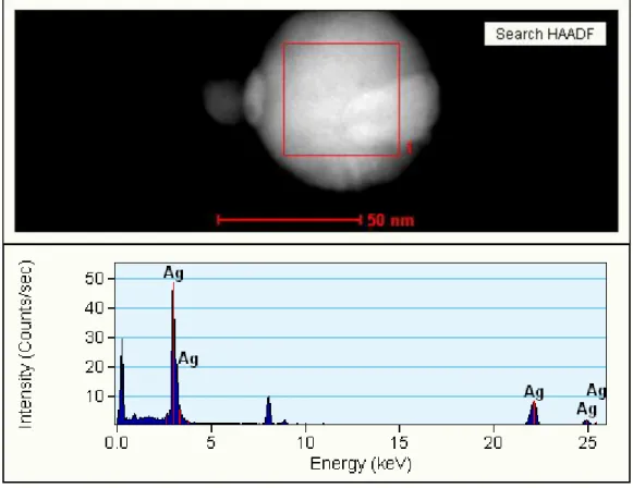
3.3.2. Elemental Composition by XPS and EDX ... 68
3.3.3. Optical Properties by UV-Vis ... 70
CHAPTER 4 SYNTHESIS AND CHARACTERIZATION OF PALLADIUM NANOPARTICLES BY PLAL ... 74
4.1. INTRODUCTION ... 74
4.2. EXPERIMENTAL ... 74
3
4.3.1. Morphology and Crystalline Structure by TEM... 79
4.3.1.1. Pd Nanoparticles: as Prepared ... 80
4.3.1.1.1. Pd Nanoparticles in Distilled Water... 80
4.3.1.1.2. Pd Nanoparticles in Methanol-Water Mixture ... 84
4.3.1.1.3. Pd Nanoparticles in SDS ... 85
4.3.1.1.4. Pd Nanoparticles in Ethylene Glycol ... 87
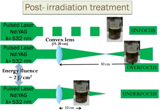
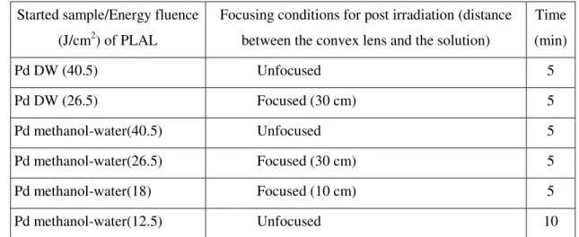
4.3.1.2. Pd Nanoparticles with Post-Irradiation and Ultrasonic Treatment... 89
4.3.1.2.1. Pd NPs in Distilled Water ... 89
4.3.1.2.2. Pd NPs in Methanol-Water Mixture ... 93
4.3.1.2.3. Pd NPs in SDS ... 95
4.3.2. Elemental Composition by EDX and XPS ... 96
4.3.2.1. Pd NPs Obtained in DW... 97
4.3.2.2. Pd NPs Obtained in Methanol-Water Mixture ... 98
4.3.2.3. Pd NPs Obtained in SDS ... 100
4.3.2.4. Pd NPs Obtained in Ethylene Glycol ... 102
4.3.3. Optical Properties by UV-Vis ... 103
4.3.3.1. As-prepared Pd NPs ... 103
4.3.3.1.1. Pd NPs Obtained in DW ... 104
4.3.3.1.2. Pd NPs Obtained in Methanol-Water Mixture ... 105
4.3.3.1.3. Pd NPs Obtained in SDS ... 106
4.3.3.1.4. Pd NPs Obtained in EG ... 107
4.3.3.2. Pd NPs with Post-Irradiation and Ultrasonic Treatment ... 108
4.3.3.2.1. Pd NPs Obtained in DW ... 108
4.3.3.2.2. Pd NPs Obtained in Methanol-Water Mixture ... 109
4 CHAPTER 5
SYNTHESIS AND CHARACTERIZATION OF PLATINUM NANOPARTICLES
BY PLAL ... 112
5.1. INTRODUCTION ... 112
5.2. EXPERIMENTAL SECTION ... 112
5.3. RESULTS AND DISCUSSION ... 115
5.3.1. Morphology ... 115
5.3.2. Elemental Composition by EDX and XPS ... 126
5.3.3. Optical Properties by UV-Vis ... 129
CHAPTER 6 ... 137
CONCLUSIONS ... 137
REFERENCES ... 141
FIGURE CAPTIONS ... 160
5
SUMMARY
Pulsed laser ablation as a novel technique had been employed in this work for the synthesis of Ag, Au, Pd and Pt nanoparticles with different experimental conditions. The different conditions of PLAL such as variation of laser parameters and liquid media were selected based on the literature review and previous knowledge obtained in master studies for this thesis work Silver and gold nanoparticles were obtained by PLAL in distilled water using 1064 and 532 nm output wavelengths. The experiments (single-PL, in-situ and post CW) were performed to study the effect of continuous laser irradiation over the final properties of nanoparticles synthesized by PLAL. Pd nanoparticles were synthesized by PLAL at 1064 nm of wavelength with different energy fluence (40.5, 26.5, 18, 12.5 and 8 J/cm2). Palladium nanoparticle colloidal solutions were obtained in different liquid media such as distilled water (DW), methanol-water mixture, ethylene glycol (EG) and in aqueous solution of sodium dodecyl sulphate (SDS) at 0.001 M. Pd NPs obtained by PLAL in water and methanol-water mixture were treated with pulsed laser post-irradiation using 532 of wavelength with focused and unfocused beam. Ultrasonic treatment was used as a simple alternative to re-disperse and recover the optical properties of Pd NPs obtained by PLAL after their precipitation. All the samples of Pd NPs produced by PLAL in DW, methanol-water mixture and SDS (with and without post-irradiation treatment) were taken to an ultrasonic bath treatment to explore their effect in re-dispersion and the optical properties of the ablated products. Platinum nanocolloids were synthesized by pulsed laser ablation in methanol, ethanol and acetone, respectively at different energy fluence (25, 19 and 9 J/cm2)
6
CHAPTER 1
INTRODUCTION TO NANOMATERIALS AND PULSED LASER
ABLATION IN LIQUID
1.1 GENERAL INTRODUCTION TO NANOMATERIALS
1.1.1 Introduction
Nanotechnology is an important topic that implies design, fabrication and application of nanostructures or nanomaterials, and the fundamental understanding of the relationships between physical properties or phenomena and material dimensions [1]. The recent advances in their synthesis methods, characterization techniques and applications have been of great utility. Another important aspect of nanotechnology is the miniaturization of current and new instruments, sensors and machines. We can find their applications in a great variety of areas as nanoscale electronics and optics, nanobiological systems, nanomedicine, technology, biology, disease treatments, etc. Therefore requires contribution from multidisciplinary teams of physicists, chemists, materials scientists, engineers, molecular biologists, pharmacologists, etc. In this thesis we report an easy, fast, alternative-novel and one-step method for the preparation of pure and stable (in some cases) noble metal nanoparticles, viz Pulsed Laser Ablation in liquid media; this method presents significant advantages over chemical routes as: pure metal nanoparticles production, less aggregation, non-vacuum requirements, non-toxic agents are required, size and shape tunability, between others. Pulsed laser ablation in liquid media will be described in detail in the next part,
1.1.2 Properties of Nanomaterials
7
nanomaterials are related to large fraction of surface atoms, large surface energy, spatial confinement and reduced imperfections. For examples: 1) lower melting point or phase transition temperature and appreciably reduced lattice constants of nanomaterials are due to a huge fraction of surface atoms in the total amount of atoms. 2) Mechanical properties of nanomaterials may reach one or two orders of magnitude higher than that of single crystals in the bulk form due to the reduced probability of defects. 3) Optical properties of nanomaterials can be significantly different from bulk crystals. For example, the optical absorption peak of a semiconductor nanoparticle shifts to a shorter wavelength, due to an increased band gap (by varying material dimension). Surface plasmon resonance is characteristic of metal nanoparticles, which can be appreciated in different color of metallic NPs by varying their sizes. 4) Electrical conductivity decreases with a reduced dimension due to increased surface scattering. However, electrical conductivity of nanomaterials could also be enhanced appreciably, due to the better ordering in microstructure, e. g. in polymeric fibrils. 5) Magnetic properties of nanostructured materials are different from that of bulk materials. Ferromagnetism of bulk materials disappears and transfers to superparamagnetism in the nanometer scale due to the huge surface energy [1].
1.1.3 Applications
Nanomaterials had a great diversity of fields of applications due to their novel properties. These applications are based principally on size dependence of physical properties and huge surface area. Some examples or area of applications can be mentioned as follows. In molecular electronics, which are sensors that translate unique molecular properties into electrical signal. Various electronic devices based on Au nanoparticles and Au55 clusters have been as the single electron transistor action, that contain ideally only one
8
drug in a localized area, thus minimizing the potential side effects of generalized drug therapy [3-5]. Nanobiotechnology is the use of nanostructures as highly sophisticated scopes, machines or materials in biology and/or medicine, and the use of biological molecules to assemble nanoscale structures. Some other applications of nanomaterials in biology or medicine are: fluorescent biological labels, bio detection of pathogens, detection of proteins, probing of DNA structure, tissue engineering, tumor destruction via heating (hyperthermia), separation and purification of biological molecules and cells, MRI contrast enhancement and phagokinetic studies [6, 7]. Industrial catalysis is other important field of application of nanoparticles. Their applications include as chemical manufacturing, energy conversion and storage. Catalytic activity of NPs is dependent of their size, shape and surface sites [8]. As mentioned previously, properties of materials were completely different at nanoscale dimensions. For example, bulk gold is chemically inert but gold NPs have excellent catalytic properties. Gold NPs have been reported in catalysis and photocatalysis processes due to their effectiveness in degrading and mineralizing organic compounds [9, 10]. Other metals as Pd and Pt nanoparticles exhibit excellent catalytic properties [11-14]. The energy sector is an important topic in which the principal goal is to obtain a more effective and efficient process as nano-based technology. Nanomaterials in energy applications would include: lithium-ion battery, fuel cell, light emitting diode (LED), ultracapacitor and solar cells [15] to obtain better performance. For example, nanostructures are advantageous for photoelectrochemical cell devices for high efficient conversion of light to electrical power due to its large surface area at which photoelectrochemical processes take place. The fabrication of solar cells has passed through a large number of improvement steps considering the technological and economic aspects. Different generations of solar cells had been development as based on Si wafers, thin films of amorphous Si and CIS (Copper-Indium-Selenide), nanocrystals and nano-porous materials and quantum dot solar cells based on CdSe-TiO2structures, among others
9
surface plasmon resonance typical of metal nanoparticles. These plasmon sensors were applied for example in miniaturized optical devices, sensors, photonic circuits and detection of trace molecules in chemistry and biology. The sensing techniques are based on surface-enhanced spectroscopies and surface plasmon resonances (SPRs) [19, 20]. Semiconductor nanowires (NWs) are nanomaterials that through controlled growth and organization are used on novel nanoscale photonic and electronic devices. Then semiconductor NWs offer many opportunities for the assembly of nanoscale electronic and optoelectronic devices. Electronic and optoelectronic devices are present in a great range of areas since simple household appliances and multimedia systems to communications, computing, and medical instruments [21, 22]. Bhattacharya et al. [23] reported the growth and electronic properties of InGaAs quantum dots for optoelectronic devices. Self-organized In(Ga)As/GaAs quantum dots are grown by molecular beam expitaxy (MBE) or metal-organic vapor phase epitaxy (MOVPE) on GaAs, InP, and other substrates and are being incorporated in microelectronic and opto-electronic devices. Another important field is nano-electromechanical systems (NEMS) which are suitable for a multitude of technological applications such as ultrafast sensors, actuators, and signal processing components [24, 25]. However, an important NEMs engineering challenge is the detection of displacements in the picometer or even femtometer range at gigahertz frequencies [26]. By this brief summary, it is possible to note that the applications of nanotechnology in different fields have distinctly different demands and challenges, which require different approaches. And their technological advances depend on many factors as understanding nanomaterial properties, nano-manipulation and characterization techniques, among others.
1.1.4 Synthesis of Nanomaterials
10
11
12
attraction and capillary forces. Self-assembled monolayers are molecular assemblies that are formed spontaneously by the immersion of an appropriate substrate into a solution of an active surfactant in an organic solvent. One of the important applications of self-assembly is the introduction of various desired functionalities and surface chemistry to the inorganic materials. Self-assembled organic monolayers are widely used to link different materials together; to the synthesis and fabrication of nanomaterials and nanostructures, particularly the core-shell structures [39-41]. Laser ablation of solid materials has been studied intensively due to their great potential in laser-based material processing [42]. In general techniques of laser processing of nanostructures are simple, quick, one-step and green, and produce materials having surfaces free from chemical contamination [43]. Laser-based nanomaterial processing methods can produce 0D, 1D, 2D, and 3D nanostructures in the gaseous or liquid phases, and can produce nano-/microstructures at the selective sites of bulk solid materials. Pulsed laser deposition [44, 45], laser vaporization controlled condensation (LVCC) [46], laser pyrolysis [47-49], laser chemical vapor deposition [50, 51], laser-induced direct surface writing for nano-/microfabrication [52, 53], two-photon polymerization [54], laser-induced forward transfer (LIFT) [55], laser ablation in liquids (LALs) [56, 57], laser-induced melting [58-60], laser fragmentation for resizing and reshaping of particles [61-68] and laser-induced photo dissociation of liquid precursors [69] are some of these processes. Laser ablation is easily carried out in conventional deposition chambers with vacuum or filled gas. Laser ablation has proven to be an effective technique for the deposition of complex films including complex metal oxides such as high Tc
13
from any type of chemical contamination. Then nanomaterials obtained by PLAL are excellent for their use in biocompatibility, functionalization, drug loading, environmental sensing and medical applications.
1.2 PULSED LASER ABLATION IN LIQUID
1.2.1 Introduction
Pulsed laser ablation in liquid (PLAL) is an important method for the synthesis of nanoparticles with tunable morphology and size. After the invention of the first pulsed Ruby laser by Maiman [76] laser-matter interactions has been studied by many researchers. Pulsed laser ablation in a vacuum or dilute gas was the technique which preceded PLAL [77]. Pulsed laser ablation in liquid is a versatile technique with the capacity to use different target materials (elemental, alloys, powders) and most recently researchers used different target geometry [78, 79] to improve the productivity of the laser ablation process. Another remarkable advantage is the devoid of toxic agents in the synthesis of nanoparticles as in comparison with chemical methods. There are many reports on synthesis of stable nanomaterials in colloidal solution by PLAL without the addition of any surfactant for size control. It is possible to use liquids which allow applications of biocompatibility [80, 81] (water, alcohols, etc.) of the nanomaterials synthesized by PLAL. A relevant advance in this topic, is the use of liquid flow [82, 83] or liquid with special properties (polymers [84] or supercritical fluids [85, 86]). The variation of implicit parameters of PLAL as: laser wavelength, pulse duration, frequency and fluence are other important aspects to produce a wide variety of nanomaterials [73, 74, 77, 87].
1.2.2 Fundamental Aspects of PLAL
PLAL has become a feasible synthesis technique of nanomaterials in colloidal solution by choosing appropriate target material, confining liquids and laser parameters. In comparison with other conventional methods of synthesis as vapor deposition, vapor phase transport, pulsed laser ablation in vacuum and hydrothermal methods, PLAL has interesting advantages. These advantages are:
1) clean and green synthesis, the ablated products are usually obtained without by-products and no need of posterior purification;
14
3) confined conditions of high pressure and temperature in the plasma plume which favors the formation of unusual metastable phases.
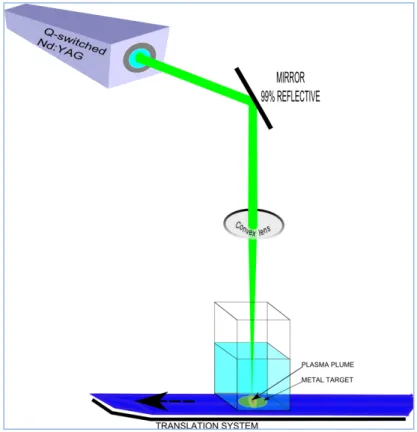
The yield of nanoparticles is dependent not only of the laser parameters (pulse duration, frequency, energy and wavelength) but also of the target properties and the liquid media. The final properties of nanoparticles are also characteristics of the plasma plume properties. The general configuration of PLAL is a laser beam focused onto a metal target which is submerged in liquid environment to produce colloidal suspensions of the nanomaterials synthesized. Figures 1 and 2 show the typical setup configurations of horizontal and vertical PLAL, respectively. In these configurations, the laser beam was focused and directed with convex lens and mirrors (99% reflective), respectively. To vary the energy fluence over the metal target, the distance between convex lens and sample is changed. Energy fluence (in J/cm2 units) was defined as the laser energy output (Joules) per unit area (cm2). Our laser laboratory is equipped with a Q-switched Nd:YAG pulsed laser operating at 1064 nm for the first harmonic and 532 nm for the second harmonic. The laser beam properties are: 10 ns pulse width, 10 Hz pulse repetition and output energy of 600 and 230 mJ/pulse for IR and visible wavelength, respectively. Two continuous wave lasers (CNI Lasers), one of 532 nm (variable to 0-10 Watts of laser power) and the other of 457 nm (fixed at 4 Watts of laser power) of wavelength are available. An optical power and energy meter (Model PM100D, Thorlabs Inc.) was used to measure the output energy of the Nd:YAG pulsed laser and the continuous lasers. To improve the ablation productivity and to avoid continuous irradiation at same place of the target, a translation system (designed by mechatronics students of FIME, UANL) with linear movement was employed. The
15
Figure 1. General experimental set-up of pulsed laser ablation in liquid (PLAL) in horizontal configuration.
16
1.2.3 Proposed Mechanisms
The process of laser interaction with the target is similar for pulsed laser deposition in vacuum (PLD) and for pulsed laser ablation in liquid (PLAL). The production of plasma and formation of a strong confinement of the emission species is characteristic in both processes. The crucial difference is the free expansion of plasma in vacuum in PLD, while in PLAL the plasma is confined by the liquid layer. The liquid layer delays the expansion of the plasma plume, generating a high plasma pressure and temperature in the irradiated zone [77]. This leads to the vaporization of both the solid target and the liquid, which allows the formation of novel materials because the products can contain atoms from the target and the liquids. The basic mechanism and the particle formation of PLAL starts with the absorption of the laser pulse by the target. An analytical study of the energy deposition process was described by S. Hachimoto et al. [88], in three stages. First, part of the laser beam was absorbed by the electronic system. Second, after a few hundreds of femtoseconds, a thermal equilibrium was reached to transfer the electron energy to the lattice via electron-phonon coupling. Third, the lattice energy is given to the surrounding liquid, resulting in the cooling of the NPs. Due to the nature of the pulsed laser irradiation, PLAL is very fast and far-from equilibrium process. Therefore all metastable and stable phases formed in the ablation process could be maintained in the final products [89]. This phenomenon resulted for the laser-matter interaction which is largely determined by: the
17
1 mg of ablated products requires 105 laser pulses, that correspond to ablation times of 20 minutes at 10 Hz (nanosecond or picoseconds) or 100 seconds at 1 KHz (femtoseconds) [90]. When the laser beam impacts the target, its surface reflects part of this incoming energy. The absorbed energy (photons) is transferred to the electrons in the target, leading to the formation of hot carriers which transfer their energy to the ions into the material (page 114 of [56]). The ions and the electron hot carriers eventually reach equilibrium (when the temperature of the electron gas Te and the lattice Tl are equal) in a timescale τE=
10-12– 10-11s [91]. This timescale is an important factor to determine the possible routes to ablation, thermal or non-thermal for “long” or “short” pulses. When the laser pulse is absorbed by the target, a plasma plume is created. This plasma plume contains the ablated material and expands into the liquid media. Due to the confining effect of the liquid media, the plasma plume expands and collapses continuously releasing energy to the liquid solution and emitting a shockwave. During this event a cavitation bubble is generated, which also expands and collapses in a time scale of hundreds of microseconds emitting a second shockwave [92]. Fabbro et al. studied the thermodynamics of the plasma plume created by PLAL by emission spectroscopy [93]. Thermal evaporation, is the proposed
mechanism for long pulses as in the case of nanosecond pulsed laser, (where τL » τE)
electron and phonons reach at equilibrium during the heating stage. Then the material ejection and phase change is dominated by thermal process. Due that the heat diffusion from the lattice takes place on a timescale shorter than the pulse width. Liu et al. reported the laser induced heating and melting (not vaporized) of the target material via heat conduction inside the material. Where the temperature distribution is governed by the heat conduction equation:
�� ���� = ∇ ∙ �∇� + 1 − � � ���� Eq. 1
Where, ρ= density, Cp= specific heat, K= thermal conductivity and T= temperature. The second term represents the source which is the laser energy absorbed by the material at a depth z from the surface. R= surface reflectivity, Io= laser irradiance and α= absorption
18
of the liquid media, the plasma plume cools and release energy to the liquid media during the expansion. NPs are formed during this process due the condensation of the vapor atoms. The nucleation and growth of the NPs are related principally by the laser parameters. The nucleation time is double that the laser pulse width used (for nanosecond lasers). There is an inverse relation between the pressure and time of nucleation and a direct relation between the velocity and the temperature of nucleation [94]. Explosive boiling takes places when a very high energy pulsed laser irradiate the target, as ultra-short pulses (femtoseconds and picoseconds). The transmitted energy to the electrons by femtosecond laser pulses is on a time-scale much shorter than the electron-phonon thermalization process. Then for ultra-short pulses, the material is into a highly non-equilibrium state and temperature of the electron gas that is much higher than the lattice temperature. By thermodynamic analysis, if the surface target is heated upper the limit of its thermodynamic stability during ultra-short pulse laser, the surface experiment a rapid transition from superheated liquid to a mixture of vapor and liquid droplets. Another approach to PLAL technique is the size reduction or shape modification by the irradiation of colloidal solutions by a selected pulsed laser beam. Either with nanosecond [63, 66-68], femtosecond [62, 95] or picosecond [61, 96] pulsed laser irradiation. Different mechanism had been proposed to fragmentation process. Coulomb explosion model presumes ejection of a quite number of electrons to generate multiply ionized NPs to undergo spontaneous fission because of the charge repulsion. Werner and Besner [62, 95] reported that femtosecond laser-induced fragmentation is dominated by the Coulomb explosion mechanism. Photothermal evaporation is another mechanism of fragmentation process. Pyatenko et al. [97] reported that the photothermal mechanism prevails at low laser intensities in their studies based on the observation of a size reduction of chemically prepared aqueous gold NPs by exposure of the SPR band to various intensities and numbers of 532 nm nanosecond pulsed laser beam. Two temperature model proposed that the transferred laser energy into the electron and lattice sub-systems is described by two temperatures, electron temperature (Te) and lattice temperature (Tl) to determine the energy distribution inside the system.
19
evaporation of NPs was not predominant compared to Coulombic explosion resulting from thermionic electron emission and photoelectric effect.
1.2.4 Methodologies
Pulse laser ablation in liquid media had been successfully used for the synthesis of different materials in a great variety of liquid media. This section will present some alternative configurations in the set-up of the PLAL experiments. These modifications are reported with the firm intention to improve the production of nanoparticles or to modify the final properties of the ablated products.
20
particles are in use in various functional materials such as cell markers and photonic materials.
1.2.5 Applications
Nanomaterials obtained by PLAL in colloidal solution had important characteristics as purity, stability without surfactants and narrow size distribution. Biotechnology applications: In biotechnology research [6], had been reported nano-gold applications [102] as the conjugation with biomolecules (for example in the case when antibody, DNA or aptamer-targeting) and fluorescence imaging [103]. The use of PLAL products showed important results in medical technology due their high reactivity and less toxicity (than chemically synthesized). S. Barcikowski et al. [81, 104, 105] reported some studies of in-situ conjugation during laser ablation of gold nanoparticles with biomolecules. They demonstrated that laser-ablation-based in situ conjugation is a rapid, one-step production method in comparison to the conventional bio-functionalization using chemical synthesis, being highly pure gold colloids possessed some advantages for biological applications as an efficient binding to biomolecules (higher yield), higher ligand load (relevant to specificity of targeting) and less cleaning effort (no interfering chemical residuals). Catalysis: The great variety and the purity of nanoparticles produced by PLAL imply advantages for catalytic application. The absence of a ligand layer due the electrostatic stabilization of laser-generated particles is a positive effect on the affinity of nanoparticles to the carrier surface. Nanoparticles synthesized by PLAL had higher deposition efficiency in comparison with chemically synthesized nanoparticles (containing residual citrate) [106]. The use of metal nanoparticles also had been explored in the catalytic reduction of CO and photocatalytic hydrogen production [107]. Wu et al. [108] examined the effect of hydrogen-thermal reduction process time on TiO2–Pd based catalysts for the performance of
21
PLAL also had been explored to produce nanoparticle-polymer nanocomposites due the purity of laser-generated colloids which allow the binding of the particles to the polymer. For example, Ana Menéndez-Manjón et al. [84] reported the formation of ligand-free gold– silver nanoparticle alloy polymer composites by picosecond laser ablation in liquid monomer.
1.2.6 Materials
PLAL, as mentioned is an effective, rapid, simple and versatile technique that could be used to synthesize a great variety of nanostructures with various compositions (metals, alloys, oxides, carbides, hydroxides, etc.) and morphologies (nanoparticles, nanocubes, nanorods, nanocomposites, etc.). Another important branch of PLAL is post laser irradiation of suspended nanomaterials that can be applied to further modify their size, shape, and composition. This section is a brief summary of the different materials reported by PLAL. Pulsed laser ablation of noble metals as Ag, Au, Pd, Pt, etc. had been reported by many authors and will be discussed in detail in the next part. Metal oxide compounds formed via the reaction of ablated metal with liquid media are another important research field [113]. For example the synthesis of ZnO [114-119], TiO2 or TiO [120-126], CuO [59,
127-130], Aluminum oxide [131, 132], SnO [133], Sb2O3 [134, 135], NiO [136], Bi2O3
[137], Fe3O4/FeO [138-141]. Also different morphologies also had been reported as ZnO
22
synthesized by PLAL are Cyanopolyynes in acetonitrile [156], linear carbon chain colloid mixed with silver nanoparticles [157], core-shell CdS/carbon NPs [158], carbon-based nanostructures [98] as (diamond-like carbon or nanodiamonds) [159] and carbon NPs [160]. The versatility of PLAL technique allowed the synthesis of a great diversity of nanomaterials, nanocompounds, nanocomposites, etc. For this reason, the field of synthesis, methodologies and future applications will be helpful to fulfill the new requirements and scientific curiosity.
1.3 METAL NANOPARTICLES BY PLAL – AN OVERVIEW
1.3.1
IntroductionIn recent years, nanotechnology have been attracted much attention due to all novel properties and applications of the nanostructured materials. Nanotechnology implies design, fabrication and application of nanostructures or nanomaterials [1]. The understanding of the relationship between physical properties and material dimensions is fundamental for all the potential applications. Nanotechnology has a broad range of potential applications as nanoscale electronics and optics, nanobiological systems, nanomedicine, etc. Materials or structures at nanometer scale possess new physical properties or exhibit new physical phenomena. For example, some noble metals at nanometer scale have a surface resonant plasmon at a frequency in the visible light range. Due the Mie`s theory which explained the red color of gold nanoparticle colloidal in 1908 by resolving Maxwell´s equation for an electromagnetic light wave interacting with small metallic spheres. By resolving these equations they concluded that the plasmon resonance depends explicitly on the particle size, r. The larger the particles, the more important the high-order modes as the light can no longer polarize the nanoparticles homogeneously. These higher-order modes peak at lower energies. Therefore, the plasmon band red shifts with increasing particle size. At the same time, the plasmon bandwidth increases with increasing particle size. The situation concerning the size dependence of the optical absorption spectrum is more complicated for smaller nanoparticles for which only the dipole term is important. For nanoparticles much smaller than the wavelength of incident light (2r < max/10), only the dipole oscillation contributes to the extinction cross-section
23
24
Actually, noble metal nanoparticles are of interest in various technological areas like sensing, catalysis, electronic and plasmonics due some features as: i) the presence of optically activate plasmonic modes which originate intense absorption and scattering bands in the visible-near infrared interval, the local field amplification due to plasmon resonance is also at the basis of surface enhanced Raman scattering (SERS); ii) the easy surface functionalization with a wide series of organic molecules, and iii) the chemical and physical stability and biocompatibility. Important catalytic properties derive from the chemical stability and surface chemistry of noble metal nanoparticles [90].
1.3.2
Gold Nanoparticles in Colloidal Solution25
Au-26
27
28
29
system to do online characterization of the production of nanoparticles by nanosecond pulsed laser ablation and to complement the information obtained by TEM. Other important feature for this work was the use of a double pulsed laser system as the reported by De Giacomo [98] and V. S. Burakov [99]. They reported that the time delay between the laser pulses increases the productivity of the ablation process. The variation of wavelength also was reported for the synthesis of gold nanoparticles as O. V. Overschelde et al. [212]. They reported the synthesis of gold nanoparticles by 248 nm of excimer KrF nanosecond pulsed laser ablation in water and some alcohols. Werner et al. [213] added high pressure to control the size of gold nanoparticles by laser-induced size reduction. They observed an effect of the energy fluence on the size reduction of these gold NPs especially at high pressures. They ascribed their results to the formation of a supercritical water layer surrounding the liquid droplet NP transformed by laser heating. They previously worked with laser-induced fragmentation of gold nanoparticles by a femtosecond pulsed laser. They proposed that the Coulombic explosion was the dominant mechanism in the presented studies [95]. Another methodology is laser melting in liquids to synthesize submicron-sized spherical particles. Tsuji [58] reported the fabrication of gold sub-micron spherical particles by laser melting using citrate as stabilizer. Also reported the synthesis of gold sub-micron particles by this technique in another less toxic stabilizer as NaCl solution [101].
1.3.3
Silver Nanoparticles in Colloidal SolutionVarious methods have been developed for the synthesis of silver nanoparticles. One of them is by the UV illumination [214] of aqueous solution containing AgClO4, acetone,
30
31
deionized water and ethanol and concluded that high polar molecules provided a strong surrounding electrical double layer, which prevents growth, aggregation and precipitation. A. Pyatenko et al. [227] reported the synthesis of silver NPs by PLAL in pure water and studied the effect of laser power and spot size (which was related to energy fluence). They observed that at high laser power and small spot sizes, small silver nanoparticles with a narrow size distribution were obtained. R. A. Ganeev et al. [228] also reported the synthesis of Ag NPs by PLAL in different liquid media (ethylene glycol, water and ethanol). They observed that the viscosity of surrounding liquid had an important effect on the stabilization of structural and nonlinear optical properties of Ag nanocolloids. F. Mafune et al. [229] reported the synthesis of silver NPs by PLAL (532 nm) using different surfactants to study the stability of nanocolloids. In another study [230], they observed the formation of smaller silver NPs with increase in the concentration of sodium dodecyl sulfate and with a decrease in the irradiation laser power. There are reports on the synthesis of complex structures, alloys or nanostructures by PLAL technique as follows. Z. Yan et al. [231] obtained the formation of Ag-Ag2O complex structures by excimer laser ablation of Ag in water.
32
lamp excitation. Silver colloidal were synthesized by using auto-polymerizable resin and AgNO3 in an ethanol solution. They produced spherical silver nanoparticles (5–8 nm) with
the combination of pulsed laser ablation with UV–visible illumination. Another important example is the report of K. G. Stamplecoskie et al. [234] of a photo-chemically synthesis method of silver nanoparticles with the shape control of these NPs under the irradiation of a light emitting diode at different wavelength. Pulsed laser ablation in liquid had many advantages, as result a great number of applications and specific studies had been reported; F. Neri and co-workers published results on the enhanced nonlinear optical response of linear carbon chain colloid mixed with silver nanoparticles [157]. E. Messina et al. [147] reported the synthesis of gold/silver alloy by PLAL in colloidal solution. The obtained solution was observed with tuneable plasmon resonance and proposed for applications in bio-sensing experiments, frequency selected photo thermal therapy, nonlinear optics, SERS applications, and spectroscopic characterization of nanomaterials and composites. Other applications of silver nanoparticles in colloidal solution synthesized by PLAL had been reported as; in biological field some applications as for mosquito control [235], antimicrobial [236], protein reduction [237]. SERS enhancement due to the superficial plasmon resonance of novel metals like silver also was reported, for example: palladium-doped silver nanoparticles [238]. In photo-catalysis field, in some composites like titanium dioxide nanotubes modified with silver and palladium nanoparticles [154], catalytic activity of silver nanoparticles supported on manganese oxide [171].
1.3.4
Palladium Nanoparticles in Colloidal Solution33
these reasons, research has been focused on the development of this nanocatalysis by the formation with fine control of both size and shape. Catalytic activity of the nano-catalyst in the nano-colloid depends not only on size and shape; another important characteristic is the surface environment, which is often dramatically influenced by the use of reducing agents. The use of protective agents had been reported as n-alkanethiols, phosphines, linear polymer such as poly(vinylpyrrolidone), polyvinyl alcohol [14], sodium polyacrylate and dendrimers. The catalytic activity of Pd nanoparticles are well understood, however the costs of this material has been increasing in the last years. Therefore the studies in which Pd is employed as nanoparticles can allow the fabrication of chemical sensors using a very small amount of this material. Pt and Pd NPs of different morphologies have been reported in the growing area of shape-controlled synthesis and shape-dependent catalytic studies. Pt and Pd NPs have a face-centered-cubic (FCC) crystal structure. The literature reports calculations that predicted thermodynamic equilibrium shape for an FCC nanocrystal is a truncated octahedron bound by six {100} and eight {111} planes. There are catalytic studies showing shape-dependent effects of Pt and Pd NPs [13], between them faceted polyhedral nanocrystals, porous-like, branched nanostructures, 1-D nanorods and nanowires. Pd nanoparticles had been coated with cationic isocyanides, thiolated -cyclodextrin, N,N,N-trimethyl (8-mercapto octyl) ammonium chloride, mercapto ammonium ligand, polyvinyl alcohols [14], among others. Palladium nanoparticles are applied in many industrial applications as in the reduction of automobile pollutants and catalyst for many organic reactions, such as C-C coupling of Heck, Suzuki, hydrogenation of alkenes and allylamines. Palladium nanoparticles have a special sensitivity to absorb hydrogen, make them appropriate as gas sensor [12, 239] and as hydrogen storage materials in fuel cells or batteries. There are reports on the use of Pd NPs as hydrogen and humidity sensors which were fabricated by laser ablation direct writing [240]. Wei-Fang et al. reported the photo catalytic performance of TiO2-Pd based nanoparticles [108]. Jianzhong
34
nanoparticles synthesized. Some research groups studied the effect of the different ablation parameters over the final properties of the Pd nanomaterials and their potential applications. Pd nanoparticles produced by PLAL can be separated mainly in two groups: first when they used an IR laser irradiation with different conditions. J. Pou and co-workers produced pure Pd nanoparticles using both, CW (monomode Ytterbium doped fiber laser YDFL, @ 1075 nm, irradiance ranged 2 x 10⁵ and 106 W/cm2) and pulsed laser (Nd:YAG @1069 nm, 1-2
ms pulse width, 10 Hz, pulse energy 2-8 J) ablation in de-ionized water [242]. M. Muniz-Miranda et al. [243] reported the production of palladium nanoparticles by pulsed laser
ablation in water. Laser ablation was performed using a NdμYAG pulsed laser ( = 1064 nm, τ= 1β ns, 10 Hz, laser energy γ-158 mJ) in an aqueous solution (with and without the addition of SDS) and an energy fluence between 1.6-2000 J/cm2. Through the variation of the fluence, it was observed the optimal conditions for the NPs productions at an intermediate fluence (~21 J/cm2) and by strongly defocusing the laser beam. Pd NPs had SERS activity observed by spectroscopic analysis of their precipitates. Soghra Mirershadi et al. [244] studied the wavelength dependence on the nanoparticle formation mechanisms during PLAL. They applied an IR Nd:YAG pulsed laser ( = 1064 nm) and UV ArF excimer laser ( = 1λγ nm) for the synthesis of Pd NPs in deionized water. Both lasers had
35
structure, elemental composition, chemical state and optical properties of the ablated products.
1.3.5
Platinum Nanoparticles in Colloidal SolutionPlatinum and its alloy nanoparticles have attracted much attention because of their potential application in many catalytic reactions as to eliminate NO generated in combustion process [247]. There are various synthesis routes for the fabrication of Pt and Pt-compounds at nanoscale as: Henglein et al. [248] studied and compared three different methods for the preparation of Pt nanoparticles: radiolysis, hydrogen reduction and citrate reduction. By pulsed laser ablation deposition had been synthesized Pt compounds with different application in catalysis, as Daniel Guay and co-workers [249] synthesized a series of PtxSn100-x NPs by PLD method by varying the background pressure during the
deposition. They reported the characterization of their physic-chemical and electrochemical properties. It was found that the surface composition and structure of the resulted catalysts NPs has a high effect on the electrocatalytic activity of the catalysis for ethanol oxidation. Synthesis of Pt and Pt compounds by PLAL have been reported by different researches groups as; Zheng-Wen Fu et al. [250] reported the preparation of highly dispersed platinum nanoparticle-graphene nanosheet (PtNP-GNS) hybrid colloidal solution by pulsed laser ablation in liquid. A NdμYAG pulsed laser ( = γ55 nm, frequency 10 Hz, τ= 5 ns, fluence β
J/cm2) was used for the ablation of a platinum plate (99.99%) which was immersed in 5 ml of aqueous graphene dispersions. Air cathodes were prepared by dispersing drops of PtNP-GNS onto the ITO substrate and drying in air for the film deposition. PtNP-PtNP-GNS hybrids showed a high electro-catalytic activity for discharge and charge process as air electrode which is important for applications for Li-air battery. Stephan Barcikowski et al. [150] were successful in the synthesis of charged Pt-Ir alloy nanoparticles by femtosecond laser ablation in acetone. These PtIr obtained nanoparticles had the same composition as the Pt9Ir
target used in PLAL and was confirmed by EDX (in TEM), STEM-EDX maps, HRTEM and SAED. These NPs were used to make electrodes by electrophoretic deposition with enhanced surface roughness in comparison with other techniques. The ablation process was developed with a femtosecond laser system ( = 800 nm, τ= 1β0 ps, pulse energy = γ00 J/,
36
system employed was a Q-switched NdμYAG ( = 1064, 5γβ and β66 nm, τ= 5 ns, 10 Hz) for the ablation of a Pt target immersed in citrate and some polymers as PEG (poly ethylene glycol), PVA (polyvinyl alcohol) and PVP (polyvinylpyrrolidone) as stabilizing agents. They observed shape modifications of the Pt NPs which were related with the wavelength selected. The nanoparticles were employed to develop films capable of assisting the laser deposition ionization (LDI) of a model peptide. Stable Pt nanoparticles were synthesized [252] by pulsed laser ablation in water and aqueous solution of sodium dodecyl sulfate (SDS). The abundance of the Pt NPs was changed with the concentration of surfactant. The
laser system used in this experiment was a NdμYAG pulsed laser ( = 5γβ nm and 1064 nm, 10 Hz). Naoto Koshizaki et al. [253-255] reported a complete study of Pt NPs produced by pulsed laser ablation in liquids. Laser ablation was carried out at normal incidence of the
laser beam NdμYAG ( = 1064, 5γβ and γ55 nm, τ= 10, 8 and 7 ns, respectively) of a platinum metal immersed in filtered, doubly de-ionized water (> 18 MΩ). They covered
37
morphology, size, size distribution, structure, elemental composition, chemical state and optical properties of the ablated products.
1.3.6
Other Metal Nanoparticles Synthesized by PLALThere are nanomaterials of other metals, non-metals, lantanides, semiconductors etc. that had been synthesized by PLAL in different liquid media. This is due to the great advantages of this technique and the potential applications of the obtained nanoparticles/nanocomposites. PLAL of a Pb target in primary alcohols [256] resulted in synthesis of (Pb3(CO3)2(OH)2). The second harmonic of an Nd:YAG laser was used for the
PLAL experiments to obtain the nanostructured hydrocerussite with hexagonal morphology. Santillan et al. [127] synthesized Cu and Cu oxide nanoparticles by femtosecond pulsed laser ablation of Cu in water and acetone. They reported significant differences in the composition and structure depending on the liquid media and the energy fluence used. Hongqiang Wang et al. [59] reported the synthesis of submicrometer sized particles of CuO by pulsed laser ablation of colloidal NPs. They proposed the heating-melting fusion as the predominant mechanism. They reported that the size and phase of ablated products could be controlled by the input laser fluence. Seung Won Lee et al. [128] also reported the synthesis of CuO nanofluid to critical heat flux (CHF) measurements. They used a Nd:YAG laser system at 532 nm of wavelength for 8 hours. P. G. Kuzmina et al. [257] produced porous nanoparticles of Al or Ti by PLA in ethanol, water and n-propanol saturated with hydrogen. They used two different pulsed laser systems, a Ti:Sapphire (180 fs at =800 nm) and a NdμYAG (70 ns at =1064 nm). They reported that the morphology of NPs and its cavities depend on the nature of the metal and on the pulse duration. Cobalt sulfate nanoparticles [258] were synthesized by PLAL using a pure Cobalt slice as metal target immersed in SDS solution. They used a Nd:YAG laser system with the fundamental wavelength for 1 hour of irradiation. Important changes in the optical and magnetic properties of the synthesized NPs were observed in comparison with the bulk material. Qiang Li et al. [259] reported the formation of core–shell TaxO@Ta2O5 (x =1/2)
composite nanoparticles by the irradiation of tantalum metal plate in ethanol. In the characterization to the photocatalytic activity of the TaxO/Ta2O5 core–shell NPs present
higher activity compared with pure micrometer-scale Ta2O5 powders. In the case of
38
compound as Tomonari Wakabayashi et al. [156] which reported the synthesis of Cyanopolyynes by pulsed laser ablation in liquid acetonitrile. The carbon powders were irradiated with an Nd:YAG 532 nm laser system at 0 °C during 5 hours. In the same group of carbon materials, S. Z. Mortazavi et al. [260] reported the synthesis of graphene by pulsed laser ablation in liquid nitrogen. They used a q-switched Nd:YAG laser at 1064 nm of wavelength and observed the relation between the number of graphenes and the energy fluence used. They proposed that the mechanism for the graphene formation is the penetration of liquid nitrogen and the subsequent expansion to gas phase during laser heating. Also there are studies on some lantanides as reported by K. Hasna et al. [261] describing the synthesis of Europium doped HAp (hydroxyapatite (Ca10(PO4)6(OH)2)
nanoparticles for applications as imaging of tumor cells and also for targeted drug delivery. They reported the laser ablation in liquid phase of europium doped HAp target using the third harmonic of the Nd-YAG laser system at a wavelength of 355 nm. Some results on semiconductor NPs synthesized by PLAL is described as follows: Nikolai V. Tarasenko et al. [262] reported the synthesis of Ga-Si nanoparticles by PLA in ethanol. They used a
NdμYAG pulsed laser at 1064 nm and 8 ns of pulse width. The Gd−Si nanoparticles
39
40
1.4 HYPOTHESIS
The morphology and properties of noble metal nanoparticles synthesized by pulsed laser ablation in liquid media is determined by laser parameters, ablation parameters (energy fluence, liquid media and ablation time) and in-situ irradiation process.
1.5 OBJECTIVES
1. To synthesize Au and Ag nanoparticles by PLAL.
2. To study the effect of in-situ irradiation of continuous laser during the pulsed laser ablation of Ag and Au metal targets in different liquid media.
3. To characterize Ag and Au nanoparticles by TEM, SAED and UV-Visible absorption spectroscopy.
4. To characterize chemical state and elemental composition of Ag and Au nanoparticles by XPS and EDX spectroscopy.
5. To synthesize Pd and Pt nanoparticles by PLAL.
6. To characterize Pd and Pt nanoparticles by TEM, SAED and UV-Vis absorption and XPS analysis.
7. To study the effect of ablation parameters (energy fluence) in the formation of Pd and Pt nanoparticles.
41
1.6 JUSTIFICATION
42
CHAPTER 2
SYNTHESIS AND CHARACTERIZATION OF GOLD
NANOPARTICLES BY PLAL
2.1.INTRODUCTION
43
laser was expanded over the colloidal solution near to the irradiated zone of the metal target, where the plasma plume and the initial stage of the ablated material occur. A detailed description of experimental conditions is given below. The shape, mean size, size distributions, elemental composition and optical properties of gold nanoparticles synthesized at different irradiation configurations were examined. These nanoparticles were characterized by transmission electron microscopy (TEM), X-ray photoelectron spectroscopy (XPS) and UV-Visible absorption spectroscopy.
2.2.EXPERIMENTAL SECTION
44
45
Figure 3. a) Photo of experimental set-up for PLAL using 1064 nm. The inset corresponded to PLAL experiment with in-situ / CW532 nm irradiation; b) schematic of vertical
configuration (1064 nm).
46
Figure 5. Au colloidal solutions obtained by PLAL for 1064 and 532 nm of wavelength under different configurations.
2.3.RESULTS AND DISCUSSION
Gold nanocolloids obtained by PLAL experiments (single, in-situ and post) were red color and stable for 6 months after their synthesis. UV-Vis absorption measurements were carried out after PLAL experiments in a Vis-NIR spectrometer (Shimadzu UV-1800) in a wavelength range of 200 to 1000 nm. For the optical measurements, the colloidal samples were taken in a quartz cuvette of 1 cm of length, using water as reference. Immediately after each experiment, a TEM grid was prepared by placing a drop of the colloidal solution on a copper grid and dried at ambient temperature. TEM analysis was performed in a FEI Titan G2 80-300 in bright field (BF) and STEM mode (with the high angle annular dark field and EDX detector). For XPS analysis a few drops of each solution were placed on a Cu tape and dried at room temperature. The samples were subjected to X-ray photoelectron spectroscopy (XPS) analysis in a Thermo Scientific K-Alpha XPS instrument. The samples were excited by a monochromatized Al Kα X-ray radiation of energy 1486.6 eV. All the spectral peaks were recorded with reference to C 1s peak (284.6 eV).
2.3.1.Morphology by TEM
47
48
by self- absorption condition. Then in-situ and post CW 457 irradiation had an effect over the growth of gold nanoparticles. Probably for in-situ CW 457, the additional energy helped the formation of nanowires meanwhile; only small nanoparticles were produced by post CW 457 irradiation.
Figure 6. TEM micrographs and size distribution of Au NPs obtained in DW by PLAL using 1064 nm in Single, In-situ and Post-irradiation configuration with CW lasers of 532
49
Figure 7. TEM micrographs and size distribution of Au NPs obtained in DW by PLAL using 532 nm in Single, In-situ and Post-irradiation configuration with CW lasers of 532
50
Figure 8. TEM micrographs of Au NPs obtained in DW by PLAL using 532 nm in Single, In-situ and Post-irradiation configuration with CW lasers of 532 and 457 nm
51
structure was not affected by the in-situ or post-irradiation of the different CW lasers (532 and 457).
Figure 9. HRTEM and SAED micrographs of Au NPs obtained in DW by PLAL using 1064 nm in Single, In-situ and Post-irradiation configuration with CW lasers of 532 and
52
Figure 10. HRTEM and SAED micrographs of Au NPs obtained in DW by PLAL using 532 nm in Single, In-situ and Post-irradiation configuration with CW lasers of 532 and 457
nm.
2.3.2.Elemental Composition by XPS
Elemental composition of gold nanoparticles was analyzed by XPS technique. For XPS analysis some drops of Au nanocolloids were dried at room temperature on a Cu tape. This was repeated many times to recollect sufficient material for analysis. Figure 11 shows the high resolution Au 4f for gold nanoparticles obtained by PLAL in the set of 532 nm of wavelength. The Au 4f7/2 at 83.97, 83.48 and 83.70 eV corresponded to single-PL532,
53
ones. This result shows the high purity of Au nanoparticles in colloidal solution obtained by PLAL, same as the results reported by this technique [64, 197, 276]. Sample preparation of in-situ CW (532 and 457) were performed using precipitates at bottom of the solution, meanwhile single PL532 was completely stable. Then, the little chemical shift between them can be attributed to the difference between core level binding energies of surface atoms with respect to those of bulk material (in reference to precipitates of gold in the solution) [277].
Figure 11. High resolution Au 4f XPS spectrum of gold nanoparticles obtained by PLAL using 532 nm of wavelength in distilled water with single and in-situ (532 and 457)
configurations.
2.3.3.Optical Properties by UV-Vis
54
55
Figure 12. UV-Vis Absorption spectra of gold nanoparticles synthesized by PLAL using 1064 nm in DW at different configurations of experiments (Single PL1064, In-situ CW457,
In-situ CW532, post CW457 and post CW532)
Figure 13. UV-Vis Absorption spectra of gold nanoparticles synthesized by PLAL using 532 nm in DW at different configurations of experiments (Single, In-situ 457, In-situ 532,
56
Figure 14. UV-Vis Absorption spectra of gold nanoparticles synthesized by PLAL using 532 nm in DW at different configurations of experiments (Single, In-situ 457, In-situ 532,
post 457 and post 532) after 43 days of PLAL experiments.
57
CHAPTER 3
SYNTHESIS AND CHARACTERIZATION OF SILVER
NANOPARTICLES BY PLAL
3.1.INTRODUCTION
This chapter presents the synthesis and characterization of Ag NPs by pulsed laser ablation in distilled water (DW). Effects of in-situ and post-irradiation with CW lasers to the silver nanocolloids during and after PLAL experiments were studied. Ag NPs were characterized by TEM, EDX, XPS and UV-Vis Absorption spectroscopy to explore the morphology, size, size distribution, structure, elemental composition, chemical state and optical properties of the ablated products.
3.2.EXPERIMENTAL SECTION
58
Mirrors 1 and 2 were used to direct the laser beam on to the metal target. One mirror was 99% reflective for the selected wavelength and the second one 50% reflective at this wavelength (because it was not special for 1064 nm). The output energies of the laser beam were 600 and 280 mJ/pulse for the first and the second mirror, respectively. The pulsed beam was focused with a convex lens to the target at its focal distance (200 mm) and the calculated energy fluence was 40.5 J/cm2. The CNI laser was placed near to the glass
beaker where the CW beam was expanded with a concave lens over the liquid media (for in-situ or post-irradiation experiments), keeping 4 W output power for both continuous lasers (457 and 532 nm). Figure 16 shows the laboratory experimental setup of PLAL experiments using 532 nm. For ablation procedure, a horizontal configuration was used in which the metal plate was fixed parallel to the wall of the beaker, and this was filled with 30 ml of distilled water. The liquid layer was 3 cm for all experiments. The second harmonic (532 nm) with output energy of 230 mJ/pulse was used and the energy fluence calculated was 27.6 J/cm2. The pulsed beam was focused by a convex lens onto the target at its focal distance (200 mm). The CW laser was placed near the glass beaker where the CW beam was expanded with a concave lens over the liquid media (for in-situ or post-irradiation experiments). Both continuous lasers 457 and 532 nm were kept with an output power of 4 W. To improve the ablation productivity in all these experiments, the beaker was attached to a translation system (developed by Mechatronics students of FIME,
UANL). All ablation experiments were carried out for 10 minutes and with 50 m/s of scan
velocity. Figures 17 and 18 show the Ag colloidal solutions obtained by PLAL in the sets using 1064 and 532 nm, respectively. Immediately after each experiment a TEM grid was prepared by placing a drop of the colloidal solution obtained on a copper grid and dried it at room temperature. UV-Visible absorption measurements were carried out in a UV-Vis spectrophotometer (Shimadzu UV-1800). For XPS analysis some drops of each solution were placed on a Cu tape and dried at room temperature. These samples were analyzed by X-ray photoelectron spectroscopy, XPS (K-Alpha, Thermo Scientific). The samples were excited by a monochromatized Al Kα X-ray radiation of energy 1486.6 eV. All the spectral
59
60
Figure 16. Photo of experimental set-up for PLAL (using 532 nm) and the five configurations (single, in-situ CW and post CW).
61
Figure 18. Photograph of silver colloids synthesized by PLAL using 532 nm and in-situ and post- irradiation of continuous wave lasers of 532 and 457 nm.
3.3.RESULTS AND DISCUSSION
62
3.3.1.Morphology by TEM
63
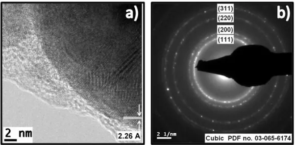
64
Figure 19. TEM micrographs and size distribution of silver nanoparticles synthesized by PLAL using 1064 nm and the in-situ and post-irradiation of the continuous wave lasers of
65
Figure 20. TEM micrographs and size distribution of silver nanoparticles synthesized by PLAL using 532 nm and the in-situ and post-irradiation of the continuous wave lasers of
66
Figure 21. Electron diffraction and high resolution TEM micrographs of silver nanoparticles synthesized by PLAL using 1064 nm and the in-situ and post-irradiation of