AIMS Neuroscience, 3 (3): 306-316. DOI: 10.3934/Neuroscience.2016.3.306 Received 16 June 2016,
Accepted 29 August 2016, Published 6 September 2016 http://www.aimspress.com/journal/neuroscience
Research article
The Network of Brodmann's Area 22 in Lexico-semantic Processing: A
Pooling-data Connectivity Study
Byron Bernal 1, *, Alfredo Ardila 2, and Monica Rosselli 3
1
Brain Institute-Radiology Department, Nicklaus Children’s Hospital, Miami, FL, USA; 2
Florida International University, Miami, FL, USA; 3
Florida Atlantic University, Davie, FL, USA
*Correspondence: Email: [email protected]; Tel: 786-268-1737
Abstract: Background and Objective: Modern neuroimaging has demonstrated that cognitive functions are based in networks of interconnected modules. The purpose of this paper was to analyze the connectivity of Brodmann's area (BA) 22 in lexico-semantic tasks. Methods: A connectivity fMRI co-activation study was performed by pooling data of lexico-semantic tasks in which BA22 (core of Wernicke's area) was reported. 283 subjects reported in 21 experiments were analyzed. Analysis of Likelihood Estimates of pooled data was utilized to generate the connectivity map; thresholds at p < 0.01 were corrected for multiple comparisons with false discovery rate. Resulting images were co-registered into MNI standard space. Results: A network consisting of 13 clusters of
activation was obtained. Main clusters were located in the left posterior temporal and left inferior frontal gyrus; in addition to these canonical areas, left insula, pre-supplementary motor area, left
BA37, primary auditory areas and left occipital lobe were also involved. Conclusions: BA22-related networks involved in lexico-semantics processing were demonstrated utilizing a pooling-data connectivity study. Significance, interpretation and limitations of the results are discussed.
1. Introduction
The contribution of Brodmann's cortical parcelation to the understanding and description of brain functions has been of critical importance in the field of neuroscience. The attempt to link histological cortical differences to specific sensory, motor and cognitive skills sounds reasonable. However, modern neuroimaging techniques have demonstrated that each of these areas in which the cortex is subdivided, may be involved in different functions. Brodmann's area (BA) 22, for example, has been found to be not only involved in receptive language tasks, as core part of the Wernicke's area, but also participating in quite distinct tasks as processing of complex sounds [1], and deductive verbal reasoning [2]. Lexical-semantic analysis, that is, the decoding of meaning from primary auditory or visual sensory input, is one important function of BA22 [3,4].
A large number of publications correlate brain function to networks; however, there is a paucity of articles examining the bases and theoretical background on this topic. Despite this lack, it is currently well accepted that cognition correlates to brain network configuration [5]. This means that any specific area (e.g., BA22) may connect with different modules, depending of the task, yielding specific network configurations from which arise distinct brain functions. Brain connectivity, the term referring to this view, may explain better complex cognitive, behavioral and neuropsychological phenomena than simple localization models.
Brain functional connectivity may be assessed by different methods. Perhaps the most popular currently is related to task-less fMRI or resting state fMRI. Fewer studies, instead, have explored brain connectivity related to tasks [6–8]. A recent described methodology that relies on pooled data analysis of coactivations found in task-related fMRI has also been described to depict brain functional networks. The method has been originally termed Meta-Analytic Connectivity Model
(MACM) [9–11]. In the present study we utilize this method to describe Wernicke's area core network involved in semantics, more specifically in lexico-semantic analysis. We have preferred to term it "pooling-data connectivity study" to avoid confusion with the standard meta-analysis methodology which usually requires broader sources.
2. Materials and Methods
of the results was utilized to filter only studies with paradigms assessing lexico-semantic functions. Twenty-two papers matched inclusion search criteria.
Exclusion criteria were used as well. Studies were excluded if they had bilingual subjects, tasks limited to expressive language, paradigms in patients, and paradigms in which language mediated the
study of different domains (e.g., inhibition, attention load) or beyond the lexico-semantic sphere (e.g. , expressive language, broad language target, etc.)
Based on these criteria 6 articles were excluded leaving 16 papers for further analysis. The
pooling data consisted of 283 subjects, 21 experiments; and 241 foci or localizations (Table 1). Activation foci associated to BA22 (search criteria) were obtained automatically from the Sleuth
software. This automatic report lists a number of clusters defined by the center of mass (in MNI coordinates), volume in mm3, maxima intensity (peak), and neighboring BA's-peaks within 5 mm of
the maxima plus and minus with respect the orthogonal coordinates (we will call them included), since these peaks are within the main cluster). Clusters are labeled accordingly with the maxima.
These coordinates, per subject/task/paper were exported as text files (pooled results) for analysis on the following step.
Statistical significance of clusters found on the pooled-data was then analyzed utilizing the Activation Likelihood Estimate-(ALE) method [12]. This step was performed with the open source
software GingerALE (http://brainmap.org). ALE treats reported peaks of activation as spatial probability distributions centered at the given coordinates. ALE computes the union of activation
probabilities for each voxel, allowing differentiation between true convergence of activation foci from random clustering (noise). ALE scores obtained from thousands of random iterations are used
to assign p-values to the observed clusters of activation. For more information on the theory of ALE the reader is advised to read the work of Eickoff et al, 2009. Our ALE maps were threshold at p < 0.01
corrected for multiple comparisons utilizing the false discovery rate method. Only clusters of 200 or
more cubic mm where accepted as valid clusters. ALE results were overlaid onto an anatomical template suitable for MNI coordinates, also provided by BrainMap.org. For this purpose we utilized
the Multi-Image Analysis GUI (Mango) (http://ric.uthscsa.edu/mango/). A mosaic of 5 × 6 transversal-cut insets of fusioned images were obtained utilizing the same tool, selecting every 3–4
images starting on axial image No. 14, and exported the mosaic to a 2D-jpg image.
3. Results
13 significant clusters of activation were found with the ALE procedure. Table 1 shows these
clusters ranked by their volume in cubic millimeters.
BA21 and BA37. The third cluster is located in the left medial frontal gyrus, more precisely in the pre-SMA corresponding with BA32. Included peaks in the cluster are left BA6 and right BA32.
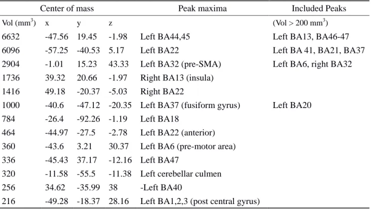
Table 1. BA22's network cluster list.
Center of mass Peak maxima Included Peaks
Vol (mm3) x y z (Vol > 200 mm3)
6632 -47.56 19.45 -1.98 Left BA44,45 Left BA13, BA46-47
6096 -57.25 -40.53 5.17 Left BA22 Left BA 41, BA21, BA37
2904 -1.01 15.23 43.33 Left BA32 (pre-SMA) Left BA6, right BA32 1736 39.32 20.66 -1.97 Right BA13 (insula)
1416 49.18 -20.37 -5.03 Right BA22
1000 -40.6 -47.12 -20.35 Left BA37 (fusiform gyrus) Left BA20 784 -26.4 -92.26 -1.19 Left BA18
464 -44.97 -27.5 -2.78 Left BA22 (anterior) 360 -43.6 3.21 30.37 Left BA6 (pre-motor area) 336 -45.43 37.17 -12.16 Left BA47
320 -11.58 -55.5 -11.38 Left cerebellar culmen 256 34.62 -35.99 38 -Left BA40
216 -49.28 -18.37 28.16 Left BA1,2,3 (post central gyrus)
ALE report. Main loci of brain connectivity of Left Brodmann Area 22 (Wernicke's Area). Conventions: x, y, z: MNI coordinates; Vol, volume of cluster in cubic millimeters as a measure of activation extent.
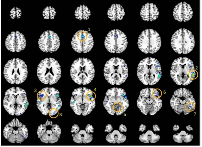
Figure 1. Functional connectivity map of BA22 by Meta-analytic Connectivity
Modeling. Insets in mosaic picture correspond to transversal descending cuts of the brain MRI template. Left hemisphere appears on the right side (Radiological convention). Clusters of activation are color coded for statistical significance from dark blue (lowest) to red (highest). Main cluster of the automatic ALE report are shown in numbered circles: BA6, 32 (1); left BA22 (2); right anterior insula (3); left BA44,45 and anterior insula (4);
left cerebellar culmen (5); left BA47 (6); left BA37 (7); left BA18 (8).
4. Discussion
We found that lexico-semantic network of BA22 consists of 13 clusters. Quite strikingly we found that the main cluster of activation includes the left infero-lateral frontal gyrus (BA44,BA45) and the anterior insula (BA13); BA44 and BA45 is the anterior language production system (Broca’s area) whilst BA46 and BA47—left dorsolateral prefrontal cortex—are likely involved in executive control of language. The insula (BA13) has been suggested to have a crucial language role by integrating the receptive and expressive functions [13].
short U fibers.
The supplementary (SMA)/pre-supplementary motor area, BA32, found included in this network (cluster 3) was unexpected as no structural connection to BA22 is known. SMA is connected structurally to BA44 through the aslant frontal fasciculus described by Catani and coworkers [14], and most likely associated with verbal fluency and initiation of speech [15]. BA32 is activated in a diversity of language-related tasks, such as verbal initiation [16], naming [17,18], and verbal fluency [19]. As such, BA32 may have a rather ancillary function in lexico-semantic analysis.
The activation of the right insula (cluster 4) is interesting. Processing of semantic violations activate the insular cortex bilaterally [20]. Further, right anterior insula activation has been described
in tasks demanding semantic analysis across different language in bilingual subjects [21]. Recent studies with transcranial magnetic stimulation have found right lateralization in temporal areas for emotional
prosody processing [22]. Thus, it seems that the right insula truly partakes in the semantic network. The connectivity to left BA37 (posterior inferior temporal gyrus, middle temporal gyrus and fusiform gyrus), corresponding to cluster 5, reveals the importance of "access to word repository" in the process of lexico-semantic analysis or the access to visuo-spatial representation of objects and ideas in this area. Finding and producing words (word retrieval and word generation) are potential functions of BA37 considering that the destruction of this area is associated with word selection anomia [23,24]; some studies indeed have found that word search results in an increased activity in this area [25,26]. Moreover, the involvement of BA37 in naming has been well documented [e.g., 27–29]. Using a similar procedure to the one reported here, it was found that left BA37 is a common node of two distinct networks—visual recognition (perception) and semantic language functions [27].
The connectivity to right BA22 (cluster 5), shows that BA22 asymmetry for language is not complete. Indeed, the ratio of activation rBA22/lBA22 is 0.23. However, it is difficult to understand the right BA22. On one hand, lesional cases seem to show a potential language function for right
BA22 as posterior aphasias recover better than anterior aphasias; but on the other hand, lesions of right temporal lobe, which includes BA22, usually do not affect language [23]. Cluster 8 shows a second peak on left BA22 which extends rostrally into the anterior half of the superior temporal gyrus, between BA38 and BA41. This area (antero-superior BA22) and BA47 process the meaning of sentences contingent to grammar construction, as explained below. It has also been described as critical for language comprehension by Turken and Dronkers [31].
The connectivity to left BA6 is most likely related to speech planning functions [32,33] as BA6 is a pre-motor area. Left BA6 has been reported activating in language processing tasks [34,35], language switching [36], object naming [37,38], lexical decisions [39] and syntactic processing [40]. It is difficult to rule out the mere effect of a speech/expressive language confound in the reported tasks, such as sub-vocalization, but functional connectivity of BA22 obtained with task-less resting-state fMRI also confirm strong BA22 to BA6 connectivity (author's observation: preliminary
results in one normal subject are available at
The connectivity to left 47 is also understood within the language network. BA47 has a function in processing the meaning of sentences contingent to grammar construction (e.g., "John gave the book to Mary" vs. "John gave Mary a book") [41]. A recent study from Ardila et al [23] has demonstrated the involvement of this area in a network of expressive/receptive language. The authors suggest that this involvement is most likely of executive type, that is, as having functions in inhibition, planning, and production of language. It seems there is an intriguing interplay between receptive language (comprehension) and motor planning via speech/language production.
The participation of the cerebellar culmen (cluster 11) in semantic processing is not clear. Although there are several publications linking the cerebellum with language functions, there is no publications linking directly the culmen to language. Of note is the interesting finding of ipsilateral, and not contralateral, activation with respect cortical motor areas activated (BA6, BA44–47), which
suggests a non-motor cerebellar involvement in lexico-semantic tasks.
Area BA40 (cluster 12) corresponds anatomically with the supramarginal gyrus. Left BA40 activates in semantic processing, mostly when complex semantic representations are required [4]. For example, left BA40 activates in sentences with transitive verbs, requiring agent and theme, (e.g.,
"The dog chased the boy") as compared to sentences with intransitive verbs, requiring only an agent as in "The dog sleeps" [42]. Left BA40 has also been found activating in task of verbal creativity [43].
Lesional transitory models like Transcranial Magnetic Stimulation validate also the role of BA40 in semantic processing. Left supramarginal gyrus stimulation (BA40) with this technique produces semantic errors [44].
The connections to BA18 (cluster 7) and BA1-3 (cluster 13) are intriguing as they are not putative language areas. Visual areas may be involved in verbal tasks as the subject "re-visualize" objects and scenes described by the verbal material; post-central gyrus may be involved as a consequence of accessing somatosensory "memories" in the meaning decoding process, in the same way that cortical motor activation has been described in semantic processing of tools [45], and activation of taste-related words includes the anterior insula, frontal operculum, lateral
orbitofrontal gyrus, and thalamus all related with task perception [46]. Likewise the motor and premotor foci of activations referred here may be explained, at least in part, by accessing repositories
of motor representations related to verbs [47,48].
Our results agree in part with the results published in the scant prior studies that utilizing different methods targeted directly or indirectly BA22 for connectivity. The comprehension network, that involves lexico-semantic analysis, and its connections have been previously demonstrated combining resting-state fMRI and Diffusion Tensor Imaging (DTI). The modules involved were BA22, BA46, BA47 and BA39 [31]. To our best of our knowledge our work is the first publication demonstrating the network of BA22 in lexico-semantic processing, utilizing pooling-data of co-activation, finding an extended network that involves left BA40, BA44, BA32, BA6 and bilateral insula.
to network configurations and not to isolated modules. The description of these networks may help to dissect the contribution of basic into more complex functions. To attach a visual referent to a word
(semantics) may require a port-of input, a lexical analyzer, a word-image repository or comparator. In addition, other modules may specifically intervene associated to the stimulus category. In that way
the core semantic network may require to access motor schemes, sensory memories, emotion
memories and valences, motor/cognitive inhibition and working memory fields.
The present study has some limitations. Our results are based in only one source data (brainmap.org). Many more articles have described BA22 activation in language tasks, but they have not been included in the Brainmap.org database. For a study to be included in Brainmap.org database, requires brain activation results to be reported in standard space coordinates (MNI or Talairach). Despite this limitation the authors estimate the number of studies/participants/experiments entering the pooling-data is large enough to provide statistical power and reflects the state of the art publications in fMRI of language.
5. Conclusions
We found that BA22 is basically connected with the anterior language production system (Broca’s area BA44 and BA45; plus BA46 and BA47—left dorsolateral prefrontal cortex—likely involved in the executive control of language), the insula (BA13) (integrative node for the receptive/temporal and production/frontal language systems), BA37 (posterior inferior temporal gyrus, middle temporal gyrus and fusiform gyrus) which represents a common node of two distinct networks—visual recognition (perception) and semantic language functions; plus some other minor connections, including visual association areas in the occipital lobe (BA18) and the pre-supplementary motor area (BA32).
Disclosures
Dr. Byron Bernal is owner and President of fMRI Consulting Inc. Reference
1. Mirz F, Ovesen T, Ishizu K, et al. (1999) Stimulus-dependent central processing of auditory stimuli: a PET study. Scand Audiol 28: 161–169.
2. Goel V, Gold B, Kapur S (1998) Neuroanatomical correlates of human reasoning. J Cogn
Neurosci 10: 293–302.
3. McDermott KB, Petersen SE, Watson JM, et al. (2003) A procedure for identifying regions preferentially activated by attention to semantic and phonological relations using functional magnetic resonance imaging. Neuropsychologia 41: 293–303.
correlates of semantic processing to visually presented words. Hum Brain Mapp 27: 915–924. 5. McIntosh AR (2000) Towards a network theory of cognition. Neural Netw Off J Int Neural Netw
Soc 13: 861–870.
6. Biswal BB, Eldreth DA, Motes MA, et al. (2010) Task-dependent individual differences in prefrontal connectivity. Cereb Cortex N Y N 1991 20: 2188–2197.
7. Caclin A, Fonlupt P (2006) Effect of initial fMRI data modeling on the connectivity reported between brain areas. NeuroImage 33: 515–521.
8. Wu X, Lu J, Chen K, et al. (2009) Multiple neural networks supporting a semantic task: an fMRI study using independent component analysis. NeuroImage 45: 1347–1358.
9. Laird AR, Eickhoff SB, Rottschy C, et al. (2013) Networks of task co-activations. NeuroImage
80: 505–514.
10. Kohn N, Eickhoff SB, Scheller M, et al. (2014) Neural network of cognitive emotion regulation--an ALE meta-analysis and MACM analysis. NeuroImage 87: 345–355.
11. Robinson JL, Laird AR, Glahn DC, et al. (2010) Metaanalytic connectivity modeling: delineating the functional connectivity of the human amygdala. Hum Brain Mapp 31: 173–184.
12. Eickhoff SB, Laird AR, Grefkes C, et al. (2009) Coordinate-based activation likelihood estimation meta-analysis of neuroimaging data: a random-effects approach based on empirical estimates of spatial uncertainty. Hum Brain Mapp 30: 2907–2926.
13. Ardila A. Bernal B, Rosselli M (2014) Participation of the insula in language revisited: A meta-analytic connectivity study. J Neurolinguistics 29: 31–41.
14. Catani M, Mesulam MM, Jakobsen E, et al. (2013) A novel frontal pathway underlies verbal fluency in primary progressive aphasia. Brain J Neurol 136: 2619–2628.
15. Martino J, de Lucas EM, Ibáñez-Plágaro FJ, et al. (2012) Foix-Chavany-Marie syndrome caused by a disconnection between the right pars opercularis of the inferior frontal gyrus and the supplementary motor area. J Neurosurg 117: 844–850.
16. Nathaniel-James DA, Fletcher P, Frith CD (1997) The functional anatomy of verbal initiation and suppression using the Hayling Test. Neuropsychologia 35: 559–566.
17. Garn CL, Allen MD, Larsen JD (2009) An fMRI study of sex differences in brain activation during object naming. Cortex J Devoted Study Nerv Syst Behav 45: 610–618.
18. Kiyosawa M, Inoue C, Kawasaki T, et al. (1996) Functional neuroanatomy of visual object naming: a PET study. Graefes Arch Clin Exp Ophthalmol Albrecht Von Graefes Arch Für Klin
Exp Ophthalmol 234: 110–115.
19. Whitney C, Weis S, Krings T, et al. (2009) Task-dependent modulations of prefrontal and hippocampal activity during intrinsic word production. J Cogn Neurosci 21: 697–712.
20. Friederici AD, Rüschemeyer S-A, Hahne A, et al. (2003) The role of left inferior frontal and superior temporal cortex in sentence comprehension: localizing syntactic and semantic processes.
Cereb Cortex N Y N 1991 13: 170–177.
spoken words in bilinguals reveals language-independent semantic representations in anterior temporal lobe. J Neurosci Off J Soc Neurosci 34: 332–338.
22. Alba-Ferrara L, Ellison A, Mitchell RL (2012) Decoding emotional prosody: resolving differences in functional neuroanatomy from fMRI and lesion studies using TMS. Brain Stimulat
5: 347–353.
23. Benson F, Ardila A (1996) Aphasia, A clinical perspective. New York: Oxford University Pres. 24. Luria A (1976) Basic problems of neurolinguistics. New York: Mouton.
25. Abrahams S, Goldstein LH, Simmons A, et al. (2003) Functional magnetic resonance imaging of verbal fluency and confrontation naming using compressed image acquisition to permit overt responses. Hum Brain Mapp 20: 29–40.
26. Friedman L, Kenny JT, Wise AL, et al. (1998) Brain activation during silent word generation evaluated with functional MRI. Brain Lang 64: 231–256.
27. Ahmad Z, Balsamo LM, Sachs BC, et al. (2003) Auditory comprehension of language in young children: neural networks identified with fMRI. Neurology 60: 1598–1605.
28. Giraud AL, Kell C, Thierfelder C, et al. (2004) Contributions of sensory input, auditory search and verbal comprehension to cortical activity during speech processing. Cereb Cortex N Y N 1991 14: 247–255.
29. Söderfeldt B, Ingvar M, Rönnberg J, et al. (1997) Signed and spoken language perception studied by positron emission tomography. Neurology 49: 82–87.
30. Ardila A, Bernal B, Rosselli M (2015) Language and visual perception associations: Meta-analytic connectivity modeling of Brodmann area 37. Behav Neurol.
31. Turken AU, Dronkers NF (2011) The neural architecture of the language comprehension network: converging evidence from lesion and connectivity analyses. Front Syst Neurosci 5: 1.
32. Fox PT, Ingham RJ, Ingham JC, et al. (2000) Brain correlates of stuttering and syllable production. A PET performance-correlation analysis. Brain J Neurol 123: 1985–2004.
33. Shuster LI, Lemieux SK (2005) An fMRI investigation of covertly and overtly produced mono-and multisyllabic words. Brain Lang 93: 20–31.
34. Basho S, Palmer ED, Rubio MA, et al. (2007) Effects of generation mode in fMRI adaptations of semantic fluency: paced production and overt speech. Neuropsychologia 45: 1697–1706.
35. De Carli D, Garreffa G, Colonnese C, et al. (2007) Identification of activated regions during a language task. Magn Reson Imaging 25: 933–938.
36. Price CJ, Green DW, von Studnitz R (1999) A functional imaging study of translation and language switching. Brain J Neurol 122: 2221–2235.
37. Baciu MV, Rubin C, Décorps MA, et al. (1999) fMRI assessment of hemispheric language dominance using a simple inner speech paradigm. NMR Biomed 12: 293–298.
38. Hirsch J, Moreno DR, Kim KH (2001) Interconnected large-scale systems for three fundamental cognitive tasks revealed by functional MRI. J Cogn Neurosci 13: 389–405.
duration and task. Brain J Neurol 117: 1255–1269.
40. Inui T, Otsu Y, Tanaka S, et al. (1998) A functional MRI analysis of comprehension processes of Japanese sentences. Neuroreport 9: 3325–3328.
41. Allen K, Pereira F, Botvinick M, et al. (2012) Distinguishing grammatical constructions with fMRI pattern analysis. Brain Lang 123: 174–182.
42. Thompson CK, Bonakdarpour B, Fix SC, et al. (2007) Neural correlates of verb argument structure processing. J Cogn Neurosci 19: 1753–1767.
43. Bechtereva NP, Korotkov AD, Pakhomov SV, et al. (2004) PET study of brain maintenance of verbal creative activity. Int J Psychophysiol Off J Int Organ Psychophysiol 53: 11–20.
44. Sollmann N, Tanigawa N, Tussis L, et al. (2015) Cortical regions involved in semantic processing investigated by repetitive navigated transcranial magnetic stimulation and object
naming. Neuropsychologia 70: 185–195.
45. Chouinard PA, Goodale MA (2010) Category-specific neural processing for naming pictures of animals and naming pictures of tools: an ALE meta-analysis. Neuropsychologia 48: 409–418. 46. Barrós-Loscertales A, González J, Pulvermüller F, et al. (2012) Reading salt activates gustatory
brain regions: fMRI evidence for semantic grounding in a novel sensory modality. Cereb Cortex N Y N 1991 22: 2554–2563.
47. Palti D, Ben Shachar M, Hendler T, et al. (2007) Neural correlates of semantic and morphological processing of Hebrew nouns and verbs. Hum Brain Mapp 28: 303–314.
48. Perani D, Cappa SF, Schnur T, et al. (1999) The neural correlates of verb and noun processing. A PET study. Brain J Neurol 122: 2337–2344.
© 2016 Byron Bernal et al., licensee AIMS Press. This is an open access article distributed under the terms of the Creative
Commons Attribution License