Risk Factors for Prognosis of Hepatocellular
Carcinoma After Curative Resection In
Patients with Low Hepatitis B Viral Load
Li-Shuai Qu, Jin-Xia Liu, Jing Zhu, Cui-Hua Lu
Department of Gastroenterology, Affiliated Hospital of Nantong University, Jiangsu Province, China. May-June, Vol. 16 No. 3, 2017: 412-420
INTRODUCTION
Hepatocellular carcinoma (HCC) is a highly prevalent and lethal cancer type being the third leading cause of can-cer-related deaths worldwide. Etiologically, HCC usually develops in chronic hepatitis B virus (HBV) carriers, es-pecially in East Asia and sub-Saharan Africa, where HBV is endemic. For these HCC patients, curative tumor resec-tion remains the most effective treatment and the progno-sis of HCC was greatly improved. However, high opportunity of intrahepatic recurrence remains a major obstruction for further improving the prognosis of HCC patients after curative resection. It has been hypothesized that early and late intrahepatic recurrence of HCC was at-tributable to two different mechanisms: intrahepatic me-tastasis and de novo multicentric carcinogenicity,
respectively. Imamura, et al. proposed a convenient frame-work to clinically differentiate each type of recurrence as ‘early’ or ‘late’ based on a cut-off of 2 years postoperatively. Potentially, risk factors including tumor size, nodule number, positive surgical margin, microvascular invasion, tumor node metastasis (TNM) stage, and Edmondson’s grade were significantly related to early HCC recurrence. With successful risk surveillance of HCC development and curative resection, the development of early recur-rence can be avoided in a higher number of patients, there-by helping them survive longer. Meanwhile, the risk factors contributing to late recurrence after surgery have not been investigated on a comprehensive basis. Although the precise mechanism underlying recurrent carcinogene-sis associated with HBV in the remaining liver in patients who have undergone curative therapy remains unclear, it is The Official Journal of the Mexican Association of Hepatology,
the Latin-American Association for Study of the Liver and the Canadian Association for the Study of the Liver
Manuscript received: Manuscript received: Manuscript received: Manuscript received:
Manuscript received: June 07, 2016. Manuscript accepted:Manuscript accepted:Manuscript accepted:Manuscript accepted:Manuscript accepted: September 25, 2016.
DOI:10.5604/16652681.1235484
A B S T R A C T A B S T R A C T A B S T R A C T A B S T R A C T A B S T R A C T
Background. Background.Background. Background.
Background. A retrospective cohort study was conducted to investigate the effect of hepatitis B surface antigen (HBsAg) level on prognosis in low viral load (< 2000 IU/mL) patients with hepatitis B-related hepatocellular carcinoma (HCC) after curative resection. Material and methods.
Material and methods. Material and methods. Material and methods.
Material and methods. A total of 192 patients with low viral load who had received curative resection of pathologically confirmed HCC were analyzed to determine the factors affecting prognosis. The risk factors for survival, early and late recurrence (2 years as a cut-off) were studied. Results.Results.Results.Results.Results. The median follow-up time was 38.5 months. The overall survival rates at 1-, 3-, and 5-year after curative resection were 94.2%, 64.0%, and 45.2%, respectively. The cumulative recurrence rates at 1-, 3-, and 5-year after curative resection were 22.4%, 46.5%, and 67.0%, respectively. Patients with high serum HBsAg levels (> 250 IU/mL) had significantly low-er survival rates than those with low HBsAg levels (HR: 1.517, 95% CI: 1.005-2.292, P = 0.047). Stratified analysis showed that pa-tients with high HBsAg levels had a significantly higher late recurrence incidence than those with low HBsAg levels (HR: 2.155, 95% CI: 1.094-4.248, P = 0.026), but did not have a significantly higher risk of early recurrence postoperatively (HR: 1.320, 95% CI: 0.837-2.082, P = 0.233). Multivariate analysis revealed that HBsAg > 250 IU/mL was an independent risk factor associated with late recurrence (HR: 2.109, 95% CI: 1.068-4.165, P = 0.032). Conclusions.Conclusions.Conclusions.Conclusions.Conclusions. HBsAg > 250 IU/mL at the time of tumor resection was an independent risk factor for late recurrence in low viral load HCC patients.
Key words. Key words.Key words. Key words.
possible that viral replication and integration of sub-ge-nomic HBV DNA fragments into the host liver cells play a key role in contributing to the carcinogenic process. Ac-cording to current opinions, there is sufficient and power-ful evidence connecting elevated serum HBV DNA levels with an increased risk of HCC recurrence and death after curative resection. In particular, patients with serum HBV DNA levels > 2,000 IU/mL at study entry have an in-creased risk of HCC recurrence. In contrast, those with HBV DNA levels < 2,000 IU/mL are usually designated inactive or low-risk HBV carriers. However, even these low viral load patients still carry a high incidence of HCC recurrence. Therefore, it is necessary to identify factors other than viral load that are predictive of HCC in these low-risk patients. Recently, the quantification of hepatitis B surface antigen (HBsAg) level has been introduced as a surrogate marker for the management of chronic hepatitis B infection. A Taiwanese cohort study demonstrated that high HBsAg levels predicted the risk of HCC in hepatitis B e antigen (HBeAg) negative patients with low HBV DNA levels. Tseng, et al. also demonstrated that high HB-sAg levels could predict disease progression in HBeAg negative patients. However, there are only a few studies as-sessing the role of HBsAg levels as a risk factor of recur-rence in HCC. Thus, the effect of HBsAg levels on HCC recurrence after curative resection in low viral load pa-tients remains uncertain.
We therefore aimed to conduct a retrospective cohort study to assess the impact of serum HBsAg levels on the prognosis of HCC among patients with low viral load (< 2,000 IU/mL). In addition, based on the hypothesis that early and late intrahepatic recurrence of HCC was attrib-utable to different mechanisms, we also investigated the risk factors for early (≤ 2 years) and late (> 2 years) intra-hepatic recurrence, separately.
MATERIAL AND METHODS
Patients
Between January 2009 and December 2011, 192 patients with HBV-related HCC who underwent curative resec-tion as the primary therapy at the Affiliated Hospital of Nantong University were enrolled in this retrospective cohort study based on the following inclusion criteria:
• Positive serum HBsAg for at least 12 months with a se-rum HBV DNA level < 2,000 IU/mL.
• Complete resection of all tumor nodules and a surgical free margin of more than 5 mm by pathological exami-nation.
• No cancerous thrombus in the portal vein (main trunk or two major branches), hepatic veins, or bile duct.
• No more than three tumor nodules. • No extrahepatic metastasis.
• No adjuvant antiviral therapy with nucleoside analogue was given before or after the operation.
• Death in the hospital not due to postoperative hepatic failure; and
• Presence of early recurrence at least 3 months postop-eratively (excluding pre-existing metastases before HCC resection).
The histological grade proposed by Edmondson and Steiner, maximal tumor size, nodule number, capsular formation around the tumor, microvascular invasion, liver cirrhosis, and extent of resection were also determined. The protocol was approved by the Ethics Committee of the Affiliated Hospital of Nantong University in accord-ance with the tenets of the Declaration of Helsinki.
Follow-up and
treatment for tumor recurrences
After surgical resection of HCC, all 192 patients were assessed every 3-6 months with dynamic contrast-en-hanced computer tomography (CT) and/or magnetic reso-nance imaging (MRI), as well as with measurement of a serum tumor marker such as AFP. Hepatic angiography was performed when recurrence was suspected. Postoper-atively, 19 patients received interferon-alpha (IFN-α) treatment, which was started at a pilot dose of 3 million units (MU) two times a week by intramuscular injection for 2 weeks, then 5 MU three times a week for 18 months. The IFN-α treatment was terminated when recurrence was confirmed. Diagnosis of intrahepatic recurrence was based on histopathologic examination of tumor tissue in 23 patients undergoing repeat hepatic resection and on the characteristic appearance of CT, MRI, and hepatic angiog-raphy in 85 patients. When recurrence was confirmed, the patients received the optimal treatment modality for each case based on the number and location of the recurrent tu-mors and liver function tests. Treatment of recurrent HCC included repeat hepatic resection, radiofrequency ablation (RFA), percutaneous ethanol injection (PEI), chemotherapy or radiotherapy, and transcatheter arterial chemoembolization (TACE) and hepatic arterial infusion chemotherapy.
Evaluation of prognostic clinical and pathological variables
aminotrans-ferase level (continuous variable), total bilirubin level (continuous variable), albumin level (continuous varia-ble), prothrombin time (continuous variavaria-ble), HBeAg sta-tus (positive or negative), serum HBsAg level (≤ 250 or > 250 IU/mL), co-existing hepatitis C virus (HCV) infec-tion (yes or no), liver cirrhosis (yes or no), Child-Pugh grade (A or B), Okuda stage (I or II), serum AFP level (≤ 400 or > 400 ng/mL), tumor size (≤ 3 or > 3 cm in di-ameter), number of tumor nodules (single or multiple), tumor capsular formation (yes or no), microvascular inva-sion (yes or no), differentiation of tumor cells (Edmond-son’s classification I/II or III/IV), postoperative IFN-α therapy (yes or no), and extent of resection (minor, < 3 segments or major, ≥ 3 segments) based on Couinaud’s no-menclature. Serum HBV DNA levels were determined using the fluorescein quantitative polymerase chain reac-tion (FQ-PCR) detecreac-tion system (TaqMan; Roche Diag-nostics, Branchburg, NJ, USA). HBsAg level was quantified by the automated chemiluminescent micropar-ticle immunoassay (Architect HBsAg, Abbott, IL, USA). The detection range of the Architect HBsAg assay was 0.05 to 250 IU/mL.
Statistical analysis
Data were presented as means ± SD, proportions, or median (range). Clinical and pathological data at the time of resection were analyzed to identify factors that influenced prognosis via the Cox proportional hazards model. The overall survival rate was calculated from the date of resec-tion to the date of death or last follow-up. The cumulative recurrence rates were calculated from the date of resection to the date when tumor recurrence was diagnosed, or if re-currence was not diagnosed at the time of study, cases were censored on the date of death or last follow-up. To investi-gate factors contributing to early and late recurrence sepa-rately, we set 2 years as the cut-off between early and late recurrence, as suggested by Imamura’s study. Survival curves were calculated by the Kaplan-Meier method and differences were compared by the log-rank test. Multivari-ate analysis was performed by the Cox proportional hazards regression model. Statistical significance was defined by a P value < 0.05. Statistical analyses were performed using the Statistical Program for Social Sciences (SPSS v.11.5 for Windows; SPSS, Inc., Chicago, IL, USA).
Table 1. Patient characteristics.
Characteristics N (%) Values
Patients (n) 192 (100)
Median age, years (range) 50 (26-78)
Male: female ratio 163:29 (84.9:15.1)
Co-existing HCV infection 5 (2.6)
Presence of cirrhosis 144 (75.0)
HBeAg seropositivity 64 (33.3)
HBsAg > 250 IU/mL 83 (43.2)
AFP > 400 ng/mL 38 (19.8)
Median baseline biochemistry and hematology (range)
Total bilirubin, μmol/L 15.8 (5.3-38.8)
Albumin, g/L 42 (30-52)
Aminotransferase, IU/L 43.5 (10-324)
Prothrombin time, s 11.4 (9.5-15.3)
Tumor size (≤ 3 cm:> 3 cm) 84:108 (43.8:56.3)
Tumor number (Single:multiple) 156:36 (81.3:18.8)
Microvascular invasion 45 (23.4)
Capsular formation 107 (55.7)
Differentiation of tumor (I/II:III/IV) 134:58 (69.8:30.2)
Child-Turcotte-Pugh grade
A 182 (94.8)
B 10 (5.2)
Okuda stage
I 179 (93.2)
I I 13 (6.8)
Type of surgical procedure (major:minor) 74:118 (38.5:61.5)
Postoperative IFN-α treatment 19 (9.9)
Median follow-up time (months) 38.5 (3-72)
Median time of recurrence (months) 24.5 (3-66)
Table 2. Factors identified on univariate and multivariate Cox regression analyses that influenced overall survival in HCC patients un-dergoing curative resection.
Factors P value Hazard ratio 95% CI
Univariate analysis
Sex (male vs. female) 0.614 1.158 0.654-2.052
Age, years 0.242 1.012 0.992-1.032
Co-existing HCV infection 0.235 0.303 0.042-2.174
Presence of cirrhosis 0.220 1.381 0.824-2.315
HBeAg seropositivity 0.766 1.068 0.692-1.649
HBsAg > 250 IU/mL 0.047 1.517 1.005-2.292
AFP > 400 ng/mL 0.408 1.229 0.754-2.004
Total bilirubin 0.134 1.022 0.993-1.053
Albumin 0.225 0.970 0.923-1.019
Aminotransferase 0.201 1.003 0.998-1.007
Prothrombin time, s 0.217 1.111 0.940-1.312
Tumor size (> 3 cm vs.≤ 3 cm) 0.020 1.673 1.086-2.577
Tumor number (multiple vs. single) 0.723 1.098 0.653-1.846
Microvascular invasion 0.015 1.751 1.115-2.750
Capsular formation 0.496 0.867 0.574-1.308
Differentiation (III/IV vs. I/II) 0.844 0.956 0.609-1.500
Child -Pugh grade (B vs. A) 0.067 2.065 0.950-4.489
Okuda stage (II vs. I) 0.139 1.795 0.827-3.896
Extent of resection (major vs. minor) 0.505 1.154 0.757-1.760
Postoperative IFN-α treatment 0.078 0.444 0.180-1.094
Multivariate analysis
Microvascular invasion 0.016 1.740 1.108-2.734
RESULTS
During a median observation period of 38.5 months (with a range of 3 to 72 months), recurrence was found in 108 patients (56.3%). Of these, 23 patients received a sec-ond resection, 7 patients received RFA, 56 patients re-ceived TACE, 14 patients rere-ceived chemotherapy or radiotherapy or PEI, and the remainder received a con-servative treatment. During follow-up, 91 patients died, 14 of whom did not have recurrent HCC but died due to oth-er causes, mainly chronic detoth-erioration of livoth-er function and massive upper digestive tract bleeding caused by es-ophageal varices. The overall survival rates at 1-, 3-, and 5-year after curative resection were 94.2%, 64.0%, and 45.2%, respectively. The cumulative recurrence rates at 1-, 3-, and 5-year after curative resection were 22.4%, 46.5%, and 67.0%, respectively. The baseline demographic, clinical, and pathological features of the study population are depicted in table 1.
Factors associated with overall survival in univariate and multivariate analyses
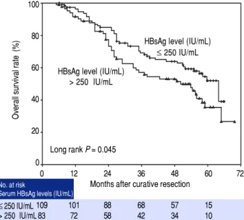
In the univariate analysis, serum HBsAg level > 250 IU/mL (hazards ratio (HR): 1.517, 95% confidence interval (CI): 1.005 - 2.292, P = 0.047), tumor size > 3 cm in diameter (HR: 1.673, 95% CI: 1.086 - 2.577, P = 0.020), and presence of microvascular invasion (HR: 1.751, 95% CI: 1.115
-2.750, P = 0.015) were significantly associated with poor overall survival postoperatively. Other clinical factors in-cluding sex, age, co-existing HCV infection, serum albu-min level, total bilirubin level, aalbu-minotransferase level, prothrombin time, HBeAg status, presence of cirrhosis, tumor number, Child-Pugh grade, Okuda stage, extent of resection, and postoperative IFN-α treatment were not as-sociated with survival (Table 2). The 1-, 3-, and 5-year overall survival rates for high HBsAg level patients (> 250 IU/mL) were 91.4%, 57.1%, and 35.4%, respectively, in contrast to 96.3%, 69.1%, and 52.0%, respectively, for low HBsAg level patients (≤ 250 IU/mL). Patients with high serum HBsAg levels had significantly lower survival rates than those with low HBsAg levels (log rank P = 0.045; Figure 1). However, in the multivariate analysis, the presence of microvascular invasion (HR: 1.740, 95% CI: 1.108 -2.734, P = 0.016) was the only independent risk factor for overall survival (Table 2).
Factors associated with early and late recurrence after curative resection
Figure 1. Figure 1. Figure 1. Figure 1.
Figure 1. Comparison of overall survival rates after curative resection between different serum HBsAg levels at the time of tumor resection (log-rank test, P = 0.045).
Figure 2. Figure 2.Figure 2.
Figure 2.Figure 2. Cumulative late recurrence (> 2 years) related to the initial HBsAg levels at the time of tumor resection (log-rank test, P = 0.022).
Table 3. Factors identified on univariate and multivariate Cox regression analyses that influenced early recurrence (≤ 2 years) in HCC patients undergoing curative resection.
Factors P value Hazard ratio 95% CI
Univariate analysis
Sex (male vs. female) 0.949 0.980 0.528-1.819
Age, years 0.343 0.989 0.968-1.011
Co-existing HCV infection 0.307 1.826 0.575-5.799
Presence of cirrhosis 0.282 1.365 0.774-2.405
HBeAg seropositivity 0.965 0.989 0.611-1.602
HBsAg > 250 IU/mL 0.233 1.320 0.837-2.082
AFP > 400 ng/mL 0.006 2.014 1.223-3.316
Total bilirubin 0.849 0.997 0.965-1.030
Albumin 0.838 1.006 0.953-1.061
Aminotransferase 0.223 1.003 0.998-1.007
Prothrombin time, s 0.231 0.886 0.727-1.080
Tumor size (> 3 cm vs.≤ 3 cm) 0.001 2.285 1.377-3.791
Tumor number (multiple vs. single) 0.642 0.868 0.477-1.579
Microvascular invasion < 0.001 2.546 1.595-4.064
Capsular formation 0.040 0.620 0.392-0.979
Differentiation (III/IV vs. I/II) 0.530 1.179 0.706-1.969
Child -Pugh grade (B vs. A) 0.490 1.377 0.555-3.418
Okuda stage (II vs. I) 0.709 0.825 0.301-2.261
Extent of resection (major vs. minor) 0.800 1.062 0.666-1.694
Postoperative IFN-α treatment 0.080 0.407 0.148-1.115
Multivariate analysis
AFP > 400 ng/mL 0.047 1.679 1.007-2.801
Tumor size > 3 cm 0.020 1.871 1.103-3.175
Microvascular invasion 0.011 1.917 1.163-3.159
Among 192 patients, 74 had recurrence within 2 years. Stratified Cox regression analysis identified four variables as factors related to early recurrence: AFP > 400 ng/mL
(HR: 2.014, 95% CI: 1.223 - 3.316, P = 0.006), tumor size > 3 cm in diameter (HR: 2.285, 95% CI: 1.377-3.791, P = 0.001), presence of microvascular invasion (HR: 2.546,
Overall survival rate (%)
100
80
60
40
20
0
0 12 24 36 48 60 72
Months after curative resection
109 101 88 68 57 15
83 72 58 42 34 10
≤ 250 IU/mL > 250 IU/mL
Long rank P = 0.045 HBsAg level (IU/mL)
> 250 IU/mL
HBsAg level (IU/mL) ≤ 250 IU/mL
Late recurrence rate (%)
100
80
60
40
20
0
0 12 24 36 48 60 72
Months after curative resection
59 46 36 8
37 29 19 5
≤ 250 IU/mL > 250 IU/mL
HBsAg level (IU/mL) ≤ 250 IU/mL Long rank P = 0.022
HBsAg level (IU/mL) > 250 IU/mL
No. at risk
Serum HBsAg levels (IU/mL) No. at risk
Table 4. Factors identified on univariate and multivariate Cox regression analyses that influenced late recurrence (> 2 years) in HCC patients undergoing curative resection.
Factors P value Hazard ratio 95% CI
Univariate analysis
Sex (male vs. female) 0.116 3.148 0.752-13.180
Age, years 0.444 1.013 0.980-1.048
Co-existing HCV infection 0.241 2.356 0.563-9.865
Presence of cirrhosis 0.033 2.821 1.089-7.310
HBeAg seropositivity 0.290 1.449 0.729-2.879
HBsAg > 250 IU/mL 0.026 2.155 1.094-4.248
AFP > 400 ng/mL 0.232 1.721 0.707-4.191
Total bilirubin 0.406 1.023 0.970-1.078
Albumin 0.551 0.975 0.898-1.059
Aminotransferase 0.757 0.999 0.990-1.008
Prothrombin time, s 0.542 0.912 0.679-1.226
Tumor size (> 3 cm vs. ≤ 3 cm) 0.108 1.749 0.885-3.456
Tumor number (multiple vs. single) 0.014 2.504 1.206-5.200
Microvascular invasion 0.101 2.012 0.872-4.640
Capsular formation 0.997 0.999 0.505-1.976
Differentiation (III/IV vs. I/II) 0.929 1.034 0.494-2.166
Child -Pugh grade (B vs. A) 0.712 1.457 0.198-10.694
Okuda stage (II vs. I) 0.432 1.780 0.423-7.493
Extent of resection (major vs. minor) 0.560 0.803 0.383-1.680
Postoperative IFN-α treatment 0.477 0.684 0.240-1.948
Multivariate analysis
HBsAg > 250 copies/mL 0.032 2.109 1.068-4.165
Multiple tumor number 0.017 2.446 1.172-5.104
95% CI: 1.595 - 4.064, P < 0.001), and presence of capsular formation (HR: 0.620, 95% CI: 0.392-0.979, P = 0.040). The presence of high HBsAg levels was not associated with a significantly higher cumulative risk of tumor recur-rence during first 2 years after curative resection (log rank P = 0.227). In the multivariate analysis, AFP > 400 ng/mL (HR: 1.679, 95% CI: 1.007 - 2.801, P = 0.047), tumor size > 3 cm in diameter (HR: 1.871, 95% CI: 1.103-3.175, P = 0.020), and presence of microvascular invasion (HR: 1.917, 95% CI: 1.163-3.159, P = 0.011) were independently asso-ciated with early tumor recurrence.
Factors related to late recurrence were investigated in 96 patients who were recurrence-free during the first 2 years. Recurrence was detected in 34 patients during fol-low-up. Univariate analysis identified three risk factors contributing to late recurrence: the presence of cirrhosis (HR: 2.821, 95% CI: 1.089 - 7.310, P = 0.033), multiple tumor number (HR: 2.504, 95% CI: 1.206 - 5.200, P = 0.014), and serum HBsAg > 250 IU/mL (HR: 2.155, 95% CI: 1.094 - 4.248, P = 0.026). Patients with high HBsAg levels at resection had a significantly higher cumulative late recurrence rate than those with low HBsAg levels (log rank P = 0.022; Figure 2). In the multivariate analy-sis, the presence of multiple tumor nodules (HR: 2.446, 95% CI: 1.172-5.104, P = 0.017) and serum HBsAg > 250 IU/mL (HR: 2.109, 95% CI: 1.068 - 4.165, P = 0.032)
were independent risk factors associated with late recur-rence.
DISCUSSION
In this retrospective cohort study, we focused primari-ly on the correlation between serum HBsAg levels and prognosis of HBV-related HCC patients with low viral load (< 2,000 IU/L) after curative resection. Multivariate analysis demonstrated that early recurrence was independ-ently associated with the presence of microvascular inva-sion, AFP > 400 ng/mL, and tumor size > 3 cm in diameter. On the other hand, the risk factors for late re-currence were high HBsAg levels (> 250 IU/mL) and the presence of multiple tumor number. However, the pres-ence of microvascular invasion was the only independent risk factor related to overall survival in low viral load sub-jects after curative resection.
viral replication. The latter is clonally independent from the primary tumor. However, there has been a paucity of studies concerning the HBV status of patients undergoing HCC resection in the past, probably due in part to the lack of interest in hepatitis virology among liver surgeons who manage these patients. Till date, several studies as-sessing the effect of HBV status on the prognosis of HCC patients receiving curative resection have been reported. Some potential viral risk factors associated with prognosis have been identified, such as seropositivity of HBeAg, high viral load, genotype, and specific viral sequence mu-tations. Among these factors, an elevated serum HBV DNA level is widely considered to be the most important risk factor for the poor prognosis of HCC patients after tumor resection, as well as for cases most suitable for in-tervention. In some case series studies on the recurrence of HBV-related HCC after curative resection, patients with a high viral load at study entry had a significantly higher risk of tumor recurrence than those with a low vi-ral load. In our previous study, during a mean follow-up period of 33.7 months after curative resection, we found that serum HBV DNA levels of 104 copies/mL or more at
poperation was an independent risk factor for HCC re-currence. However, even in these low viral load patients who received tumor resection, the risk of tumor recur-rence 2 years postoperatively remained. Recently, the in-troduction of HBsAg quantification has attracted much attention for its value in stratifying the risk of disease pro-gression and predicting the prognosis of patients with chronic HBV infection. A Taiwanese cohort study dem-onstrated that high HBsAg levels predicted the risk of HCC in HBeAg-negative patients with low HBV DNA levels.16 However, the role of HBsAg in the recurrence of
HBV-related HCC in low HBV DNA level subjects was seldom reported yet. In this study, we evaluated the role of serum HBsAg levels on the prognosis of low viral load HCC patients (< 2,000 IU/mL). Our results revealed that in a subgroup of 96 patients who were recurrence-free for first 2 years, the presence of multiple tumor nodules and HBsAg > 250 IU/mL were independent risk factors for late recurrence. However, high HBsAg levels were not as-sociated with early tumor recurrence or overall survival in the multivariate analysis. In a recent study, Huang, et al. also confirmed that a preoperative serum HBsAg ≥ 1,000 IU/mL was an independent risk factor for HCC recur-rence in patients with low HBV DNA levels. In our latest meta-analysis, despite recommended cutoffs and signifi-cant heterogeneity among the different included studies, the results still supported the opinion that the presence of high HBsAg level was associated with a high risk of late recurrence after curative resection of HCC, although the rate of early recurrence was not higher in high HBsAg pa-tients compared to the papa-tients with low HBsAg level.
The precise mechanism for recurrent carcinogenesis associated with high HBsAg levels in patients with chron-ic HBV infection remains unclear. As we known, HBsAg is the hallmark of HBV infection and was first reported by Blumberg, et al. in 1968. Measurement of HBsAg is usually used to identify HBV infection. The level of serum HB-sAg reflects the transcriptional activity of covalently closed circular DNA (cccDNA). It is generally believed that serum HBsAg concentration is significantly correlat-ed with intrahepatic amounts of total HBV DNA and cccDNA. HBsAg is mainly derived from the integrated form of HBV DNA rather than the episomal form, and pa-tients with a low viral load who have high HBsAg levels may have more hepatocytes with HBV integration than those who have low HBsAg levels. It has been speculated that the higher risk of HCC in high HBsAg level patients might be attributed to the increased genomic instability as a result of integrated viral sequences, which plays an im-portant role in hepatocarcinogenesis. Additionally, in the-ory, late recurrence (> 2 years) has been speculated to be associated with the background liver disease condition, such as hepatic inflammation and liver damage. High HB-sAg levels in patients may indicate higher cccDNA levels and confer further increased viral replication, leading to even higher HCC risk. When these lines of evidence are taken together, these studies may provide a new insight into the role of serum HBsAg levels in the recurrence of HBV-related HCC after curative resection. Based on our data, preoperative measurement of HBsAg levels in addi-tion to HBV DNA levels could be used as a potential clin-ical marker useful for the prediction of prognosis in HBV-related HCC.
Other independent risk factors associated with HCC prognosis after curative resection that were identified in this study included preoperative AFP ≥ 400 ng/mL, multi-ple tumor number, and the presence of microvascular in-vasion. These findings were consistent with those described previously. Patients with high AFP level tended to have greater tumor size, bilobar involvement, massive or diffuse types, and tumor vascular invasion. The pres-ence of microvascular invasion was consistently reported as strongly predictive of intrahepatic metastasis. Mean-while, tumor multiplicity was thought to be a variable re-flecting increased carcinogenicity of the background liver tissue. Our stratified analysis demonstrated that the pres-ence of multiple tumor nodules was an independent risk factor for late recurrence. This finding strongly supported the hypothesis that late recurrence was mainly attributable to de novo multicentric carcinogenicity.
associated with a significantly higher later recurrence rate after curative resection in low viral load patients. There are also several limitations in this study. First, HB-sAg quantification was based on a single blood sample obtained at surgery; therefore, we could not assess the risk of fluctuation in serum HBsAg levels during follow-up on the prognosis of HCC. Second, as a retrospective cohort study, it is difficult to reach a firm conclusion and a large-scale, well-designed prospective study with long-term fol-low-up should be conducted to solve this issue in the future.
In summary, we found that the independent risk factors for early recurrence after curative resection of HBV-relat-ed HCC were the presence of microvascular invasion, AFP > 400 ng/mL, and tumor size > 3 cm in diameter. Meanwhile, late recurrence was associated with high preoperative HBsAg levels and multiple tumor number at resection in low viral load HCC subjects. If the role of HBsAg levels in predicting HBV-related HCC prognosis after curative resection is further confirmed, this new potential biomarker could be incorporated into the risk calculator for HCC recurrence, particularly in patients with low viral load.
ABBREVIATIONS
• CI: Confidence interval.
• HBsAg: Hepatitis B surface antigen. • HBV: Hepatitis B virus.
• HCC: Hepatocellular carcinoma. • HR: Hazards ratio.
GRANTS
The study was financially supported by National Nature Science Foundation of China (No. 81302056), China Ministry of Health (W201202), Natural Science Foundation of Jiangsu Province (BK2012225), and Foun-dation of Jiangsu Province (WS056).
ACKNOWLEDGEMENTS
The authors thank all the patients and clinical investiga-tors who are involved in the studies included in this study.
CONFLICTS OF INTEREST
None.
REFERENCES
1. Llovet JM, Burroughs A, Bruix J. Hepatocellular carcinoma.
Lancet 2003; 362: 1907-17.
2. Kao JH, Chen DS. Global control of hepatitis B virus infec-tion. Lancet Infect Dis 2002; 2: 395-403.
3. Lavanchy D. Hepatitis B virus epidemiology, disease burden, treatment, and current and emerging prevention and control measures. J Viral Hepat 2004; 11: 97-107.
4. Imamura H, Matsuyama Y, Tanaka E, Ohkubo T, Hasegawa K, Miyagawa S, Sugawara Y, et al. Risk factors contributing to early and late phase intrahepatic recurrence of hepatocellular carcinoma after hepatectomy. J Hepatol 2003; 38: 200-7. 5. Poon RT, Fan ST, Ng IO, Lo CM, Liu CL, Wong J. Different
risk factors and prognosis for early and late intrahepatic re-currence after resection of hepatocellular carcinoma. Can-cer 2000; 89: 500-7.
6. Tung-Ping Poon R, Fan ST, Wong J. Risk factors, prevention, and management of postoperative recurrence after resec-tion of hepatocellular carcinoma. Ann Surg 2000; 232: 10-24. 7. Cha C, Fong Y, Jarnagin WR, Blumgart LH, DeMatteo RP. Predictors and patterns of recurrence after resection of hepatocellular carcinoma. J Am Coll Surg 2003; 197: 753-8. 8. Utsunomiya T, Shimada M, Taguchi KI, Hasegawa H,
Ya-mashita Y, Hamatsu T, Aishima SI, et al. Clinicopathologic features and postoperative prognosis of multicentric small hepatocellular carcinoma. J Am Coll Surg 2000; 190: 331-5.
9. Huang Y, Tong S, Tai AW, Hussain M, Lok AS. Hepatitis B vi-rus core promoter mutations contribute to hepatocarcinogen-esis by deregulating SKP2 and its target, p21.
Gastroenterology 2011; 141: 1412-21, 1421 e1-5.
10. Mun HS, Lee SA, Kim H, Hwang ES, Kook YH, Kim BJ. Novel F141L pre-S2 mutation in hepatitis B virus increases the risk of hepatocellular carcinoma in patients with chronic geno-type C infections. J Virol 2011; 85: 123-32.
11. Pan J, Clayton M, Feitelson MA. Hepatitis B virus X antigen promotes transforming growth factor-beta1 (TGF-beta1) ac-tivity by up-regulation of TGF-beta1 and down-regulation of alpha2-macroglobulin. J Gen Virol 2004; 85: 275-82. 12. Kubo S, Hirohashi K, Tanaka H, Tsukamoto T, Shuto T,
Yamamoto T, Ikebe T, et al. Effect of viral status on recur-rence after liver resection for patients with hepatitis B virus-related hepatocellular carcinoma. Cancer 2000; 88: 1016-24. 13. Hung IF, Poon RT, Lai CL, Fung J, Fan ST, Yuen MF. Recur-rence of hepatitis B-related hepatocellular carcinoma is as-sociated with high viral load at the time of resection. Am J Gastroenterol 2008; 103: 1663-73.
14. Wu JC, Huang YH, Chau GY, Su CW, Lai CR, Lee PC, Huo TI, et al. Risk factors for early and late recurrence in hepatitis B-related hepatocellular carcinoma. J Hepatol 2009; 51: 890-7.
15. Qu LS, Jin F, Huang XW, Shen XZ. High hepatitis B viral load predicts recurrence of small hepatocellular carcinoma after curative resection. J Gastrointest Surg 2010; 14: 1111-20.
16. Tseng TC, Liu CJ, Yang HC, Su TH, Wang CC, Chen CL, Kuo SF, et al. High levels of hepatitis B surface antigen in-crease risk of hepatocellular carcinoma in patients with low HBV load. Gastroenterology 2012; 142: 1140-1149 e3; quiz e13-4.
17. Tseng TC, Liu CJ, Yang HC, Su TH, Wang CC, Chen CL, Hsu CA, et al. Serum hepatitis B surface antigen levels help pre-dict disease progression in patients with low hepatitis B vi-rus loads. Hepatology 2013; 57: 441-50.
19. Huang G, Lau WY, Zhou WP, Shen F, Pan ZY, Yuan SX, Wu MC. Prediction of Hepatocellular Carcinoma Recurrence in Patients With Low Hepatitis B Virus DNA Levels and High Pr-eoperative Hepatitis B Surface Antigen Levels. JAMA Surg
2014: 149(6): 519-27.
20. Chen YJ, Yeh SH, Chen JT, Wu CC, Hsu MT, Tsai SF, Chen PJ, et al. Chromosomal changes and clonality relationship be-tween primary and recurrent hepatocellular carcinoma. Gas-troenterology 2000; 119: 431-40.
21. Sun HC, Zhang W, Qin LX, Zhang BH, Ye QH, Wang L, Ren N, et al. Positive serum hepatitis B e antigen is associated with higher risk of early recurrence and poorer survival in patients after curative resection of hepatitis B-related hepa-tocellular carcinoma. J Hepatol 2007; 47: 684-90.
22. Yang T, Lu JH, Zhai J, Lin C, Yang GS, Zhao RH, Shen F, et al. High viral load is associated with poor overall and recur-rence-free survival of hepatitis B virus-related hepatocellu-lar carcinoma after curative resection: a prospective cohort study. Eur J Surg Oncol 2012; 38: 683-91.
23. Liang TJ, Mok KT, Liu SI, Huang SF, Chou NH, Tsai CC, Chen IS, et al. Hepatitis B genotype C correlated with poor surgi-cal outcomes for hepatocellular carcinoma. J Am Coll Surg
2010; 211: 580-6.
24. Yeh CT, So M, Ng J, Yang HW, Chang ML, Lai MW, Chen TC, et al. Hepatitis B virus-DNA level and basal core promoter A1762T/G1764A mutation in liver tissue independently pre-dict postoperative survival in hepatocellular carcinoma.
Hepatology 2010; 52: 1922-33.
25. Su CW, Chiou YW, Tsai YH, Teng RD, Chau GY, Lei HJ, Hung HH, et al. The Influence of Hepatitis B Viral Load and Pre-S Deletion Mutations on Post-Operative Recurrence of Hepatocellular Carcinoma and the Tertiary Preventive Effects by Anti-Viral Therapy. PLoS One 2013; 8: e66457.
26. Qu LS, Liu JX, Zhang HF, Zhu J, Lu CH. Effect of serum hep-atitis B surface antigen levels on predicting the clinical out-comes of chronic hepatitis B infection: A meta-analysis.
Hepatol Res 2015; doi: 10.1111/hepr.12523;epub ahead of print.
27. Blumberg BS, Sutnick AI, London WT. Hepatitis and leukemia: their relation to Australia antigen. Bull N Y Acad Med 1968; 44: 1566-86.
28. Chan HL, Wong VW, Tse AM, Tse CH, Chim AM, Chan HY, Wong GL, et al. Serum hepatitis B surface antigen quantita-tion can reflect hepatitis B virus in the liver and predict
treatment response. Clin Gastroenterol Hepatol 2007; 5: 1462-8.
29. Nguyen T, Thompson AJ, Bowden S, Croagh C, Bell S, Des-mond PV, Levy M, et al. Hepatitis B surface antigen levels during the natural history of chronic hepatitis B: a perspec-tive on Asia. J Hepatol 2010; 52: 508-13.
30. Brunetto MR. A new role for an old marker, HBsAg. J Hepa-tol 2010; 52: 475-7.
31. Kumada T, Nakano S, Takeda I, Sugiyama K, Osada T, Kiriya-ma S, Sone Y, et al. Patterns of recurrence after initial treat-ment in patients with small hepatocellular carcinoma. Hepatology 1997; 25: 87-92.
32. Jaroszewicz J, Calle Serrano B, Wursthorn K, Deterding K, Schlue J, Raupach R, Flisiak R, et al. Hepatitis B surface anti-gen (HBsAg) levels in the natural history of hepatitis B virus (HBV)-infection: a European perspective. J Hepatol 2010; 52: 514-22.
33. Werle-Lapostolle B, Bowden S, Locarnini S, Wursthorn K, Petersen J, Lau G, Trepo C, et al. Persistence of cccDNA during the natural history of chronic hepatitis B and decline during adefovir dipivoxil therapy. Gastroenterology 2004; 126: 1750-8.
34. Ikai I, Arii S, Kojiro M, Ichida T, Makuuchi M, Matsuyama Y, Nakanuma Y, et al. Reevaluation of prognostic factors for survival after liver resection in patients with hepatocellular carcinoma in a Japanese nationwide survey. Cancer 2004; 101: 796-802.
35. Sumie S, Kuromatsu R, Okuda K, Ando E, Takata A, Fukushi-ma N, Watanabe Y, et al. Microvascular invasion in patients with hepatocellular carcinoma and its predictable clinico-pathological factors. Ann Surg Oncol 2008; 15: 1375-82. 36. Tangkijvanich P, Anukulkarnkusol N, Suwangool P,
Lertma-harit S, Hanvivatvong O, Kullavanijaya P, Poovorawan Y. Clinical characteristics and prognosis of hepatocellular car-cinoma: analysis based on serum alpha-fetoprotein levels. J Clin Gastroenterol 2000; 31: 302-8.
Correspondence and reprint request: Professor Cui-Hua Lu
Department of Gastroenterology, Affiliated Hospital of Nantong University, 20# Xisi Road, Nantong 226001, China.