Acoustic Radiation Force Impulse
Elastography with APRI and FIB-4 to Identify Significant
Liver Fibrosis in Chronic Hepatitis B Patients
Cheng-Hao Tseng,* Chi-Yang Chang,†,‡ Lein-Ray Mo,† Jaw-Town Lin,‡,§ Chi-Ming Tai,† Daw-Shyong Perng,† Chih-Wen Lin,† Yao-Chun Hsu†,‡,§,||,¶
* Division of Gastroenterology and Hepatology, E-DA cancer hospital/I-Shou University, Kaohsiung, Taiwan.
† Division of Gastroenterology and Hepatology, E-DA hospital/I-Shou University, Kaohsiung, Taiwan. ‡ Division of Gastroenterology and Hepatology, Fu-Jen Catholic University Hospital, New Taipei, Taiwan. § School of Medicine and Big Data Research Centre, Fu Jen Catholic University. || Center for Database Research, E-Da Hospital/I-Shou University, Kaohsiung, Taiwan. ¶ Graduate Institute of Clinical Medicine, China Medical University, Taichung, Taiwan.
September-October, Vol. 17 No. 5, 2018: 789-794
INTRODUCTION
Chronic hepatitis B (CHB) is a leading cause of end-stage liver disease and hepatocellular carcinoma around the world. Significant advances in antiviral therapy for CHB over the past two decades have resulted in the use of interferon-based therapy and nucleos(t)ide analogues (NAs), which have been shown to inhibit virus replication and may effectively improve clinical outcomes. Serum levels of viral DNA, and elevated levels of alanine ami-notransferase (ALT), in the absence of hepatic decompen-sation, liver cirrhosis, or immune suppression are
currently used as therapeutic indications for antiviral treat-ment.
Serum viral load and ALT elevation are not always con-sistent with each other, and when there is a discrepancy, major international guidelines suggest reliance on histopa-thology obtained by biopsy to guide antiviral treatment.1-3
Initiation of medication is recommended in patients with substantial liver fibrosis. The desire to avoid invasive pro-cedures has resulted in a tremendous enthusiasm for de-veloping non-invasive methods that can replace liver biopsies.4,5 Previous studies have shown that tissue
elas-tography can be used to discriminate the severity of liver
The Official Journal of the Mexican Association of Hepatology, the Latin-American Association for Study of the Liver and
the Canadian Association for the Study of the Liver
Manuscript received: Manuscript received:Manuscript received:
Manuscript received:Manuscript received: October 08, 2017. Manuscript accepted:Manuscript accepted:Manuscript accepted:Manuscript accepted:Manuscript accepted: February 03, 2018.
DOI:10.5604/01.3001.0012.3137
A B S T R A C T A B S T R A C T A B S T R A C T A B S T R A C T A B S T R A C T
Introduction and aim. Introduction and aim.Introduction and aim. Introduction and aim.
Introduction and aim. In chronic hepatitis B (CHB) patients with equivocal indication for antiviral therapy, therapeutic decision currently depends on histopathology of the liver. We aimed to evaluate if acoustic radiation force impulse (ARFI) in conjunction with aspartate transaminase to platelet ratio index (APRI) and fibrosis-4 (FIB-4) score could replace liver biopsy to indicate treatment for CHB. Material and methods.Material and methods.Material and methods.Material and methods. We prospectively enrolled 101 clinically non-cirrhotic patients whose serum alanine aminotrans-Material and methods. ferase was mildly elevated (1-2 folds above the upper normal limit) despite a high viral load (HBV DNA > 2,000 IU/mL). All partici-pants underwent liver biopsy, and measurement of ARFI, APRI and FIB-4. The ability of the markers to distinguish fibrosis ≥ METAVIR F2 was evaluated. Results.Results.Results.Results.Results. According to histopathology, liver fibrosis was METAVIR F0 in 2 (2.0%), F1 in 43 (42.6%), F2 in 34 (33.7%), F3 in 16 (15.8%), and F4 in 6 (5.9%) patients, and was correlated with ARFI (p = 0.0001), APRI (p = 0.012), and FIB-4 (p = 0.004). The six patients with cirrhosis were included for analysis, and received antiviral therapy. The C statistics of ARFI, APRI, and FIB-4 for fibrosis ≥ F2 were 0.70 (95% confidence interval [CI], 0.59-0.80), 0.62 (95% CI, 0.51-0.73), and 0.64 (0.53-0.75), respectively. The cut-off values for 95% sensitivity and 95% specificity to identify significant fibrosis were 0.97 m/sec and 1.36 m/sec for ARFI, 0.36 and 1.0 for APRI, 0.63 and 2.22 for FIB-4, respectively. Using a combination of these 3 indices, 44 pa-tients (43.6%) could be spared a liver biopsy procedure. Conclusions.Conclusions.Conclusions.Conclusions.Conclusions. A combination of ARFI, APRI, and FIB-4 may spare some CHB patients with equivocal indication for antiviral treatment a liver biopsy.
Key words. Key words.Key words. Key words.
fibrosis through measurement of shear wave velocity by acoustic radiation force impulse (ARFI) image,6 and
blood-based indices such as Aspartate transaminase to platelet ratio index (APRI)7,8 and fibrosis-4 (FIB-4)
score.9-11 However, the performance of these indices in
guiding indications for antiviral therapy in CHB has not been elucidated.
In this study, we prospectively recruited CHB patients with discrepant serum gradients of viral DNA and ALT. None of the patients had liver cirrhosis, hepatic decom-pensation, immunosuppression, or other clear indications for antiviral treatment. We performed liver biopsies and measured ARFI, APRI, as well as FIB-4 levels in all par-ticipants in order to evaluate the performance of these non-invasive methods in distinguishing patients with or without significant liver fibrosis.
MATERIALS AND METHODS
Study design and patients
This cross-sectional observational study recruited a to-tal of 101 CHB patients at a teaching hospito-tal in southern Taiwan (E-Da Hospital, Kaohsiung, Taiwan) between February 7, 2012 and July 24, 2014. Inclusion criteria were: age > 20 years, history of CHB for more than 6 months, serum HBV DNA > 2,000 IU/mL, highest serum ALT > 1 fold of upper limit of normal (ULN), but < 2 X ULN on at least two occasions (≥ 3 months apart) in the preced-ing one year. Exclusion criteria were: co-infection with human immunodeficiency virus, hepatitis C virus (HCV), or hepatitis D virus (HDV), previous exposure to antiviral treatment for CHB, prior diagnosis of liver cirrhosis by conventional ultrasound or clinical evidence of portal hy-pertension, hepatic decompensation, malignant diseases, organ transplantation, or bleeding tendency. The study protocol was approved by the institutional review board of E-Da Hospital (EMRP36100N).
Methods of measurement
Participants were interviewed and physically examined. Blood tests included hemogram, biochemistry, determi-nation of serology markers of hepatitis B virus (HBV), and determination of serum viral load. Percutaneous liver bi-opsy was done under real-time sonographic guidance. Histopathological specimens were evaluated independent-ly by an experienced pathologist (I-Wai Chang) who was blinded to the patients’ clinical information. The blood-based indices for liver fibrosis, APRI and FIB 4, were cal-culated from their original formulae as previously described.12,13 APRI was derived as follows: (aspartate
aminotransferase [AST]/ ULN)/platelets (PLT) x 100.
FIB 4 score was derived as follows: age [yr] X AST [U/L])/((PLT [109/L]) X (ALT [U/L])1/2).
ARFI was measured by the same examiner (YCH) on the day of liver biopsy. ARFI is a unique mode for tissue elasticity available in the commercialized ultrasound ma-chine (Acuson S2000; Siemens AG, Germany). ARFI measurement was focused in the right lobe of liver through intercostal spaces when patients were lying flat. The probe was placed approximately 2 cm below the liver capsule, over segments 8 or 5, and away from ductular or vascular structures. Shear wave velocity of the targeted area was captured at a motionless image when the patient held breathing. The measurement was repeated 10 times in all patients, and was represented by the median value.
Outcome measures
The primary outcome was liver fibrosis stage as evalu-ated by METAVIR scoring system. Based on current prac-tice guidelines which recommend antiviral therapy in patients with a METAVIR fibrosis score of 2 points or more (≥ F2), our participants were categorized into two groups according to their fibrosis status (the F0 or F1 group and the ≥ F2 group).
Data analysis and statistical methods
Quantitative data were summarized as mean ± standard deviation (SD) and categorical variables as percentages. Fisher’s exact test was used to compare proportions of cat-egorical variables. Unpaired and paired student’s t-tests were used to compare means of continuous variables be-tween groups and within groups respectively. The diag-nostic accuracy of blood tests and ARFI for liver fibrosis was evaluated by plotting the receiver operating character-istic (ROC) curve, and calculating the area under the curve (AUC). We reported the sensitivity, specificity, pos-itive predictive value, and negative predictive value, of the non-invasive strategy for determination of antiviral indica-tion. All tests were two-tailed. A P value less than 0.05 was considered to be statistically significant.
RESULTS
Demographic
characteristics of the study subjects
hepatitis B e-antigen positive. According to liver histopa-thology, a majority of the patients were staged at either METAVIR F1 (n = 43, 42.6%) or F2 (n = 34, 33.7%). Of note, although cirrhosis was documented on the liver tis-sue samples of 6 patients, it was not disclosed by clinical assessment prior to biopsy.
Correlation of ARFI, APRI, and FIB-4 with fibrosis determined by histopathology
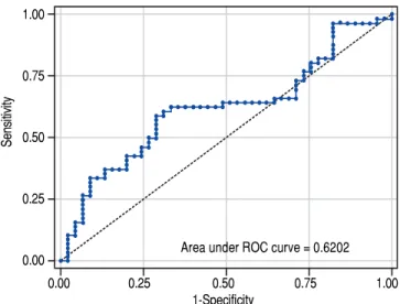
The value of ARFI measurement increased with the se-verity of liver fibrosis (Figure 1). There was a significant correlation between all 3 non-invasive assessments and fi-brosis stage, with the Spearman’s ρ of 0.38 for ARFI (p = 0.0001), 0.25 for APRI (p = 0.012), and 0.28 for FIB-4 (p = 0.004). The C statistics of ARFI (Figure 2), APRI (Figure 3), and FIB-4 (Figure 4) for fibrosis stage ≥ 2 were 0.70 (95% confidence interval [CI], 0.59-0.80), 0.62 (95% CI, 0.51-0.73), and 0.64 (95% CI, 0.53-0.75), respectively.
Cutoff values of ARFI, APRI, and FIB-4 to spare liver biopsy
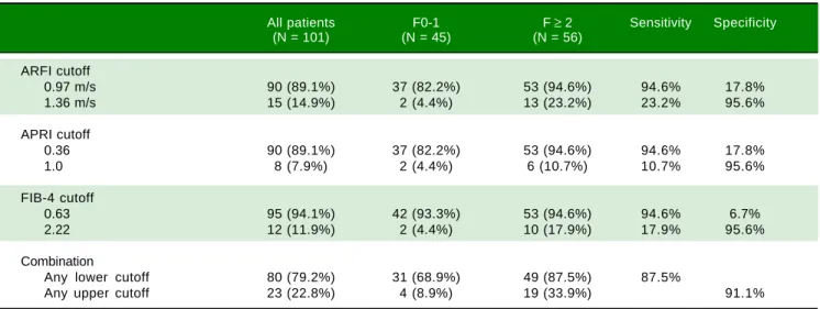
The measured value with 95% sensitivity and that with 95% specificity for fibrosis ≥ F2 were chosen as the lower and upper cutoffs to identify patients who may be spared liver biopsy, since significant liver fibrosis was very un-likely when the measurement fell below the lower cutoff but almost certain when it was above the upper one. The upper and lower cutoff points to distinguish significant
liver fibrosis were 0.97 m/sec and 1.36 m/sec for ARFI, 0.36 and 1.0 for APRI, 0.63 and 2.22 for FIB-4, respectively (Table 2). A combination of these 3 non-invasive methods would spare liver biopsy in a total of 44 (43.6%) patients who had measurements below the lower cutoff, or above the upper cutoff, with an overall sensitivity of 87.5% and specificity of 91.1%.
DISCUSSION
This prospective study showed ARFI, APRI, and FIB correlated with degree of liver fibrosis among non-cir-rhotic CHB patients with high viral load and slightly ele-vated ALT. After combination of these three methods, 44(43.6%) patient could spare liver biopsy. To our knowl-edge, it is the first article that focuses on this population, in which the degree of liver fibrosis is crucial to deter-mine antiviral therapy.
APRI and FIB-4 are biomarkers with high applicability and goof reproducibility for fibrosis evaluation. However, the value might be affected by some factors such as inflam-mation, hemolysis, and Gilbert syndrome. Liver stiffness measurement is another kind of modality.4 ARFI is an
ul-trasound-based elastography method that is integrated into a conventional ultrasound machine. Comparing to tran-sient elastography (TE), ARFI is more technic-dependent, however, it can be easily applied in a routine abdominal ultrasound practice, and it overcomes the limitations of TE, such as obesity and ascites.4 Besides, ARFI has been
shown to have a higher rate of reliable measurements
Figure 1. Figure 1.Figure 1. Figure 1.
Figure 1. The value of ARFI measurement increased with the severity of liver fibrosis.
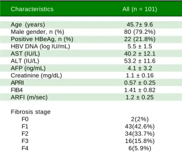
Table 1. Characteristics of participants.
Characteristics All (n = 101)
Age (years) 45.7± 9.6
Male gender, n (%) 80 (79.2%)
Positive HBeAg, n (%) 22 (21.8%)
HBV DNA (log IU/mL) 5.5 ± 1.5
AST (IU/L) 40.2 ± 12.1
ALT (IU/L) 53.2 ± 11.6
AFP (ng/mL) 4.1 ± 3.2
Creatinine (mg/dL) 1.1 ± 0.16
APRI 0.57 ± 0.25
FIB4 1.41 ± 0.82
ARFI (m/sec) 1.2 ± 0.25
Fibrosis stage
F0 2(2%)
F1 43(42.6%)
F2 34(33.7%)
F3 16(15.8%)
F4 6(5.9%)
HBV: Hepatitis B virus. AST: aspartate aminotransferase. ALT: alanine transaminase. AFP: alpha-fetoprotein. APRI: aspartate transaminase to platelet ratio index. FIB-4: fibrosis-4. ARFI: acoustic radiation force impulse.
3.0
2.5
2.0
1.5
1.0
0.5
Measure median
F0-1 F2 F3 F4
Fibrosis
1.22
1.48 1.18
1.10
*
compared to TE.5 However, it had some disadvantage
in-cluded narrow range of value, inadequate accuracy to dis-criminate intermediated stage fibrosis and, not well validated as TE, APRI, and FIB-4.5 Besides, it could be
af-fected by body mass index.14
In the present study, APRI tended to present less accu-rate result than ARFI and FIB-4, which was correlated with other reports.15,16 However, the predictive accuracy
of the three modalities were all lower than previous stud-ies. The area under RCO for significant fibrosis of ARFI, FIB-4, and APRI were 0.7, 0.64, and 0.62 respectively. Re-viewing previous studies, the area under ROC for signifi-cant fibrosis ranged from 0.70-0.91,0.74-0.76, and 0.72-0.79
for ARFI, FIB-4, and APRI respectively.14-19 In fact,
previ-ously reported results were inconsistent as well. Liu, et al.15
reported that ARFI and APRI had acceptable predictive value for fibrosis stage (area under ROC: 0.91 and 0.79). However, Tai, et at.20 reported the cirrhosis predictive accuracy of ARFI
was worse than conventional ultrasound in CHB patients, and ARFI was influenced by liver inflammation. A meta-anal-ysis17 showed APRI had limited value in CHB patients. The
different targeted population might mainly contribute to the discrepancy between our results and others. Comparing to previous reports, our patient population was very homogene-ous. Most of them were clinically non-cirrhotic CHB pa-tients with high viral load and slightly elevated ALT (1-2 folds of upper normal limit). Besides, most of the patients were classified as F1(42.6%) or F2 (33.7%). In contrast, the background conditions were very heterogeneous in previous studied with wide range ALT level and fibrosis distribution. Furthermore, the ARFI measurement was performed on the same day of liver biopsy in our study. On the other hand, the period between ARFI measurement and liver biopsy might be up to 4 weeks in one previous study.18
The strength of this prospective study was its strict inclu-sion criteria among a homogeneous and unique population. However, this study also had some important limitations. First, we recognize that the sample size is relatively small, which precludes the development of a multivariate-adjusted model. Second, external validation by an independent cohort is necessary before the reported cutoff points can be applied in daily practice. Third, as it remains debatable whether per-cutaneous needle biopsy can serve as the gold standard to stage hepatic fibrosis, we cannot exclude possible variability in the sampling of liver tissue and interpretation of data.21-23
Figure 3. Figure 3. Figure 3. Figure 3.
Figure 3. The C statistics of APRI for fibrosis stage ≥ 2 were 0.62 (95%CI, 0.51-0.73).
Figure 4. Figure 4.Figure 4. Figure 4.
Figure 4. The C statistics of APRI for fibrosis stage ≥ 2 were 0.64 (95% CI, 0.53-0.75).
Figure 2. Figure 2. Figure 2. Figure 2.
Figure 2. The C statistics of ARFI for fibrosis stage ≥ 2 were 0.70 (95%CI, 0.59-0.80).
1.00
0.75
0.50
0.25
0.00
Sensitivity
0.00 0.25 0.50 0.75 1.00
1-Specificity
Area under ROC curve = 0.6962
1.00
0.75
0.50
0.25
0.00
Sensitivity
0.00 0.25 0.50 0.75 1.00
1-Specificity
Area under ROC curve = 0.6401
1.00
0.75
0.50
0.25
0.00
Sensitivity
0.00 0.25 0.50 0.75 1.00
1-Specificity
In conclusion, our study demonstrated that measure-ments of ARFI, APRI, and FIB-4 were modestly correlat-ed with severity of liver fibrosis in a unique CHB population composed of non-cirrhotic patients with mild-ly elevated ALT despite high viral load. Combination of these non-invasive methods may spare “some” but not all patients for a liver biopsy.
ABBREVIATION
• ALT: alanine aminotransferase.
• APRI: aspartate transaminase to platelet ratio index.
• ARFI: acoustic radiation force impulse.
• AST: aspartate aminotransferase.
• AUC: area under the curve.
• CHB: chronic hepatitis B.
• CHC: chronic hepatitis C.
• CI: confidence interval.
• FIB-4: fibrosis-4.
• HBV: hepatitis B virus.
• HCV: hepatitis C virus.
• HDV: hepatitis D virus.
• NAs: nucleos(t)ide analogues.
• PLT: platelet.
• ROC: operating characteristic.
• SD: standard deviation.
• TE: transient elastography.
• ULM: upper limit of normal.
GRAND AND FINANCIAL SUPPORT
The grand is supported by E-Da Hospital (EDAHP104003), Tomorrow Medical Foundation (105-2), and the Ministry of Science and Technology (MOST-105-2314-B-650-001-MY2)
ACKNOWLEDGEMENT
The authors are grateful to “Liver Disease Prevention & Treatment Research Foundation, Taiwan” for the support of the study and Ms. Ying-Ju Lee for her efficient assist-ance
REFERENCES
1. Liaw YF, Kao JH, Piratvisuth T, Chan HL, Chien RN, Liu CJ, Gane E, et al. Asian-Pacific consensus statement on the management of chronic hepatitis B: a 2012 update. Hepatol Int 2012: 6: 531-61.
2. Sokal EM, Paganelli M, Wirth S, Socha P, Vajro P, Lacaille F, Kelly D, et al. Management of chronic hepatitis B in child-hood: ESPGHAN clinical practice guidelines: consensus of an expert panel on behalf of the European Society of Pediat-ric Gastroenterology, Hepatology and Nutrition. J Hepatol 2013: 59: 814-29.
3. Terrault NA, Bzowej NH, Chang KM, Hwang JP, Jonas MM,
Murad MH, American Association for the Study of Liver D. AASLD guidelines for treatment of chronic hepatitis B.
Hepatology 2016: 63: 261-83.
4. Castera L. Noninvasive methods to assess liver disease in
patients with hepatitis B or C. Gastroenterology 2012: 142: 1293-302 e4.
5. Castera L. Hepatitis B: are non-invasive markers of liver fi-brosis reliable? Liver Int 2014: 34(Suppl. 1): 91-6.
6. Takahashi H, Ono N, Eguchi Y, Eguchi T, Kitajima Y,
Ka-waguchi Y, Nakashita S, et al. Evaluation of acoustic radiation force impulse elastography for fibrosis staging of chronic liver disease: a pilot study. Liver Int 2010: 30: 538-45.
7. Jin W, Lin Z, Xin Y, Jiang X, Dong Q, Xuan S. Diagnostic ac-curacy of the aspartate aminotransferase-to-platelet ratio in-dex for the prediction of hepatitis B-related fibrosis: a leading meta-analysis. BMC Gastroenterol 2012: 12: 14.
8. Lin ZH, Xin YN, Dong QJ, Wang Q, Jiang XJ, Zhan SH, Sun
Y, et al. Performance of the aspartate aminotransferase-to-platelet ratio index for the staging of hepatitis C-related
fi-brosis: an updated meta-analysis. Hepatology 2011: 53:
726-36.
Table 2. Sensitivity and specificity of ARFI, APRI, FIB-4, and the combination to distinguish significant liver fibrosis (METAVIR F ≥ 2).
All patients F0-1 F ≥ 2 Sensitivity Specificity (N = 101) (N = 45) (N = 56)
ARFI cutoff
0.97 m/s 90 (89.1%) 37 (82.2%) 53 (94.6%) 94.6% 17.8%
1.36 m/s 15 (14.9%) 2 (4.4%) 13 (23.2%) 23.2% 95.6%
APRI cutoff
0.36 90 (89.1%) 37 (82.2%) 53 (94.6%) 94.6% 17.8%
1.0 8 (7.9%) 2 (4.4%) 6 (10.7%) 10.7% 95.6%
FIB-4 cutoff
0.63 95 (94.1%) 42 (93.3%) 53 (94.6%) 94.6% 6.7%
2.22 12 (11.9%) 2 (4.4%) 10 (17.9%) 17.9% 95.6%
Combination
Any lower cutoff 80 (79.2%) 31 (68.9%) 49 (87.5%) 87.5%
9. Houot M, Ngo Y, Munteanu M, Marque S, Poynard T. System-atic review with meta-analysis: direct comparisons of bi-omarkers for the diagnosis of fibrosis in chronic hepatitis C and B. Aliment Pharmacol Ther 2016: 43: 16-29.
10. Teshale E, Lu M, Rupp LB, Holmberg SD, Moorman AC, Spra-dling P, Vijayadeva V, et al. APRI and FIB-4 are good predic-tors of the stage of liver fibrosis in chronic hepatitis B: the Chronic Hepatitis Cohort Study (CHeCS). J Viral Hepat 2014: 21: 917-20.
11. Xiao G, Yang J, Yan L. Comparison of diagnostic accuracy of aspartate aminotransferase to platelet ratio index and fi-brosis-4 index for detecting liver fibrosis in adult patients with chronic hepatitis B virus infection: a systemic review and meta-analysis. Hepatology 2015: 61: 292-302.
12. Wai CT, Greenson JK, Fontana RJ, Kalbfleisch JD, Marrero JA, Conjeevaram HS, Lok AS. A simple noninvasive index can predict both significant fibrosis and cirrhosis in patients with chronic hepatitis C. Hepatology 2003: 38: 518-26. 13. Sterling RK, Lissen E, Clumeck N, Sola R, Correa MC,
Mon-taner J, M SS, et al. Development of a simple noninvasive in-dex to predict significant fibrosis in patients with HIV/HCV coinfection. Hepatology 2006: 43: 1317-25.
14. Park MS, Kim SW, Yoon KT, Kim SU, Park SY, Tak WY, Kweon YO, et al. Factors influencing the diagnostic accura-cy of acoustic radiation force impulse elastography in pa-tients with chronic hepatitis B. Gut and Liver 2016: 10: 275. 15. Liu Y, Yang G, Liu J, Yao S, Yuan J, Li S, Le X, et al. Optimal
linear combination of ARFI, transient elastography and APRI for the assessment of fibrosis in chronic hepatitis B. Liver
International 2015: 35: 816-25.
16. Xiao G, Yang J, Yan L. Comparison of diagnostic accuracy of aspartate aminotransferase to platelet ratio index and fi-brosis-4 index for detecting liver fibrosis in adult patients with chronic hepatitis B virus infection: A systemic review and meta-analysis. Hepatology 2015: 61: 292-302.
17. Jin W, Lin Z, Xin Y, Jiang X, Dong Q, Xuan S. Diagnostic ac-curacy of the aspartate aminotransferase-to-platelet ratio in-dex for the prediction of hepatitis B-related fibrosis: a leading meta-analysis. BMC Gastroenterology 2012: 12: 14. 18. Friedrich-Rust M, Buggisch P, Knegt Rd, Dries V, Shi Y,
Mat-schenz K, Schneider M, et al. Acoustic radiation force im-pulse imaging for non-invasive assessment of liver fibrosis in chronic hepatitis B. J Viral Hepat 2013: 20: 240-7. 19. Enomoto M, Morikawa H, Tamori A, Kawada N. Noninvasive
assessment of liver fibrosis in patients with chronic hepatitis
B. World J GastroenterolWJG 2014: 20: 12031.
20. Tai D-I, Tsay P-K, Jeng W-J, Weng C-C, Huang S-F, Huang C-H, Lin S-M, et al. Differences in Liver Fibrosis Between Patients With Chronic Hepatitis B and C. J Ultrasound Med 2015: 34: 813-21.
21. Bedossa P, Dargère D, Paradis V. Sampling variability of liver fibrosis in chronic hepatitis C. Hepatology 2003: 38: 1449-57. 22. Poynard T, Halfon P, Castera L, Charlotte F, Bail B, Munteanu
M, Messous D, et al. Variability of the area under the receiv-er opreceiv-erating charactreceiv-eristic curves in the diagnostic evalua-tion of liver fibrosis markers: impact of biopsy length and fragmentation. Alimentary Pharmacology & Therapeutics 2007: 25: 733-9.
23. Rousselet MC, Michalak S, Dupré F, Croué A, Bedossa P, Saint-André JP, Calès P. Sources of variability in histological scoring of chronic viral hepatitis. Hepatology 2005: 41: 257-64.
Correspondence and reprint request: Yao-Chun Hsu, M.D., Ph.D.
Department of Internal Medicine and Center for Database Research, E-Da Hospital/I-Shou University, 1, E-Da Rd., Kaohsiung 824, Taiwan; Tel.: +886-7-615-1100, Ext.: 5903, Fax: