PNPLA3 rs738409 causes steatosis according to viral &
IL28B genotypes in hepatitis C
Javier Ampuero,* José A. Del Campo,* Lourdes Rojas,* José R. García-Lozano,† Ricard Solá,‡ Raúl Andrade,§ José A. Pons,|| José M. Navarro,¶ José L. Calleja,**
María Buti,†† María F. González-Escribano,† Xavier Forns,‡‡ Moisés Diago,§§ Javier García-Samaniego,|||| Manuel Romero-Gómez*
* Unit for Clinical Management of Digestive Diseases and ciberehd, Valme University Hospital, Sevilla, Spain.
† Immunology Unit, Virgen del Rocio Hospital, Sevilla, Spain. ‡ Mar Hospital, Barcelona, Spain. § Digestive Unit and ciberehd. Virgen de la Victoria University Hospital, Málaga, Spain. || Virgen de la Arraixaca University Hospital, Murcia, Spain. ¶ Costa del Sol Hospital, Marbella, Spain.
** Puerta de Hierro Hospital, Madrid, Spain. ††Hepatology Unit and ciberehd. Vall d’Hebrón Hospital, Barcelona, Spain. ‡‡ Liver Unit and ciberehd, Clinic Hospital. IDIBAPS, Barcelona, Spain. §§ Digestive Department, Valencia Hospital, Spain.
|||| Digestive Unit and ciberehd. Carlos III Hospital, Madrid, Spain.
ABSTRACT
Background. Hepatitis C virus (HCV) is associated with a higher prevalence of steatosis compared to the general population. Aim. Our aim was to assess the impact of PNPLA3 rs738409 G-allele on steatosis in HCV patients. Material and methods. We included 474 HCV patients treated with peginterferon plus ribavirin. PNPLA3 rs738409 was genotyped and patients were classified according to alleles and genotypes. Steatosis was detected in 46.4% (220/474). Fibrosis was assessed by Scheuer score. Gene expression was analyzed in Huh7.5 and Huh7 cells using Real Time-PCR. Results. PNPLA3 allele-G was associated with steatosis [54.1% (126/233) vs. 39% (94/241)] (p = 0.0001). In HCV-1, allele-G was related to steatosis [50.6% (82/162) vs. 32.3% (53/164)] (p = 0.001), but did not in HCV-3 [61.9% (26/42) vs. 62% (31/50)] (p = 0.993). PNPLA3 allele-G was associated with steatosis in patients with IL28B-CT/TT [57.7% (82/142) vs. 37.1% (56/151)] (p = 0.0001), but did not in IL28B-CC [47.8% (43/90) vs. 42% (37/88)] (p = 0.442). Independent variables associated with steato-sis were: PNPLA3 G-allele [O.R. 1.84 (CI95%: 1.06-3.21); p = 0.007], age [O.R. 1.04 (CI95%: 1.01-1.07); p = 0.017], HCV-genotype 3 [O.R. 2.46 (CI95%: 1.30-4.65); p = 0.006], HOMA > 4 [O.R. 2.72 (CI95%: 1.27-5.82); p = 0.010]. Since PNPLA3 RNA could not be detected on PBMC from HCV patients, an in vitro analysis was performed. Huh7.5 cells infected with JFH1 had a decreased PNPLA3 gene expression (fold inhibi-tion = 3.2 ± 0.2), while Huh7 cells presented increased PNPLA3 gene expression (fold inducinhibi-tion = 1.5 ± 0.2).
Conclusion. PNPLA3 allele-G modulated the development of steatosis, particularly in patients with HCV-1 and IL28B-CT/TT genotype, but was not associated with SVR. Metabolic but not viral steatosis seems to be PNPLA3 regulated. Gene interaction may result in differential PNPLA3 gene expression levels in HCV infection.
Key words. Single nucleotide polymorphism. Viral replication. Huh7.5 cells. Liver biopsy. Dyslipidemia.
Correspondence and reprint request: Prof. Manuel Romero-Gómez, M.D., Ph.D. Full Professor, Unit for the Clinical Management of Digestive Diseases and CIBERehd, Hospital Universitario de Valme. Avenida de Bellavista s/n, Sevilla 41014, Spain.
Ph.: (+34) 955-015761. Fax: (+34) 955-015899 E-mail: [email protected]
Manuscript received: February 02, 2014. Manuscript accepted: March 24, 2014. INTRODUCTION
Liver steatosis is frequent among patients with chronic hepatitis C virus; its prevalence is higher
isoleucine-to-methionine substitution at position 148 in the hu-man patatin-like phospholipase domain containing 3 (PNPLA3) rs738409 C>G single nucleotide poly-morphism (SNP) was identified as the strongest de-terminant of hepatocyte steatosis. Several candidate gene studies have demonstrated that PNPLA3 al-lele-G influences on liver fat accumulation. Addi-tionally to NAFLD, PNPLA3 I148M sequence variant has been extensively associated with NASH,5 fibrosis progression,6 alcoholic liver dis-ease7 and hepatocellular carcinoma.8 On the other hand, IL28B polymorphism CC has been related to higher SVR rate. However, its relationship with liv-er steatosis remains unclear, although suggested CC genotype could be related to lower steatosis degree.9
The primary aim of this study was to evaluate the association of PNPLA3 I148M variant with steato-sis and fibrosteato-sis severity in a Spanish cohort of hepa-titis C patients, while the second aim was to seek the interaction among genetic, metabolic and viral factors on steatosis development.
MATERIAL AND METHODS
In this cross-sectional study, 474 consecutive pa-tients with chronic hepatitis C infection, selected for peginterferon (PEG-IFN) and ribavirin (RBV) thera-py, were enrolled from 14 Spanish Hospitals be-tween October 2002 and January 2010. All patients followed standard treatment duration (24 or 48 week duration) according to HCV genotype. Pa-tients received PEG-IFN-alpha (either Pegintron; Shering-Plough 1.5 mcg/kg/wk or Pegasys; Roche 180 mcg/wk) combined with RBV 1,000 mg/day if body weight was 75 kg or less, or 1,200 mg if body weight was greater than 75 kg. PEG-IFN and RBV dose modification followed standard criteria and pro-cedures.10 Inclusion criteria were: patients > 18 years of age diagnosed as having chronic hepatitis C with HCV-RNA positive, who had criteria for com-mencing antiviral therapy in clinical practice; clas-sified as genotype 1, 2, 3 or 4. Exclusion criterium was the presence of any coexisting chronic liver dis-ease (including HIV and HBV infection). All pa-tients completed the scheduled treatment.
All patients provided written informed consent for the collection and storage of peripheral blood mono-nuclear cells, as well as host DNA testing for re-search proposes consistent with the current study. The database for this analysis included clinical and demographic data extracted from the original clini-cal database, which included data about liver, kid-ney and metabolic profiles (particularly, glucemic
and lipid metabolism). The study was approved by the Ethics Committee of each center and was con-ducted in accordance with the provisions of the dec-laration of Helsinki and good Clinical Practice Guidelines.
Viral load and HCV genotyping
At baseline, all patients had a quantitative meas-ure of serum or plasma HCV-RNA performed by polymerase chain reaction assay using the COBAS AmpliPrep/COBAS TaqMan HCV Test (Roche Diag-nostics GMBH, Mannheim) with a lower limit detec-tion of 15 UI/mL. A qualitative or quantitative measurement of serum or plasma HCV-RNA was performed at week 4, 8, 12, 24 and 48 and follow-up weeks 4, 12, and 24. HCV genotyping was performed by reverse hybridization (Versant HCV® 2.0 Assay LiPA, Siemens) in all patients.
PNPLA3 genotyping
The rs738409 SNPs were analyzed using the Ste-pOnePlus Real Time PCR System (Applied Biosys-tems, Foster City, USA) with a TaqMan SNP Genotyping Assay developed together with Applied Biosystems using published sequences from the NCBI Entrez SNP Database (www.ncbi.nlm. nih.gov/sites/entrez) (rs738409: 5’-AAGGAGGGA-TAAGGCCACTGTA-3’ as forward and 5’-CTT-TCACAGGCCTTGGTATGTTC-3’ as reverse primer).
IL28B genotyping
The genomic region associated with HCV treat-ment response lies on chromosome 19 and contains multiple SNPs in linkage disequilibrium around the IL28B gene. We selected the most strongly associat-ed SNP, rs12979860, locatassociat-ed 3-kb upstream of the IL28B gene, for genotyping in the cohorts by RT-PCR using the LightCycler® 480 System (Roche Di-agnostic).
Histological features
All of patients underwent to liver biopsy before therapy. Histologic evaluation was carried out by the same pathologist at each Hospital Center by Scheuer scoring:11
• F3: many bridging fibrosis. • F4: cirrhosis.
Steatosis was defined as > 5% of hepatocytes with macrovesicular steatosis.
Cell cultures and gene expression analysis
Huh7.5 (rs12979860 genotype CT) and Huh7 (rs12979860 genotype CC) cells were grown in DMEM culture medium supplemented with 10% FBS, antibiotics, L-Glutamine and Non-Essential aminoacids. Cells were incubated at 37 oC, 5% CO
2. Serum from patients infected by HCV-genotypes 1 and 3 with high viral load (> 107 IU/mL) were used for cell culture infection. Total RNA was extracted from cellular lysates using standard protocols (TRIsure™, BIOLINE, London, UK). JFH1 full ge-nomic replicon was used to infect Huh7 and Huh7.5 cells, using 1 particle per cell. After infection, cells were incubated (37 oC, 5% CO
2) for 48 h and then collected for total RNA isolation. We have performed the respective retro-transcription reactions using commercially available kits (Qiagen, Invitrogen, Carlsbad, CA, USA). Gene expression was analyzed by semi-quantitative real-time PCR using a illumi-na® Eco™ cycler. Primers for ACC (Acetyl-CoA Car-boxylase), APOB (Apolipoprotein B), DGAT1 (Diacylglycerol O-Acyltransferase 1), DGAT2, FASN (Fatty Acid Synthase), LDLr (LDL cholesterol re-ceptor), MTP (Microsomal triacylglycerol transfer protein), PPARG (Peroxisome Proliferator-activated Receptor Gamma), PNPLA3 (Patatin-like phospholi-pase domain containing 3) and SREBP (Sterol Regu-latory Element-Binding Protein) genes were obtained from Qiagen (Hilden, Germany). GAPDH (Glyceraldehyde-3-phosphate Dehydrogenase) was used as a house-keeping gene.
Statistical analysis
Statistical analyses and graphs were done with SPSS (19.0, SPSS Inc., Chicago, IL). All values are presented as means ± SD. Comparisons between groups were made using the Mann-Whitney U test, the Student t-test or ANOVA for continuous varia-bles, and the Chi-square or the Fisher exact proba-bility test for categorical data. Variables that showed significance p < 0.10 in univariate analysis were entered into backward logistic regression anal-ysis. The multivariate models were constructed se-quentially with variables entered one at a time and a significance level of 0.05 was used to remove them
from the model, except age and sex, which were in-cluded in all the models.
RESULTS
Baseline epidemiological and biochemical features of overall cohort are shown in table 1. Gender dis-tribution was 64.8% (307/474) males and 35.2% (167/474) females, with a mean age of 43.4 ± 9.81 years of age.
Table 1. Baseline characteristics of overall cohort.
Age 43.4 ± 9.81
Gender distribution
Males 64.8% (307/474)
Females 35.2% (167/474)
IL28B
CC 37.8% (178/471)
CT 49.9% (235/471)
TT 12.3% (58/471)
PNPLA3
GG 5.1% (24/474)
GC 44.1% (209/474)
CC 50.8% (241/474)
Steatosis 46.4% (220/474)
Fibrosis
F0 4% (19/474)
F1 54.6% (259/474)
F2 25.7% (122/474)
F3 9.1% (43/474)
F4 6.5% (31/474)
HCV genotype
1 68.8% (326/474)
2 2.5% (12/474)
3 19.4% (92/474)
4 9.3% (44/474)
Ethnicity
Caucasians 82.8% (332/401)
Non-Caucasians 17.2% (69/401)
Sustained virological response 52.1% (247/474)
High viral load 69.8% (331/474)
Body mass index 25.73 ± 4.6
HOMA-IR 2.9 ± 2.74
AST (IU/mL) 65.54 ± 48.27
ALT (IU/mL) 79.13 ± 64.17
GGT (IU/mL) 76.81 ± 91.52
57.1
43.3 55.6
37.8 85.7
25
58.365.1 55
33.3
46.1 45.2
64.3
42
Figure 1. Steatosis (%), according to PNPLA3 allele, by
IL28B genotype distribution. p = 0.910
p = 0.0001
HCV-genotype distribution was:
• 68.8% (326/474) patients with genotype 1. • 2.5% (12/474) patients with genotype 2. • 19.4% (92/474) patients with genotype 3; and • 9.3% (44/474) patients with genotype 4.
PNPLA3 polymorphism was genotyped:
• GG 5.1% (24/474). • GC 44.1% (209/474).
• CC 50.8% (241/474); G allele 49.2% (233/474) vs. 50.8% (241/474) non-G alelle.
IL28B polymorphism was analyzed:
• CC 37.8% (178/471). • CT 49.9% (235/471), and
• TT 12.3% (58/471); IL28B-CC 37.8% (178/471) vs. 62.2% (293/471) IL28B-CT/TT.
Patients showing > 800.000 IU/mL [69.8% (331/ 474)] were considered as high baseline viral load. St-eatosis was observed in 46.4% (220/474) of patients.
Fibrosis stage was measured:
• 4% (19/474) F0. • 54.6% (259/474) F1. • 25.7% (122/474) F2. • 9.1% (43/474) F3; and • 6.5% (31/474) F4.
According to ethnicity, 82.8% (332/401) were Caucasians and 17.2% (69/401) were from other eth-nicities.
The PNPLA3 allele-G did not show steatosis in patients with IL28B-CC [47.8% (43/90) vs. 42% (37/ 88) in PNPLA3 non-allele-G] (p = 0.442). However, in patients with IL28B-CT/TT steatosis was more prevalent in PNPLA3 allele-G (57.7%; 82/142) than in non-allele-G (37.1%; 56/151) (p = 0.0001) (Figure 1). In the overall cohort (all HCV-genotypes), stea-tosis was present in 54.1% (126/233) in patients with PNPLA3 allele-G, while it was detected in 39% (94/241) in non-allele-G (p = 0.0001). In HCV-geno-type 1 (n = 326), PNPLA3 allele-G influenced on the presence of steatosis (50.6%; 82/162) vs. non-allele-G (32.3%; 53/164) (p = 0.001). PNPLA3 allele-G was associated with steatosis in non-3 genotype (52.4%; 100/191 vs. 33%; 63/191) (p = 0.0001). In contrast, allele-G did not show any impact on steatosis in HCV-genotype 3 (61.9%; 26/42 vs. 62%; 31/50)
(p = 0.993) (Figure 2). On the other hand, HOMA was higher in patients with steatosis (3.92 vs. 2.49; p = 0.013), as well as body mass index (26.06 vs. 25.16; p = 0.015). In multivariate analysis, varia-bles independently associated with steatosis were: PNPLA3 allele-G [O.R. 1.84 (CI95%: 1.06-3.21); p = 0.007], age [O.R. 1.04 (CI95%: 1.01-1.07); p = 0.017], HCV-genotype 3 [O.R. 2.46 (CI95%: 1.30-4.65); p = 0.006] and HOMA > 4 [O.R. 2.72 (CI95%: 1.27-5.82); p = 0.010] (Table 2).
In contrast, PNPLA3 did not influence on fibrosis severity neither SVR when patients were stratified by viral or IL28B genotypes (data not shown). Indeed, PNPLA3 allele-G achieved SVR in 50.2% (117/233), while non-allele-G in 53.9% (130/241) (p = 0.417).
In vitro analysis (Figure 3) showed that Huh7.5 cells (CT genotype) infected by HCV-genotype 1 had significantly increased gene expression (> 1.5 fold induction) for DGAT1 and DGAT2 genes. In Huh7 cells (CC genotype) APOB, DGAT1, DGAT2, LDLR and SREBP were found induced. Infection by
HCV-Figure 2. Steatosis (%), according to PNPLA3 allele, by
HCV-genotype distribution. HCV: hepatitis C virus. 100
80
60
40
20
0
G Allele Non-G Allele
G Allele Non-G Allele 100
80
60
40
20
0
IL28B-CC IL28B-CT/TT
Overall cohort HCV-1 HCV-2 HCV-3 HCV-4
n = 376 n = 245 n = 11 n = 79 n = 41
Table 2. Univariate and multivariate analysis according to steatosis.
Univariate analysis Multivariate analysis
Age (years) 45.05 vs. 42.55; p = 0.001 OR 1.04 (CI95% 1.01-1.07); p = 0.017 Gender distribution (males vs. females) 52.4% vs. 48.7%; p = 0.370
Body mass index 26.06 vs. 25.16; p = 0.015
HOMA > 4 66.1% vs. 45.3%; p = 0.005 OR 2.72 (CI95% 1.27-5.82); p = 0.010 PNPLA3 allele-G 54.1% vs. 39%; p = 0.0001 OR 1.84 (CI95% 1.06-3.21); p = 0.007 IL28B-(CC vs. CT/TT) 50.9% vs. 51.4%; p = 0.904
HCV-genotype (3 vs. 1) 65% vs. 47.6%; p = 0.001 OR 2.46 (CI95% 1.30-4.65); p = 0.006
AST (IU/mL) 67.72 vs. 62.90; p = 0.216
ALT (IU/mL) 79.89 vs. 82.43; p = 0.619
GGT (IU/mL) 79.14 vs. 71.71; p = 0.281
Triglycerides (mg/dL) 108.41 vs. 96.09; p = 0.016
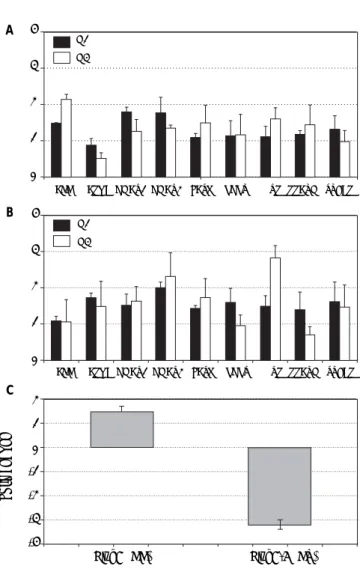
Figure 3. Gene expression (fold induction) in Huh7.5 (IL28B
CT genotype) (A) and Huh7 (IL28B CC genotype) (B) cells infec-ted with JFH1 particles. Lipid metabolism relainfec-ted gene ex-pression was analyzed by semi-quantitative real time-PCR (see methods for details). C. PNPLA3 gene expression in Huh7 and Huh7.5 cells infected with JFH1 particles. Non-infected cell cultures are used as control. Data shown are mean va-lues from three independent experiments. G1: genotype 1. G3: genotype 3.
A
C B
genotype 3 promoted increased gene expression of ACC, FASN and MTP in Huh7.5 while DGAT1, DGAT1, FASN and MTP were significantly induced in Huh7 cells. PNPLA3 gene expression was also analyzed in these cells (Figura 3C): in Huh7.5 (IL28B CT genotype) cells infected with JFH1, a de-creased gene expression was observed (fold inhibi-tion = 3.2 ± 0.2). By contrary, in Huh7 cells harboring IL28B CC genotype, the opposite effect was detected: PNPLA3 gene expression was induced (fold induction = 1.5 ± 0.2).
DISCUSSION
We reported an association between PNPLA3 rs738409 allele-G and liver steatosis in a large co-hort of chronic hepatitis C patients. Indeed, the im-pact of PNPLA3 polymorphisms on steatosis depends on viral and IL28B genotypes. Steatosis is known to negatively impact on the natural history of HCV infection as it accelerates the progression to cirrhosis. Metabolic, enviromental, genetic and viral factors have been implicated on steatosis develop-ment. However, the weight of each of them in a de-terminate patient remains elusive. Patients with PNPLA3 allele-G, overweight, HOMA > 4 and viral genotype 3a were independently associated with st-eatosis. PNPLA3 allele-G was associated with stea-tosis in patients infected by HCV-genotype 1, showed a trend in genotype 2 and genotype 4, but was not related to steatosis in genotype 3a. A link between this gene and steatosis was reported by Trépo, et al., but genotype 3 patients were not in-cluded.12 Our data confirm previous results reported by Valenti, et al.13 and Cai, et al.14 that evaluated the effect of PNPLA3 on liver fat and fibrosis in HCV patients. The rs738409 GG genotype was close-ly associated with steatosis across all viral
geno-4
3
2
1
0
ACC APOB DGAT1 DGAT2 FASN LDLR MTP PPARG SREBP G1
G3
4
3
2
1
0
ACC APOB DGAT1 DGAT2 FASN LDLR MTP PPARG SREBP G1
G3
2 1 0 -1 -2 -3 -4
Fold change
types except for genotype 3a. However, Rembeck, et al., did not find differences in the prevalence of stea-tosis between HCV-genotypes 2 and 3 in patients with PNPLA3 rs738409 allele-G, in spite of HCV-genotype 2 showed an increased insulin resistance.15 These data could indicate two types of liver steatosis in chronic hepatitis C patients:
• Metabolic steatosis promoted by genetic factors (PNPLA3 allele-G) in HCV-genotypes 1, 2 and 4. • Viral steatosis related to HCV-genotype 3.
On the other hand, PNPLA3 polymorphisms in-fluenced on steatosis depending on IL28B genotype. PNPLA3 allele-G increased the risk of steatosis in patients with IL28B-CT/TT but did not in genotype IL28B-CC. The rational for considering the effect of PNPLA3 on steatosis according to IL28B genotype was to identify some factor which explains the high-er presence of steatosis in IL28B-CT/TT patients. In fact, Valenti, et al. observed that IL28B-CC protected from steatosis in PNPLA-GG patients, but did not in those negative for the PNPLA3 G variant at risk, in non genotype-3 patients.16 Tillman, et al. demon-strated that the presence of steatosis was lower in IL28B-CC compared to non-CC patients in two inde-pendent HCV-genotype 1 cohorts.17 Recently, simi-lar results have been documented by Agundez, et al.18 Therefore, the different effect of PNPLA3 allele-G according to IL28B genotype could explain, at least in part, the higher prevalence of steatosis in IL28B-CT/TT patients. Our in vitro results showed that cells harboring IL28B-CC genotype induced greater gene expression than IL28B-CT genotype cells in HCV genotype 3 infection, in particular for MTP (Figure 3). As pointed out by Mirandola, et al.19 there is a complex relationship between MTP and HCV: since MTP is essential for the assembly and secretion of HCV, MTP expression should be up-regulated to facilitate its propagation during early infection stages, although MTP mRNA levels are re-duced in chronic HCV patients independently of HCV genotype.20 Pharmacologic inhibition of MTP has been proposed as a potential antiviral strategy for HCV.21
PNPLA3 is a member of the patatin-like phos-pholipase family which encodes a transmembrane protein expressed in stellate cells but more promi-nently in hepatocytes, where interacts with lipids. The mechanism by which variation in PNPLA3 af-fects liver triglyceride content is not completely known. The PNPLA3 rs738409 G-allele was sug-gested to impair triglyceride hydrolysis in
hepato-cytes and favour the increase of triglycerides accu-mulation.22 Both Huh7.5 (IL28B CT genotype) and Huh7 (IL28B CC genotype), harbor the GG geno-type for rs738409 polymorphism, being a good sys-tem for studying gene interaction with IL28B genotypes. Our results demonstrated that JFH1 in-creased PNPLA3 expression in favorable IL28B gen-otype cells. Since lower PNPLA3 gene expression and activity are associated to steatosis development, the interaction between IL28B, PNPLA3 polymor-phism and HCV infection could explain the grade of steatosis and sustained viral response.
On the other hand, it has been suggested that ac-cumulation of fat in the hepatocyte cytoplasm in form of large lipid droplets favors virion assembly. Otherwise, DGAT1 and DGAT2 enzymes catalyze the final step in triglyceride biosynthesis and are es-sential in lipid droplet biogenesis, especially DGAT1.23 Interestingly, DGAT1 (required for effi-cient viral particle formation24) and DGAT2 genes were found induced in genotype 1 compared to geno-type 3 in cells with IL28B-CT genogeno-type. This effect was not observed in IL28B-CC cells, indicating a host-virus genetic interaction.
PNPLA3 polymorphisms were not associated with sustained virological response. This fact con-firms data from previous studies that showed PNP-LA3 rs738409 allele-G did not influence on treatment response.25,26 Nevertheless, PNPLA3 poly-morphisms have been associated with increased risk of hepatocellular carcinoma in alcoholic cirrhosis.27 HCC is developed in some cirrhotic patients despite SVR.28 If PNPLA3 genotype, through promoting steatosis, could be implicated on this process requires further studies.
This study has some limitations. First, the design of the study (cross-sectional) is not the appropiate method to stablish cause-effect relationships. How-ever, most of data are consistent with hypothesis about PNPLA3. Second, the number of patients with genotype non-1 was limited, particularly HCV-genotypes 2 and 4 (HCV-1 genotype is the most prevalent genotype in Spain), which could underesti-mated the effect of PNPLA3 on liver steatosis in these HCV-genotypes.
Indeed, lipids-related gene expression was modulated by viral genotype according to IL28B background in vitro. These findings support that steatosis is the final result of the interaction between host and viral factors.
ABBREVIATIONS
• APOB: apolipoprotein B.
• ACC: acetyl-CoA carboxylase.
• DGAT: diglyceride acyltransferase.
• FASN: fatty acid synthase.
• HCC: hepatocellular carcinoma.
• HCV: hepatitis C virus.
• LDLr: LDL receptor.
• MTP: microsomal triaglyceride transfer protein.
• NAFLD: non-alcoholic fatty liver disease.
• NASH: non-alcoholic steatohepatitis.
• PEG-IFN: peginterferon.
• PNPLA3: patatin-like phospholipase domain
containing 3.
• PPARG: peroxisome proliferator-activated
re-ceptor gamma.
• RBV: ribavirin.
• SNP: single nucleotide polymorphism.
• SREBP: sterol regulatory element-binding protein.
• SVR: sustained viral response.
SPECIFIC AUTHOR CONTRIBUTION
• Planning and conducting the study: Javier Am-puero, Manuel Romero-Gomez.
• Drafting the manuscript: Javier Ampuero, Jose Antonio del Campo, Manuel Romero-Gomez. • Interpreting data: Javier Ampuero, Jose Antonio
del Campo, Manuel Romero-Gomez.
• Performing in vitro studies: Jose Antonio del Campo, Lourdes Rojas.
• Collecting data: Jose Raúl García-Lozano; Ricard Solá; Raúl Andrade; José Antonio Pons; Jose María Navarro; Jose Luis Calleja; María Buti; María Francisca González-Escribano; Xavier Forns, Moisés Diago, Javier García-Samaniego.
Guarantor of the article: Manuel Romero-Gomez.
FINANCIAL SUPPORT
None.
POTENTIAL COMPETING INTEREST
None.
REFERENCES
1. Browning JD, Szczepaniak LS, Dobbins R, Nuremberg P, Horton JD, Cohen JC, Grundy SM, et al. Prevalence of he-patic steatosis in an urban population in the United Sta-tes: impact of ethnicity. Hepatology 2004; 40: 1387-95. 2. Castéra L, Hézode C, Roudot-Thoraval F, Bastie A, Zafrani
ES, Pawlotsky JM, Dhumeaux D. Worsening of steatosis is an independent factor of fibrosis progression in untrea-ted patients with chronic hepatitis C and paired liver biopsies. Gut 2003; 52: 288-92.
3. Romero-Gómez M, Castellano-Megias VM, Grande L, Irles JA, Cruz M, Nogales MC, Alcón JC, et al. Serum leptin levels correlate with hepatic steatosis in chronic hepatitis C. Am J Gastroenterol 2003, 98: 1135-41.
4. Romeo S, Kozlitina J, Xing C, Pertsemlidis A, Cox D, Pen-nacchio LA, Boerwinkle E, et al. Genetic variation in PN-PLA3 confers susceptibility to nonalcoholic fatty liver disease. Nat Genet 2008; 40: 1461-5.
5. Sookoian S, Pirola CJ. Meta-analysis of the influence of I148M variant of patatin-like phospholipase domain contai-ning 3 gene (PNPLA3) on the susceptibility and histological severity of nonalcoholic fatty liver disease. Hepatology 2011; 53: 1883-94.
6. Petit JM, Guiu B, Masson D, Duvillard L, Jooste V, Buffier P, Bouillet B, et al. PNPLA3 polymorphism influences liver fi-brosis in unselected patients with type 2 diabetes. Liver Int 2011; 31: 1332-6.
7. Stickel F, Hampe J. Genetic determinants of alcoholic liver disease. Gut 2012; 61: 150-9.
8. Guyot E, Sutton A, Rufat P, Laguillier C, Mansouri A, Mo-reau R, Ganne-Carrié N, et al. PNPLA3 rs738409, hepato-cellular carcinoma occurrence and risk model prediction in patients with cirrhosis. J Hepatol 2013; 58: 312-8. 9. Petta S, Rosso C, Leung R, Abate ML, Booth D, Salomone F,
et al. Effects of IL28B rs12979860 CC genotype on meta-bolic profile and sustained virologic response in patients with genotype 1 chronic hepatitis C. Clin Gastroenterol Hepatol 2013; 11: 311-7.
10. McHutchison JG, Lawitz EJ, Shiffman ML, Muir AJ, Galler GW, McCone J, Nyberg LM, et al. Peginterferon Alfa-2b or Alfa-2a with Ribavirin for Treatment of Hepatitis C Infec-tion. N Engl J Med 2009; 361: 580-93.
11. Scheuer PJ. Classification of chronic viral hepatitis: a need for reassessment. J Hepatol 1991; 13: 372-3. 12. Trépo E, Pradat P, Potthoff A, Momozawa Y, Quertinmont
E, Gustot T, Lemmers A, et al. Impact of patatin-like phos-pholipase-3 (rs738409 C>G) polymorphism on fibrosis pro-gression and steatosis in chronic hepatitis C. Hepatology 2011; 54: 60-9.
13. Valenti L, Rumi M, Galmozzi E, Aghemo A, Del Menico B, De Nicola S, Dongiovanni P, et al. Patatin-like phospholipase domain-containing 3 I148M polymorphism, steatosis, and li-ver damage in chronic hepatitis C. Hepatology 2011; 53: 791-9.
14. Cai T, Dufour JF, Muellhaupt B, Gerlach T, Heim M, Mora-dpour D, Cerny A, et al. Viral genotype-specific role of PN-PLA3, PPARG, MTTP, and IL28B in hepatitis C virus-associated steatosis. J Hepatol 2011; 55: 529-35. 15. Rembeck K, Maglio C, Lagging M, Christensen PB, Färkkilä
M, Langeland N, Buhl MR, et al. PNPLA 3 I148M genetic va-riant associates with insulin resistance and baseline viral load in HCV genotype 2 but not in genotype 3 infection. BMC Med Genet 2012: 14: 82.
steatosis in chronic hepatitis C non genotype-3 patients. J Hepatol 2012; 56: 1209-10; author reply 1210-2. 17. Tillmann HL, Patel K, Muir AJ, Guy CD, Li JH, Lao XQ,
Thompson A, et al. Beneficial IL28B genotype associated with lower frequency of hepatic steatosis in patients with chronic hepatitis C. J Hepatol 2011; 55: 1195-200. 18. Agúndez JA, García-Martin E, Maestro ML, Cuenca F,
Mar-tínez C, Ortega L, Carballo M, et al. Relation of IL28B gene polymorphism with biochemical and histological featu-res in hepatitis C virus-induced liver disease. PLoS One. 2012;7(5):e37998. Doi: 10.1371/journal.pone.0037998. 19. Mirandola S, Bowman D, Hussain MM, Alberti A. Hepatic
steatosis in hepatitis C is a storage disease due to HCV interaction with microsomal triglyceride transfer protein (MTP). Nutr Metab (Lond) 2010; 7: 13.
20. Mirandola S, Realdon S, Iqbal J, Gerotto M, Dal Pero F, Bor-toletto G, Marcolongo M, et al. Liver microsomal triglyce-ride transfer protein is involved in hepatitis C liver steatosis. Gastroenterology 2006; 130: 1661-9.
21. Ye J. Reliance of host cholesterol metabolic pathways for the life cycle of hepatitis C virus. PLoS Pathog 2007; 3: e108.
22. He S, McPhaul C, Li JZ, Garuti R, Kinch L, Grishin NV, Co-hen JC, et al. A sequence variation (I148M) in PNPLA3 as-sociated with nonalcoholic fatty liver disease disrupts triglyceride hydrolysis. J Biol Chem 2010; 285: 6706-15.
23. Harris C, Herker E, Farese RV Jr, Ott M. Hepatitis C virus core protein decreases lipid droplet turnover: a mecha-nism for core-induced steatosis. J Biol Chem 2011; 286: 42615-25.
24. Herker E, Harris C, Hernandez C, Carpentier A, Kaehlcke K, Rosenberg AR, Farese RV Jr, et al. Efficient hepatitis C virus particle formation requires diacylglycerol acyltrans-ferase-1. Nat Med 2010; 16: 1295-8.
25. Valenti L, Aghemo A, Stättermayer AF, Maggioni P, De Ni-cola S, Motta BM, Rumi MG, et al. Implications of PNPLA3 polymorphism in chronic hepatitis C patients receiving pe-ginterferon plus ribavirin. Aliment Pharmacol Ther 2012; 35: 1434-42.
26. Clark PJ, Thompson AJ, Zhu Q, Vock DM, Zhu M, Patel K, Harrison SA, et al. The association of genetic variants with hepatic steatosis in patients with genotype 1 chronic hepatitis C infection. Dig Dis Sci 2012; 57: 2213-21. 27. Trepo E, Guyot E, Ganne-Carrie N, Degre D, Gustot T,
Franchimont D, Sutton A, et al. PNPLA3 (rs738409 C>G) is a common risk variant associated with hepatocellular carcinoma in alcoholic cirrhosis. Hepatology 2012; 55: 1307-8.