Portal vein thrombosis in patients with cirrhosis:
just a common finding or a predictor of poor outcome?
Omar D. Borjas-Almaguer,* Carlos A. Cortez-Hernández,** Emmanuel I. González-Moreno,** Francisco J. Bosques-Padilla,** José A. González-González,** Aldo A. Garza,** Juan A. Martínez-Segura,** Diego García-Compean,** Juan V. Alejandre-Loya,** Jesús García-García,** Guillermo Delgado-García,* Héctor J. Maldonado-Garza**
* Internal Medicine Department and ** Gastroenterology Division. Hospital Universitario “Dr. José Eleuterio González”. Universidad Autónoma de Nuevo León. Monterrey, México.
A B S T R A C T A B S T R A C T A B S T R A C T A B S T R A C T A B S T R A C T
Background & Aims. Background & Aims.Background & Aims. Background & Aims.
Background & Aims. It is unclear whether portal vein thrombosis (PVT) unrelated to malignancy is associated with reduced sur-vival or it is an epiphenomenon of advanced cirrhosis. The objective of this study was to assess clinical outcome in cirrhotic patients with PVT not associated with malignancy and determine its prevalence. Material and methods.Material and methods.Material and methods.Material and methods. Retrospective search in oneMaterial and methods. center from June 2011 to December 2014. Results.Results.Results.Results.Results. 169 patients, 55 women and 114 men, median age 54 (19-90) years. Thirteen had PVT (7.6%). None of the patients received anticoagulant treatment. The PVT group was younger (49 [25-62] vs. 55 [19-90] years p = 0.025). Child A patients were more frequent in PVT and Child C in Non-PVT. Median Model for End Stage Liver Disease (MELD) score was lower in PVT (12 [8-21] vs. 19 [7-51] p ≤ 0.001) p ≤ 0.001). There was no difference between upper gastrointes-tinal bleeding and spontaneous bacterial peritonitis in the groups. Encephalopathy grade 3-4 (4 [30.8%] vs. 73 [46.8%] p = 0,007) and large volume ascites (5 [38.5%] vs. 89 [57.1%] p= 0,012) was more common in non-PVT. Survival was better for PVT (16.5 ± 27.9 vs. 4.13 ± 12.2 months p = 0.005). Conclusions: We found that PVT itself does not lead to a worse prognosis. The most reliable predictor for clinical outcome remains the MELD score. The presence of PVT could be just an epiphenomenon and not a marker of advanced cirrhosis.
Key words. Key words.Key words. Key words.
Key words. Cirrhosis. Portal vein thrombosis. Prognosis. MELD Score. Child-Pugh Score.
November-December, Vol. 15 No. 6, 2016: 902-906
INTRODUCTION
Liver cirrhosis is a global health problem with an esti-mated prevalence in the United States of 0.27%.1 In
Mexico, it is the fourth and second place in global mor-tality and mormor-tality in productive age, respectively.2
Sev-eral predictors of mortality have been described in cirrhosis, with the most reliable being the Child-Pugh and Model for End Stage Liver Disease (MELD) score.3
The coagulation system in patients with liver disease suf-fers several changes, going to pro-thrombotic or predis-position to bleeding depending on the predominating factors.4 Nowadays, there is a wide availability of
imag-ing techniques used routinely in patients with cirrhosis to assess complications. This has caused an increase in the diagnosis of portal vein thrombosis (PVT) not
relat-ed to malignancy. Portal vein thrombosis has been re-ported from 1 to 22% in different studies.5,6 Increased
gastrointestinal hemorrhage and intestinal infarction has been linked to PVT.7 However it is unclear whether
non-malignancy related PVT is associated with reduced survival or if it is epiphenomenon of advanced liver dis-ease. Some studies have found similar survival rates and even lower mortality in patients with PVT.8,9 In Mexico
the prevalence of PVT or the clinical outcome in these patients is unknown. Therefore, we decided to conduct this study assessing as the primary objective its relevance on clinical outcome: such as upper gastrointestinal bleeding, spontaneous bacterial peritonitis (SBP), grade 3 or 4 encephalopathy, large volume ascites and overall survival and secondary outcome the prevalence of PVT not associated to malignancy.
The Official Journal of the Mexican Association of Hepatology, the Latin-American Association for Study of the Liver and
the Canadian Association for the Study of the Liver
Manuscript received: Manuscript received: Manuscript received: Manuscript received:
Manuscript received: January 18, 2016. Manuscript accepted:Manuscript accepted:Manuscript accepted:Manuscript accepted:Manuscript accepted: May 03, 2016
MATERIAL AND METHODS
We conducted a retrospectively search from June 2011 to December 2014 of patients treated with the ICD 10 diagnosis of “fibrosis and liver cirrhosis” or “other cirrho-sis” in a single center (Hospital Universitario, UANL). Liver cirrhosis was diagnosed by physical examination, laboratory exams, radiologic study and/or histopathology. We excluded patients with known malignancy; incom-plete medical record or those patients who did not have at least one follow up at this institution. Hepatocellular car-cinoma and other primary malignancies were excluded with physical examination, upper endoscopy, and radio-logic examination (computed tomography). In our pa-tients, no other cause of PVT was suspected.
For each patient we registered age, gender, etiology of cirrhosis, CHILD and MELD score, hemoglobin, plate-lets, albumin, globulin, aspartate aminotransferase (AST), alanine aminotransferase (ALT), alkaline phosphatase (ALP), total and indirect bilirubin, glucose, creatinine, coagulation panel, diagnosis of PVT and last follow up at the hospital. The diagnosis of PVT was made by Doppler ultrasonography (GE Logic 7, 2007) or contrast-enhanced tomography, both interpreted by a radiologist. Portal vein thrombosis was defined when there was endoluminal ma-terial with partial or total absence of flow or the presence of cavernous transformation.
Data were retrieved on an Excel spread (Microsoft Of-fice 2013) and analyzed with SPSS software Version 20 for Windows (IBM Corp. 1989-2011). Normality was studied using the Shapiro-Wilk test. Categorical variables were presented as percentages and frequencies and continuous variables as means and standard deviations or medians and minimum and maximum range. Student’s t test and the Mann-Whitney U test were used to compare continuous variables according to normality. Categorical variables were compared with χ2. Kaplan-Meier was used to assess
survival and frequency of clinical outcomes. A two tailed-P value < 0.05 was considered statistically significant.
RESULTS
General characteristics
We identified 189 patients, however, we excluded 15 patients with a diagnosis of malignancy and 5 with an in-complete medical record. Therefore, we included 169 pa-tients. Fifty-five women and 114 men, median age was 54 (19-90) years. Thirteen patients (7.6%) presented PVT, 8 had a diagnosis made by ultrasonography and contrast en-hanced tomography and 5 only with ultrasonography. Nine patients had total thrombosis, 3 had partial thrombosis and 1 had cavernous degeneration. None of the patients with PVT received anticoagulation treatment. The analysis was
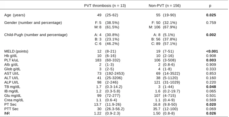
Table 1. Demographic characteristics.
PVT thrombosis (n = 13) Non-PVT (n = 156) p
Age (years) 49 (25-62) 55 (19-90) 0.025
Gender (number and percentage) F: 5 (38.5%) F: 50 (32.1%) 0.759 M: 8 (61.5%) M: 106 (67.9%)
Child-Pugh (number and percentage) A: 4 (30.8%) A: 8 (5.1%) 0.002
B: 3 (23.1%) B: 56 (37.8%) C: 6 (46.2%) C: 89 (57.1%)
MELD (points) 12 (8-21) 19 (7-51) <0.001
Hb g/dL 10 (6-16) 10 (2-16) 0.908
PLT k/uL 183 (60-332) 106 (3-508) 0.003
Alb g/dL 2 (1-3) 2 (0.8-6) 0.909
Glob g/dL 3 (2-5) 4 (1-8) 0.333
AST UI/L 73 (192-2453) 69 (14-3522) 0.853
ALT UI/L 41 (25-3206) 38 (5-1120) 0.160
ALP UI/L 98 (2-246) 121 (31-1029) 0.220
TB mg/dL 1.7 (0.3-14.2) 3 (1-44) 0.048
IB mg/dL 1.2 (0.3-5.8) 1.6 (0.2-19.7) 0.065
Glu mg/dL 99 (72-277) 107 (4-715) 0.501
Crea mg/dL 1.1 (0.6-4) 1.1 (0.4-9) 0.569
PT Sec 13.7 (11.9-26) 16.8 (9.8-50) 0.020
PTT Sec 30 (26.3-56.2) 35.7 (12-100) 0.069
INR 1.22 (0.9-2.3) 1.50 (0.8-8) 0.026
divided in two groups, patients with PVT and patients without PVT. The patients with PVT were younger (49 vs.
55 years p = 0.025), and male gender was predominant in both groups (8 [61.5%] and 106 [67.9%], respectively) without being statistically significant (Table 1). The pre-dominant etiology of cirrhosis was alcohol in both groups (Table 2). Child A was more frequent in the PVT group (4 [30.8%] vs. 8 [5.1%]), and Child C in the non-PVT group (6 [46.2%] vs. 89 [57.1%]). MELD score mean was lower among PVT patients (12 [8-21] vs. 19 [7-51] p= <0.001). Prothrombin time, INR and total bilirubin also were low-er in PVT patients. Finally, platelet levels wlow-ere highlow-er in PVT patients (183,000 [60,000-332,000] vs. 106 [3,000-508,000] p= 0.003).
Clinical outcome
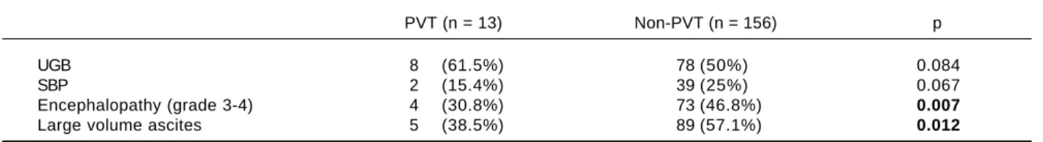
We performed Kaplan-Meier analysis for upper gas-trointestinal bleeding, spontaneous bacterial peritonitis, encephalopathy, large volume ascites and global survival. The frequency of upper gastrointestinal bleeding (8 [61.5%] vs. 78 [50%]) and spontaneous bacterial peritonitis (2 [15.4%] vs. 39 [25%]) was not statistically different be-tween groups (p = 0.84 and p = 0.67 respectively). How-ever, the rate of encephalopathy (grade 3-4) requiring inhospital treatment was better in the PVT group (4 [30.8%] vs. 73 [46.8%] p= 0.007). Large volume ascites was also less frequent in the PVT group (5 [38.5%] vs. 89 [57.1%] p= 0.012) (Table 3). One patient (8%) in PVT and 56 (36%) in non-PVT died during follow up (p = 0.063). Global survival was better for patients with PVT (16.5 ± 27.9 vs. 4.13 ± 12.2 months p = 0.005) (Figure 1).
DISCUSSION
Acute decompensating precipitants that lead to organ failure in patients with cirrhosis have a mortality of 30%.10
Importantly, mortality is higher in previously compensat-ed patients than in those with previous decompensation. Precipitating factors such as infections, PVT, surgery, and hepatocellular carcinoma generally trigger decompensat-ing events.11 Several factors can lead to the development of
PVT in patients with cirrhosis, primarily from a reduction in portal blood flow and hypercoagulability.12
The aim of this study was to assess the relevance on clinical outcome in cirrhotic patients with PVT not asso-ciated with malignancy and its prevalence. In Mexico, to our knowledge, there are no studies assessing this out-come. The prevalence in our study was 7.6%, similar to the range previously reported in the literature.5,13,14 Contrary
to previous studies,15-17 we found that patients with PVT
are younger.
Male gender and alcohol etiology were predominant, similar to what has been previously found in our coun-try.18,19 This is due to the trends of alcohol consumption
in Mexico.19 We found more patients being classified as
cryptogenic cirrhosis compared to the literature,9,16,20 but
in our country, most of the patients are evaluated without specialized studies due to economic burden, therefore there could be less patients with this diagnosis. The pa-tients in the PVT group had lower Child-Pug and MELD score compared with the patients in non-PVT. This could be explained in part because patients with non-PVT are more likely to visit a hospital due to symptomatology caused by cirrhosis complications following poor hepatic
Table 3. Frequency of clinical outcomes.
PVT (n = 13) Non-PVT (n = 156) p
UGB 8 (61.5%) 78 (50%) 0.084
SBP 2 (15.4%) 39 (25%) 0.067
Encephalopathy (grade 3-4) 4 (30.8%) 73 (46.8%) 0.007
Large volume ascites 5 (38.5%) 89 (57.1%) 0.012
* Some patients had 2 or more complications.
Table 2. Etiology of cirrhosis.
Etiology Global (n = 169) PVT (n = 13) Non-PVT (n = 156)
Alcohol 98 (58%) 6 (46.2%) 92 (59%)
Viral 10 (5.9%) 1 (7.7%) 9 (5.8%)
Autoimmune 11 (6.5%) 3 (23.1%) 8 (5.1%)
Drug 4 (2.4%) 0 4 (2.4%)
Cryptogenic 34 (20.1%) 2 (15.4%) 32 (20.5%)
NASH 6 (3.6%) 1 (7.7%) 5 (3.2%)
reserve. However, we did not explore the presence of abdominal pain because of the retrospective design. Nonetheless, the MELD score in the patients in our group without PVT was significantly higher compared to previous studies.9,15,20 PT, INR and total bilirubin were
lower in the PVT group while the platelet was count high-er, this could be due to the state of advanced disease in pa-tients without PVT.
Upper gastrointestinal bleeding (UGB) did not differ between groups, even though it has been described as a common presentation among patients with PVT;7,21 thus,
we do not consider PVT a risk factor for UGB as previ-ously reported.7 The frequency of spontaneous bacterial
peritonitis was the same, and to date, there is only a case report describing this finding in patients with PVT.22
En-cephalopathy requiring in-hospital treatment was more frequent in patients without PVT contrasting previous studies.9,17 This could be explained due to the disparity
of MELD score in both groups. In our study, we did not assess recanalization after diagnosis of PVT; these pa-tients have been described to achieve less frequently he-patic encephalopathy.23 Nonetheless, it has been
proposed that patients with PVT develop high levels of ammonia due to porto-systemic shunts leading to sub-clinical neurological abnormalities compatible with minimal hepatic encephalopathy.24 Contrary to our
find-ings, these patients do not seem to be at more risk for presenting hepatic encephalopathy. Large volume ascites was also more frequent in non-PVT patients. Moderate ascites has been described previously in patients with PVT;9,15 however, we believe this finding could be the
result of advanced liver stage in the non-PVT group. Global mortality was less for patients with PVT, a finding consistent with previous reports.9,15
Recently, Hugenholtz, et al. remarked the importance of anticoagulation in patients with cirrhosis in different settings including PVT;25 however, we found that PVT
by itself does not carry a worse prognosis in terms of survival, UGB, SBP, large volume ascites or encephalop-athy, even if it does not receive anticoagulation treat-ment.8 The reliable variable to predict outcomes remains
the MELD score. Most of our patients had MELD scores above 14 and all of our findings could be explained by di-minished hepatic function. The presence of PVT could be just an epiphenomenon and not a marker of advanced liver disease.
To our knowledge, this is the first study to report the prevalence of PVT in Mexico, and confirms many findings described previously about PVT. However, our study also has some limitations. It is a single center retrospective study, we did not assess recanalization of PVT and we did not have the information of mutations that predispose to thrombosis.4,17 This study opens the scenario to calculate
the size for prospective studies and randomized clinical trials to evaluate the benefits and harms of anticoagulation treatment.
ABBREVIATIONS
• ALP: alkaline phosphatase. • ALT: alanine aminotransferase. • AST: aspartate aminotransferase.
• MEDL: Model for End Stage Liver Disease. • PVT: portal vein thrombosis.
• UGB: upper gastrointestinal bleeding.
CONFLICT OF INTEREST
None.
FINANCIAL SUPPORT
None.
Figure 1. Figure 1.Figure 1.
Figure 1.Figure 1. Survival in months.
1.0
0.8
0.6
0.4
0.2
0.0
0 20 40 60 80 100
Months
Survival 1.0
Months 0 1-20 21-40 41-60 61-80 81-100
PVT (no at risk) 13 8.5 2.5 1.5 1 0.5 Non-PVT (no at risk) 156 110.5 11 3.5 2 1
PVT (no. of events) 0 1 0 0 0 0
Non-PVT (no. of events) 0 51 3 0 1 1
p = 0.005
Non-PVT PVT
REFERENCES
1. Scaglione S, Kliethermes S, Cao G, Shoham D, Durazo R, Luke A, Volk ML. The Epidemiology of Cirrhosis in the United States: A Population-based Study. J Clin Gastroenterol 2015; 49: 690-6.
2. SINAS. Mortalidad. Información tabular. Available from: http://sinais.salud.gob.mx/mortalidad/: 2008; 2008 [updated 2011; cited 2015 03/05/2015].
3. Schuppan D, Afdhal NH. Liver cirrhosis. Lancet 2008; 371: 838-51.
4. Valla DC, Rautou PE. The coagulation system in patients with end-stage liver disease. Liver Int 2015; 35(Suppl. 1): 139-44. 5. Nonami T, Yokoyama I, Iwatsuki S, Starzl TE. The incidence of portal vein thrombosis at liver transplantation. Hepatology 1992; 16: 1195-8.
6. Ogren M, Bergqvist D, Bjorck M, Acosta S, Eriksson H, Sternby NH. Portal vein thrombosis: prevalence, patient characteristics and lifetime risk: a population study based on 23,796 consecutive autopsies. World J Gastroenterol 2006; 12: 2115-9.
7. Amitrano L, Guardascione MA, Brancaccio V, Margaglione M, Manguso F, Iannaccone L, Grandone E, et al. Risk factors and clinical presentation of portal vein thrombosis in patients with liver cirrhosis. J Hepatol 2004; 40: 736-41.
8. John BV, Konjeti R, Aggarwal A, Lopez R, Atreja A, Miller C, Zein NN, et al. Impact of untreated portal vein thrombosis on pre and post liver transplant outcomes in cirrhosis. Ann Hepatol 2013; 12: 952-8.
9. Berry K, Taylor J, Liou IW, Ioannou GN. Portal vein thrombo-sis is not associated with increased mortality among patients with cirrhosis. Clin Gastroenterol Hepatol 2015; 13: 585-93. 10. Moreau R, Jalan R, Gines P, Pavesi M, Angeli P, Cordoba J, Durand F, et al. Acute-on-chronic liver failure is a distinct syn-drome that develops in patients with acute decompensation of cirrhosis. Gastroenterology 2013; 144: 1426-37, 37 e1-9. 11. Tsochatzis EA, Bosch J, Burroughs AK. Liver cirrhosis.
Lan-cet 2014; 383:1749-61.
12. Raja K, Jacob M, Asthana S. Portal vein thrombosis in cirrho-sis. J Clin Exp Hepatol 2014; 4: 320-31.
13. Englesbe MJ, Schaubel DE, Cai S, Guidinger MK, Merion RM. Portal vein thrombosis and liver transplant survival benefit. Liver Transpl 2010; 16: 999-1005.
14. Ponziani FR, Zocco MA, Senzolo M, Pompili M, Gasbarrini A, Avolio AW. Portal vein thrombosis and liver transplan-tation: implications for waiting list period, surgical ap-proach, early and late follow-up. Transplant Rev 2014; 28: 92-101.
15. Girleanu I, Stanciu C, Cojocariu C, Boiculese L, Singeap AM, Trifan A. Natural course of nonmalignant partial portal vein thrombosis in cirrhotic patients. Saudi J Gastroenterol 2014; 20: 288-92.
16. Chung JW, Kim GH, Lee JH, Ok KS, Jang ES, Jeong SH, Kim JW. Safety, efficacy, and response predictors of anticoagu-lation for the treatment of nonmalignant portal-vein thrombo-sis in patients with cirrhothrombo-sis: a propensity score matching analysis. Clin Mol Hepatol 2014; 20: 384-91.
17. Gabr MA, Bessa SS, El-Zamarani EA. Portal vein thrombosis in Egyptian patients with liver cirrhosis: Role of methylenetet-rahydrofolate reductase C677T gene mutation. Hepatol Res 2010; 40: 486-93.
18. Gonzalez-Gonzalez JA, Garcia-Compean D, Vazquez-Elizondo G, Garza-Galindo A, Jaquez-Quintana JO, Maldona-do-Garza H. Nonvariceal upper gastrointestinal bleeding in patients with liver cirrhosis. Clinical features, outcomes and predictors of in-hospital mortality. A prospective study. Ann Hepatol 2011; 10: 287-95.
19. Roman S, Zepeda-Carrillo EA, Moreno-Luna LE, Panduro A. Alcoholism and liver disease in Mexico: genetic and environ-mental factors. World J Gastroenterol 2013; 19: 7972-82. 20. Luca A, Caruso S, Milazzo M, Marrone G, Mamone G, Crino
F, Maruzzelli L, et al. Natural course of extrahepatic nonma-lignant partial portal vein thrombosis in patients with cirrho-sis. Radiology 2012; 265: 124-32.
21. Farmer AD, Saadeddin A, Holt CE, Bateman JM, Ahmed M, Syn WK. Portal vein thrombosis in the district general hospi-tal: management and clinical outcomes. Eur J Gastroenterol Hepatol 2009; 21: 517-21.
22. Eleftheriadis N, Makris P. Portal vein thrombosis in a patient with HCV cirrhosis and combined hemophilia A and throm-bophilia V Leiden. Ther Clin Risk Manag 2010; 6: 539-41. 23. Delgado MG, Seijo S, Yepes I, Achecar L, Catalina MV,
Gar-cia-Criado A, Missing author, et al. Efficacy and safety of anticoagulation on patients with cirrhosis and portal vein thrombosis. Clin Gastroenterol Hepatol 2012; 10: 776-83. 24. Minguez B, Garcia-Pagan JC, Bosch J, Turnes J, Alonso J,
Rovira A, Córdoba J. Noncirrhotic portal vein thrombosis exhibits neuropsychological and MR changes consistent with minimal hepatic encephalopathy. Hepatology 2006; 43: 707-14.
25. Hugenholtz GC, Northup PG, Porte RJ, Lisman T. Is there a rationale for treatment of chronic liver disease with anti-thrombotic therapy? Blood Rev 2015; 29: 127-36.
Correspondence and reprint request: Emmanuel I. González-Moreno, MD Gastroenterology Division. Hospital Universitario
“Dr. José Eleuterio González”.
Universidad Autónoma de Nuevo León. Monterrey, México. Madero y Gonzalitos S/N, Monterrey,
Nuevo León, México. 64460 México Tel. and Fax: +011 52 (81) 8333-3664