current perspectives
Rosa E Quiroz-Castañeda, " Jorge L Folch-Mallol
Laboratorio de Biología Molecular de Hongos, Centro de Investigación en Biotecnología,Universidad Autónoma del Estado de Morelos Ave. Universidad 1001 Col. Chamilpa, Cuernavaca 62209, Morelos, México E-mail: [email protected]
ABSTRACT
Lignocellulose constitutes a raw material with considerable potential for the production of fermentable sugars and the generation of biofuels. In nature, lignocellulosic waste from forestry, agriculture and gardening acts as the pre-ferred carbon source of a number of bacteria and fungi endowed with the required ligninolytic machinery. These hydrolytic activities could potentially be complemented with those from other proteins that remodel the structure of cell walls, such as expansins, a group of proteins originally identified in plants that have the capacity of relaxing cell wall tension to allow cell growth. Expansins participate in processes where remodeling of the plant cell wall is required: organogenesis, fruit ripening, and growth of the pollen tube, among others. Expansins and expansin-like proteins have been proposed to act by disrupting the hydrogen bonds binding together cellulose fibrils and cellulose and other polysaccharides through a non-enzymatic process, enhancing subsequent degradation. In this manuscript, a review on plant cell wall composition and ligninolytic enzymes from cell wall-degrading bacteria and fungi is pre-sented. Proteins with expansin-like activity, their properties and their potential application to enhance sugar release from lignocellulosic material are also reviewed.
Keywords: lignocellulose, cellulases, expansins, fungi, biofuels
Biotecnología Aplicada 2011;28:205-215
RESUMEN
Proteínas que remodelan y degradan la pared celular vegetal: perspectivas actuales. El material
ligno-celulósico constituye una materia prima potencial para la obtención de azúcares fermentables y biocombustibles. Algunas bacterias y hongos con cualidades ligninolíticas, pueden utilizar los desechos lignocelulósicos de la naturaleza (forestales, agrícolas y de jardín) como fuente de carbono. Tal actividad de degradación podría complementarse con la actividad de las proteínas que remodelan la pared celular, como las expansinas, identificadas en plantas. Estas proteínas pueden relajar los componentes de la pared celular y promover el crecimiento. Participan en procesos de desarrollo como la organogénesis, la abscisión, la maduración de los frutos, el crecimiento del tubo polínico, entre otros, donde ocurren modificaciones de la pared celular. Se ha planteado que las proteínas del tipo expan-sinas rompen los puentes de hidrógeno que unen los filamentos de celulosa y la celulosa con otros polisacáridos, mediante un proceso no enzimático, que favorece la posterior degradación de la pared celular. En este trabajo se hace una revisión bibliográfica acerca de las características de la pared celular vegetal y su composición, así como de las enzimas ligninolíticas de bacterias y hongos que la degradan, las propiedades y el potencial que tienen las proteínas con actividad tipo expansina para hacer más eficiente la liberación de azúcares reductores del material lignocelulósico.
Palabras clave: lignocelulosa, celulasas, expansinas, hongos, biocombustibles
I
ntroduction
Lignocellulose, the principal and most abundant component of the renewable biomass produced by photosynthesis, is synthesized at an estimated rate of some 200 billion tons per year [1-2]. This biopoly-mer is formed principally by cellulose, hemicelluloses and lignin [1], in addition to small amounts of pectin, ash and proteins [2]. Cellulose is the most abundant polymer of our planet. In nature, it is found mainly as a structural component of plant and algal cell walls, although cellulose is also produced by some animals, such as tunicates, and several bacteria [3]. It does not accumulate in the environment, due to the existence of cellulolytic fungi and bacteria degrading some of the components of plant cell walls; a process that, while extremely slow, plays a major role in carbon recycling within the biosphere [4]. Obviously, the existence of formidable reserves of lignocellulose that are continu-ously renewed represents an excellent opportunity for
the development of biofuels, as a sustainable alterna-tive to the fossil fuels currently in use [5].
Exploitation of cellulosic biomass is, however, cur-rently limited by the absence of low-cost technologies for its processing. A promising strategy for this pur-pose is the use of cell wall remodeling and degrading enzymes from ligninolytic organisms [3].
S
tructure of the cell wall
and composition of lignocellulose
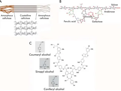
The cell wall is a highly ordered structure formed, mainly, by cellulose, hemicelluloses and lignin, a phe-nolic polymer (Figure 1) [6]. Exact identity and rela-tive abundances of each of these polymers vary even within the same plant, depending on age, tissue and growth stage [1]. Cell walls are structured so as to en-able them to play a wide array of disparate, sometimes opposing roles. They provide resistance tomechani-Zhang YH, Lynd LR. Toward an ag-1.
gregated understanding of enzymatic hydrolysis of cellulose: noncomplexed cel-lulase systems. Biotechnol Bioeng. 2004; 88(7):797-824.
Zhong R, Ye ZH. Regulation of cell wall 2.
biosynthesis. Curr Opin Plant Biol. 2007; 10(6):564-72.
Lynd LR, Weimer PJ, van Zyl WH, Preto-3.
rius IS. Microbial cellulose utilization: fun-damentals and biotechnology. Microbiol Mol Biol Rev. 2002;66(3):506-77.
Aro N, Pakula T, Penttilä M. Transcrip-4.
tional regulation of plant cell wall degrada-tion by filamentous fungi. FEMS Microbiol Rev. 2005;29(4):719-39.
Gray KA, Zhao L, Emptage M. Bio-5.
ethanol. Curr Opin Chem Biol. 2006;10(2): 141-6.
cal stress, shape the cell and protect it against many pathogens; at the same time, they must be reason-ably fl exible to withstand shear forces, and permeable enough to allow the passage of signaling molecules into the cell [7].
C
ellulose
Cellulose, the most abundant natural polymer, is highly stable and insoluble in water. It constitutes the princi-pal component of plant cell walls, accounting for 50% of the dry weight of wood. Its degree of polymeriza-tion varies according to its origin, and may range from 2000 to 25,000 monomers [7-8]. Cellulose is formed by D-glucose monomers condensed together through β-1,4 glycosidic bonds, forming cellobiose molecules (β-1,4-linked glucose dimer) that are, in turn, linked together into straight, non-branched chains [9]. In nature cellulose is seldom found as single, isolated chains; forming fi laments instead from the very mo-ment it is synthesized. These fi laments, denominated microfi brils, may contain from 36 to over 1200 cel-lulose chains, and have diameters of 5 to 15 nm [7]. Cellulose chains are held together in the microfi bril through hydrogen bonds and Van der Waals forces, forming a crystalline, organized structure that is re-fractory to hydrolysis in certain areas of the microfi bril (Figure 2A) [9]. Highly ordered, crystalline regions are interspersed with regions containing disorganized or amorphous cellulose, which constitute 5 to 20% of the microfi bril. Amorphous regions are more sus-ceptible to enzymatic degradation [10-11]. Cellulose is a highly resistant substrate that is, in turn, tightly associated with hemicelluloses and lignin, forming a structure that is very resistant to degradation. Degrad-ing lignin, therefore, is a feat accomplished only by a few cellulolytic organisms [8].
H
emicellulose
Hemicellulose constitutes from 25 to 30% of wood by dry weight. It is a complex heteropolysaccharide composed mainly of pentoses (D-xylose and L-ara-binose) and hexoses (glucose, mannose and D-galactose), usually acetylated and forming branched chains, in addition to 4-O-methylglucuronic, D-galac-turonic and D-glucuronic acids, condensed through β-1,4 and, occasionally, β-1,3 glycosidic linkages [9]. These short lateral branches, formed by different sug-ars, make hemicellulose less refractory to a number of treatments (Figure 2B) [9, 12]. The components of hemicellulose are also classifi ed as xylans, xyloglu-cans, mannans, glucomannans and gluxyloglu-cans, bonded together through β-1,3 or β-1,4 linkages. Xylans are the most abundant component of hemicellulose (over 70% by weight). They are formed by D-xylose units condensed through β-1,4 linkages, and may carry different substitutions, originating arabinoxylans (if substituted with arabinose), such as those found in grasses, or glucuronoxylans and glucuronoarabinoxy-lans (if substituted with glucose or glucose-arabinose, respectively), which represent the main constituents of the secondary wall of dicotyledonous plants. In addition to xylose, xylans may contain arabinose, glucuronic acid or 4-O-methyl ether-glucuronic acid, and acetic, ferulic or p-coumaric acids. Exact compo-sition and branching frequency depend on the origin
Golgi
Cellulose
Pectin
Lignin
Hemicellulose
Xyloglucan Arabinoxylan
Rhamnogalacturonan I
Xylogalacturonan Homogalacturonan
Arabinan
Rhamnogalacturonan II
Vesicles
Cytoplasm
Plasma membrane Cellulose synthase
complex (CESA)
Figure 1. Composition of the plant cell wall. Formed basically the polysaccharides cellulose, hemice-llulose and pectin together with lignin, a phenolic polymer. Lignin forms a matrix where the polysac-charides are embedded. Modified from Cosgrove [63].
A B
Amorphous
cellulose Crystalline cellulose Amorphous cellulose
Ferulic acid Galactose
Arabinose Xylose
Glucose
C
Coumaryl alcohol
Sinapyl alcohol
Coniferyl alcohol
of hemicellulose [13]. In hard woods from deciduous trees such as poplar, birch and elm, hemicellulose is formed mainly by xylans where 60 to 70% of residues are acetylated, whereas the soft woods of conifers such as pine and cedar sport hemicelluloses composed mainly of glucomannans [14].
The mannans and galactomannans of hemicellu-lose have a core structure of β-1,4-linked mannose residues, which is randomly branched with mannose and glucose residues in glucomannans. There are structural differences between hemicelluloses from different species and even different cell types in the same individuals [14-15]. The most important role of hemicellulose is to bond together lignin and cellulose
fi bers, thus providing rigidity to the cellulose-hemi-cellulose-lignin mesh. Lignin and hemicellulose are linked together mainly by ester bonds between ara-binose residues in hemicellulose and hydroxyl groups in lignin residues, whereas cellulose binds to hemicel-lulose through hydrogen bonds [16].
L
ignin
Lignin is one of the most abundant polymers in na-ture after cellulose and hemicellulose. It is highly re-sistant to chemical or biological degradation, provid-ing structural support to the cell wall, decreasprovid-ing its permeability and conferring resistance to the attack of microorganisms. Together, lignin and hemicellulose form an amorphous matrix imbibing cellulose fi bers to protect them from degradation [17].
Structurally, lignin is a water-insoluble, irregular, branched heteropolymer formed through the polymer-ization of three phenylpropane-type aromatic alcohols (coumaryl, coniferyl and sinapyl alcohols) through C-C bonds and esters involving the aromatic rings. This polymer, with constitutes 20 to 30% of wood by weight, protects and confers rigidity to the structural polysaccharides (cellulose and hemicellulose) (Figure 2C) [4, 18-19]. The main constituent of lignin in soft woods is coniferyl alcohol; in hard woods, this place is occupied by coumaric and sinapinic acids instead [12]. Lignin is the component of lignocellulosic ma-terial exhibiting the highest resistance to degradation, which limits its application and that of the polysac-charides it protects. The number of microorganisms able to mineralize this substance is really small [18].
L
ignocellulose-degrading
microorganisms
There are natural organisms that can degrade lignocel-lulose. Some of these are aerobic cellulolytic bacteria of the Actinomycetales order (Phylum Actinobacteria)
living in soils, water, humus, agricultural waste such as sugarcane bagasse and decaying leaves [20]. En-zyme systems composed of cellulases and xylanases capable of degrading cell wall components have been described in aerobic bacteria such as Pseudomonas fl uorescens subsp. cellulosa, Streptomyces lividans
and Cellulomonas fi mi [21-23]. Anaerobic bacteria of
the Clostridiales order (Phylum Firmicutes)
gener-ally found in soils, decaying plant waste, the rumen of ruminant animals, termite guts, compost, waste water and wood processing plants, also contain cellulolytic enzyme complexes denominated cellulosomes [20]. Some cellulolytic anaerobic bacteria, such as
Butyr-ivibrio fi brisolvens, Fibrobacter succinogenes, Rumi-nococcus fl avefaciens, Clostridium cellulovorans, C. cellulolyticum y C. thermocellum are also endowed with cellulases and xylanases [24-26].
Extreme environments also host a number of cel-lulolytic microorganisms, such as the Antarctic bacte-rium Pseudoalteromonas haloplanktis [27].
Among fungi, the most effi cient at using wood as substrate are the basidiomycetes, considered the principal taxonomic group involved in the degra-dation of wood with all its components [3, 17, 28]. However, the ability to utilize lignocellulosic mate-rial is widely distributed among fungi, from chytri-diomycetes to basichytri-diomycetes. The chytrichytri-diomycetes include a number of anaerobic species living in the gastrointestinal tract of ruminants [29]. Anaeromy-ces, CaecomyAnaeromy-ces, Neocallimastix, Orpinomyces and Piromyces constitute the fi ve most studied genera
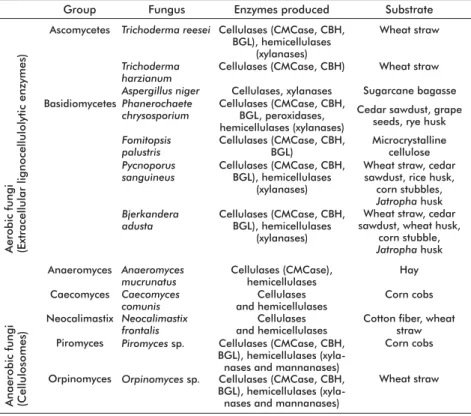
of anaerobic fungi [30]. Unlike aerobic fungi, their anaerobic counterparts are often endowed with large multienzyme complexes of cellulases and hemicellu-lases similar to those of bacterial cellulosomes [31-32] (Table 1). Examining the taxonomic composition of cellulolytic fungi inhabiting the decaying leaves and rotting woods of forest soils, zygomycetes are represented by a single genus, Mucor, while
asco-mycetes and basidioasco-mycetes are represented by gen-era such as Chaetomium, Trichoderma, Aspergillus, Fusarium, Coriolus, Phanerochaete, Schizophyllum, Volvariella, Pycnoporus and Bjerkandera [3, 33-35].
Two of the most studied fungi, due to their industrial relevance, are Trichoderma reesei and Phanerochaete chrysosporium [20].
Table 1. Some aerobic and anaerobic cellulolytic fungi, enzymes they produce and subs-trates they degrade. Compiled from Dashtban M, Schraft H, Qin W [28] and Quiroz-Castañeda RE, et al. [33]
Aerobic fungi (Extracellular lignocellulolytic enzymes)
Anaerobic fungi (Cellulosomes)
Ascomycetes
Basidiomycetes
Anaeromyces
Caecomyces
Neocalimastix
Piromyces
Orpinomyces
Trichoderma reesei
Trichoderma harzianum Aspergillus niger Phanerochaete chrysosporium
Anaeromyces mucrunatus Fomitopsis palustris
Caecomyces comunis Pycnoporus sanguineus
Neocalimastix frontalis Bjerkandera adusta
Piromyces sp.
Orpinomyces sp.
Wheat straw
Wheat straw
Sugarcane bagasse Cedar sawdust, grape
seeds, rye husk
Hay Microcrystalline
cellulose
Corn cobs Wheat straw, cedar sawdust, rice husk, corn stubbles, Jatropha husk
Cotton fiber, wheat straw Wheat straw, cedar sawdust, wheat husk,
corn stubble, Jatropha husk
Corn cobs
Wheat straw
Substrate
Cellulases (CMCase, CBH, BGL), hemicellulases
(xylanases) Cellulases (CMCase, CBH)
Cellulases, xylanases Cellulases (CMCase, CBH,
BGL, peroxidases, hemicellulases (xylanases)
Cellulases (CMCase), hemicellulases Cellulases (CMCase, CBH,
BGL)
Cellulases and hemicellulases Cellulases (CMCase, CBH,
BGL), hemicellulases (xylanases)
Cellulases and hemicellulases Cellulases (CMCase, CBH,
BGL), hemicellulases (xylanases)
Cellulases (CMCase, CBH, BGL), hemicellulases
(xyla-nases and manna(xyla-nases) Cellulases (CMCase, CBH, BGL), hemicellulases
(xyla-nases and manna(xyla-nases)
Enzymes produced Fungus
Group
Fry SC. Plant Cell Walls. In: Encyclo-6.
pedia of Life Sciences [Internet]. Chich-ester: John Wiley & Sons Ltd: 2001 Apr [cited 2011 May 17]. Available in: http:// www.els.net/WileyCDA/ElsArticle/refId-a0001682.html
Levy I, Shani Z, Shoseyov O. Modifi-7.
cation of polysaccharides and plant cell wall by endo-1,4-beta-glucanase and cellulose-binding domains. Biomol Eng. 2002;19(1):17-30.
Hildén L, Johansson G. Recent devel-8.
opments on cellulases and carbohydrate-binding modules with cellulose affinity. Biotechnol Lett. 2004;26(22):1683-93.
Pérez J, Muñoz-Dorado J, de la Rubia T, 9.
Martínez J. Biodegradation and biological treatments of cellulose, hemicellulose and lignin: an overview. Int Microbiol. 2002; 5(2):53-63.
Atalla R. The Structures of Native Cel-10.
luloses. 10th international symposium on wood and pulping chemistry. TAPPI Press. 1993;1:608-14.
Béguin P, Aubert JP. The biological 11.
degradation of cellulose. FEMS Microbiol Rev. 1994;13(1):25-58.
Martínez AT, Speranza M, Ruiz-Dueñas 12.
FJ, Ferreira P, Camarero S, Guillén F, et al. Biodegradation of lignocellulosics: micro-bial, chemical, and enzymatic aspects of the fungal attack of lignin. Int Microbiol. 2005;8(3):195-204.
Saha BC. Hemicellulose bioconversion. 13.
E
nzymes involved in the hydrolysis
of lignocellulose
Cellulases are O-glucoside hydrolases (GH) hydro-lyzing the β-1,4 linkages of cellulose. They are pre-dominantly found among prokaryotes and fungi [8]. More than a dozen fungal species producing cellulases have been described, including Trichoderma viride, T. harzianum, T. atroviride, T. reesei, Fusarium solani, Aspergillus niger, A. terreus, and P. chrysosporium,
well known for their cellulolytic abilities[18]. Cel-lulase genes have also been identifi ed in the marine yeast Aureobasidium pullulans [36]. Curiously, a
cel-lulase gene family (GH45), perhaps acquired through horizontal gene transfer, has been found in Bursaph-elenchus xylophilus, a nematode infecting pine wood,
[37]. GH are classifi ed into cellulase families on the basis of aminoacid sequence similarity [38]. Out of the currently existing 122 families, 14 correspond to cellulases. Most cellulases, together with other gluco-side hydrolases, have a structure comprised of a cata-lytic module, a highly O-glycosylated linker, and a cellulose-binding module (CBM) [3]. This last domain facilitates cellulose hydrolysis by holding the catalytic module in close proximity to its substrate [39]. Cel-lulases are classifi ed, depending on their enzymatic activity, in three major groups: exoglucanases, endo-glucanases and β-glucosidases; some of which have been crystallized, enabling the determination of their tri-dimensional structure [40].
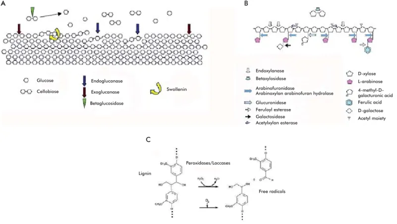
Exoglucanases or cellobiohydrolases (CBH) (EC 3.2.1.74; 1,4-β-D-glucan-glucanhydrolase and EC 3.2.1.91; 1,4-β-D-glucan-cellobiohydrolase) catalyze the successive hydrolysis of residues from the reduc-ing and non-reducreduc-ing ends of the cellulose polysaccha-ride, releasing cellobiose molecules as main product of the reaction [4]. These enzymes account for 40 to 70% of the total component of the cellulase system, and are able to hydrolyze crystalline cellulose (Figure 3A). They are monomeric proteins with a molecular weight ranging from 50 to 65 kDa, although there are smaller variants (41.5 kDa) in some fungi, such as Sclerotium rolfsii [41]. Exoglucanases have low levels of
glycosy-lation (from 12% to none at all), their optimum pH is 4 to 5, and their optimum temperature varies from 37 to 60 °C, depending on the specifi c enzyme-substrate combination [42-43]. Exoglucanases form part of the cellulolytic arsenal of the fungi causing white and soft rot and the plant pathogen S. rolfsii, but are found only
in some of the basidiomycetes causing the brown rot, such as Fomitopsis palustris [44].
Endoglucanases (EG) (EC 3.2.1.4; 1,4-β -D-glucan-4-glucanhydrolase) randomly cleave internal linkages in amorphous cellulose fi laments, generating ran-domly sized oligosaccharides and creating new chain ends that can in turn be attacked by exoglucanases [4]. Available evidence indicates that these are the en-zymes that initiate the cellulolytic process, randomly cleaving internal linkages at amorphous regions of
Glucose A
A B
Cellobiose
Endoglucanase
Endoxylanase D-xylose
L-arabinose 4-methyl-D-galacturonic acid Ferulic acid D-galactose Acetyl moiety Exoglucanase
Betaxylosidase
Swollenin
Betaglucosidase
C
Lignin
Peroxidases/Laccases
Free radicals Arabinofuronidase
Glucuronidase Feruloyl esterase Galactosidase Acetylxylan esterase
Arabinoxylan arabinofuran hydrolase
Figure 3. Enzymes participating in the degradation of cell wall components. A) Degradation of cellulose. Three types of cellulolytic activities synergize to degrade this material efficiently. Swollenin activity might enhance the efficiency of the process by relaxing the structure of cellulose in a non-hydrolytic manner. B) Degradation of hemicellulose. Endoxylanases and xylosidases are the main enzymes involved in this process. Additional accessory enzymes are required to degrade the side branches of the heteropolysaccharide. C) Degradation of lignin. The process involves laccases and peroxidases catalyzing oxidative reactions and generating highly unstable free radicals that favor further depolymerization. Images taken from Aro et al. [4].
Kumar R, Singh S, Singh OV. Bio-14.
conversion of lignocellulosic biomass: biochemical and molecular perspectives. J Ind Microbiol Biotechnol. 2008;35(5): 377-91.
Scheller H, Ulvskov P. Hemicelluloses. 15.
Annu Rev Plant Biol. 2010;61:263-89.
Laureano-Perez L, Teymouri F, Aliza-16.
deh H, Dale BE. Understanding factors that limit enzymatic hydrolysis of biomass: characterization of pretreated corn stover. Appl Biochem Biotechnol. 2005;121-124: 1081-99.
Sánchez C. Lignocellulosic residues: 17.
biodegradation and bioconversion by fun-gi. Biotechnol Adv. 2009;27(2):185-94.
Cunningham RE, López GD. Etanol de 18.
lignocelulósicos: Tecnología y perspectivas. Santa Fe: Universidad de Santiago de Compostela, Servicio de Publicaciones e Intercambio Científico; 1994.
Hammel KA. Extracellular free radical 19.
biochemistry of ligninolytic fungi. New J Chem. 1996;20:195-8.
Doi RH. Cellulases of mesophilic 20.
microorganisms: cellulosome and noncel-lulosome producers. Ann New York Acad Sci. 2008;1125:267-79.
Khanna S, Gauri. Regulation, purifica-21.
the cellulose fi ber and creating new reducing and non-reducing ends that are susceptible to the action of cel-lobiohydrolases [45]. Endoglucanases are monomeric enzymes with a molecular weight that ranges from 22 to 45 kDa, although some fungi such as S. rolfsii and
Gloeophyllum sepiarium have endoglucanases twice this size [46]. In general, endoglucanases are not gly-cosylated; however, they sometimes may have rela-tively low amounts of carbohydrate (from 1 to 12%) [42]. Optimum pH is usually 4 to 5; the only known endoglucanase with a neutral pH optimum is that from the basidiomycete Volvariella volvacea, expressed in recombinant yeast [47]. Their optimum temperature ranges from 50 to 70 °C [48]. Endoglucanases have been successfully isolated from the basidiomycetes causing white and brown rot, from the plant patho-gen S. rolfsii, from the yeast Rhodotorula glutinis and from the termite symbiont Termitomyces sp. [42].
Exhaustively hydrolyzing cellulose also requires the action of β-glucosidases (BGL) (EC 3.2.1.21), which hydrolyze cellobiose, releasing two molecules of glucose and thereby provide a carbon source that is easy to metabolize [4]. Fungi causing white and brown rot, mycorrhizal fungi, plant pathogens and yeast all produce these enzymes [42]. β-glucosidases have molecular weights ranging from 35 to 640 kDa; they can be monomeric, reaching molecular weights of ap-proximately 100 kDa, or exist as homo-oligomers, as is the case in the yeast Rhodotorula minuta [49]. Most β-glucosidases are glycosylated; in some cases, as that of the 300 kDa BGL from Trametes versicolor, glyco-sylation may be superior to 90%. Their optimum pH ranges from 3.5 to 5.5, and their optimum temperature ranges from 45 to 75 °C [28]. The activity of cellu-lase enzyme systems is much higher than the sum of the activity of its individual subunits; a phenomenon known as synergism [3]. Cellulase systems are not just simply a conglomerate of enzymes with components from all three cellulase types, but act coordinately to effi ciently hydrolyze cellulose fi bers [3].
Hemicellulose is degraded into monosaccharides and acetic acid. Xylans, the main carbohydrate of hemicellulose, require the coordinated action of sev-eral hydrolytic enzymes, such as xylanases and acces-sory proteins, for their degradation [9]. Most hemi-cellulases are glycoside hydrolases, although some of them are carbohydrate esterases hydrolyzing the ester bonds linking acetate or ferulic acid with branched sugars.
Xylanases are the main enzymes participating in the degradation of hemicellulose. This group includes the endoxylanases (EC 3.2.1.8; endo-1,4-β-D-xylanases) which act on the main carbohydrate chain, hydrolyz-ing the linkages between xylan units and releashydrolyz-ing oligosaccharides.
β-xylosidases (EC 3.2.1.37; xylan 1,4-β-xylosidase) release xylose by cleaving the bonds of xylan oligo-saccharides [4, 9]. Degrading hemicellulose also re-quires accessory enzymes such as xylan esterases, fer-ulic and coumaric esterases, α-arabinofuranosidases and α-4-methyl glucuronosidases, among others, which act in a synergic fashion to hydrolyze hemicel-lulose effi ciently (Figure 3B) [17]. Like cellulases, xylanases also have a modular structure with catalytic and substrate-binding domains, where the fi rst
deter-mines specifi city and reactivity towards the substrate, while the second facilitates binding of the enzyme to the substrate [9]. Genes coding for endoxylanases and β-xylosidases have been cloned from different Asper-gillus species, as well as from Penicillium, Agaricus bisporus and Magnaporthe grisea [50-51].
Hydrolases also comprise carbohydrate esterases, which catalyze the O- and N-deacetylation of sub-strates such as xylan, chitin and some peptidoglycans. There is a carbohydrate esterase in Aspergillus sp., denominated feruloyl esterase, which increases the release of sugars from lignocellulose by removing the ferulic acid residues cross-linking hemicellulose
fi bers, thus destabilizing the structure and making it more susceptible to the action of hydrolytic enzymes [52-53]. It has also been described that the levels of acetyl xylan esterase of the fungus Volvariella volva-cea increase when it is grown in the presence of oat xylan, chitin, cellulose, cellobiose, lactose or galac-tose as carbon source [54]. Selig et al. [55] demon-strated that both esterase and xylanase activities are capable of improving the effi cacy of a cellobiohydro-lase, acting in synergism to degrade lignocellulosic material.
Lignin depolymerization involves extracellular ox-idative enzymes that release highly unstable products that later undergo oxidation reactions [56]. White rot-causing fungi are the most effi cient organisms regard-ing lignin degradation. They are endowed with perox-idases and laccases that participate in ligninolysis[9]. Two groups of peroxidases have been characterized: lignin peroxidase (LiP; EC 1.11.1.14) and manganese-dependent peroxidase (MnP; EC 1.11.1.13); both are oxidoreductases catalyzing hydrogen peroxide-de-pendent oxidative reactions that involve phenolic and non-phenolic compounds and are essential for lignin degradation [9].
Lignin peroxidase is a glycoprotein with a heme group on its active center. It is the most effective per-oxidase, and can oxidize phenolic and non-phenolic lignin compounds, amines, aromatic ethers and aro-matic polycyclic compounds [17].
Manganese-dependent peroxidase is also a gly-coprotein. It uses manganese as substrate, oxidizing it from Mn2+ to Mn3+. The latter is in turn a strong
oxidant that reacts with phenolic lignin compounds [57]. Biochemical and molecular studies have found a third type of peroxidase, originally described for the
fi rst time in the basidiomycete Pleurotus eryngii [58]. This enzyme has received the name of versatile per-oxidase (VP), as it combines the activities of manga-nese-dependent peroxidases and lignin peroxidases. A PV was found in the fungus Bjerkandera adusta
strain UAMH 8258,where its synthesis is stimulated by exogenously administered organic acids such as glycolate, glyoxalate and oxalate [59]. This enzyme effi ciently oxidizes Mn2+ to Mn3+ andcan oxidize a
number of substrates in the absence of Mn, such as 2,6-dimetoxyphenol, guaiacol, ABTS, 3- hydroxyan-thranilic acid, o-anisin and p-anisidine. Its affi nity for veratryl alcohol is comparable to that of LiP from other fungi [60].
Laccases (EC 1.10.3.1; p-diphenol dioxygen oxi-doreductase) are polyphenol oxidases bearing four copper ions in their active center that catalyze the
Braithwaite KL, Black GW, Hazlewood 22.
GP, Ali BR, Gilbert HJ. A non-modular endo-beta-1,4-mannanase from Pseudomonas fluorescens subspecies cellulosa. Biochem J. 1995;305(Pt 3):1005-10.
Arcand N, Kluepfel D, Paradis FW, 23.
Morosoli R, Shareck F. Beta-mannanase of Streptomyces lividans 66: cloning and DNA sequence of the manA gene and characterization of the enzyme. Biochem J. 1993;290(Pt 3):857-63.
Murty MV, Chandra TS. Purification 24.
and properties of an extra cellular xylanase enzyme of Clostridium strain SAIV. Antonie van Leeuwenhoek. 1992;61(1):35-41.
Lin LL, Thomson JA. An analysis of the 25.
extracellular xylanases and cellulases of Butyrivibrio fibrisolvens H17c. FEMS Micro-biology Letters. 1991;84(2):197-204.
Tomme P, Warren RA, Gilkes NR. Cel-26.
lulose hydrolysis by bacteria and fungi. Adv Microb Physiol. 1995;37:1-81.
Sonan GK, Receveur-Brechot V, Duez 27.
C, Aghajari N, Czjzek M, Haser R, et al. The linker region plays a key role in the adaptation to cold of the cellulase from an Antarctic bacterium. Biochem J. 2007; 407(2):293-302.
Dashtban M, Schraft H, Qin W. Fungal 28.
bioconversion of lignocellulosic residues; opportunities & perspectives. Int J Biol Sci. 2009;5(6):578-95.
Lee SS, Ha JK, Kang HS, Mcallister TA, 29.
Cheng KJ. Overview of energy metabolism, substrate utilization and fermentation characteristics of ruminal anaerobic fungi. Korean J Anim Nutr Feedstuffs. 1997; 21(4):295-314.
Nicholson MJ, Theodorou MK, Brook-30.
man JL. Molecular analysis of the anaero-bic rumen fungus Orpinomyces - insights into an AT-rich genome. Microbiology. 2005;151(Pt 1):121-33.
Eberhardt RY, Gilbert HJ, Hazlewood 31.
GP. Primary sequence and enzymic prop-erties of two modular endoglucanases, Cel5A and Cel45A, from the anaerobic fungus Piromyces equi. Microbiology. 2000;146(Pt 8):1999-2008.
Steenbakkers PJM, Li XL, Ximenes 32.
EA, Arts JG, Chen H, Ljungdahl LG, et al. Noncatalytic docking Domains of cel-lulosomes of anaerobic fungi. J Bacteriol. 2001 Sep;183(18):5325-33.
Quiroz-Castañeda RE, Balcázar-López 33.
E, Dantán-González E, Martinez A, Folch-Mallol J, Martínez C. Characterization of cellulolytic activities of Bjerkandera adusta and Pycnoporus sanguineus on solid wheat straw medium. Electr J Biotechnol. 2009 Oct 15 [cited 2011 May 17];12(4)[about 13 p.]. Available from: http://www.ejbio-technology.cl/content/vol12/issue4/full/3/ index.html
Ding S, Ge W, Buswell JA. Cloning of 34.
multiple cellulase cDNAs from Volvariella volvacea and their differential expression during substrate colonization and fruit-ing. FEMS Microbiol Lett. 2006;263(2): 207-13.
Koseki T, Mese Y, Fushinobu S, Ma-35.
oxidation of many phenolic and non-phenolic com-pounds in the presence of mediators, coupling the reduction of molecular oxygen to water [4]. These enzymes oxidize lignin and generate highly unstable aromatic radicals that favor its depolymerization through breakage of C4 ether bonds, breakage of aro-matic rings and demethoxylation [17] (Figure 3C). They have been found in basidiomycetes such as P. chrysosporium, Pleurotus ostreatus, T. versicolor and
Pycnoporus sanguineus [61].
E
xpansins: plant cell wall-remodeling
proteins
In addition to lignocellulose-degrading enzymes, there are also enzymes involved in remodeling the cell wall, denominated expansins, which facilitate its later degradation. Expansins increase the extensibility and relax the tension of plant cell walls. They were
fi rst identifi ed through studies of the mechanism in-volved in pH-dependent extension of plant cell walls (‘acid growth’) [62]. Plant cell wall pH is usually de-termined by the activity of an H+ ATPase localized
to the plasma membrane, which pumps protons onto the cell wall; it ranges from 5.5 to 4.5. Low extra-cellular pH (lower than 5.5) causes the cell wall to relax, due mainly to the action of expansins, whose optimum pH is acid [63]. McQueen-Mason et al. [62] isolated two proteins from the cell walls of cucumber hypocotyls that induced the extension of previously heat-inactivated cell walls. Cloning and sequencing of expansin genes [64] revealed, after sequence homol-ogy searches in genomic and expressed sequence tags (EST) databases, that expansins are coded by large multigene families present from bryophytes to angio-sperms [65]. Expansins are also present in monocoty-ledonous plants (rice, maize), dicotymonocoty-ledonous plants (Arabidopsis), ferns and mosses [66].
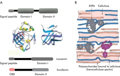
After the discovery of expansins, Cosgrove et al.[67] found that group I allergens from grass pol-len have regions of signifi cant similarity with the aminoacid sequence of expansins, and demonstrated that maize pollen extracts exhibited expansin activ-ity when applied in vitro to plant cell walls; a fi nding that was later corroborated for group I allergens from pollen of other grasses. These proteins with expansin activity secreted by pollen have been proposed to par-ticipate in softening of the stigma and tissues of the style to facilitate the penetration of pollen through the pollen tube [68]. Expansins have no hydrolytic activ-ity (glucosidase) and therefore, the bonds they break, if any, are most likely non-covalent [69]. In fact, ex-pansins have been suggested to work by breaking hy-drogen bonds between cellulose fi bers or between cel-lulose and other polysaccharides (xyloglucans), using a non-enzymatic mechanism [70-71] (Figure 4B). Ex-pansins have molecular weights ranging from 25 to 28 kDa and, like cellulases, have a two-domain modular structure and an approximately 20 aminoacids-long amino-terminal signal peptide [63]. Sequence identity among members of the expansin family is only 20 to 40%, although the degree of sequence conservation is higher in domain I [71].
Domain I occupies the amino-terminal part of the protein, and adopts a DPBB (Double Psi Beta Barrel) structure. It is homologous to the catalytic domain of
members of glucoside hydrolase family 45 (GH45), which includes mainly β-1,4-endoglucanases of fun-gal origin. The DPBB domain of members of this family adopts a six-stranded beta barrel structure forming a substrate-binding groove [72]. There are a number of cysteines residues in this domain that are conserved among members of the GH45 family, and are involved in disulfi de bonding in the case of fungal enzymes. Despite the presence of the GH45 catalytic domain in expansins, no hydrolytic activity has been detected for the latter [72]. Domain II, at the C-terminal end, is homologous to group II pollen al-lergens from grasses. Some authors have speculated that this might be a polysaccharide-binding domain, due to the presence of aromatic and polar aminoacids on the protein surface, where two tryptophans and one tyrosine would form a planar platform of aromatic residues favoring such binding [63, 73]. Domain II folds as a β-sandwich formed by two sheets of four anti-parallel β strands each (Figure 4A). In fact, a β-sandwich formed by 3 to 6 β strands per sheet is the most common fold in carbohydrate-binding modules of proteins binding substrates such as crystalline cel-lulose or chitin [74]. Whitney et al. [75] incubated a
compound of cellulose and xyloglucans of bacterial origin with a cucumber expansin and detected a rapid relaxation of the structure of said compound. This re-sult, obtained through the use of “artifi cial cell walls”, suggests that expansins modulate the binding between cellulose fi bers and xyloglucans, relaxing or breaking the bonds keeping them together. Recently, Wei et al.
[71] reported that a cucumber α-expansin synergizes with a pectin lyase by breaking the hydrogen bonds between pectin and xyloglucans.
A total of four families are included in the ex-pansin superfamily: α expansins (EXPA), β expansins (EXPB), α-expansin like-proteins (EXLA) and β-expansin like-proteins (EXLB) [72]. The EXPA Signal peptide
Signal peptide Domain I
Loosenin
Swollenin Polysaccharides bound to cellulose(hemicellulose-pectin) Domain I
EXPA
CBD
Cellulose
A B
Domain II
Domain II
Figure 4. Domain structure of expansins and possible functions. A) Structure of plant expansins, with a signal peptide and domains I and II, showing their three-dimensional structure. Other proteins with expansin activity, such as loosenins and swollenins have only domain I or II. Swollenins have a cellulose-binding domain (CBD). B) Possible function of expansins and expansin-like proteins. It has been described that expansins break hydrogen bonds between adjacent cellulose filaments or between cellulose and other polysaccharides. Structures in A and B taken from Sampedro and Cosgrove [72].
Chi Z, Chi Z, Zhang T, Liu G, Li J, 36.
Wang X. Production, characterization and gene cloning of the extracellular enzymes from the marine-derived yeasts and their potential applications. Biotechnol Adv. 2009;27(3):236-55.
Kikuchi T, Jones JT, Aikawa T, Kosaka 37.
H, Ogura N. A family of glycosyl hydrolase family 45 cellulases from the pine wood nematode Bursaphelenchus xylophilus. FEBS Lett. 2004;572(1-3):201-5.
CAZy. Carbohydrate-Active Enzymes. 38.
Glycoside Hydrolase family classification [Internet]. Marseille: AFMB - CNRS - Uni-versités Aix-Marseille I & II. c1998-2011 – [updated 2011 Nov 18, cited 2011 Nov 21]. Available from: http://www.cazy.org/ Glycoside-Hydrolases.html
Divne C, Ståhlberg J, Teeri TT, Jones 39.
TA. High-resolution crystal structures reveal how a cellulose chain is bound in the 50 A long tunnel of cellobiohydrolase I from Trichoderma reesei. J Mol Biol. 1998; 275(2):309-25.
Stone B. Cellulose: Biogenesis and 40.
Biodegradation. In: Encyclopedia of Life Sciences [Internet]. Chichester: John Wiley & Sons Ltd: 2005 Sep [cited 2011 May 17]. [cited 2011 May 17]. Available in: http:// www.els.net/WileyCDA/ElsArticle/refId-a0003297.html
Sadana JC, Patil RV. 1,4-beta-D-glucan 41.
cellobiohydrolase from Sclerotium rolfsii. Methods Enzymol. 1988;160:307-14.
Baldrian P, Valásková V. Degradation of 42.
cellulose by basidiomycetous fungi. FEMS Microbiol Rev. 2008;32(3):501-21.
Hamada N, Ishikawa K, Fuse N, 43.
familyincludes proteins participating in the relaxation and extension of plant cell walls through a pH-depen-dent mechanism. These proteins would participate in developmental processes such as organogenesis [76], the degradation of cell walls during the ripening of fruits [77-79] and other processes where extending the cell wall is crucial [66, 80-81]. The EXPB fam-ily includes group I pollen allergens from grasses. These proteins are secreted by pollen and have been suggested to soften the tissues of the stigma and style to facilitate the penetration of the pollen tube [67]. EXPB proteins, unlike EXPA members, relax specifi -cally the cell walls of grass cells, probably refl ecting differences regarding the organization of cell walls between grasses and dicotyledonous plants [67]. An HFD motif has been found in domain I of EXPA and EXPB family members that is known to form part of the active site of endoglucanases. EXLA and EXLB do not have this sequence motif, which suggests that their mode of action differs to that of the other ex-pansins [72].
The EXLA and EXLB families are comprised of proteins identifi ed by sequence analysis which, despite possessing the two-domain organization typical of ex-pansins, have a number of divergent sequence features that separate them from the EXPA and EXPB families [82]. EXLA family members have a conserved CDRC motif towards the N-terminal end of domain I, and an approximately 17 aminoacid long extension towards the C-terminal end of domain II that is not found among the remaining expansin families [72]. A recent report by Dermatsev et al. [83] ascribed an important role to a tomato EXLB protein during early stages of the interaction with the mycorrhizal fungus Glomus intraradices, based on the fact that transiently silenc-ing the transcription of the ELXB protein caused a re-duction in spore formation and arbuscular expansion. Another group included in the superfamily is the X-like expansin family (EXLX), comprised of proteins exhibiting weak sequence homology with the domains of EXPA and EXP members, and identifi ed in organ-isms other than plants [82], such as the mucilaginous fungus Dictyostelium [84] and the bacteria Bacil-lus subtilis, Clavibacter michiganensis and Hahella chejuensis [85-87] .
The denomination of expansin or expansin-like is reserved for proteins exhibiting both domain I and do-main II. Proteins with only one of these dodo-mains are not classifi ed as expansins [82].
B
iological role of expansins
The role of expansins has been studied using a diverse array of experimental approaches, ranging from im-munohistochemistry, gene expression analysis and ec-topic expression of expansin genes to gene silencing with antisense technology and transgenic plants.
Expansin immunohistochemistry
Immunohistochemistry has been used to locate ex-pansins to meristems and growth zones of plant roots and stems, as well as to forming leaf primordia in apical meristems and epidermal cell walls of form-ing roots [76, 88]. Expansins have been found to be distributed evenly along the cell wall, and are not restricted to specifi c sites or the cell
wall-cytoplas-mic membrane interface. In some occasions, Golgi vesicles are also labeled with expansin antibodies, indicating that expansins travel to the cell wall via the secretory route [72].
Immunofl uorescence studies in fi ne roots from maize have shown expansins to accumulate at the cytoplasm and cell wall of emerging primordia [89]. Expansins of the EXPB family are quite abundant in pollen from prairie plants, and have been found both on pollen surface and intracellularly [90].
Gene expression analysis
Expansin gene expression studies by Northern blot and in situ hybridization have shown these genes to be subject to differential regulation depending on or-gan, tissue and cell type under study, responding dif-ferently to plant hormone treatments, light and pol-lination. These studies have revealed that expansins are involved in a number of events, going from ger-mination, fruit ripening and pollination to growth responses under fl ood conditions. For instance, Re-inhardt et al. (88) found that gene LeExp18, coding for a tomato α-expansin, was expressed during the formation of visible leaf primordia in the apical mer-istems of tomato plants [88]. Another α-expansin is differentially up-regulated during fruit ripening in tomato and strawberry [78-79]. Similarly, transcripts from an α-expansin accumulate in the endosperm during the germination of tomato seeds, possibly to remodel the cell wall and facilitate the appearance of the radicle [91]. In maize, fi ve genes coding for α and β expansins exhibited differential regulation pat-terns between seedlings and adult plants for different organs. In rice internodes, gibberellin (GA) induces the expression of fi ve β-expansin genes, whose levels correlate positively with growth rate [92-93].
Ectopic expression of expansin genes
Pien et al. [94] induced the local expression of the
cucumber expansin gene CsEx29 in incipient leaf
primordia of the apical meristems of tobacco leaves. The results showed that the expansin induced early growth of leaf primordia with a change in phylotaxis (arrangement of leaves along the shoot) for the apical meristem.
Applying expansins to tomato leaf primordia forced their growth, resulting in deformed leaves, as described by Fleming et al. [95]. The exogenous
de-livery of expansins to Arabidopsis hypocotyls
stimu-lates their expansion to a degree comparable to that achieved when applying auxin at 1 μM [81].
Use of antisense sequences and expansin transgenic plants
Although the multigene nature of expansin families has hindered the task of determining the biological role of individual genes, there are some results worth discussing in this area. Cho and Cosgrove [96], for in-stance, used antisense sequences for an Arabidopsis α
expansin, observing that reductions in its steady state levels were accompanied by signifi cant reductions in growth rate. Also, suppressing the expression of the
tomato gene LeEXPA1 during ripening results in fi
rm-er fruits that can be stored for longrm-er prm-eriods [97]. In most cases, silencing expansin genes leads to growth
Yoon JJ, Kim YK. Degradation of crys-44.
talline cellulose by the brown-rot basidi-omycete Fomitopsis palustris. J Microbiol. 2005;43(6):487-92.
Lynd LR, Cushman JH, Nichols RJ, 45.
Wyman CE. Fuel ethanol from cellulosic biomass. Science. 1991;251(4999):1318-23.
Sadana JC, Lachke AH, Patil RV. Endo-46.
(1-4)-beta-D-glucanases from Sclerotium rolfsii -purification, substrate specificity, and mode of action. Carbohydr Res. 1984; 133:297-312.
Ding SJ, Ge W, Buswell J. Secretion, 47.
purification and characterisation of a recombinant Volvariella volvacea endo-glucanase expressed in the yeast Pichia pastoris. Enzyme Microbial Technol. 2002; 31:621-6.
Valásková V, Baldrian P. Degradation 48.
of cellulose and hemicelluloses by the brown rot fungus Piptoporus betulinus - production of extracellular enzymes and characterization of the major cellulases. Microbiology. 2006;152:3613-22.
Onishi N, Tanaka T. Purification and 49.
properties of a galacto- and gluco-oli-gosaccharide-producing betaglycosidase from Rhodotorula minuta IFO879. J Fer-ment Bioeng. 1996;82(5):439-43.
Polizeli ML, Rizzatti AC, Monti R, 50.
Terenzi HF, Jorge JA, Amorim DS. Xylanases from fungi: properties and industrial appli-cations. Appl Microbiol Biotechnol. 2005; 67(5):577-91.
Kimura I, Sasahara H, Tajima S. 51.
Purification and characterization of two xylanases and an arabinofuranosidase from Aspergillus sojae. J Ferment Bioeng. 1995;80(4):334-9.
Hermoso JA, Sanz-Aparicio J, Molina 52.
R, Juge N, González R, Faulds CB. The crystal structure of feruloyl esterase A from Aspergillus niger suggests evolutive functional convergence in feruloyl esterase family. J Mol Biol. 2004;338(3):495-506.
Ramírez L, Arrizon J, Sandoval G, 53.
Cardador A, Bello-Mendoza R, Lappe P, et al. A new microplate screening method for the simultaneous activity quantification of feruloyl esterases, tannases, and chloroge-nate esterases. Appl Biochem Biotechnol. 2008;151(2-3):711-23.
Liu X, Ding S. Molecular characteriza-54.
tion of a new acetyl xylan esterase (AXEII) from edible straw mushroom Volvariella volvacea with both de-O-acetylation and de-N-acetylation activity. FEMS Microbiol Lett. 2009;295(1):50-6.
Selig MJ, Knoshaug EP, Adney WS, Him-55.
mel ME, Decker SR. Synergistic enhance-ment of cellobiohydrolase performance on pretreated corn stover by addition of xylanase and esterase activities. Bioresour Technol. 2008;99(11):4997-5005.
Boerjan W, Ralph J, Baucher M. Lignin 56.
biosynthesis. Annu Rev Plant Biol. 2003; 54:519-46.
Ikehata K, Buchanan I, Smith D. Recent 57.
inhibition, whereas excessive ectopic expression leads to abnormal growth.
Expansins have been identifi ed as an important player in developmental processes requiring a de-crease of cell wall tension, such as fruit ripening [77, 97], the formation of xylem [98], abscission during the development of parasite plants [99], seed germi-nation [91], the penetration of the pollen tube through the stigma [67, 100], association with mycorrhizal fungi [83], the development of nitrogen-fi xing nod-ules in legumes [101], the development of parasite plants [102], and rehydration of the resurrection plant,
Craterostigma plantagineum, which coils when dry and extends when hydrated [103].
Some plants adapted to aquatic environments elon-gate markedly when fl ooded, which correlates with the activation of expansin genes [69, 104]. Rice, for instance, when fl ooded and subjected to hypoxia, in-creases the expression of an α expansin, and increases growth of the coleoptile. Similarly, there was a posi-tive correlation between growth of cotton fi bers and the expression of α expansin genes during the early stage of elongation [105-106].
Expansin genes are also induced under drought; for instance, leaf shoots of the temperature-tolerant grass
Agrostis scabra induce the AsEXP1 gene, coding for an expansin-like protein, after a 1 hour exposure to heat stress [107]. In the resurrection plant, C. plan-tagineum, there is a correlation between the extension of leaves during dehydration and increased levels of α-expansin transcripts in the cell walls of leaves, sug-gesting a role for these proteins in the regulation of leaf growth during dehydration. The involvement of expansins in drought and dehydration-related process-es must, however, be invprocess-estigated further [103].
T
axonomic diversity of expansins
Expansins and expansin-like proteins have been de-tected in angiosperms such as Arabidopsis thaliana, Oryza sativa, Zea mays and Triticum aestivum [65, 93, 108-109], gymnosperms such as pine and pop-lar, ferns such as Regnellidium diphyllum and Mar-silea quadrifolia and the moss Physcomitrella patens.Some members of the expansin superfamily have been found even in a potato-infecting nematode, Glo-bodera rostochiensis, where they are hypothesized to favor the infection process [108, 110-112] (Table 2). Active EXLX members have been also described in
Dictyostelium discoideum, the bacteria B. subtilis, Xy-lella fastidiosa and C. michiganensis, and the marine bacterium H. chejuensis [84-87, 108].
O
ther proteins with expansin-like
activity
The expansin-like proteins described for fungi such as
T. reesei, which utilizes plant material, may obviously
participate in the degradation of cellulose. In other words, fungal expansins and expansin-like proteins may be involved in plant pathogenesis or in the degra-dation of cell walls, aimed at using its components as carbon source [108]. Proteins with expansin-like ac-tivity denominated swollenins have been identifi ed in ascomycete fungi such as Trichoderma and Aspergil-lus [113-115] (Table 2). Saloheimo et al.[114] cloned
and expressed in S. cerevisiae a swollenin gene from
T. reesei denominated swo1, coding for a protein that modifi es the structure of cellulose in swollen regions of cotton fi bers (hence the name) without releasing re-ducing sugars. Swo1 is a fungal expansin-like protein, containing a pollen allergen domain and a cellulose-binding domain.
Other fungal swollenins include the one described for Trichoderma asperellum [115]. This protein has been ascribed an important role in the process of colo-nization of cucumber roots, since its overexpression increases the colonization success rate.
Proteins with expansin activity could be used to improve the effi ciency of cellulose bioconversion processes. For example, a swollenin purifi ed from
Aspergillus fumigates has been used in combination with cellulases to facilitate the saccharifi cation of microcrystalline cellulose (Avicel) [113]. Kim et al. [85] also described the synergism of an EXLX from
B. subtilis in the enzymatic hydrolysis of cellulose. Recently, Quiroz-Castañeda et al. [116] cloned and characterized a new protein with expansin activity from the basidiomycete fungus Bjerkandera adusta,
denominated loosenin (LOOS1). This protein only contains a DPBB domain, and is able to relax the structure of cotton, enhancing up to 7.5-fold the rate of release of reducing sugars from agave (Agave te-quilana) fi ber. Given the optimum pH of LOOS1 (pH 5), it could be applied to processes of saccharifi cation from natural substrates, facilitating the release of re-ducing sugars together with cellulases and
expansin-Table 2. Organisms containing members of the expansin superfamily and other expan-sin-like proteins
Members of the expansin superfamily
Source
EXPA EXPB EXLA EXLB
Organism
Arabidopsis 26 6 3 1 [63]
Rice 33 18 4 1 [85]
Poplara 27 3 2 4 [87]
Papayaa 15 3 1 0 [96]
Physcomitrella patens 27 7 0 0 [84] Globodera rostochiensis 0 1 0 0 [86]
Zea mays 5 8 0 0 [82]
Triticum aestivuma 30 0 0 0 [97]
Pinus taeda 7 1 1 1 [85]
Proteins with expansin-like activity
Trichoderma reeseia Trichoderma reesei Bjerkandera adusta Aspergillus nidulans Aspergillus fumigatus
Hahella chejuensis Trichoderma asperellum
Ricea
Postia placentaa
Laccaria bicolora
Pleurotus ostreatusa
Neurospora crassaa Dictyostelium discoideuma Bacillus subtilis
Organism
3 1 1 1 1
1 1
80 7
11
1
2 5 1
No. Protein type
Expansin-like protein Swollenin
Loosenin Swollenin Swollenin
EXLX Swollenin
Expansin-like protein Expansin-like protein
Expansin-like protein
Expansin-like protein
Expansin-like protein Expansin-like protein
EXLX
Source
[98] [88] [90] [99] [87]
[80] [89]
[57] [100]
http://genome.jgi-psf.org/ Lacbi1/Lacbi1.home.html http://genome.jgi-psf.org/ Pleos PC15_2/PleosPC15_2.home.html
http://www.broadinstitute.org/
annotation/genome/neurospo-ra/MultiHome.html [81] [78]
a Sequences identified in the corresponding genome whose activity has not been evaluated.
Camarero S, Sarkar S, Ruiz-Dueñas 58.
FJ, Martínez MJ, Martínez AT. Description of a versatile peroxidase involved in the natural degradation of lignin that has both manganese peroxidase and lignin peroxidase substrate interaction sites. J Biol Chem. 1999;274(15):10324-30.
Wang Y, Vazquez-Duhalt R, Pickard 59.
MA. Manganese-lignin peroxidase hybrid from Bjerkandera adusta oxidizes polycyclic aromatic hydrocarbons more actively in the absence of manganese. Can J Microbiol. 2003;49(11):675-82.
Mester T, Field JA. Characterization 60.
of a novel manganese peroxidase-lignin peroxidase hybrid isozyme produced by Bjerkandera species strain BOS55 in the absence of manganese. J Biol Chem. 1998;273(25):15412-7.
Dantán-González E, Vite-Vallejo 61.
O, Martínez-Anaya C, Méndez-Sánchez M, González MC, Palomares LA, et al. Production of two novel laccase isoforms by a thermotolerant strain of Pycnoporus sanguineus isolated from an oil-polluted tropical habitat. Int Microbiol. 2008 Sep;11(3):163-9.
McQueen-Mason S, Durachko DM, 62.
Cosgrove DJ. Two endogenous proteins that induce cell wall extension in plants. Plant Cell. 1992;4(11):1425-33.
Cosgrove DJ. Loosening of plant 63.
like proteins. For example, it might be used as an addi-tive to obtain fermentable sugars from lignocellulose. The idea of using plant expansins in saccharifi cation processes has, in fact, been patented [117].
B
iotechnological potential
of lignocellulose
Using lignocellulose materials means, in the fi rst place, gaining access to the hemicellulose and cellulose em-bedded within the lignin matrix. The timber and wood industries, as well as gardening activities and agricul-ture, generate huge amounts of lignocellulosic left-overs every year [28] that can be potentially put to use in the preparation of animal fodder or the generation of biofuels. In 2008 alone, two countries, the United States and Brazil, accounted for 90% approximately (out of a total of 12.3 billion gallons) of the global production of ethanol. Brazil produces ethanol from sugarcane juice, employing a total of 344 production plants, whereas the United States obtains ethanol from cornstarch, employing 217 production plants [17].
The use of sugarcane juice or cornstarch for pro-ducing ethanol has turned out to be highly controver-sial though, as it competes with the production of food and is relatively expensive, making ethanol less cost-effi cient than currently used fossil fuels.
These problems have prompted a search for alterna-tive raw materials to produce biofuels, among which li-gnocellulosic materials occupy a relevant position. One illustration of the potential of lignocellulosic materials for the production of biofuels is provided by a plant run in Canada by Iogen Corporation, which produces over 3 million liters of ethanol from 30 tons of wheat, oat and rye straw per day [118, 119]. As mentioned above, the degradation of lignocellulosic biomass de-pends on the accessibility of cellulose and hemicellu-lose contained therein and the subsequent hydrolysis of these polysaccharides into 5- and 6-carbon sugars, which are then turned into ethanol by fermentation. In-bicon, a Danish company, has built a proof-of-concept plant for producing ethanol from plant biomass with an annual capacity of 1.4 million gallons, and Japan-owned Nippon Oil has built plants for the production of biofuels from cellulose that are slated to produce some 67 billion gallons by 2014 [118]. In Europe, the production of bioethanol from lignocellulosic waste has increased with the participation of Finnish compa-nies such as SEKAB, with expertise in the use of sug-arcane bagasse and sawdust for this purpose. Recently, in India, Praj Industries has built two plants processing up to 2 tons per day of corn stubbles, cobs and bagasse as well as agricultural waste and sawdust, producing ethanol from processes based on pre-treatments that effi ciently remove and separate lignin from its accom-panying polysaccharides [118].
The current option for obtaining biofuels in Latin America is the use of non-cellulosic compounds. This production has seen its volume increase in the last years, and biofuels are currently produced from
Shcherban TY, Shi J, Durachko DM, Guil-64.
tinan MJ, McQueen-Mason SJ, Shieh M, et al. Molecular cloning and sequence analysis of expansins-a highly conserved, multigene family of proteins that mediate cell wall exten-sion in plants. Proc Natl Acad Sci USA. 1995; 92(20):9245-9.
cane and wheat by countries such as Brazil, Colombia, Paraguay and Costa Rica. In Mexico, the production of ethanol as biofuels is limited to two plants with an annual output of 2 million liters, obtained from sugar-cane. In Argentina ethanol is produced from molasses and sorghum. In Central American countries like El Salvador, Nicaragua and Guatemala, the production of bioethanol is not signifi cant and the necessary in-frastructure is still under construction; however, there is government support for developing this industry [120]. The main challenge posed by the conversion of biomass into ethanol is to increase yields to the point that it becomes competitive, from a cost perspective, with currently used fossil fuels [28]. Yields, however, are limited by the lignin matrix protecting cellulose and hemicellulose from degradation, in addition to the presence of crystalline regions in cellulose hinder-ing its hydrolysis. These barriers are currently dealt with using chemical and physical pretreatments that constitute, therefore, a necessary prerequisite for its degradation. Such pretreatments employ high temper-atures and extreme pH; a costly and often ineffi cient option. In addition, they often generate compounds that inhibit later fermentative stages, such as furans and phenolic derivatives [121].
Expansins constitute a potentially useful tool in the utilization of lignocellulosic biomass with bio-technological purposes, as they would optimize the degradation of cellulosic material by relaxing the ten-sion of cell walls under slightly acid conditions (pH ≤ 5.0), similar to those optimal for the action of most cellulases. The pH normally employed in current sac-charifi cation and fermentation processes for the pro-duction of biofuels falls precisely within this range (4.8 to 5), and therefore expansins might be used as additives to enhance the effi ciency of enzymatic sac-charifi cation [122].
C
onclusions
There are advantages to using lignocellulosic material over sugar (sugarcane or starch) as starting material for the production of bioethanol, including, but not limited to, its availability and low cost. One of its fun-damental limitations, however, is the need to degrade lignocellulose; a material that, owing to its composi-tion and structure, is refractory to degradacomposi-tion by most organisms and is therefore seldom used as carbon source in nature. It is possible to release fermentable sugars from plant biomass with the use of cellulolytic and xylanolytic enzymes. Recently, it has been de-scribed that proteins with expansin activity from fungi, bacteria and plants can be used to remodel plant cell walls, making them more amenable to enzymatic deg-radation and increasing considerably the effi ciency of their release. Applying these new proteins and increas-ing the effi ciency of these processes would enable the production of larger amounts of sugars from lignocel-lulosic waste, which would then become a very attrac-tive starting material for obtaining biofuels.
Li Y, Jones L, McQueen-Mason S. Ex-65.
pansins and cell growth. Curr Opin Plant Biol. 2003;6(6):603-10
Lee Y, Choi D, Kende H. Expansins: ever-66.
expanding numbers and functions. Curr Opin Plant Biol. 2001;4(6):527-32.
Cosgrove DJ, Bedinger P, Durachko DM. 67.
Li LC, Cosgrove DJ. Grass group I pollen 68.
allergens (b-expansins) lack proteinase activity and do not cause wall loosening via proteoly-sis. Eur J Biochem. 1999; 263(1):33-40.
Cho HT, Kende H. Expression of Expansin 69.
Genes Is Correlated with Growth in Deepwater Rice. Plant Cell. 1997;9(9): 1661-71.
McQueen-Mason S, Cosgrove DJ. Dis-70.
ruption of hydrogen bonding between plant cell wall polymers by proteins that induce wall extension. Proc Natl Acad Sci USA. 1994;91(14):6574-8.
Wei W, Yang C, Luo J, Lu C, Wu Y, Yuan S. 71.
Synergism between cucumber alpha-expansin, fungal endoglucanase and pectin lyase. J Plant Physiol. 2010; 167(14):1204-10.
Sampedro J, Cosgrove DJ. The expansin 72.
superfamily. Genome Biol. 2005; 6(12):242.
Cosgrove DJ. Relaxation in a high-stress 73.
environment: the molecular bases of exten-sible cell walls and cell enlargement. Plant Cell. 1997;9(7):1031-41.
Kerff F, Amoroso A, Herman R, Sauvage 74.
E, Petrella S, Filée P, et al. Crystal struc-ture and activity of Bacillus subtilis YoaJ (EXLX1), a bacterial expansin that promotes root colonization. Proc Natl Acad Sci USA. 2008;105(44):16876-81.
Whitney SE, Gidley MJ, McQueen-75.
Mason SJ. Probing expansin action using cellulose/hemicellulose composites. Plant J. 2000;22(4):327-34.
Cho HT, Cosgrove DJ. Regulation of 76.
root hair initiation and expansin gene ex-pression in Arabidopsis. Plant Cell. 2002; 14(12):3237-53.
Rose JKC, Lee HH, Bennett AB. Expression 77.
of a divergent expansin gene is fruit-specific and ripening-regulated. Proc Natl Acad Sci USA. 1997;94(11):5955–60.
Rose JK, Cosgrove DJ, Albersheim P, Darvill 78.
AG, Bennett AB. Detection of expansin proteins and activity during tomato fruit ontogeny. Plant Physiol. 2000; 123(4):1583-92.
Civello PM, Powell AL, Sabehat A, 79.
Bennett AB. An expansin gene expressed in ripening strawberry fruit. Plant Physiol. 1999;121(4):1273-80.
Li LC, Bedinger PA, Volk C, Jones AD, 80.
Cosgrove DJ. Purification and character-ization of four beta-expansins (Zea m 1 isoforms) from maize pollen. Plant Physiol. 2003;132(4):2073-85.
Cosgrove DJ, Li LC, Cho HT, Hoffmann-81.
Benning S, Moore RC, Blecker D. The grow-ing world of expansins. Plant Cell Physiol. 2002;43(12):1436-44.
Kende H, Bradford K, Brummell D, Cho HT, 82.
Cosgrove D, Fleming A, et al. Nomenclature for members of the expansin superfamily of genes and proteins. Plant Mol Biol. 2004; 55(3):311-4.
Dermatsev V, Weingarten-Baror C, 83.
Resnick N, Gadkar V, Wininger S, Kolotilin I, et al. Microarray analysis and functional tests suggest the involvement of expansins in the early stages of symbiosis of the arbuscular mycorrhizal fungus Glomus intraradices on tomato (Solanum lycopersicum). Mol Plant Pathol. 2010;11(1):121-35.
Darley CP, Li Y, Schaap P, McQueen-Mason 84.
SJ. Expression of a family of expansin-like proteins during the development of Dictyos-telium discoideum. FEBS Lett. 2003;546(2-3):416-8.
Kim ES, Lee HJ, Bang WG, Choi IG, Kim 85.
KH. Functional characterization of a bacterial expansin from Bacillus subtilis for enhanced
enzymatic hydrolysis of cellulose. Biotechnol Bioeng. 2009;102(5):1342-53.
Laine MJ, Haapalainen M, Wahlroos T, 86.
Kankare K, Nissinen R, Kassuwi S, et al. The cellulase encoded by the native plasmid of Clavibacter michiganensis ssp. sepedonicus plays a role in virulence and contains an expansin-like domain. Physiol Mol Plant Pathol. 2000;57(5):221-33.
Lee HJ, Lee S, Ko HJ, Kim KH, Choi IG. An 87.
expansin-like protein from Hahella chejuensis binds cellulose and enhances cellulase activity. Mol Cells. 2010; 29(4):379-85.
Reinhardt D, Wittwer F, Mandel T, Kuhle-88.
meier C. Localized upregulation of a new expansin gene predicts the site of leaf for-mation in the tomato meristem. Plant Cell. 1998;10(9):1427-37.
Baluska F, Salaj J, Mathur J, Braun M, 89.
Jasper F, Samaj J, et al. Root hair formation: F-actin-dependent tip growth is initiated by local assembly of profilin-supported F-actin mesh-works accumulated within expansin-enriched bulges. Dev Biol. 2000;227(2):618-32.
Staff IA, Taylor PE, Smith P, Singh MB, Knox 90.
RB. Cellular localization of water soluble, al-lergenic proteins in rye-grass (Lolium perenne) pollen using monoclonal and specific IgE an-tibodies with immunogold probes. Histochem J. 1990;22(5):276-90.
Chen F, Bradford KJ. Expression of an 91.
expansin is associated with endosperm weak-ening during tomato seed germination. Plant Physiol. 2000;124(3):1265-74.
Lee Y, Kende H. Expression of
92. β-expansins
is correlated with elongation of internodes in deepwater rice. Plant Physiol. 2001;127(2): 985-97.
Wu Y, Meeley RB, Cosgrove DJ. Analysis 93.
and expression of the α-expansin and β -expansin gene families in maize. Plant Physiol. 2001;126(1):222-32.
Pien S, Wyrzykowska J, McQueen-Mason 94.
S, Smart C, Fleming A. Local expression of expansin induces the entire process of leaf development and modifies leaf shape. Proc Natl Acad Sci USA. 2001 Sep 25;98(20): 11812-7.
Fleming AJ, McQueen-Mason S, Mandel T, 95.
Kuhlemeier C. Induction of leaf primordia by the cell wall protein expansin. Science. 1997; 276(5317):1415-8.
Cho HT, Cosgrove DJ. Altered expression 96.
of expansin modulates leaf growth and pedicel abscission in Arabidopsis thaliana. Proc Natl Acad Sci USA. 2000;97(17):9783-8.
Brummell DA, Harpster MH, Civello PM, 97.
Palys JM, Bennett AB, Dunsmuir P. Modifica-tion of expansin protein abundance in tomato fruit alters softening and cell wall polymer metabolism during ripening. Plant Cell. 1999; 11(11):2203-16.
Gray-Mitsumune M, Mellerowicz EJ, Abe 98.
H, Schrader J, Winzéll A, Sterky F, et al. Ex-pansins abundant in secondary xylem belong to subgroup A of the alpha-expansin gene family. Plant Physiol. 2004;135(3):1552-64.
Belfield EJ, Ruperti B, Roberts JA, Mc-99.
Queen-Mason S. Changes in expansin activity and gene expression during ethylene-promot-ed leaflet abscission in Sambucus nigra. J Exp Bot. 2005;56(413):817-23.
100. Pezzotti M, Feron R, Mariani C. Pollina-tion modulates expression of the PPAL gene, a pistil-specific beta-expansin. Plant Mol Biol. 2002;49(2):187-97.
101. Giordano W, Hirsch AM. The expres-sion of MaEXP1, a Melilotus alba expansin
gene, is upregulated during the sweetclover-Sinorhizobium meliloti interaction. Mol Plant Microbe Interact. 2004;17(6):613-22.
102. O’Malley RC, Lynn DG. Expansin message regulation in parasitic angiosperms: mark-ing time in development. Plant Cell. 2000; 12(8):1455-65.
103. Jones L, McQueen-Mason S. A role for expansins in dehydration and rehydration of the resurrection plant Craterostigma plantag-ineum. FEBS Lett. 2004;559(1-3):61-5.
104. Colmer TD, Peeters AJ, Wagemaker CA, Vriezen WH, Ammerlaan A, Voesenek LA. Expression of alpha-expansin genes during root acclimations to O2 deficiency in Rumex palustris. Plant Mol Biol. 2004;56(3):423-37.
105. Huang J, Takano T, Akita S. Expression of alpha-expansin genes in young seedlings of rice (Oryza sativa L.). Planta. 2000;211(4): 467-73.
106. Ruan YL, Llewellyn DJ, Furbank RT. The control of single-celled cotton fiber elonga-tion by developmentally reversible gating of plasmodesmata and coordinated expression of sucrose and K+ transporters and expansin. Plant Cell. 2001;13(1):47-60.
107. Xu J, Tian J, Belanger FC, Huang B. Identification and characterization of an expansin gene AsEXP1 associated with heat tolerance in C3 Agrostis grass species. J Exp Bot. 2007;58(13):3789-96.
108. Li Y, Darley CP, Ongaro V, Fleming A, Schipper O, Baldauf SL, et al. Plant expansins are a complex multigene family with an an-cient evolutionary origin. Plant Physiol. 2002; 128(3):854-64.
109. Lin Z, Ni Z, Zhang Y, Yao Y, Wu H, Sun Q. Isolation and characterization of 18 genes encoding alpha- and beta-expansins in wheat (Triticum aestivum L.). Mol Genet Genomics. 2005;274(5):548-56.
110. Carey RE, Cosgrove DJ. Portrait of the expansin superfamily in Physcomitrella patens: comparisons with angiosperm expansins. Ann Bot. 2007;99(6):1131-41.
111. Sampedro J, Carey R, Cosgrove D. Genome histories clarify evolution of the expansin superfamily: new insights from the poplar genome and pine ESTs. J Plant Res. 2006;119(1):11-21.
112. Kudla U, Qin L, Milac A, Kielak A, Mais-sen C, Overmars H, et al. Origin, distribution and 3D-modeling of Gr-EXPB1, an expansin from the potato cyst nematode Globodera ros-tochiensis. FEBS Lett. 2005;579(11):2451-7.
113. Chen XA, Ishida N, Todaka N, Nakamura R, Maruyama J, Takahashi H, et al. Promotion of efficient Saccharification of crystalline cellulose by Aspergillus fumigatus Swo1. Appl Environ Microbiol. 2010;76(8):2556-61.
114. Saloheimo M, Paloheimo M, Hakola S, Pere J, Swanson B, Nyyssönen E, et al. Swolle-nin, a Trichoderma reesei protein with sequence similarity to the plant expansins, exhibits disruption activity on cellulosic materials. Eur J Biochem. 2002;269(17):4202-11.
115. Brotman Y, Briff E, Viterbo A, Chet I. Role of swollenin, an expansin-like protein from Trichoderma, in plant root colonization. Plant Physiol. 2008;147(2):779-89.
116. Quiroz-Castañeda RE, Martínez-Anaya C, Cuervo-Soto LI, Segovia L, Folch-Mallol JL. Loosenin, a novel protein with cellulose-disrupting activity from Bjerkandera adusta. Microb Cell Fact. 2011;10:8.