Transjugular intrahepatic portosystemic shunt is associated
with significant changes in mitral inflow parameters
Radek Pudil,* Rudolf Praus,* Petr Hulek,** Vaclav Safka,** Tomas Fejfar,** Martina Vasatova,*** Vaclav Jirkovsky**
*Charles University Prague, Faculty of Medicine, 1st Department of Medicine, Hradec Kralove, Czech Republic. **Charles University Prague, Faculty of Medicine, 2nd Department of Medicine, Hradec Kralove, Czech Republic.
***Department of Clinical Biochemistry and Diagnostics, University Hospital Hradec Kralove, Czech Republic.
Correspondence and reprint request: Prof. MUDr. Radek Pudil, Ph.D. Charles University Prague, Faculty of Medicine, 1st Department of Internal Medicine and Cardiology
Sokolska 585, 500 05 Hradec Kralove, Czech Republic Tel.: 420 495 833 249. Fax: 420 495 832 006 E-mail: pudilradek@yahoo.com
Manuscript received: June 11, 2012. Manuscript accepted: November 05, 2012. INTRODUCTION
A transjugular intrahepatic portosystemic shunt (TIPS) is a percutaneous created connection within the liver between the portal and systemic circula-tion. A TIPS is placed to reduce portal pressure in patients with severe complications of portal
hyper-tension (variceal bleeding, refractory ascites and he-patorenal syndrome).1,2 However, TIPS placement
can be associated with potential complications and negative side effects.3 TIPS placement is associated
with increased heart rate, cardiac output and plas-ma volume, and reduced systemic vascular resistan-ce ad arterial blood pressure.4-6 Furthermore, liver
cirrhosis per se is frequently associated with cardiac dysfunction and abnormalities in the central, splan-chnic and peripheral circulation, and hemodynamic changes caused by humoral and nervous dysregula-tion.7-9 Transjugular intrahepatic portosystemic
shunt (TIPS) insertion represents stressful situa-tion in this group of patients. Thus, the prognosis of the patients after TIPS procedure can be affected by the development of heart failure, especially at those who have preexisting myocardial dysfunction.
ABSTRACT
Introduction. Liver cirrhosis is associated with hyperdynamic circulation which can result in heart failure. Transjugular intrahepatic portosystemic shunt (TIPS) due to increase of cardiac output is a stressful stimu-lus for cardiovascular system. Therefore, new methods for early detection of heart failure are needed. Transmitral flow is a marker of diastolic dysfunction. Aim. To analyze short- and long-term effect of TIPS procedure on transmitral flow. Material and methods. 55 patients (38 men and 17 women, 55.6 ± 8.9 years) with liver cirrhosis treated with TIPS were enrolled in the study. Echocardiography was performed before, 24 h, 7, 30 and 180 days after the procedure. During 6 month follow up 22 patients died. Results. Left ven-tricle end-diastolic diameter was increasing during the follow-up [baseline: 47 (44.7-51.2) mm, day 7: 50 (46.5-51.3) mm, p < 0.05; day 30: 49.5 (46.7-55.2) mm, p < 0.01; 6 months: 52.5 (48.3-55.2) mm, p < 0.01)]. The peak early filling velocity (E) was significantly increasing [before: 75.5 (60.5-87.3) cm/s, 24 h: 88 (74.3-109.7), p < 0.01; day 7: 89 (81.5-105) p < 0.01; 1 month: 94 (82.7-108.5) p < 0.01; 6 month: 91 (80.1-120.2) p < 0.01]. Peak late atrial filling velocity (A) significantly increased within 24 h after the procedure: 85.1 (76.2-99.5) vs. 91.2 (81.5-104.5) cm/s, p < 0.05. The E/A ratio was increasing during the follow up (baseline: 0.88, 24 h after: 0.89, 1 week: 1.0, 30 days: 1.13, 6 month: 1.06 p < 0.01). Conclusion. Hemodynamic changes following TIPS procedure can be monitored using echocardiography. Transmitral flow analysis can serve as a useful tool for evaluating of diastolic function in these patients.
It has been shown, that diastolic dysfunction is rela-tively frequent in patients with liver cirrhosis and is associated with increased mortality.10,11 The
diasto-lic dysfunction can progress to severe heart failu-re.12-14 Therefore, early diagnosis of diastolic heart
failure could help to identify risk patients. Diastolic function can be evaluated using Doppler analysis of transmitral flow.12 The aim of our study was to
in-vestigate short- and long-term effects of TIPS on transmitral flow parameters with the aim to identify risk patients.
MATERIAL AND METHODS
Study population
The study population consisted of fifty-five conse-cutive patients (38 men and 17 women, aged 55.6 ± 8.9 years, range 37-74) with liver cirrhosis treated with elective transjugular portosystemic shunting. The causes of liver disease were alcohol con-sumption in 36 (65%) pts., viral hepatitis in 7 (13%) pts., non-alcoholic steatohepatitis in 6 (11%) pts., autoimmune hepatitis in 2 (4%) pts., and un-known origin in 4 (7%) pts.
A standard procedure was used for TIPS inser-tion, which was performed under sedation. The conventional angioplasty and deployment of bare metal stents (Wallstent; Boston Scientific, Natick, Massachusetts), nondedicated ePTFE-covered stent grafts (Jostent; Jomed, Rangendingen, Germany; and Ella stent graft; Ella, Hradec Kralove, Czech Republic), and dedicated ePTFE-covered stent grafts (Viatorr; W. L. Gore and Associates) were performed. The decision on the type of intervention (conventional angioplasty, bare metal stent, nonde-dicated or denonde-dicated ePTFE-covered stent graft) was solely that of the operator. The diameter of the balloon or stent used was 10 or 12 mm in all cases. Patient demographic characteristics are shown in table 1. Patency of the shunt was evaluated with Doppler ultrasound of the liver tissue during regular follow-up. All patients were in a stable condition, with no gastrointestinal bleeding being recorded during the 15 days preceding TIPS insertion.
Medication affecting hemodynamics, such as
β-blockers and vasodilators, was stopped for at least 10 days before TIPS. Diuretics were kept constant during the week before TIPS insertion.
The study protocol conforms to the ethical guidelines of the 1975 Declaration of Helsinki, and was approved by Ethical committee of our institution. Written infor-med consent was obtained from each patient.
Echocardiography
All echocardiography were taken by two experien-ced cardiologists who were unaware about the study protocol. The detailed M-mode, two-dimensional, co-lor Doppler, and PW Doppler analyses were perfor-med on resting subjects in regular setting by HP Sonos 5500 (Hewlett Packard, USA) imaging system with a 2.5 MHz transducer. Echocardiography was performed before TIPS procedure, 24 h, 7, 30 and 180 days after the procedure.
All parameters were taken on the basis of the American Society of Echocardiography standards.15
Data were obtained from the parasternal, apical and subcostal views. We measured left ventricular
end-Table 1. Patient demographic characteristics.
Sex
Men, n (%) 38 (69)
Women, n (%) 17 (31)
Age
All patients (x ± SD) 55.6 ± 8.9
Women (x ± SD) 55.1 ± 9.4
Men (x ± SD) 55.8 ± 8.7
Etiology
Alcohol, n (%) 36 (65)
Viral, n (%) 7 (13)
NASH, n (%) 6 (11)
Autoimmune, n (%) 2 (4)
Unknown, n (%) 4 (7)
Child - Pugh score (points, x ± SD) 8.7 ± 1.5 Child classification
A, n (%) 7 (13)
B, n (%) 32 (58)
C, n (%) 16 (29)
Indications for TIPS
Recurrent bleeding, n (%) 13 (23) Refractory ascites, n (%) 40 (73)
PH hydrothorax, n (%) 2 (4)
Stents used
ePTFE covered stents n (%) 29 (53) Bare metallic stents n (%) 26 (47) Portosystemic gradient (mmHg)
Before TIPS median (min; max) 25 (5; 34) After TIPS median (min; max) 8,5 (3; 18) TIPS dysfunction
Up to 1 month, n (%) 4 (7)
Up to 6 months, n (%) 11 (20)
diastolic (LVEDD) and end-systolic diameters (LVESD), end-diastolic septal and posterior wall thickness, left atrium (LA) diameter, right ventricle (RV) diameter and inferior vena cava (IVC) diame-ter. Modified Simpson’s method was used to determi-ne LV ejection fraction.
Mitral flow velocities were evaluated by PW Do-ppler from apical four-chamber view with the sample volume positioned at the tip of the mitral leaflets and at an angle as parallel to mitral flow as possi-ble. The following parameters were measured: peak early filling velocity (E), peak late atrial filling velo-city (A), E/A ratio and deceleration time of E veloci-ty (DT-E). Isovolumic relaxation time (IVRT) was derived by placing the cursor of Doppler in the left ventricle outflow tract to simultaneously display the end of aortic ejection and the onset of mitral inflow. Impaired left ventricular relaxation was defined as reduction in the E/A ratio (< 1) and a prolongation of DT-E (> 220 ms).
Tricuspid regurgitation jet velocity was obtained using continuous-wave Doppler, and the systolic pressure gradient across the tricuspid valve was calculated using the modified Bernoulli equation.
All Doppler parameters were recorded at horizon-tal speed of 50 mm/s. The average values obtained at least three consecutive cardiac cycles were taken in to the consideration in statistical analysis.
Hemodynamic monitoring
All patients were monitored by using Swan-Ganz pulmonary catheter inserted in pulmonary circula-tion before TIPS procedure. Only local anesthesia using lidocaine was used. The hemodynamic para-meters, i.e. heart rate, blood pressure, cardiac output (CO), cardiac index (CI), systemic vascular resistance (SVR), pulmonary vascular resistance (PVR), pulmonary artery pressure (PAP), pulmona-ry capillapulmona-ry wedge pressure (PCWP) were measured before and 24 h after the TIPS procedure. The base-line data were compared with the reference values of our laboratory.
STATISTICAL ANALYSIS
Statistical analysis was performed by Statistica 5 programme (Tulsa, USA). Normally distributed va-riables are expressed as means ± standard de-viation, while non-normally distributed variables are expressed as median (interquartile range). Cate-gorical variables are presented as percentages. Con-tinuous variables were compared using Student’s
t-or Mann-Whitney tests, where appropriate. Ft-or ca-tegorical variables, comparisons between the groups were made using χ2 test. Kaplan-Meier survival
analysis was used to calculate the survival of the patients. A p value of < 0.05 was considered statisti-cally significant.
RESULTS
Patient characteristics
Patient demographic characteristics are shown in table 1. The study group consisted of 55 patients (38 men, 17 women), the mean age was 55. 6 ± 8.9 years. All patients had sinus rhythm. The mean Child Pugh score was 8.7 ± 1.5 points.
Hemodynamic parameters (pulmonary artery catheterization)
We found increase in pulmonary capillary wedge pressure (9.5 ± 1.9 vs. 10.7 ± 3.6 mmHg, p < 0.05), cardiac output (7.2 ± 1.7 vs. 9.5 ± 1.9 L/min, p < 0.001), cardiac index (4.3 ± 2.3 vs. 5.2 ± 1.1 L/min/m2,
p < 0.05). Before the TIPS procedure, high cardiac index was present in 17 (31%) patients, 24 h after the procedure, 44 (84%) patients had higher cardiac index. Before the procedure, mean systemic vascular resistance was significantly decreased compare to normal values (908.2 ± 276.8 vs. 1,170 ± 270 dyn·sec·cm-5, p < 0.01), and after the procedure
sig-nificantly decreased (908.2 ± 276.8 vs. 631 ± 132.7 dyn·sec·cm-5, p < 0.001). Pulmonary vascular
resis-tance was without significant change (89.3 ± 51.6
vs. 82.3 ± 43.3 dyn·sec·cm-5, p ns.).
Echocardiography
30 days after the procedure: 18.7 (16.5 to 19.8) mm, p < 0.05; 180 days after the procedure: 19 (18.2 to 20.5), p < 0.05]. Right ventricle and left ventricle end-systolic diameters were without any significant change. Parameters of the function of the left ven-tricle (ejection fraction and fractional shortening) were without significant changes (Table 2).
Mitral inflow parameters
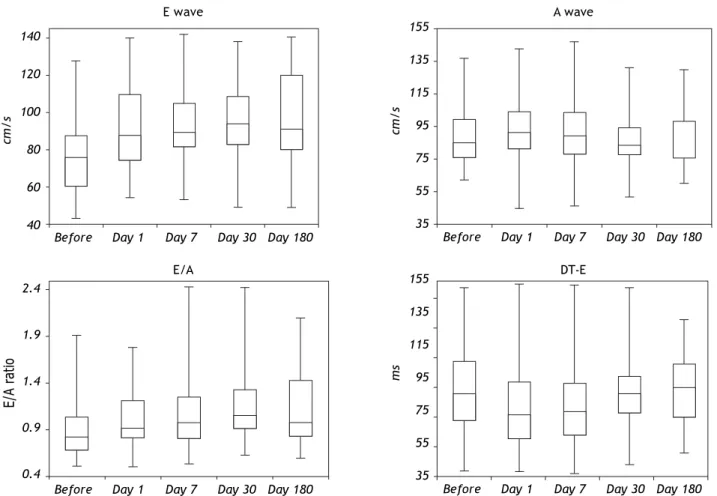
Table 3 shows mitral inflow parameters. During 6 month follow-up, the peak early filling velocity (E) was significantly increasing. Peak late atrial filling velocity (A) significantly increased within 24 h after the procedure [85.1 (76.2-99.5) vs. 91.2 (81.5-104.5) cm/s, p < 0.05]. The E/A ratio was abnormal at the beginning of the study. However, during the follow-up was significantly increasing and became normal (Figure 1). Isovolumic relaxation time was increa-sing during the follow-up period. Impaired left ven-tricular relaxation was present in 25 (45%) patients before the procedure. During the follow-up, the number of the patients with impaired left ventricular relaxation was significantly decreasing (Table 4). There were no patients with other types of mitral fil-ling patterns (pseudonormalisation or restrictive filling pattern).
Diastolic dysfunction and
survival of the patients with liver cirrhosis
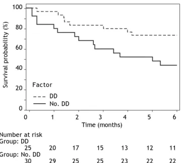
During 6 months follow-up 22 patients died. Out of 25 pts with diastolic dysfunction (DD), 14 pts (56%) died. Out of 30 patients without diastolic dys-function (no DD), 8 (26%) died. The survival of the patients with diastolic dysfunction was significantly decreased (DD vs. no DD: HR: 0.54, 95% CI: 0.31-0.94, p 0.0149). Figure 2 shows Kaplan-Meier cur-ves of survival.
DISCUSSION
The main findings of this study are:
• Hemodynamic abnormalities (e.g. hyperdynamic circulation, lower systemic vascular resistance, impaired relaxation of the left ventricle) are fre-quent in patients with liver cirrhosis.
• The transjugular portosystemic shunting is potent stimulus which can deteriorate hemocircu-lation (further increase of the cardiac output, in-crease of the pulmonary capillary wedge pressure, decrease of the systemic vascular resistance). • TIPS procedure affects pulsed-wave Doppler
parameters of mitral inflow (increase of peak
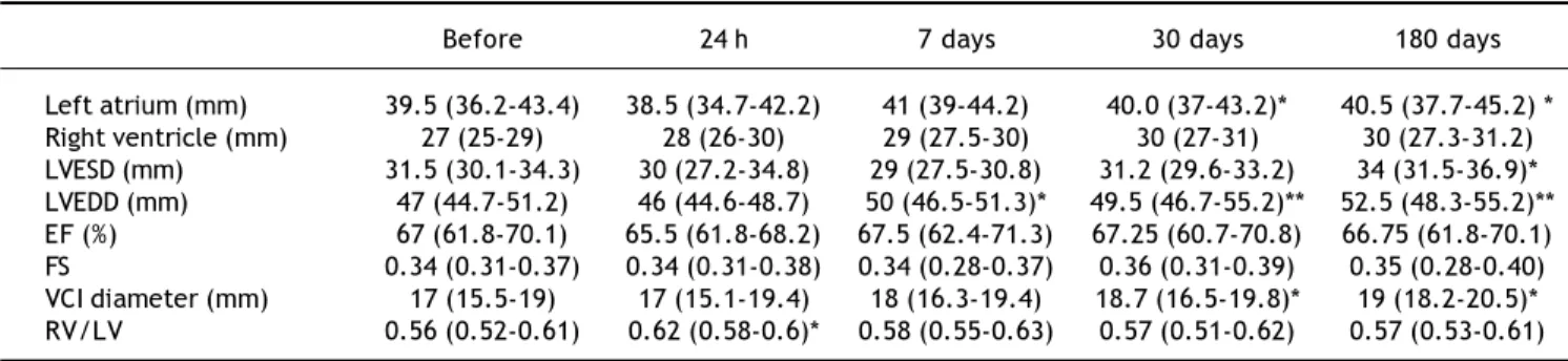
Table 2. Basic echocardiographic parameters in patients before and after the TIPS procedure during 6 month follow-up.
Before 24 h 7 days 30 days 180 days
Left atrium (mm) 39.5 (36.2-43.4) 38.5 (34.7-42.2) 41 (39-44.2) 40.0 (37-43.2)* 40.5 (37.7-45.2) * Right ventricle (mm) 27 (25-29) 28 (26-30) 29 (27.5-30) 30 (27-31) 30 (27.3-31.2) LVESD (mm) 31.5 (30.1-34.3) 30 (27.2-34.8) 29 (27.5-30.8) 31.2 (29.6-33.2) 34 (31.5-36.9)* LVEDD (mm) 47 (44.7-51.2) 46 (44.6-48.7) 50 (46.5-51.3)* 49.5 (46.7-55.2)** 52.5 (48.3-55.2)** EF (%) 67 (61.8-70.1) 65.5 (61.8-68.2) 67.5 (62.4-71.3) 67.25 (60.7-70.8) 66.75 (61.8-70.1) FS 0.34 (0.31-0.37) 0.34 (0.31-0.38) 0.34 (0.28-0.37) 0.36 (0.31-0.39) 0.35 (0.28-0.40) VCI diameter (mm) 17 (15.5-19) 17 (15.1-19.4) 18 (16.3-19.4) 18.7 (16.5-19.8)* 19 (18.2-20.5)* RV/LV 0.56 (0.52-0.61) 0.62 (0.58-0.6)* 0.58 (0.55-0.63) 0.57 (0.51-0.62) 0.57 (0.53-0.61)
The data are expressed as median (25-75 percentiles). LVESD: left ventricular end-systolic diameter. LVEDD: left ventricular end-diastolic diameter. EF: ejection fraction of the left ventricle. FS: fractional shortening; VCI: inferior vena cava diameter. RV/LV: right and left ventricle ratio. *p < 0.05. **p < 0.01.
Table 3. Doppler mitral inflow parameters.
Before Day 1 Day 7 Day 30 Day 180
E (cm/s) 75.5 (60.5-87.3) 88 (74.3-109.7)** 89 (81.5-105)** 94 (82.7-108.5)** 91 (80.1-120.2)** A (cm/s) 85.1 (76.2-99.5) 91.2 (81.5-104.5)* 89 (78.4-103.5) 83.5 (77.5-94.5) 86 (75.2-98.1)
E/A 0.887192 0.887192 1** 1.13253** 1.05814**
DT-E (ms) 210 (165-265) 175 (134-230)* 180 (140-227.5) 210 (177.5-240.1) 220 (170-260) IVRT (ms) 70.2 (60.3-80.4) 75 (62.5-89.5)* 76 (63.4-89.6)* 77.5 (62.6-95.8)* 75 (59.8-91.5)*
early filling velocity and normalization of E/A ratio).
• The presence of diastolic dysfunction is associa-ted with significantly increased mortality.
Liver cirrhosis is frequently associated with hy-perdynamic syndrome which comprises increased heart rate, cardiac output and plasma volume, and reduced systemic vascular resistance and arterial blood pressure.16-18 We observed increased cardiac
output in 17 (31 %) pts. and decreased systemic vascular resistance in 28 (51%) pts. before the procedure. High cardiac output is a result of increa-se in venous return, heart rate and contractility,
Figure 1. Box plots of mitral inflow pulsed wave Doppler parameters. The lowest, second lowest, middle, second highest and highest box points represent the minimum, 25th percentile, median, 75th percentile and maximum respectively.
which are controlled by the autonomic nervous system. Cardiac output can be increased also by vasodilatation, the presence of arteriovenous communications, expanded blood volume and increased sympathetic nervous activity.7,8
In our study, cardiac output and cardiac index in-creased and systemic vascular resistance dein-creased as a result of the creation of postosystemic shunt. This was accompanied with increase in pulmonary artery wedge pressure. All these findings indicate that TIPS procedure is a potent factor that may affect blood circulation.
Diastolic dysfunction is relatively frequent in pa-tients with liver cirrhosis.11,13,18 Recent studies of left
Table 4.
Before Day 1 Day 7 Day 30 Day 180
Impaired relaxation n (%) 25 (45) 18 (33) * 12 (22)** 11 (20)** 12 (22) **
Impaired relaxation pattern was defined as E/A < 1 and DT-E > 220ms. *p < 0.05. **p < 0.01. 140
120
100
80
60
40
cm/s
155
135
115
95
75
55
35
cm/s
Before Day 1 Day 7 Day 30 Day 180 Before Day 1 Day 7 Day 30 Day 180
E wave A wave
E/A DT-E
2.4
1.9
1.4
0.9
0.4
E/A ratio
155
135
115
95
75
55
35
ms
ventricular filling in cirrhosis support the presence of a subclinical myocardial disease with diastolic dys-function.19 It has been shown that myocardial
fibro-sis and increased myocardial mass lead to increased stiffness of the myocardial wall resulting in impaired ventricular filling and diastolic dysfunction.8,10
Fur-thermore, the TIPS procedure provides additional burden on cardiovascular system.
In our study, the diastolic dysfunction (impaired relaxation) was present in 45 % of patients before the procedure. 24 h later, the E/A ratio increased and the number of patients with impaired relaxation decreased. Further increase was observed until the end of the follow-up. This was caused by the increa-se of peak early filling velocity (E). In contrast to E wave, the peak late atrial filling velocity (A) only slightly increased 24 h after the TIPS procedure. De-celeration time had biphasic response: a rise in the first 24 h was followed by a decrease to baseline value. It is not easy to explain these changes. In our study, major changes were observed in increasing velocity of the E wave. It is known that E velocity is influenced by left atrial pressure at mitral valve ope-ning, the relative driving force between the left atrium and left ventricle, minimal left ventricular diastolic pressure, compliance of the left atrium and the rate of ventricular relaxation. Creation of the porto-systemic shunt leads to the changes in a num-ber of parameters (increase in cardiac output, cardiac index, heart rate, etc.), but the most important seems to be a decrease in systemic vascular resistance.13,20
Figure 2. Kaplan-Meier curves of survival during 6 months follow-up according to presence of diastolic dysfunction (DD).
Furthermore, diastolic dysfunction is probably associated with poor outcome of these patients. Caz-zaniga, et al. showed that diastolic dysfunction (E/A < 1) is associated with poor survival in patient un-dergoing TIPS procedure in a group of 32 pts. with liver cirrhosis.19
In our study, we confirmed the association of diastolic dysfunction and increased mortality in pa-tients with liver cirrhosis. Diastolic dysfunction may be a significant factor in the development of heart failure, may precede systolic dysfunction in patients with cirrhosis, and may play a part in the pathogenesis of sodium fluid retention in cirrho-sis.7,10
Study limitations: the diastolic dysfunction was evaluated using pulsed-Doppler techniques. The tis-sue Doppler analysis was not performed. During fo-llow-up, the patients may develop TIPS dysfunction (decreased patency or closure of the porto-systemic shunt) that can affect the hemodynamic parameters. This factor was eliminated by Doppler ultrasound of the liver tissue during regular follow-up.
Despite the limitations, the study showed:
• Liver cirrhosis is associated with the increased number of the diastolic dysfunction.
• Diastolic dysfunction is associated with decrea-sed survival.
• Transjugular porto-systemic shunt creation is as-sociated with significant hemodynamic changes. • The main change was in pulsed-wave Doppler
mi-tral inflow analysis (increase in E velocity) and it was probably associated with decreased systemic vascular resistance.
Today, a great effort is placed on efforts to impro-ve quality of life and survival of patients with liimpro-ver cirrhosis treated with TIPS. Therefore, the risk stratification and proper selection of the patients for TIPS procedure are very important.21-23 It has been
shown, that hemodynamic monitoring before and af-ter the TIPS procedure is very helpful for selecting of the patients.24 Our study confirmed the need of
hemodynamic monitoring of patients undergoing TIPS procedures and showed, that echocardiogra-phy as a non-invasive method can be very useful for this purpose.
ABBREVIATIONS
• A wave: peak late atrial filling velocity. • CI: cardiac index.
• CO: cardiac output.
0 1 2 3 4 5 6
Time (months) 100
80
60
40
20
0
Survival probability (%)
Number at risk Group: DD
25 20 17 15 13 12 11
Group: No. DD
30 29 25 25 23 22 22
• DT-E: deceleration time of E velocity. • E wave: peak early filling velocity wave. • EF: ejection fraction.
• FS: fractional shortening. • IVC: inferior vena cava.
• IVRT: isovolumic relaxation time. • LAD: left atrium diameter.
• LV: left ventricle.
• LVEDD: left ventricular end-diastolic diameter. • LVESD: end-systolic diameter.
• PAP: pulmonary artery pressure.
• PCWP: pulmonary capillary wedge pressure. • PVR: pulmonary vascular resistance.
• PW: pulsed wave Doppler. • RV: right ventricle.
• SVR: systemic vascular resistance.
• TIPS: transjugular intrahepatic portosystemic shunt.
ACKNOWLEDGEMENT
Supported by the programme PRVOUK P37/03.
REFERENCES
1. García-Pagán JC, Caca K, Bureau C, Laleman W, Appenrodt B, Luca A, Abraldes JG, et al. Early use of TIPS in patients with cirrhosis and variceal bleeding. N Engl J Med 2010; 362: 2370-9.
2. Boyer TD, Haskal ZJ. American Association for the Study of Liver Diseases. The role of transjugular intrahepatic portosystemic shunt (TIPS) in the management of portal hypertension: update 2009. Hepatology 2010; 51: 306. 3. Huonker M, Schumacher YO, Soricher S, Keul J, Rossle M.
Cardiac function and haemodynamics in alcoholic cirrhosis and effects of the transjugular intrahepatic portosyste-mic stent shunt. Gut 1999; 44: 743-8.
4. Lotterer E, Wengert A, Fleig WE. Transjugular intrahepa-tic portosystemic shunt: short-term and long-term effects on hepatic and systemic hemodynamics in patients with cirrhosis. Hepatology 1999; 29: 632-9.
5. Merli M, Valeriano V, Funaro S, Attili AF, Masini A, Efrati C, De CS, et al. Modifications of cardiac function in cirr-hotic patients treated with transjugular intrahepatic portosystemic shunt (TIPS). Am J Gastroenterol 2002; 97: 142-8.
6. Montgomery A, Ferral H, Vasan R, Postoak DW. MELD score as a predictor of early death in patients undergo-ing elective transjugular intrahepatic portosystemic shunt (TIPS) procedures. Cardiovasc Intervent Radiol
2005; 28: 307-12.
7. Wong F, Liu P, Lilly L, Bomzon A, Blendis L. Role of cardiac structural and functional abnormalities in the pathogene-sis of hyperdynamic circulation and renal sodium reten-tion in cirrhosis. Clin Sci 1999; 97: 259-67.
8. Møller S, Henriksen JH. Cardiovascular complications of ci-rrhosis. Gut 2008; 57; 268-78.
9. Henriksen JH, Fuglsang S, Bendtsen F, Christensen E, Møller S. Dyssynchronous electrical and mechanical sys-tole in patients with cirrhosis. J Hepatol 2002; 36: 513-20.
10. Finucci G, Desideri A, Sacerdoti D, Bolognesi M, Merkel C, Angeli P, Gatta A. Left ventricular diastolic function in li-ver cirrhosis. Scand J Gastroenterol 1996; 31: 279-84. 11. Umgelter A, Reindl W, Geisler F, Saugel B, Huber W, Berger
H, Schmid RM. Effects of TIPS on global end-diastolic volu-me and cardiac output and renal resistive index in ICU pa-tients with advanced alcoholic cirrhosis. Ann Hepatol
2010; 9: 40-5.
12. Zile MR, Brutsaert DL. New concepts in diastolic dysfunc-tion and diastolic heart failure: part I. Diagnosis, progno-sis and measurements of diastolic function. Circulation
2002; 105: 1387-93.
13. Aurigemma GP, Gaasch WH. Diastolic heart failure. N Engl J Med 2004; 351: 1097-105.
14. Little WC, Kitzman DW, Cheng CP. Diastolic dysfunction as a cause of exercise intolerance. Heart Fail Rev 2000; 5: 301-6.
15. Nagueh SF, Appleton CP, Gillebert TC, Marino PN, Oh JK, Smiseth OA, Waggoner AD, et al. Recommendations for the Evaluation of Left Ventricular Diastolic Function by Echo-cardiography. J Am Soc Echocardiogr 2009; 22: 107-33. 16. Ortiz-Olvera NX, Castellanos-Pallares G, Gómez-Jiménez
LM, Cabrera-Muñoz ML, Méndez-Navarro J, Morán-Villota S, Dehesa-Violante M. Anatomical cardiac alterations in liver cirrhosis: An autopsy study. Ann Hepatol 2011; 10: 321-6. 17. Blendis L, Wong F. Is there a cirrhotic cardiomyopathy?
Am J Gastroenterol 2000; 11: 3026-8.
18. Pozzi M, Carugo S, Boari G, Pecci V, de Ceglia S, Maggiolini S, Bolla GB, et al. Evidence of structural and functional cardiac abnormalities in cirrhotic patients with and without ascites. Hepatology 1997; 26: 1131-7.
19. Cazzaniga M, Salerno F, Pagnozzi G, Dionigi E, Visentin S, Ci-rello I, Meregaglia D, et al. Diastolic dysfunction is associa-ted with poor survival in cirrhotic patients with transjugular intrahepatic portosystemic shunt. Gut 2007; 56: 869-75. 20. Gilbert JC, Glantz SA. Determinants of left ventricular
fil-ling and of the diastolic pressure–volume relation. Circ Res
1989; 64: 827-52.
21. Malinchoc M, Kamath PS, Gordon FD, Peine CJ, Rank J, Borg PC. A model to predict poor survival in patients un-dergoing transjugular intrahepatic portosystemic shunts.
Hepatology 2000; 31: 864-71.
22. Kamath PS, Wiesner RH, Malinchoc M, Kremers W, Ther-neau TM, Kosberg CL, D Amico G, et al. A model to predict survival in patients with end-stage liver disease. Hepatolo-gy 2001; 33: 464-70.
23. Said A, Williams J, Holden J, Remington P, Gangnon R, Mus-at A, Lucey MR. Model of end stage liver disease score predicts mortality across a broad spectrum of liver disea-se. J Hepatol 2004; 40: 897-903.