SUMMARY
Extensive cellular and behavioral studies have led to the postulation that memories are encoded by changes in synaptic strength between neurons, as demonstrated by the correlation between the term changes in animal’s behavior and long-term changes in neuronal connections underlying a specific behavior in invertebrate animals, or even in vertebrate animals, where cellular models of synaptic plasticity using genetic approaches, such as Long-Term Potentiation (LTP) and Long-Term Depression (LTD), have been shown to depend on long-term changes in synaptic activity implicated in behavioral learning and memory. Long-term memory (LTM) is crucial for animal’s survival and thus represents a mechanism that underlies fundamental neurobiological events in the nervous system of vertebrate and non-vertebrate species including the human. Long-term changes in synaptic connectivity as well as long-term behavioral changes (both activities that underlie several of the properties of LTM and used as a parameter to explain the long-lasting enhancement of neuronal function after a stimulus) have been demonstrated to rely on signals that initially occur in the cell body. LTP is a form of synaptic plasticity widely accepted as a cellular model for stabilization of synapses in neurobiological phenomena such as development and learning and memory. Much of the experimen-tal work concerning LTP in learning has been focused on the NMDA receptor dependent forms of LTP. But several questions have arisen regarding if LTP equals memory. If LTP has a real role in memory, a more appropriate hypothesis should be stated by postulating that activity-dependent synaptic plasticity and multiple forms of memory known to exist, share a common core; that is, the synaptic plasticity and memory, hypothesis states that activity dependent synaptic plasticity is induced at appropriate synapses during memory formation. Synaptic plasticity is a physiological phenomenon that induces specific patterns of neural activity sustained by chemical and molecular mechanisms, that gives rise to changes in
synaptic efficacy and neural excitability which long outlast the events that trigger them. Based on the several properties of synaptic plasticity discovered, LTP may be proposed as a suitable neuronal mechanism for the development of several memory systems, including initial encoding and storage of memory traces and initial phases of trace consolidation over time. Such memory processing made up by LTP or LTD most probably occur as a network specific process, making LTP a universal mechanism for encoding and storage of memory traces and what gets encoded is part of a network property rather than mechanisms working at individual synapses. For example, the type of information processed at the hippocampus is quite different from the information processed by the amygdala, and such information should remain if the mechanisms of plasticity operating in each brain area are conserved. Decades of research have demonstrated that LTP in the hippocampus is induced by synaptic activity and that cytoplasmic membrane-bound molecule(s) are required to transduce extracellular signals mediated by receptor-activation into activation of intracellular signaling processes. Most of these processes depend on intracellular calcium activity, and thereby on calcium-dependent mechanisms that are recruited for LTP induction and expression. For instance, NMDA receptors have been shown to be essential for initiation of LTP, but expression of this phenomenon in brought primarily by AMPA receptors. Induction of LTP in CA1 hippocampal region has been shown to depend on increases of intracellular calcium and activation of specific calcium-dependent molecules such as the calcium/ calmodulin-dependent protein kinase (CaMKII), whose cell expression is confined predominantly at postsynaptic densities. Moreover, long-term expression of LTP requires protein synthesis, where transient signals will be linked to activation of specific genes that ultimately will determine growth and remodeling of potential active synapses. Different types of synapses may express and use a different set of molecules mediating activation of intracellular signaling pathways for initiating and maintaining
U
NDERSTANDING
THE
NEUROBIOLOGICAL
MECHANISMS
OF
LEARNING
AND
MEMORY
:
CELLULAR
,
MOLECULAR
AND
GENE
REGULATION
IMPLICATED
IN
SYNAPTIC
PLASTICITY
AND
LONG
-TERM
POTENTIATION
. P
ART
IV A
Philippe Leff*, Isaac Retana*, Adriana Arias-Caballero*, Enrique Zavala*, Frida Loria*, Lenin Pavón*, Benito Antón*
*Laboratorio de Neurobiología Molecular y Neuroquímica de Adicciones. Instituto Nacional de Psiquiatría Ramón de la Fuente. Corresponding:
synaptic plasticity. Several studies have demonstrated that neuronal modifications of neurotransmitter receptors or membrane-re-ceptor subunits at postsynaptic densities, represent one of the neuronal mechanisms by which neurons regulate their synaptic strength. For instance, it has been demonstrated that neuronal dendrites are able to regulate their own transmembrane receptor synthesis in response to external stimuli (i.e., GluR2 subunit of AMPA receptor) and such molecular mechanisms, posed important implications in the understanding of how individual synapses are selectively strengthened. In addition, recent experiments have demonstrated that specific intracellular signaling molecules (i.e., neuronal Synaptic GTPase-activating protein or SynGAP) are selectively expressed and enriched at excitatory synapses.
Interestingly enough are the evidences that demonstrate that different subsets of protein kinases (MAPKs, SAPKs, MAPKAKs, p38MAPK, etc.) and intracellular signaling pathways activate transcription factors (AP-1 complex, CREB) that regulate the expression of different immediate early genes (IEG) which are crucial for neuronal development, glutamate receptor trafficking to specific synapses and for LTP induction. Much of the neurochemical and molecular changes that occurr in synaptic plasticity may be well associated with dynamic morphological changes in spine synapses as suggested to participate in the development and consolidation of LTP. In addition, glial cells, known to participate in the excitatory neurotransmission in the CNS besides their conceptualized cellular function, as elements for structural support and homeostasis, may play an important role in synaptic plasticity and thereby may regulate the information processed in the brain.
As hippocampal LTP has been the target of intensive molecular genetic analysis, several studies have demonstrated that LTP is altered when particular single genes are knocked out or overexpressed in null mutant mice or transgenic mice. Such studies have led to the amazing observation that variations in LTP exist within natural inbred mouse strains.
Key words: Long-term potentiation, long-term memory hippocampus, synaptic platicity, synapse speaficity, protein synthesis, protein kinases, gene transcription.
RESUMEN
Extensos estudios celulares y conductuales han llevado a la postulación de que la memoria es codificada por cambios en la fuerza sináptica entre las neuronas, como lo ha demostrado la correlación entre los cambios a largo plazo en la conducta de los animales y en las conexiones neuronales que generan una conduc-ta específica, en animales invertebrados o vertebrados, en los que los modelos celulares de plasticidad sináptica, usando aproxima-ciones genéticas como el fenómeno de potenciación de largo pla-zo (LTP), o el fenómeno de la depresión de largo plapla-zo (LTD), han demostrado que dependen de cambios a largo plazo en la actividad sináptica implicada en las conductas de aprendizaje y memoria.
La memoria de largo plazo (LTM) es crucial para la sobrevivencia de los animales y representa un mecanismo fundamental para los eventos neurobiológicos en el sistema nervioso de las especies de vertebrados e invertebrados, incluyendo el del humano. Los bios a largo plazo en la conectividad sináptica, así como los cam-bios conductuales de largo plazo (ambas actividades son
respon-sables de varias propiedades que caracterizan el fenómeno de LTM y se usan como parámetros funcionales para explicar el au-mento de la actividad neuronal dependiente de estímulos) han demostrado que las señales ocurren inicialmente en el cuerpo celular. El fenómeno biológico de LTP es una forma de plastici-dad sináptica ampliamente aceptada como un modelo celular que promueve la estabilización de los sinapsis activas y que participan en eventos neurobiológicos como el desarrollo, el aprendizaje y la memoria.
Una gran mayoría de los trabajos experimentales concernientes al fenómeno biológico del LTP en el aprendizaje, se ha enfocado a la actividad funcional de los receptores glutamatérgicos, tipo NMDA. Si bien muchas preguntas han surgido con respecto de si el fenómeno de TLP es equivalente a la función de memoria, esto es, si el fenómeno de TLP juega un papel real y preponderante en la función de memoria, entonces, una hipótesis apropiada debería establecer el postulado de que el fenómeno LTP como la activi-dad dependiente de los eventos de plasticiactivi-dad sináptica y de múl-tiples formas de memoria que existen, compartan un denomina-dor común. Esto permite postular la hipótesis que sugiere que la actividad dependiente de la plasticidad sináptica es inducida en sinapsis particulares y específicas durante la formación del apren-dizaje y la consolidación de la memoria.
La plasticidad sináptica es un fenómeno fisiológico que induce patrones específicos de actividad neuronal, sostenidos por meca-nismos químicos y moleculares, que dan origen a cambios en la eficiencia sináptica y en la excitabilidad neuronal, que perdura por más tiempo que los eventos que los originan. Basados en algunas propiedades de plasticidad sináptica recientemente estu-diadas y documentadas, el fenómeno de LTP puede ser propuesto como un mecanismo neuronal para el desarrollo de algunos siste-mas de memoria que incluye la codificación inicial, el almacena-miento de la memoria y las primeras fases de la consolidación de la misma. Si el procesamiento funcional de la memoria es mediado por el fenómeno LTP o LTD, muy probablemente ocurre como un proceso específico, dentro de una red de circuitos neuronales, situando al fenómeno de LTP como un mecanismo universal para la codificación y almacenaje de la memoria. Asimismo, la codifica-ción sería parte de una propiedad de red neuronal más que de un mecanismo neuronal de contactos sinápticos individuales. Por ejemplo, el tipo de información procesada en el hipocampo es muy diferente de la información procesada por la amígdala y esta información puede permanecer si el mecanismo de plasticidad que opera en cada región del cerebro se conserva con el tiempo.
kinasa-dependiente de calcio en la neurona ha sido ampliamente demostrada en las densidades postsinápticas (PSD).
Por otra parte, la expresión a largo plazo del fenómeno LTP requiere la síntesis de proteínas, en donde las señales transitorias pueden estar ligadas a la activación de genes específicos que deter-minarán en última instancia el crecimiento y la remodelación de sinapsis potencialmente activas. Diversos tipos de sinapsis pue-den expresar y hacer uso de diversos grupos de moléculas proteicas que participan en la activación de diferentes vías de señalamiento intracelular y que, por igual, son responsables de las fases iniciales y de sostenimiento de los eventos de plasticidad sináptica. Varios estudios han demostrado que las modificaciones neuronales de los receptores específicos de unión de alta afinidad, de diferentes neurotransmisores o de las subunidades proteicas, que componen estos receptores membranales en las densidades postsinápticas (PSD), representan uno de los mecanismos celulares por los cua-les las neuronas regulan su actividad de reforzamiento sináptico. Por ejemplo, se ha demostrado que las dendritas neuronales pue-den regular su propia síntesis de receptores proteicos membranales en respuesta a estímulos externos (por ejemplo, la subunidad GluR2 del receptor glutamatérgico, AMPA), y tales mecanismos moleculares implican importantes planteamientos en la compren-sión de cómo las sinapsis individuales se consolidan selectivamente. Mas aún, recientes experimentos han demostrado que moléculas que participan en vías de señalamiento intracelular (v.g., la proteí-na sináptica neuroproteí-nal con actividad de GTPasa, denomiproteí-nada como SynGAP) están selectivamente expresadas y enriquecidas en neuronas que median respuestas sinápticas excitatorias.
Es interesante constatar que estos estudios han demostrado que diversos subgrupos de proteínas Kinasas (v.g., MAPKs, SAPKS, MAPKAKs, p38MAPK), implicadas en la activación de diversas vías de señalamiento intracelular, son responsables de la actividad funcional de distintos factores de trascripción (v.g., com-plejo AP-1, C-Fos, Jun, CREB), que a su vez regulan la expresión de múltiples genes de expresión temprana (intermediate early genes [IEG, por sus siglas en inglés]) que son cruciales para el desarrollo neuronal, para la regulación del transporte vesicular de receptores glutamatérgicos a sinapsis específicas, así como para la inducción del fenómeno de LTP. Gran parte de los cambios neuroquímicos y moleculares que ocurren en los eventos de plasticidad sináptica se puede asociar con cambios morfocelulares dinámicos en las espi-nas sinápticas, como diversos estudios han demostrado durante el desarrollo y la consolidación del fenómeno de LTP. Además, si bien diversos trabajos experimentales han demostrado la partici-pación de las células gliales en la neurotransmisión excitatoria en el SNC, estas células, además de ejercer una función celular am-pliamente conceptualizada, como elementos de soporte estructu-ral y de homeostasis, poseen un papel crucial en los eventos de plasticidad sináptica, de tal forma que también regulan la infor-mación procesada en el cerebro de los mamíferos, incluyendo los sistemas neuronales de especies de invertebrados. Si bien el fenó-meno de LTP en el hipocampo ha sido el blanco de mayor inten-sidad de estudio, y en particular en el análisis genético molecular, donde a este respecto varios estudios han demostrado que el fenómeno de LTP está alterado cuando los genes particulares son inhabilitados permanentemente (knockout) o temporalmente (knockdown) en su expresión funcional y/o sobreexpresados en ratones mutantes nulos o en ratones transgénicos. Estos estudios han llevado a observaciones interesantes que demuestran que dentro de las diferentes cepas naturales del ratón existen variacio-nes naturales en la expresión del fenómeno de LTP.
Palabras clave: Potenciación a largo plazo, memoria a largo plazo, hipocampo, plasticidad sináptica, especificidad sináptica, síntesis proteica, proteínas kinasas, transcripción génica.
INTRODUCTION
The understanding of the neural basis of learning and memory has been established since several decades ago. Before LTP discovery there was Hebb´s postulate for learning, based on the theoretical work of Cajal and many others (Chapman, 2002). This theory postulates that patterns of activity between neurons cause lasting changes to synapses between neurons. Thus, based on this concept, it would be feasible to map the circuits responsible for a particular form of learning and identifying those synapses within such circuits that show plasticity and, further on, specify the molecular mechanisms responsible for that plasticity (Chapman, 2002). Although, several investigations were focused in identifying the neural circuits implicated in learning and memory (Thompson, 1986), through the discovery of Long-term potentiation, LTP, most of the experi-mental work moved towards the description of the neurobiological mechanisms in synaptic plasticity and their implications in learning and memory (Chapman, 2002), by means of pharmacological tools. For example, the discovery that LTP induction could be blocked by NMDA-receptor antagonist, AP5, led to conducting a series of experiments in the effects of AP5 on a variety of learning tasks (Chapman, 2002).
The increasing knowledge of different sorts of molecules and players identified and molecularly characterized implicated in different synaptic plasticity events, showed that most of the identified components were not readily amenable to pharmacological intervention (due that specific inhibitors were either not available or target molecules were inaccessible to pharmacological treatment to applied drugs)(Chapman, 2002). Application of targeted gene deletion techniques, and the generation of transgenic mutant mice, to the study of synaptic plasticity, and thus, to the learning and memory processing functions, led to exciting and fascinating results, that otherwise, would not be approachable by pharmacological treatment (Chapman, 2002). For instance, two landmark studies that used these techniques, by observing the effects of the knocking-out of two specific genes that encode for two intracellular proteins, αCaMKII (Ca2+
mutant mice, demonstrating the significant importance of the hippocampus, when exposed to lesion.
Moreover, the resultant learning and memory deficits shown by these animals matched with the impairment of LTP in the hippocampus (Chapman et al., 2003). Therefore these results confirm previous pharmacological work, demonstrating that unreachable molecules were able to become accessible for a much better understanding of the molecular and cellular mechanisms that regulate synaptic plasticity and, thus, different forms of learning and memory (Chapman, 2002). Thus application of the knock out techniques offered a useful tool for generating a large number of mice with targeted deletions of genes that encoded proteins known to be implicated in learning and memory processing (Mayford and Kandel, 1999). Thus, the knock out technique approach led to the generation of mutant mice with expression of variable cell phenotypes, in whose targeted deleted genes included neurotransmitter receptors, protein kinases, nuclear hormone receptors, and transcription factors. molecules known to be implicated in synaptic plasticity, learning and memory function (Chapman, 2002). LTP represents the best model for learning related plasticity in the CNS of mammals. Several evidences support the hypothesis that activity-dependent synaptic plasticity plays a significant role or at least shares important features with learning and memory (Martin et al., 2000). Although LTP occurs in several brain structures, this phenomenon has been investigated and characterized at the hippocampus, due to its relative simple anatomy and its importance in the formation of new declarative memories (Soderling and Derkach, 2000; Orban et al., 1999). Nonetheless, no convincing demonstration has been yet evidenced that LTP is the mechanism of learning and memory, because there may be different mechanisms and expressions of use-dependent plasticity in different forms of learning (Chapman et al., 2003). Moreover, by acknowledging which features of activity-dependent plasticity are common in forebrain synapses and used in different type of synapses, it could be possible to enlarge our spectrum of the nature of neuronal plasticity and behavior (Chapman et al., 2003).
LTP is the increase in strength of synaptic transmission observed after application of tetanic stimulation. This potentiation that lasts for as long as it can be measured, meaning hours or even days, is the best model available for studying learning and memory on a cellular basis (Edwards, 1995). The initial induction of LTP depends on influx of Ca2+ through NMDA receptor-channel
while the cell is depolarized (via activation on AMPA receptors). Synaptic potentiation can be divided in several temporal stages that rely on different cellular
and molecular mechanisms: Short-term potentiation (lasting 15-30 min) and early-phase of LTP (E-LTP)(stable for 2-3 h) do not require gene expression and therefore, protein synthesis de novo; while late phase LTP (L-LTP) (lasting from 6-8 h in hippocampal slices) does(Malenka, 1994). In such context, activation of NMDA receptors alone results in short-term potentiation that decays in less than an hour, so that in order to sustain LTP for long periods of time, additional factors are necessary for establishment of LTP. Thus, the extra factor that may be presumed to be involved is the metabotropic glutamate receptor (Edwards, 1995). Moreover, important to note is that changes at the intracellular environment in the postsynaptic cell, should be taken into account, for the LTP model to occur. Although early phases of induction of LTP in hippocampal formation can be understood, evidences of later phases of LTP are still unclear. It should be clear, that for expression of LTP many arguments have been postulated to be either a presynaptic or post-synaptic phenomenon.
Thus, in order to explain many inconsistencies of LTP, both neurochemical and molecular aspects need to be described in addition to the electrophysiological and anatomical data to allow certain contradictory inconsistencies of LTP to be compatible; where is the locus of expression of LTP. The interaction of presynaptic and postsynaptic events is crucial for the development and establishment of LTP, in brain structures showing active-dependent synaptic plasticity. Several studies have demonstrated that application of protein kinase inhibitors to the postsynaptic cell prevents development of LTP. Similar results have been obtained with protein synthesis inhibitors. A change in synaptic strength could well result of the addition of postsynaptic membrane receptors or increase in the probability of release of neurotransmitter or increase in the number of synapses between activated cells (Edwards, 1995). Using quantal analysis to approach this question, different studies have demonstrated that LTP is dependent on presynaptic effects (i.e., increased in miniature frequency) while others have demonstrated effects at the postsynaptic level (i.e., increased in miniature amplitude) or even both. Moreover, inconsistencies have been observed whether AMPA or NMDA receptor mediated components of synaptic current, both increase presynaptic or postsynaptic activity or just AMPA-mediated component increases (postsynaptic) (Edwards, 1995).
(see box 1 for full description of the nonsynaptic neurotransmitter system mediated by NO). Blocking the synthesis of these products shown by different groups has either no effects or blocks the induction phase of LTP. Thus, this set of conflicting results drives to the clue to mechanisms implicated in this phenomenon, that suggest a common mechanism for LTP, based on electrophysiological and anatomical evidences (Edwards, 1995).
Theoretical considerations based on the Hopfield model of associative memory, via the formation of cell ensembles (Hopfield, 1982) explain that reciprocal connections between two active neurons are simultaneously strengthened. This model has been extremely useful to understand several insights of the neuronal mechanisms of learning and memory (Tsodyks, 2002), based on previous studies that demonstrated that the direction of modification of information between two neurons depends on the tem-poral order in the firing of presynaptic and postsynaptic neurons. For instance, LTP requires a causal relationship, where presynaptic firing must have occurred before postsynaptic modification, and conversely, LTD expression requires the opposite relationship (Tsodyks, 2002). Thus, application of such theoretical concept using dual whole cell-patch clamp, on hippocampal neurons, investigators demonstrated and confirmed that the direction of modification depends on the tem-poral ordering, with a precision of milliseconds (Markram et al., 1997; Tsodyks, 2002). This novel mechanism used to describe this form of plasticity was defined as “spike-timing dependent plasticity or STDP”. Although the functional implications of such a model are not clear, theoretical studies predict that this model of neuronal plasticity underlies important effects on sequence learning, predictive learning, and the balance between excitatory and inhibitory effects between neurons and neural circuits, particularly the synaptic pathways embedded into neocortical circuits (Tsodyks, 2002).
At the molecular level, the formation of long-term memory is thought to depend on long-lasting changes in synaptic efficacy and reorganization of neuronal networks. Most of the mechanisms underlying such plasticity are mostly dependent on a genetic program in neurons, that drive de novo synthesis of proteins, as demonstrated from the extensive works that have shown that pharmacological inhibition of protein synthesis, impair long-term memory, leaving short-term memory intact (Davis and Squire, 1984; Meiri and Rosenblum, 1998). These results have allowed the proposition that the regulation of transcription of specific genes during learning is required for the esta-blishment of a permanent memory trace (Bozon et
al., 2002). Several evidences have demonstrated that LTP and long-lasting forms of synaptic plasticity depend on gene transcription (Nguyen et al., 1994), as well as on the novo protein synthesis (Otani and Abraham, 1989; Frey and Moris, 1997). In addition, several studies have demonstrated that both LTP and learning induce transcription of a variety of genes in specific areas in the brain of mammals (Bozon et al., 2002). Thus, these evidences support the idea that LTP is an important mechanism for long-term information storage in the brain (Bozon et al., 2002).
Transcription requirement for synapse specificity in long-term memory (LTM)
Extensive cellular and behavioral studies have led to the postulation that memories are encoded by changes in synaptic strength between neurons, as demonstrated by the correlation between the long-term changes in animal’s behavior and long-term changes in neuronal connections underlying a specific behavior in invertebrate animals, or even in vertebrate animals, where cellular models of synaptic plasticity using genetic approaches, such as LTP and LTD, have been shown to depend on long-term changes in synaptic activity implicated in behavioral learning and memory (Sossin, 1996). In Hebbian models of synaptic plasticity and memory, neurons modify their synaptic strengths between presynaptic and postsynaptic activity (Marr et al., 1971), such that individual synapses of a neuron can change independently, implying that most neurons make specific connections to multiple postsynaptic partners as they receive multiple presynaptic inputs (Sossin, 1996).
Long-term changes in synaptic connectivity as well as long-term behavioral changes (both activities that underlie several of the properties of LTM and are used as a parameter to explain the long-lasting enhancement of neuronal function after application or entry of a stimulus), have been demonstrated to rely on signals that initially occur in the cell body (Sossin et al., 1996). For instance, studies in Aplysia, Drosophila
and mice have shown that specific activation of the nuclear transcription factor, CREB (see below), is required for the formation of LTM (Montarolo et al., 1986; Bourtchulasze et al., 1994). This nuclear transcription factor is known to be activated by different signal-transduction pathways that depend on the increasing levels of cAMP and Ca2+, and therefore, as
establish-ment of long-term changes in synaptic plasticity (Sossin et al., 1996). For instance, experiments performed in
Aplysia have demonstrated that the facilitatory neurotransmitter 5-HT lead to the formation of LTM, even at the expense that short-term facilitation is abolished. Such data demonstrates that signals arriving from the cell body are sufficient for the establishment of LTM (Emptage & Carew, 1993; Clark & Kandel, 1993).
In the same extent, experiments performed in
Drosophila, have shown that LTM for odor discrimination, not only requires activation of CREB, which relies on the transferring of signals from the cell body, but also is independent of other intermediary forms of memory (Dezazzo & Tully, 1995; Yin et al., 1994). Thus, such experiments show that changes that result in activation of signals of the cell body that finally result in the increase of CREB activity are sufficient to induce a LTM, even in a single training episode that is unlikely to generate sustained changes at the synapse (Bartsh et al., 1995; Yin et al., 1995). In a similar context, experiments performed using rat hippocampus slices, have demonstrated that cAMP leads to LTM, that requires transcription, without short term effects on synaptic strength (Huang & Kandel, 1995). These results show that local synaptic changes are not required for formation and establishment of certain forms of LTM as shown to occur in these species, including the brain of mammals (Sossin, 1996). Several models of synapse specificity have been offered to explain the mechanisms that allow the activity of nuclear signals to be expressed only at specific synapses and thus facilitating the formation of LTM (Sossin, 1996). But several questions arise when trying to solve how signals emanating from the cell body “know” which synapse need to be activated. In such context, it can be postulated as an initial hypothesis that signals arriving from the cell body must interact with molecules that tagged specific synapses as a result of local activity in such synapses, which, thus, implies the presence of specific molecules in previous activated synapses and thereby, predicts no changes in synaptic strength should occur in the absence of synaptic signals. These models, as will be explained below, predict that even in the presence of synaptic signals, changes will occur at both inactive synapses and active ones as well, providing a small degree of synapse specificity (Sossin, 1996).
a)Molecular and cellular mechanisms for synapse specificity in the establishment of LTM.- One way to establish synaptic specificity is to sort vesicles containing new molecules that convey specific targeting information that recognizes only activated synapes and thus would not interact with inactive ones (i.e., contigency model, Sossin,
1996). An alternate model explains that vesicles transporting novel molecules have no specificity targeting information for activated synapses, but accumulate at any specific synapse, due to the increased activity of the protein machinery involved in vesicle fusion in such synapses. Thus, in the absence of synaptic signals, vesicles would impinge all synapses equally, leading to cell-wide LTM (Sossin, 1996).
Several works have demonstrated that mRNAs signals can be routed from the cell body to dendrites (Spilker et al., 2002; Boekhoff et al., 1997; Kiebler & DesGroseillers, 2000), thus supporting the concept that such tagged molecules can be routed to specific activated synapses (referred to sorting model) and/or novel mRNAs may either accumulate at active and inactive synapses as well (referred to accumulation model, Sossin, 1996). Moreover, cell-derived signals can be sorted to all synapses, but limit their activation to specifc synapses, thus, mRNAs sorted equally to several synapses, will be regulated and translated only at precise selective synapses. In a similar context, proteins that require phosphorylation (see section below) or proteolysis for activity could be sorted in unmodified form, but only those synapses that contain active kinases or proteases will activate these molecules. Thus, models that explain the activation of specific synapses predict that no changes will occur in the absence of synaptic activation, and therefore, the mechanisms for its activation rely independently on the type of signals sorted from the cell body; activation of synapses will occur if such synapses have been previously activated, expressing the molecular machinery needed for its activation, while inactive synapses are not enables to do so (Sossin, 1996).
Like wise, a model has been offered which explains that strengthening of synaptic connections might not require signals from the cell body in an initial stage, and thus, molecules arriving from the cell body might be recruited only later to stabilize synaptic changes. This model (i.e., consolidation model) describes the situation of a smooth transition for synapse activation from an independent-transcriptional stage to a later complete dependent-transcriptional stage; this model is inconsistent with the cell-wide LTM, in the absence of synaptic changes, as no consolidation changes are required in the wide LTM model (Sossin, 1996).
at inactive synapses. Thus, the accumulation model that explains the cell-wide LTM in the absence of synaptic tagging, is opposite to the synapse specificity, as they support the strengthening of inactive synapses as well (Sossin, 1996).
b)Synapse specificity retained in LTM.- Certain mRNA transcripts, encoding protein information might be crucial for producing LTM, whereas others, under control of different transcriptional factors (see below) may act in concert with several synaptic molecules. For instance, transcriptional activation of mRNAs in the presynaptic cell, and sorted to synapses through axonal transport, could eventually be sufficient to induce a cell-wide LTM. In turn, transcriptional activation of specific mRNAs in the postsynaptic cell, referring to signals that need to be transported through dendrites, might be necessary but no sufficient to establish a cell-wide LTM, but lead to synapse-specific LTM (Sossin, 1996). In this context, several studies using different learning paradigms have demonstrated supporting evidences of this hypothesis. For instance, in Aplysia
sensory neurons, LTM is known to be encoded by presynaptic facilitation, where protein synthesis is crucial and occurs only at the presynaptic cell in order to establish LTM (Trudeau & Castellucci, 1995). Conversely, paradigms that show synapse specificity such as LTP in the CA1 region of the hippocampus (Nguyen et al., 1994) require activation of the postsynaptic cell as demonstrated by the activation and expression of immediate early genes (IEG) in the postsynaptic cell after LTP (Wisden et al., 1990; Cole et al, 1989). Thus, these studies demonstrate that synapse specificity may be easier to achieve in the postsynaptic cell basically due that RNA sorting and transport as well as local translation of mRNAs may occur in dendrites compared to presynaptic terminals where no evidences have been shown yet (Sossin, 1996).
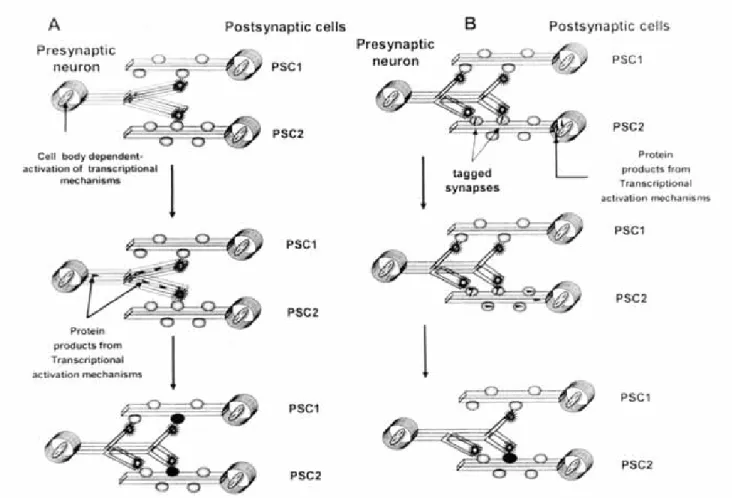
Thus, based on the previous hypothesis just described, a general model for learning can be established. This model requires first the initial activation of transcription mechanisms in presynaptic cells that will result in the generation of new synapses with postsynaptic members. These synapses will serve as substrates for synapse-specific changes induced by the postsynaptic cell (figure 1A). Thus, long-term changes in synaptic strength will only occur in new generated synapses, as demonstrated by several experimental observations that show the selective activation of novel synapses or silent synapses (Charpier et al., 1995; Liao et al., 1996; Malinow, 1991). For instance, synaptic responses formed during behavioral adaptations in birds, are pharmacologically different from pre-existing synaptic responses establishing that these “learned” synaptic
responses represent novel synaptic connections that could have been generated by transcriptional activation of the presynaptic cell (Sossin, 1996). If such synapses were transient or unstable, then learning processing would emulate the process of early synaptic development where a large number of temporary connections are pruned by competitive mechanisms that follow Hebb´s postulates (Goodman & Shatz, 1993; Sossin, 1996). Moreover, it has been demonstrated that critical stages during neuronal development are highly regulated by limiting the stimulation and activation of transcription mechanisms in presynaptic neurons, and thereby, limiting the substrates for further neuronal plasticity (Sossin, 1996). As neurons are anatomically interconnected as both presynaptic or postsynaptic cells, activation of transcription mechanisms in neurons will be used for the purpose to select specific inputs and stimulate the generation of new outputs, that eventually will facilitate the propagation of associations from neuron to neuron (Sossin, 1996).
In summary, LTM is crucial for animal’s survival and thus represents a mechanism that underlies fundamen-tal neurobiological events in the nervous system of vertebrate and non-vertebrate species including the human. Several models have been offered to explain the mechanisms that lead to LTM, or the increased ability of neurons to consolidate LTM, when signals generated from presynaptic transcriptional activation reach the synapse. In such context, mechanisms for localizing signals that arrive from the cell body to the synapse will require specific synaptic marks or tagging. Some situations will show that signal raised from the soma will be enough to consolidate LTM in neurons, thus establishing that LTM is not a synapse-specific event. However, to describe if LTM is a presynaptic or postsynaptic phenomenon, will depend on the origin of the transcriptional signal. In this context, transcription mechanisms that originate signals in the presynaptic cell will lead to cell-wide LTM, while the activation of transcription mechanisms in the postsynaptic cell will be driven itself to the generation of synapse-specific LTM, as demonstrated by induction and establishment of either LTP and LTD (Sossin, 1996).
adult brain as well as in the developing brain (Fields & Itoh, 1996). Several experiments have demonstrated that neural impulses of appropriate patterns regulate the expression of specific CAMs in neurons as shown in the mouse dorsal-root ganglia, alter the cell-cell adhesion and produce structural reorganization of axon terminals in culture. In the same context, learning studies in chicks, LTP in the rat hippocampus, and synaptic plasticity studies in Aplysia, have shown to be associated with changes in the expression of CAMs, and disruption of CAM activity (i.e., antibody blockade of CAMs) may result in the blocking of synaptic plasticity events,
for instance, LTP. Moreover, as different experiments have demonstrated, learning deficits may result from blockade of CAMs function or in transgenic mutant mice lacking specific CAMs (for a complete review see Fields & Itoh, 1996).
Anatomical features of synapses implicated in induction and stabilization of LTP
Morphological and anatomical studies have demonstrated that different types of synapses (e.g.,
stubby spines, thin spines and mushroom spines)are commonly observed in neurons involved in brain areas where LTP is commonly studied and they all received simple synapses. Mushroom spines are structurally more complex synapses with one or more hooves or perforations splitting the active zone. Such perforations are discontinuities in both the postsynaptic density opposing the presynaptic grid. These discontinuities have been explained to be caused by the slight curvation and narrowing of the synaptic cleft in an area where densities are absent. In other cases finger-like projections push-up into the presynaptic terminal forming a spinule (figure 1B and 1C). This discontinuity observed within the active zone produces no densities or clustering of vesicles in the area where perforation appears (Edwards, 1995). Thus, the presence of perforation synapses in spine synapses has allowed the proposition that this structure favors the connection between different groups of active zones. Furthermore, the appearance of such structure has led to the proposition that a diffusion barrier in the cleft at the perforation site allows the independent function of each active zone present within a perforated synapse. Such interpretation leads to the concept that a single synapse may turn into a multiple, functional independent active zone (Edwards, 1995). These anatomical observations have led to the proposition of the synaptic plasticity model implicated in LTP induction and formation, as well as to the explanation of the well observed miniature synaptic currents occurring in the CNS that result in skewed distribution of amplitude (Edwards, 1995).
a)Relationship between postsynaptic receptor number and the observed skewed miniature distribution.- Several evidences support that GABA or glutamate neurotransmitter concentration at the synaptic cleft is sufficiently high as 1 mM, as concluding from the analysis of the fast rise of central synaptic currents and several other concurrent evidences (note that the concentration of these neurotransmitters in synaptic vesicles is at least 60 mM, 1200 molecules/vesicle)(Burger et al., 1989). Several lines of evidence have demonstrated that at least 1 mM of neurotransmitter (glutamate or GABA) is necessary to mimic synaptic currents as demonstrated by fast application of transmitter to out-sided membrane-patches and analysis of the time-course of action of different NMDA-receptor antagonists, showing that a high concentration of neurotransmitter in the synaptic cleft is necessary for virtual saturation of postsynaptic receptors (Clements et al., 1992; Tong and Jahr, 1994). However, the quantal size in fast transmission synapses measured in the brain gives a very small value of around 20 receptor-channels. In such a context, it seems that an apparent mismatch
Fig. 1B. Schematic representation of the anatomical structure of a spine synapse. As depicted in the figure, the synapse consists of a presynaptic bouton and the postsynaptic element, the spine. Both bouton and spine are closely apposed and separated by the synaptic cleft (measuring less than 15 nm in width) . The spine synapse contains a postsynaptic density (PSD) below the postsynaptic membrane, and a presynaptic grid extending into the bouton opposite to the PSD. Vesicles (≅ 40-50 nm) concentrated at the presynaptic bouton are clustered into the presynaptic grid, fitting within the grid elements in preparation to be fused with presynaptic membrane (docking mechanism). Thus, these structural features present at the synapse form the active zone.
Several anatomical and ultrastructural studies fusion mechanisms of vesicles, using electron microscopy, have shown that as synaptic vesicles fused and open to release their contents into the synaptic cleft, increase the space where they docked, distorting the grid, and thereby decrease the probability of release from other presynaptic areas besides the active zone (Triller and Korn, 1985). All spines contains a smooth endoplasmic reticulum (sER) (a subcellular structure implicated in the sequestration and storage of Ca2+, whose release into the cytoplasm is
regulated via the activation of inositol triphosphate [IP3] receptors) associated with microfilaments that run from this organelle in the spine neck up to the PSD. Moreover, the spine apparatus present in the postsynaptic element seems to be continous with the sER, and several reports have suggested that this subcellular apparatus is similar to the Golgi apparatus localized in the cell body. In close proximity to the PSD, coated vesicles might be observed as they might be involved in the insertion and removal of membrane and membrane-receptors from the spine (see papers referring to agonist induced rapid receptor-endocytosis for several G-protein coupled receptors). (Fi-gure and text adapted from Edwards, 1995; and modified from the original by the principal author of the present paper.)
between neurotransmitter released and membrane-re-ceptor-channels opened occurs in active synapses. Several explanations have been given to explain such inconsistencies. First, besides the high concentration of transmitter found in the synaptic cleft, diffusion mechanisms and binding of membrane-receptor uptake sites at presynaptic terminals, limit or prevent most of the neurotransmitter from reaching the thousands of membrane-receptors covering postsynaptic surface. This explanation seems unlikely, based on the anatomical observation that the synaptic geometry is quite large to allow for the diffusion of the neurotransmitter out of the cleft before reaching
postsynaptic membrane-receptors.
Furthermore, uptake blockers have little or no effect on the amplitude of postsynaptic currents as observed in both slice or culture preparation of specific brain regions (Isaacson and Nicoll, 1993; Sarantis et al., 1993). The second explanation to be offered consists in the fact that only a limited number of postsynaptic receptors are present at the postsynaptic membrane opposite to the release sites of the neurotransmitter in the presynaptic terminal (localized in the postsynaptic density) and therefore, this virtually “quantal cluster” of receptors is activated by the release of a single vesicle containing a quantal size of neurotransmitters. In fact,
there is an actual limit in the space available for the postsynaptic density; even in the largest simple densities observed, no more than 1000 receptor proteins can be packed. As uptake sites would have to outnumber postsynaptic membrane receptors considerably to limit the quantal size, then the limitation of quantal size would have to result from a limitation in the receptors number. This explanation that accounts for the small quantal size being caused by the availability of small quantal clusters of receptors on the postsynaptic membrane, represents the simplest explanation consistent with the electrophysiological data obtained. Moreover, in support of this last hypothesis, several studies have demonstrated that in both glutamate and GABA synapses the variation of quantal size estimated from the evoked amplitude distribution is quite low (Edwards et al., 1990; Jonas et al., 1993). If miniature currents occur as a result of some sort of local changes, depending on the influx of Ca2+ or voltage-changes at
the presynapsis, it would be expected that this changes might affect the whole bouton. Thus, a bouton containing multiple release sites, with the probability of release from each active site, would result in proportional miniatures caused by a simultaneous release of more than one vesicle. In this context, the miniature-amplitude potential distribution in perforated synapses would show a multiquantal or skewed distribution as opposed to unperforated synapses where miniature-amplitude potentials would be Gaussian (Edwards, 1995). For instance, the miniature distribution of potentials at the soma results as the sum of distribution from synapses impinging on the cell, resulting in a skewed miniature distribution. Stimuli plasticity events are induced at active synapses. Several reports have demonstrated that plastic changes in acti-ve synapses occur in response to stimuli that are remarkably consistent (Calverley and Jones, 1990). Indistinctly of the kind of stimulus applied, whether these might be a tetanic stimulus, used to induce widespread LTP, or a subthreshold stimulus to induce kindling, or a variety of behavioral stimuli, training or experience paradigms, all have the property to induce an increase in the size of the postsynaptic densities, increasing the proportion of perforated synapses (fi-gures 1B and 1C). Some authors have suggested that plastic changes at synapses in individual neurons involve a cycle between simple synapses and perforated synapses, in which the latter represents neural and synaptic connections with higher efficiency (Geinisman, 1993).
b)Perforated synapses and model of LTP. Taking into account that perforated synapses may represent several separate active zones, it follows that the anatomical observations of the changes in postsynaptic density and the increases
in number of stimulus-induced-perforated synapses, might suggest that the effect of LTP is basically to increase the average number of release sites/bouton (Edwards, 1995). Assuming that the quantal size is limited by the number of postsynaptic receptors as explained above, LTP events can be interpreted then as presynaptic, postsynaptic or a combination of both, depending on the time of observation, and of several parameters and conditions of the experiment taking place (Edwards, 1995):
1)Postsynaptic changes. Once glutamate is released onto the postsynaptic membrane, binding and activation of both ionotropic and metabotropic glutamate will occur. Activation of ionotropic receptors would occur rapidly, and influx of calcium through NMDA recep-tor will induce the insertion or activation of a new number of AMPA receptor-channels into the postsynaptic membrane. This mechanism will result in a postsynaptic change, as observed by quantal analysis, producing an increase in the size of the amplitude but not in the frequency of miniature currents, and also in a selective increase in the AMPA-receptor mediated component of the fast synaptic response. This postsynaptic event, will not be modified or affected by metabotropic glutamate receptor blockers or antagonists (Manzoni et al., 1994) and furthermore, during this stage, the potentiation observed would tend to be reversible. Moreover, if the further steps did not occur, the added receptors on the postsynaptic density would diffuse away (Edwards, 1995).
2) Activation of metabotropic glutamate receptors. Activation of metabotropic glutamate receptors results in the activation of inositol 1,4,5 triphosphate (IP3) and the release of calcium from intracellular stores (Fossier et al., 1999). Both of these factors and other activated systems, would be responsible for triggering intraspinal mechanisms (at the spine apparatus) resulting in elongation of microfilaments attached to postsynaptic density, enhancing the bending up of the structure and the perforation of the bouton (Edwards, 1995).
enough, this dynamic effect is based primerily on the structural organization of the spine synapse and the active zone within. All pre and postsynaptic components included in the active zone are associated so tightly that when preparing synaptosome fractions, the postsynaptic density is tightly attached to the presynaptic terminal. In this context, microfilament extension to the postsynaptic membrane would result in a spinule formation, as observed from electron micrograph of perforated synapses (figure 3). As the presynaptic grid is bent and perforated, inhibition of transmitter release would be limited by distortion and discontinuity of the presynaptic grid. Under such circumstances, two independent release sites might be formed, each one containing separate postsynaptic– receptor clusters, positioned opposite to each presynaptic grid formed.
Initially, the separation in the cleft might be minimal, where transmitter released from the presynaptic ter-minal might diffuse freely between the two opposite releasing sites. These changes, as estimated by quantal analysis, would represent mixed pre and postsynaptic stage, conformed by an increase in number of release sites and apparent quantal size (Edwards, 1995). The final configuration would result in the complete functional separation of two or more active zones, with the bend in the cleft (or spinule formation in ex-treme cases) forming a sufficient diffusion barrier to prevent overlap of transmitter onto the neighboring active zone (at least at the peak of the current)(Edwards, 1995). Thus far, a whole synapse change results in postsynaptic receptor changes and in an increased number of release sites. Quantal analysis of such plastic changes (as estimated in an increase in “n” meaning increase of releasing sites) would be interpreted in an increase of miniature frequency and an increase in both NMDA and AMPA receptor-mediated components of the synaptic currents (Edwards, 1995). Additionally, in the case that metabotropic glutamate receptors be activated in the absence of activation of NMDA receptors, these physical changes (taking place at both pre and postsynaptic densities) would occur, and because no extra receptors are added of any other factors as well, one of the releasing sites would form a silent active zone, that would have no membrane receptors at the postsynaptic site opposite to the release site. On activation of ionotropic NMDA receptors, these empty postsynaptic densities would receive receptors, and the effect in quantal analysis would result, in an apparent presynaptic effect. Thus, in the general configuration, metabotropic glutamate receptors would be acting as a “switch” (Edwards, 1995).
Overall, this theory of synaptic plasticity provides a plausible model for LTP, resulting in a model that
results, in the first place, in a postsynaptic potentiation, with further changes to an apparently presynaptic phenomenon (Edwards, 1995). Moreover, time course changes of the physical changes described above would be dependent on the exact state of the tissue preparation. For instance, patch-clamp procedures used for recording fast postsynaptic potentials on single ionotropic receptors (Byrne, 1999) would not be able to wash out microfilaments but readily would wash out factors that affect the rate of synaptic growth (Edwards, 1995). Under such a context, several conditions should be taken into account when experi-mental manipulations are performed (e.g., solution osmolarity, Ca2+ buffering, temperature, pH;
parameters that are crucial for the effects of membrane flexibility, the rate of fiber extension or contraction, and the metabolic state of the cell) at early and late phases of LTP, so as to maintain relative constant ex-perimental conditions, that otherwise would induce subtle variations in the rate of the underlying plastic changes just described (Edwards et al., 1995).
Acknowlegments
The publication of this paper was supported by Fundación Gonzalo del Rio Arronte and Instituto Nacional de Psiquia-tría Ramón de la Fuente, Proyecto 2040.
REFERENCES
1. BARTSCH D, GHIRARDI M, SKEHEL PA, KARL KA, HERDER SP, CHEN M, BAILEY CH, KANDEL ER: Aplysia CREB2 represses long-term facilitation: relief of repression converts transient facilitation into long-term functional and structural change. Cell, 83(6):979-992, 1995. 2. BOURTCHULADZE R, FRENGUELLI B, BLENDY J, CIOFFI D, SCHUTZ G, SILVA AJ: Deficient long-term memory in mice with a targeted mutation of the cAMP-responsive element-binding protein. Cell, 79(1):59-68, 1994. 3. BOZON B, DAVIS S, LAROCHE S: Regulated transcription of the immediate-early gene Zif268: mechanisms and gene dosage-dependent function in synaptic plasticity and memory formation. Hippocampus, 12(5):570-7, 2002.
4. BURGER PM, MeHL E, CAMERON PL, MAYCOX PR, BAUMERT M et al. : Synaptic vesicles immunoisolated from rat cerebral cortex contain high levels of glutamate. Neuron, 3(6):715-20, 1989.
5. BYRNE HJ: Postsynaptic potentials and synaptic integration. In: Fundamentals Neuroscience. Zigmond JM, Bloom EF, Landis CS, Roberts LJ, Squire RL (eds). Academic Press, pp: 345-362, New York, 1999.
6. CALVERLEY RK, JONES DG: Contributions of dendritic spines and perforated synapses to synaptic plasticity. Brain Res Brain Res Rev, 15(3):215-249, 1990.
7. CHAPMAN PF: Giving drugs to knockout mice: can they do that? Trends Neurosciences, 25(6):277- 279, 2002. 8. CHAPMAN PF, RAMSAY MF, KREZEL W, Knevett SG:
KORN H: «Latent» inhibitory connections become functional during activity-dependent plasticity. Proc Natl Acad Sci USA, 92(1):117-120, 1995.
10. CLARK GA, KANDEL ER: Induction of long-term facilitation in Aplysia sensory neurons by local application of serotonin to remote synapses. Proc Natl Acad Sci USA, 90(23):11411-15, 1993.
11. CLEMENTS JD, LESTER RA, TONG G, JAHR CE, WESTBROOK GL: The time course of glutamate in the synaptic cleft. Science, 258(5087):1498-501, 1992. 12. COLE AJ, SAFFEN DW, BARABAN JM, WORLEY PF:
Rapid increase of an immediate early gene messenger RNA in hippocampal neurons by synaptic NMDA receptor activation. Nature, 340(6233):474-476, 1989.
13. DAVIS HP, SQUIRE LR: Protein synthesis and memory: a review. Psychol Bull, 96(3):518-59, 1984.
14. DEZAZZO J, TULLY T: Dissection of memory formation: from behavioral pharmacology to molecular genetics. Trends Neurosci, 8(5):212-218, 1995.
15. EDWARDS FA, KONNERTH A, SAKMANN B: Quantal analysis of inhibitory synaptic transmission in the dentate gyrus of rat hippocampal slices: a patch-clamp study. J Physiol, 430:213-49, 1990.
16. EDWARDS FA: LTP- a structural model to explain the inconsistencies. Trends Neurosciences, 18:250- 255, 1995. 17. EDWARDS FA: LTP:A structural model to explain the
inconsistencies. Trends Neurosciences, 18:250-255, 1995. 18. EMPTAGE NJ, CAREW TJ: Long-term synaptic facilitation
in the absence of short-term facilitation in Aplysia neurons. Science, 262(5131):253-256, 1993.
19. FIELDS D, ITOH K: Neural cell adhesion molecules in activity- dependent development and synaptic plasticity. Trends Neuroscience, 19:473-480, 1996.
20. FREY U, MORRIS RG: Synaptic tagging and long term potentiation. Nature, 385:533-536, 1997.
21. GEINISMAN Y: Perforated axospinous synapses with multiple, completely partitioned transmission zones: proba-ble structural intermediates in synaptic plasticity. Hippocampus, 3(4):417-433, 1993.
22. GOODMAN CS, SHATZ CJ: Developmental mechanisms that generate precise patterns of neuronal connectivity. Cell (72 Supl):77-98, 1993.
23. GRANT SGN et al.: Impaired long- term potentiation, spatial learning, and hippocampal development in fyn mutant mice. Science, 258:1903-1910, 1992.
24. HOPFIELD JJ: Neural networks and physical systems with emergent collective computational abilities. Proc Natl Acad Sci USA, 79(8):2554-8, 1982.
25. HUANG YY, KANDEL ER: D1/D5 receptor agonists in-duce a protein synthesis-dependent late potentiation in the CA1 region of the hippocampus. Proc Natl Acad Sci USA, 92(7):2446-50, 1995.
26. ISAACSON JS, NICOLL RA: The uptake inhibitor L-trans-PDC enhances responses to glutamate but fails to alter the kinetics of excitatory synaptic currents in the hippocampus. J Neurophysiol,70(59):2187-91, 1993.
27. JONAS P et al: Quantal components of unitary EPSCs at the mossy fiber synapse on CA3 pyramidal cells of rat hippocampus. J Physiol (Lond.), 474:615-663, 1993. 28. LIAO D, HESSLER NA, MALINOW R : Activation of
postsynaptically silent synapses during pairing-induced LTP in CA1 region of hippocampal slice. Nature, 375(6530):400-404, 1995.
29. MALEKA RC: Synaptic plasticity in the hippocampus: LTP and LTD. Cell, 78:535-538, 1994.
30. MALINOW R: Transmission between pairs of hippocampal slice neurons: quantal levels, oscillations, and LTP. Science. 252(5006):722-724, 1991.
31. MANZONI O, WEISSKOPF MG, NICOLL RA: MCPG antagonizes metabotropic glutamate receptors but not long-term potentiation in the hippocampus. Eur J Neurosci, 6(6):1050-54, 1994.
32. MARKRAM H, LUBKE J, FROTSCHER M, SAKMANN B: Regulation of synaptic efficacy by coincidence of postsynaptic APs and EPSPs. Science, 275(5297):213-215, 1997.
33. MARR D: Simple memory: a theory for archicortex. Philos Trans R Soc Lond B Biol Sci, 262(841):23-81, 1971. 34. MARTIN SJ, GRIMWOOD PD, MORRIS RJ: Synaptic
plasticity and memory: an evaluation of hypothesis. Annu Rev Neurosci, 23:649-711, 2000.
35. MAYFORD M, KANDEL ER: Genetic approaches to memory storage. Trends Genet, 15:463-470, 1999. 36. MEIRI N, ROSENBLUM K: Lateral ventricle injection of
the protein synthesis inhibitor anisomycin impairs long-term memory in a spatial memory task. Brain Res, 789(1):48-55, 1998.
37. MONTAROLO PG, GOELET P, CASTELLUCCI VF, MORGAN J, KANDEL ER, SCHACHER S: A critical period for macromolecular synthesis in long-term heterosynaptic facilitation in Aplysia. Science, 234(4781):1249-54, 1986.
38. NGUYEN PV, ABEL T, KANDEL E: Requirement of a critical period of transcription for induction of a late phase of LTP. Science, 265:1104-1107, 1994.
39. NGUYEN PV, ABEL T, KANDEL ER: Requirement of a critical period of transcription for induction of a late phase of LTP. Science, 265(5175):1104-07, 1994.
40. ORBAN PC, CHAPMAN PF, BRAMBILLA R: Is the Ras-MAPK signalling pathway necessary for long-term memory formation? Trends Neurosci, 22(1):38-44, 1999.
41. OTANI S, ABRAHM WC: Inhibition of potein synthesis in the dentate gyrus, but not the entorhinal cortex, block maintenance of long term potentiation in rats. Neurosci Lett, 106:175-180, 1989.
42. SARANTIS M, BALLERINI L, MILLER B, SILVER RA, EDWARDS M, ATTWELL D: Glutamate uptake from the synaptic cleft does not shape the decay of the non-NMDA component of the synaptic current. Neuron, 11(3):541-9, 1993.
43. SILVA AJ, STEVENS CF, TONEGAWA S, WANG Y: Deficient hippocampal long-term potentiation in alpha-calcium- calmodulin kinase II mutant mice. Science, 257:201-206, 1992b.
44. SILVA AJ, PAYLOR R, WEHNER JM, TONEGAWA S: Impaired spatial learning in alpha- calcium- calmodulin kinase II mutant mice. Science, 257:206-211, 1992a.
45. SODERLING TR, DERKACH VA: Postsynaptic protein phosphorylation and LTP. Trends Neurosci, 23(2):75-80, 2000. 46. SOSSIN W: Mechanisms for the generation of synapse specificity in long- term memory: the implications of a requirement for transcription. Trends Neuroscience, 19(6):215-218, 1996.
47. THOMPSON RF: The Neurobiology of learning and memory. Science, 233:941-947, 1986.
48. TONG G, JAHR CE : Multivesicular release from excitatory synapses of cultured hippocampal neurons. Neuron, 12(1):51-9.
49. TRUDEAU LE, CASTELLUCCI VF: Postsynaptic modifications in long-term facilitation in Aplysia: upregulation of excitatory amino acid receptors. J Neuroscience, 15(2):1275-84, 1995.
51. WISDEN W, ERRINGTON ML, WILLIAMS S, DUNNETT SB, WATERS C, HITCHCOCK D, EVAN O, BLISS TV, HUNT SP: Differential expression of immediate early genes in the hippocampus and spinal cord. Neuron, 4(4):603-614,1990.
52. YIN JC, WALLACH JS, DEL VECCHIO M, WILDER EL, ZHOU H, QUINN WG, TULLY T: Induction of a dominant
negative CREB transgene specifically blocks long-term memory in Drosophila. Cell, 79(1):49-58, 1994.
53. YIN JC, DEL VECCHIO M, ZHOU H, TULLY T: CREB as a memory modulator: induced expression of a dCREB2 activator isoform enhances long-term memory in Drosophila. Cell, 81(1):107-115, 1995.
It has been established since a long time that brain functioning is based primarily on chemical neurotransmission, and that communication between neurons is mostly dependent via synaptic contacts (Kiss & Vizi, 2001). However, several studies being undertaken in the past few decades have set the idea that interneuronal communication can be mediated too by nonsynaptic transmission, which seems to be of physiological importance as the synaptic transmission, whose function depends on the presynaptic release of neurotransmitters and postsynaptic activation of membrane ligand-receptors. These studies have led to the conceptualization of the existence of a complete new form of nonsynaptic communication in the CNS, mediated through the activity of nitric oxide (NO), between glutamatergic and monoaminergic neurons, showing that NO participates in the regulation of monoamine-mediated transmission (Kiss, 2000; Kiss & Vizi, 2001). The chemical mechanism by which neuronal NO regulates monoamine-neurotransmission is through the functional inhibition of monoamine transporters. Thus, NO is able to reversibly inhibit the uptake of dopamine (DA), noradrenaline (NA) and 5-HT into striatal and hippocampal synaptosomes as well as PC12 cells without affecting the recognition site of transporters (Pogun et al., 1994; Lonart & Johnson, 1994,1995; Kaye et al., 1997; Asano et al., 1997). In a such context, functional release experiments have clarified that inhibition of the neuronal nitric oxide synthase (nNOS) induced a significant increase of the carrier-mediated release of NA induced by DMPP in rat hippocampal slices, showing that in the absence of NO the reverse transport of NA is increased (Kiss et al., 1996, 1997). Parallel experiments, using microdialysis
in vivo, have demonstrated that the striatal release of NA in anesthetized rats is decreased after application of NOS inhibitor, L-NAME (L-nitro-arginine-methyl ester), suggesting that the uptake mechanism of the monoamine is more efficient in the absence of NO
BOX 1.- NITRIC OXIDE: A NOVEL MESSENGER LINKING SYNAPTIC AND NON SYNAPTIC TRANSMISSIONSYSTEMS
(Kiss et al., 1999). These sets of experiments have led to conceptualize that NO exerts a tonic inhibitory effect on monoamine transporters, and that the inhibitory action of NO is independent of the actual direction of transport (meaning that, it can be observed during normal uptake process and reverse transport) (Kiss & Vizi, 2001). Other studies have revealed that the mechanism by which NO produces its inhibitory effect monoamine transporters is through S-nitrosylation mechanism on cysteine residues (Cys 351) as demonstrated by the significant decrease in the uptake of [3H]NA by the human NA transporter (NET), after transfecting its corresponding cDNA into Chinese hamster ovary cells (Kaye et al. 2000). Other studies have established that NO does not affect directly DA uptake, whose functional inhibition is mediated by reactive oxygen species (ROS) such as peroxynitrite produced from reactions of NO and other free radicals, that may target cysteine residues of DAT (Fleckenstein et al., 1997).
a).- Synthesis of NO and its dependency on Glutamate-mediated transmission.- NO is synthesized from L-Arg by nNOS. Although different isoforms exist for this enzyme, the endothelial form, which is responsible for the cardiovascular effects, an inducible form, produced by macrophages and implicated in several immunological processes and the neuronal isoform (nNOS) located in a specific group of neurons, but is constitutively synthesized by few percent of neurons that are responsible for the specific actions of NO on monoamine neurotransmission. As NO synthesis is actually dependent on calmodulin-mediated process, its synthesis must be preceded by an increase of intracellular Ca2+ concentration (Griffith & Stuehr,
a specific postsynaptic density protein (PSD95), and thus allows the enzyme to be exposed directly to the influx of calcium ions entering through the voltage-gated sensitive calcium channel of the activated NMDA receptor (Brenman & Breht, 1997; Kiss & Vizi, 2001) (see figure 1 adjacent to Box 1). Calcium influx mediated via activation of other activated receptors might be too diluted by the time they reach the surrounding milieu of nNOS, and thus the resultant effect is that the enzyme can be turned on through the activation of NMDA receptors. These studies, therefore, support several evidences that demonstrate that the endogenous synthesis of NO occurs on synapses expressing nNOS coupled to NMDA-recep-tor, reflecting its dependency on the activity of glutamate mediated transmission (Kiss & Vizi, 2001) (figure 1).
b). - Nonsynaptic transmission of NO and its dependence on glutamate neurotransmission. Glutamate is the major excitatory neurotransmitter of the brain whose activity is mainly dependent on synaptic interactions due to its high-binding affinity receptors being expressed within synapses. Conversely, most monoaminergic varicosities release transmitters without having synaptic connections with the extrasynaptic space. Besides that most of the glutamate release (synaptic spillover of glutamate) is efficiently captured by neuronal and glial temperature regulated-uptake processes (Asztely et al., 1997; Kullman & Asztely, 1998), recent experiments demonstrats that the spillover of glutamate exists at body temperature, whose diffusion is very limited due to the neuronal and glial uptake mechanisms. These data lead to the assumption that small amounts of the synaptic released glutamate reach their specific receptors
Box 1. Schematic representation of Nitric Oxide synthesis in the CNS. Presynaptic depolarization of glutamatergic neuron leads to the release of glutamate to synaptic cleft activating the NMDA receptor at the postsynaptic neuron, and with the concomitant influx of Ca2+ (as shown) activates nitric oxide synthase (nNOS) coupled to the NMDA-receptor via the PSD95
located extrasynaptically or in neighboring synapses (Rusakov et al., 1999; Kiss & Vizi, 2001). NO is a highly diffusible gas that permeates biological membranes easily, and within a very short period of activity (in the range of seconds) can diffuse a couple of hundred micrometers (Gally et al., 1990). These data make feasible to categorize NO, as a chemical mediator of nonsynaptic interactions (Kiss & Vizi, 2001). As NO is synthesized by postsynaptic neurons by nNOS, its diffusion may affect the functioning of neighboring neurons in an area surrounding the synaptic site of its release (NO cloud). Therefore, NO may affect monoamine uptake in the surrounding milieu by changing the inhibitory tone of monoamine transporters, and thereby producing an increase of extracellular concentration of monoamines; a signal represented by the activation of glutamate-mediated neurotransmission (Kiss & Vizi, 2001) (figure 1 in this box). Thus, NO represents a functional extension of the glutamate neurotransmission, allowing glutamate to participate in a long-distance range in nonsynaptic interactions (Kiss & Vizi, 2001).
Summaryzing, NO-mediated communication represents a novel neurotransmission system because it allows the information originating from glutamatergic system to reach monoaminergic systems at long-range sites (e.g., varicosities) that do not express specific glutamate receptors. In such a context, monoaminergic systems respond to the activation of glutamate-mediated transmission, via NO synthesis, and through its diffusion effect monoamine transporter activity. This glutamate-NO interneuronal communication represents a novel form of neurotransmission in the brain, mediated through a nonsynaptic interaction without receptor activation (Kiss & Vizi, 2001).
REFERENCES
1. ASANO S, MATSUDA T, NAKASU Y, MAEDA S, NOGI H, BABA A: Inhibition by nitric oxide of the uptake of [3H]serotonin into rat brain synaptosomes. Jpn J Pharmacol, 75(2):123-128, 1997.
2. ASZTELY F, ERDEMLI G, KULLMANN DM: Extrasynaptic glutamate spillover in the hippocampus: dependence on temperature and the role of active glutamate uptake. Neuron, 18(2):281-293,1997.
3. BACH P: Neurotransmission in the brain by diffusion through the extracellular fluid: a review. Neuroreport, 4(4): 343-350, 1993.
4. BRENMAN JE, BREDT DS : Synaptic signaling by nitric oxide. Curr Opin Neurobiol, 7(3):374-378, 1997.
5. FLECKENSTEIN AE, METZGER RR, BEYELER ML, GIBB JW, HANSON GR: Oxygen radicals diminish dopamine transporter function in rat striatum. Eur J Pharmacol, 334(1):111-114, 1997.
6. GALLY JA, MONTAGUE PR, REEKE GN Jr, EDELMAN GM: The NO hypothesis: possible effects of a short-lived, rapidly diffusible signal in the development and function of the nervous system. Proc Natl Acad Sci USA, 87(9):3547-3551, 1990.
7. GRIFFITH OW, STUEHR DJ: Nitric oxide synthases: properties and catalytic mechanism. Annu Rev Physiol, 57:707-736, 1995.
8. KAYE DM, GRUSKIN S, SMITH AI, ESLER MD: Nitric oxide mediated modulation of norepinephrine transport: identification of a potential target for S-nitrosylation. Br J Pharmacol, 130(5): 1060-1064, 2000.
9. KAYE DM, WIVIOTT SD, KOBZIK L, KELLY RA, SMITH TW: S-nitrosothiols inhibit neuronal norepinephrine transport. Am J Physiol, 272(2 Pt 2):H875-H883, 1997. 10. KISS JP, HENNINGS EC, ZSILLA G, VIZI ES: A possible
role of nitric oxide in the regulation of dopamine transporter function in the striatum. Neurochem Int, 34(4):345-350,1999. 11. KISS JP, SERSHEN H, LAJTHA A, VIZI ES: Inhibition of neuronal nitric oxide synthase potentiates the dimethylphenylpiperazinium-evoked carrier-mediated release of noradrenaline from rat hippocampal slices. Neurosci Lett, 215(2):115-118,1996.
12. KISS JP, VIZI ES: Nitric oxide: a novel link between synaptic and nonsynaptic transmission. Trends Neurosci, 24(4):211-215, 2001.
13. KISS JP, WINDISCH K, BALLA A, SERSHEN H, LAJTHA A: Dual effect of DMPP on the resting release of noradrenaline from rat hippocampal slices. Brain Res Bull, 43(3):257-262, 1997.
14. KISS JP: Role of nitric oxide in the regulation of monoaminergic neurotransmission. Brain Res Bull, 52(6):459-466, 2000.
15. KULLMANN DM, ASZTELY F: Extrasynaptic glutamate spillover in the hippocampus: evidence and implications. Trends Neurosci, 21(1):8-14, 1998.
16. LONART G, JOHNSON KM: Characterization of nitric oxide generator-induced hippocampal [3H]norepinephrine release. II. The role of calcium, reverse norepinephrine transport and cyclic 3',5'-guanosine monophosphate. J Pharmacol Exp Ther, 275(1):14-22, 1995.
17. POGUN S, BAUMANN MH, KUKAR MJ: Nitric oxide inhibits [3H]dopamine uptake. Brain Res, 641(1):83-91, 1994. 18. RUSAKOV DA, KULLMANN DM, STEWART MG: Hippocampal synapses: do they talk to their neighbours? Trends Neurosci, 22(9):382-388, 1999.