Este artículo puede ser consultado en versión completa en http://www.medigraphic.com/rma
www.medigraphic.org.mx
Perioperative management of ventricular assist device
patients undergoing non-cardiac surgery
Alina Grigore, MD, MHS, FASE*
* Associate Professor of Anesthesiology. Director, Division of Cardiothoracic Anesthesiology University of Maryland School of Medicine Baltimore, MD
C
CONFERENCIAS MAGISTRALES
Vol. 34. Supl. 1 Abril-Junio 2011 pp S288-S292
Congestive heart failure (CHF) is a pathophysiologic state of inadequate myocardial contraction and/or relaxation leading to decreased cardiac output and inadequate organ perfusion. CHF, a leading cause of morbidity and mortality, can result from a variety of insults to the myocardial tissue. According to American College of Cardiology/American Heart Association (ACC/AHA) guidelines for the evaluation and management of chronic heart failure there are four classes based on stages of the syndrome (Table I)(1). In the early stages of the disease,
ventricular contractility is maintained by adrenergic stimu-lation, renin-angiotensin-aldosterone activation, and other neurohormonal and cytokine system responses(2,3). However,
as the disease progresses and these compensatory mechanisms cease to provide benefit, ventricular dilation and fibrosis occur and cardiac function deteriorates. This produces a chronic state of low perfusion, ACC/AHA Class D (Table II). New York Heart Association (NYHA) functional classification is also used to assess the severity of functional limitations and correlates fairly well with prognosis (Table II).Strategies for treating end-stage CHF aim to improve quality of life, limit disease progression, and prolong life. Medical therapies such as angiotensin-converting enzyme inhibitors (ACEI), b block-ers, diuretics, inotropic agents, and antiarrhythmics represent the usual standard of care for CHF management. However, even multidrug regimens may not prevent progression toward Class D CHF; when this occurs, there is a greater than 75% two-year mortality risk, with surgical intervention being the only effective treatment. According to ACC/AHA guidelines, the only established surgical treatment option for advanced heart failure is transplantation(1).
Certainly, cardiac transplantation represents the definitive therapy for terminal CHF; it is associated with excellent 1-year survival (> 80%), 5-year survival (60%), and functional ca-pacity(4). However, whereas > 10,000 patients are on a heart
transplant waiting list, fewer than 2,200 donor hearts are available each year(5). Also, these organs are usually reserved
for patients < 65 years of age, even though older patients have the highest prevalence of CHF. Furthermore, patients with comorbidities are often ineligible for transplantation. It is this mismatch between the increasing number of potential candidates for cardiac transplantation and the relatively fixed
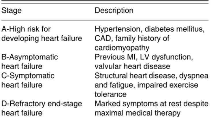
Table I. ACC/AHA classification of chronic heart failure(1)
Stage Description
A-High risk for Hypertension, diabetes mellitus, developing heart failure CAD, family history of
cardiomyopathy
B-Asymptomatic Previous MI, LV dysfunction, heart failure valvular heart disease
C-Symptomatic Structural heart disease, dyspnea heart failure and fatigue, impaired exercise
tolerance
D-Refractory end-stage Marked symptoms at rest despite heart failure maximal medical therapy
Table II. New York Heart Association heart failure symptom classification system.
NYHA Class Level of Impairment
I No symptom limitation with ordinary physical activity
II Ordinary physical activity somewhat limited by dyspnea
III Exercise limited by dyspnea at mild work load IV Dyspnea at rest or with very little exertion
www.medigraphic.org.mx
number of donors, as well as the large number of acute CHFdeaths, that continues to stimulate the search for alternative surgical therapies.
Ventricular assist devices (VADs) are designed to connect to the heart or to be placed within the heart to assume some of the workload and to allow the ventricle to rest, undergo reverse remodeling, and recover some of its contractile function. It has been previously demonstrated that the myocardium has the capacity to repair itself during a period of unloading(6,7),
after which some patients are able to resume a normal lifestyle and no longer need cardiac transplantation.
The basic design of the LVAD system is simple: in-flow is through a conduit from the left ventricular (LV) apex to a pump implanted in the pre-peritoneal space, and output from the pump is directed through another conduit into the ascending aorta. Current models have a power cable/driveline that exits the abdominal wall in the right lower abdomen to connect with an external power pack and system controller. Currently available LVADs do not provide oxygenation of the blood, nor removal of waste products.
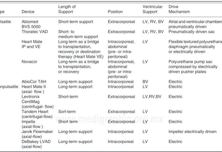
Based on the device related blood flow characteristics, VADs could be classified as nonpulsatile or pulsatile (Table III). When the site of implantation is taken into account me-chanical assist devices could be categorized as extracorpo-real or intracorporeal.. Most of the extracorporeal devices, nonpulsatile and pulsatile, are now used for short to medium-term support. Nonpulsatile devices were designed with either centrifugal or axial flow patterns. Axial flow pumps present the advantage of being small, silent, with no valves, fully implantable, they work in concert with the heart and improve the position of the left ventricle on the Frank-Starling curve. Among axial flow devices Heart Mate II is the most commonly used devices in United States.
PRE-OPERATIVE CONSIDERATIONS
Baseline clinical status
The preoperative clinical status of an LVAD-supported pa-tient depends primarily on the amount of end-organ damage sustained during low-output states prior to VAD implantation,
Table III. Types of ventricular assist devices (VADs).
Length of Ventricular Drive
Type Device Support Position Support Mechanism
Pulsatile Abiomed Short-term support Extracorporeal LV, RV, BV Atrial and ventricular chambers
BVS 5000 pneumatically driven
Thoratec VAD Short- to Extracorporeal LV, RV, BV Pneumatically driven sac medium-term support
Heart Mate Long-term as a bridge Intracorporeal, LV Flexible textured polyurethane IP and VE to transplantation, abdominal diaphragm pneumatically
recovery or destination (pre- or intra- or electrically driven therapy (Heart Mate VE) peritoneal)
Novacor Long-term as a bridge Intracorporeal, LV Polyurethane pump sac to transplantation, abdominal compressed by electrically or recovery (pre- or intra- driven pusher plates
peritoneal)
AbioCor TAH Long-term support Intracorporeal BV Electric Nonpulsatile Heart Mate II Long-term support Intracorporeal LV Electric
(axial- flow )
Levitronix Short-term Extracorporeal LV,RV,BV Electric CentiMag
(centrifugal- flow)
Tandem Heart Sort-term Extracorporeal LV Electric (centrifugal-flow)
Impella Short term Extracorporeal LV Electric (axial-flow )
Jarvik Flowmaker Long-term support Intracorporeal LV Impeller electrically driven (axial-flow)
DeBakey LVAD Long-term support Intracorporeal LV Electric (axial-flow)
www.medigraphic.org.mx
post-implantation complications, and the present surgicalproblem. Many LVAD-supported patients are ambulatory and otherwise uncompromised. Others may exist with varying degrees of renal, hepatic, pulmonary, and/or central nervous system insufficiency. Therefore, careful preoperative evalua-tion of all major organ systems is essential, because any further deterioration in the perioperative period may preclude full re-covery or disqualify a patient from later heart transplantation.
Anticoagulation
One of the most serious complications of extracorporeal cir-culation through the VAD is thromboembolism. During the perioperative period anticoagulation should be maintained with intravenous heparin. Anesthesiologists must determine (perhaps in consultation with the surgeon and the physician managing the VAD) a safe anticoagulation regimen for the perioperative period. The majority of surgical procedures (with the likely exception of neurosurgical cases) can proceed safely in the presence of anticoagulation, however scrupulous attention to hemostasis is required intraoperatively. Fresh frozen plasma or cryoprecipitate may be infused to decrease the level of anticoagulation toward the lower limit of manufac-turer’s recommendations, but it is not advisable to completely reverse anticoagulation. Frequent measurements of partial thromboplastin time (PTT) are important to balance the po-tential complications of hemorrhage and thrombus formation.
Infection
Adherence to strict aseptic technique is mandatory for all invasive procedures and prophylactic perioperative antibiot-ics are routinely employed. While LVAD patients frequently develop infections in their abdominal site of implantation or along the drive-line tunneled through their skin, infection of the LVAD itself is a catastrophic complication, as they are very large foreign bodies that cannot be adequately sterilized with antibiotics.
Pacemakers and implantable cardioverter-defibrillators (ICDs)
Preoperative considerations and practices regarding pacemak-ers and ICDs are much the same in LVAD-supported patients as in other patients. The pacemaker mode is ascertained and it is interrogated for proper functioning. Usually, atrio-ventric-ular sequential pacing will be in use (DDD or DOO mode), because this frequently preserves RV output (and therefore LV filling) in these patients. Magnets should be available in case of pacemaker malfunction. Modern pacemakers will usually convert to an asynchronous mode (e.g., AOO, VOO, or DOO) when a magnet is applied, and should revert to
their prior programming when the magnet is removed. In any event, pacemaker dependent patients should have their device interrogated postoperatively to assure proper functioning. Many, but not all LVAD patients will have an ICD. Unipo-lar electrocautery emits a high-frequency signal that could potentially be interpreted as ventricular fibrillation, resulting in unnecessary defibrillatory discharges. Consequently, ICDs are usually deactivated in the immediate preoperative setting, assuming that a defibrillator is immediately available. Where possible, bipolar electrocautery should be preferentially used. In an emergency situation, a magnet may be used to deacti-vate the ICD. Most ICDs (Medtronics, St. Jude) will remain deactivated as long as the magnet remains in place.
Advanced cardiac life support (ACLS) protocols
Cardiovascular collapse in LVAD patients is treated with standard advanced cardiac life support (ACLS) protocols. However, one should never perform chest compressions on a VAD-supported patient because dislodgement of intracardiac cannulae will result in certain death.
Specific intra-operative considerations
LVAD-supported patients will be transported to the operat-ing room with the device runnoperat-ing on battery power. As with all critical life-support equipment in the operating room, an LVAD must be connected to a reliable power supply as soon as feasible because battery life is limited.
Anesthetic management
The pump’s pre-peritoneal location places the LVAD-sup-ported patient at increased risk for aspiration. Consequently, «full stomach» precautions (e.g., gastric acid prophylaxis and rapid sequence induction with cricoid pressure) should be considered. Extubation criteria for the LVAD-supported patient are the same as in any other patient.
While LVADs do not specifically contraindicate any par-ticular anesthetic agents, the anesthetic plan must consider the potentially dysfunctional unassisted right ventricle (RV), and particular attention must be paid to optimizing RV preload, after load, and inotropic support as required. The anesthetic drugs chosen should be appropriate for the planned operation, and should take into account any alterations of physiology resulting from insufficiency of, or prior injury to, major organ systems. For example, succinylcholine may be contraindicated in patients with recent cerebrovascular accidents, and it may be disadvantageous to use pancuronium in the patient with renal insufficiency.
www.medigraphic.org.mx
Este documento es elaborado por Medigraphic
must be individualized. Inotropes, vasodilators, andvasopres-sors are administered to achieve optimal hemodynamics. LVADs are typically set to automatically eject as soon as the blood chamber is full. Thus, the faster the device fills, the faster it pumps and the higher the pump output. Hypovolemia predictably results in slow pump filling, decreased LVAD out-put, and hypotension. Consequently, the goal of fluid manage-ment is to maintain normal or slightly elevated intravascular volume. Markedly increased systemic vascular resistance (SVR) impairs forward flow, resulting in incomplete pump emptying, which leads to stagnation of blood in the pump and increased risk of thrombosis. Therefore, maintenance of normal or slightly low SVR is desirable.
Depth of anesthesia is often judged, in part, by hemody-namic parameters, but tachycardia and hypotension are not re-liable indicators of the depth of anesthesia in LVAD-supported patients. As discussed above, LVADs eject as soon as the blood chamber fills, and it is this rate of ejection which con-stitutes the LVAD-supported patient’s pulse rate. One should therefore consider that absence of tachycardia is not indicative of an adequate depth of sedation. Similarly, intraoperative tachycardia (should it occur) is reflective only of the speed of LVAD filling, and is not a reliable sign of light anesthesia. On the other hand, while relative hypertension is most likely reflective of relative volume overload and higher pump out-puts, it could also reflect heightened adrenergic activity with increased systemic vascular resistance accompanying light anesthesia. Nevertheless, lack of an acutely increased blood pressure with surgical stimulation is not always a reliable indicator of adequate depth of anesthesia. Finally, one should also consider that the pulse rate is rarely going to be the same as the ECG-derived heart rate in this population, as this may have implications for automated record keeping systems.
Surgical positioning
The effect of surgical positioning on venous return must be considered, as adequate preload is the most important factor in maintaining LVAD output. Fluid management must be individualized, and inotropes, vasodilators, and vasopressors should be used as necessary to continuously create optimal hemodynamics.
Intraoperative monitoring
Electrocardiograph (ECG), pulse oximetry, end-tidal carbon dioxide, temperature, and blood pressure are standard for patients undergoing general anesthesia, and the LVAD-supported patient is no exception. LVAD command consoles offer continuous digital readouts of the effective cardiac output (VAD-output). Arterial pressure monitoring catheters are generally inserted for accurate monitoring, since patients
with axial flow devices have low pulse pressure which leads to inaccurate blood pressure cuff and pulse oximetry reading. Procedures anticipated to produce large swings in blood pres-sure (e.g., resection of a pheochromocytoma), for frequent arterial blood sampling (e.g., during thoracic surgery with one-lung ventilation) or hemodynamically unstable patients also require invasive blood pressure reading.
Central venous pressure (CVP) monitoring is used when large fluid shifts are anticipated. As explained above, op-timal LVAD function depends on adequate intravascular volume. However, for a variety of reasons, LVADs increase the risk of RV failure. Firstly, the high output from an LVAD will increase RV preload, and sometimes this alone is enough to cause RV failure in patients with moderate-to-severe RV dysfunction. Secondly, decompression of the left ventricle by an LVAD causes a leftward shift of the interventricular septum, resulting in altered RV geometry, increased RV compliance, and decreased RV contractil-ity(8). Thirdly, moderate-to-severe tricuspid regurgitation
occasionally results from displacement of the papillary muscle attached to the interventricular septum as it is shifted leftward. Finally, while an optimally functioning LVAD will reduce RV after load and often improve RV function in patients with normal pulmonary vascular resistance (PVR), patients with fixed, elevated PVR may actually experience an increased RV after load, due to increased right-sided and PA flows(9). In addition to monitoring CVP to detect
developing RV failure and guide fluid management, central access is useful for drug infusions and the potential intro-duction of a transvenous pacing wire. Additionally, one can calculate SVR in the LVAD-supported patient with an indwelling CVP monitor by substituting the VAD output for the cardiac output in the hemodynamic formula. The calculation would then be as follows:
SVR = [(MAP – CVP)/LVAD output] x 80 dynes-sec/cm(5)
In general, however, central catheters are a potential source of sepsis, and should be avoided when not absolutely necessary.
www.medigraphic.org.mx
the anticipated benefits. If the patient has a CVP catheter,SVR can be calculated without a PAC as outlined above and, though it is not quantitative, one can surmise that the SVR has abruptly increased in the LVAD-supported patient when
the residual volume in the pump abruptly increases. Trans-esophageal echocardiography (TEE) is the intra-operative monitor of choice if there is concern about failure of an unassisted ventricle.
REFERENCES
1. Hunt SA, Baker DW, Chin MH, et al. ACC/AHA Guidelines for the evaluation and management of chronic heart failure in the adult: execu-tive summary: a report of the American College of Cardiology/American Heart Association Task Force on Practice Guidelines (Committee to Revise the 1995 Guidelines for the Evaluation and Management of Heart Failure): developed in collaboration with the International Society for Heart and Lung Transplantation; endorsed by the Heart Failure Society of America. Circulation 2001;104:2996-3007.
2. Francis GS, Goldsmith SR, Levine TB, et al. The neurohumoral axis in congestive heart failure. Ann Int Med 1984;101:370-377.
3. Levine B, Kalman J, Mayer L, et al. Elevated circulating levels of tumor necrosis factor in severe chronic heart failure. New Engl J Med 1990;323:236-241. 4. Taylor DO, Edwards LB, Boucek MM, Trulock EP, Keck BM, Hertz
MI. The Registry of the International Society for Heart and Lung Transplantation: twenty-first official adult heart transplant report-2004. J Heart Lung Transplant 2004;23:796-803.
5. Miller LW, Lietz K. Candidate selection for long-term left ventricular assist device therapy for refractory heart failure. J Heart Lung Transplant 2006;25:756-64.
6. Frazier OH, Benedict CR, Radovancevic B, et al. Improved left ven-tricular function after chronic left venven-tricular unloading. Ann Thorac Surg 1996;62:675–681.
7. Grigore A, Poindexter B, Vaughn WK, Nussmeier N, Frazier OH, Cooper JR, Gregoric I, Buja LM, Bick R. Alterations in Alpha adrenoreceptor density and localization after mechanical left ventricular unloading with the Jarvik flowmaker left ventricular assist device. Journal of Heart and Lung Transplantation 2005;24:609-613.
8. Santamore WP, Gray LA. Left ventricular contributions to right ventricular systolic function during LVAD support. Ann Thorac Surg 1996;61:350-6.