Genomic instability associated to impairment of Fe-S
clusters synthesis in Saccharomyces cerevisiae
yeast cells
José Carlos Aires Maria
Dipòsit Legal: L.1237-2014 http://hdl.handle.net/10803/275980
Genomic instability associated to impairment of Fe-S clusters synthesis in Saccharomyces cerevisiae yeast cells està subjecte a una llicència de Reconeixement-NoComercial-SenseObraDerivada 3.0 No adaptada de Creative Commons
Les publicacions incloses en la tesi no estan subjectes a aquesta llicència i es mantenen sota les condicions originals.
Genomic instability associated to
impairment of Fe-S clusters
synthesis in
Saccharomyces
cerevisiae
yeast cells
José Carlos Aires Maria
(Graduate in Microbiology)
Thesis submitted to fulfill the requirements for the
degree of Doctor, held under the scientific guidance of
Ao meu anjo da guarda
It is like life and destiny: we think we know the story, but
Pedra filosofal
Eles não sabem que o sonho é uma constante da vida tão concreta e definida
como outra coisa qualquer,
como esta pedra cinzenta em que me sento e descanso, como este ribeiro manso em serenos sobressaltos, como estes pinheiros altos
que em verde e ouro se agitam, como estas aves que gritam em bebedeiras de azul. Eles não sabem que o sonho é vinho, é espuma, é fermento,
bichinho álacre e sedento, de focinho pontiagudo, que fossa através de tudo num perpétuo movimento. Eles não sabem que o sonho
é tela, é cor, é pincel, base, fuste, capitel, arco em ogiva, vitral, pináculo de catedral, contraponto, sinfonia, máscara grega, magia, que é retorta de alquimista,
mapa do mundo distante, rosa dos ventos, infante, caravela quinhentista, que é Cabo da Boa Esperança, ouro, canela, marfim,
florete de espadachim, bastidor, passo de dança, Colombina e Arlequim, passarola voadora, pára-raios, locomotiva, barco de proa festiva, alto-forno, geradora, cisão do átomo, radar, ultra som, televisão
desembarque em foguetão na superfície lunar. Eles não sabem, nem sonham,
que o sonho comanda a vida. Que sempre que um homem sonha o mundo pula e avança
como bola colorida
entre as mãos de uma criança.
A
A
CK
C
KN
N
OW
O
WL
LE
ED
DG
GM
ME
EN
NT
TS
S
Researchers are awkwardly social beings, but, in spite of that, they can not live isolated from the society in which they work. They are afflicted by the same injustices, struggles and lack of social awareness as other citizens, have problems, questions, suffer from loneliness, are happy in various degrees, receive a certain degree of support in the pursuit of their goals, have or have no friends. Despite the difficulties with which PhD candidates are confronted (economic, isolation, loneliness), the thesis presented here and the investigation that led to it had numerous supporters - some unexpected, some more expected, and others because of intense friendships. It is, therefore, my pleasure to list each supporter and gratefully acknowledge their contribution.
This thesis is not only the result of extensive hours of study, reflection and work during the many phases that were required for its completion. It is also the culmination of an academic goal that I pursue. It would not have been possible without the help of a considerable number of people.
Començo agraint als meus directors, primer al Dr Enric Herrero per acceptar-me en el seu laboratori i donar-me la possibilitat d'entrar en aquesta aventura que va començar fa gairebé 5 anys. Gràcies per la teva paciència, la teva preocupació pel meu benestar i per compartir la teva saviesa i, sobretot, aquesta inquietud contagiosa que t'envolta. En segon lloc, la Dra Gemma Belli que més que una consellera vaig tenir el plaer de tenir-te com una companya. Gràcies pel teu consell, la crítica i la teva disposició a suavitzar sempre els mals resultats que estava tenint. També una paraula d'agraïment a la Dra Celia Casas per la seva preocupació constant en fer les coses bé i més que res en la raó per fer-les.
Una paraula afectuosa als meus companys de laboratori que amb el temps es van convertir en amics, la Laia, la Judit, la Maria, el Jordi, el David, l’ Andrés, el Sergi i el Fernando per fer d'aquest meu segon llar un lloc més agradable.
També vull agrair a les meves segones mans perquè sense la seva participació aquest treball no es va fer d'una manera suau, em refereixo a la Mireia, a la Esther, a la Meri i a la Sílvia, les hi dono les gràcies per la seva ajuda i disponibilitat.
Agradeço aos meus pais, às minhas madrinhas (em principal a “madrinha” Cecília) e ao resto da minha família por me terem dado todas as oportunidades para que pudesse chegar onde estou. Uma palavra de apreço ao “pai” Guerra que foste tu o principal culpavél de estar nesta situação e mais recentemente ao meu “veio” Zé Luis que com a tua boa disposição contagiante fizeste desta minha travessia um passeio na praia de Copacabana.
do ar luso que tanto falta sinto.
La Moma i l’Anabel per la seva amistat, suport i constant preocupació per mi. A la Ana y a José mi "tocayo" guatemalteco por aguantarme y por hacereme sentir que no soy un extraño, sino uno más como ellos. José con tu buen gusto por los coches, comida, música y programas de tele me has hecho olvidar que eres de los maristas.
To Charu, Disha, Upasana and Arindam for accepting me into your circle of friends and show me the beauty that is the Indian culture. I am eternally grateful to consider me as a friend.
To Hiren, Rinku and Venky for sharing a bit of their lives with me.
Ana con tu “broken english” y amistad, gracias por compartir las cenas, alegrías y penas.
Alejandra, Ayax y Eric, guardaré vuestra amistad mexicana en el corazón con mucho cariño.
Bernarda, ha sido con un enorme placer conocerte y muchas gracias desde el corazón por tu amistad.
Ingrid, estimada amiga gràcies per l'acollida afectuosa a Avignon i Dublín. En tots dos viatges ens has fet sentir com a casa. Sergi gràcies per ser un bon guia i per ajudar-me en els aspectes tècnics d'aquesta tesi.
A la familia Camarasa, René, Sandra, madre, Tere, Lay, Moni y Héctor por haberme hecho sentir parte de vuestra familia muy divertida y peculiar en la que me considero un miembro más.
Aan Paula, die ik beschouw als een goede vriend, een speciaal woord van dank voor het laten meebeleven van vele onvergetelijke en ontstressende wandelingen.
Ao “Ti Alex” que de Barcelona foste a melhor recordação que mantenho no coração. Quem diria que uma década depois estaria nesta posição de defender uma tese, hein? E a sua respectiva e doce QiaoQiao que com a sua amizade nos cativou para sempre.
A minha “primeira filhota” Tânia que desde o nosso encontro aqui em Lleida nunca mais deixamos de estar em contacto. Um abraço forte ao Bruno e ao vosso rebento.
Aos meus velhos amigos de sempre, Sidónio, Sérgio, Pedro, Martinha, Pedro Reis e Chicão que como eu sabem o que é ser emigrante e o que isso representa.
Por último mas não menos importante, a minha família portuguesa aqui em Lleida, Filipe, Tânia e Alex, um muito, mas muito obrigado por me terem adotado como mais um membro da família e me fazerem sentir amado e mimado.
Distracted like I am, more names should appear here but it would extend the number of pages and increase the price of this thesis and that my advisor would not thing so funny, so...
Grx5 is one of eight proteins of the glutaredoxin (GRX) family in Saccharomyces cerevisiae. GRXs are thiol oxidoreductases widely spread among prokaryotic and eukaryotic organisms. Yeast Grx5 is located at the mitochondrial matrix and participates in the synthesis of iron-sulphur clusters (ISCs), which are co-factors required for several essential cellular processes, such as respiration, photosynthesis, nitrogen fixation, ribosome biogenesis, regulation of gene expression and DNA-RNA metabolism. Cells lacking Grx5 display several phenotypes: (i) inability to grow on minimal medium or in the presence of a non-fermentable carbon source, (ii) hypersensitivity to external oxidants, (iii) iron accumulation inside the cell, (iv) increase in protein oxidative damage, and (v) defects in the activity of enzymes requiring ISCs as cofactors, like aconitase or succinate dehydrogenase. These phenotypes are similar to those of other ISC mutants. Some ISC proteins are involved in DNA metabolism, more precisely in DNA replication and/or repair processes. Recent works reported that defects in ISC metabolism compromise the stability of the cellular genome, and this instability is associated to human predisposition towards multiple types of cancers. Using the yeast grx5 mutant as a model we wanted to gain further insight in the relationship between genomic instability and defects in ISC biosynthesis.
La proteína Grx5 de Saccharomyces cerevisiae es una de las ocho proteínas de la familia de las glutaredoxinas (GRXs) en esta especie de levadura. Las GRXs son tiol oxidoreductasas ampliamente distribuídas entre organismos procariotas y eucariotas. La proteína Grx5 de levadura se encuentra en la matriz mitocondrial y participa en la síntesis de los centros hierro-azufre (ISCs), que son co-factores necesarios para varios procesos celulares esenciales, tales como la respiración, la fotosíntesis, la fijación de nitrógeno, la biogénesis de los ribosomas, la regulación de la expresión génica y el metabolismo del ADN-ARN. Las células de levadura que carecen de Grx5 muestran varios fenotipos: (i) incapacidad para crecer en medio mínimo o en presencia de una fuente de carbono no fermentable, (ii) hipersensibilidad a oxidantes externos, (iii) acumulación de hierro dentro de la célula, (iv) aumento del daño oxidativo en las proteínas, y (v) defectos en la actividad de enzimas que requieren ISCs como cofactores, tales como la aconitasa o la succinato deshidrogenasa. Estos fenotipos son similares a los de otros mutantes en la biogénesis de ISCs. Algunas proteínas asociadas a ISCs están involucradas en el metabolismo del ADN, más precisamente en su replicación o en procesos de reparación del mismo. Trabajos recientes demostraron que los defectos en el metabolismo de ISCs comprometen la estabilidad del genoma celular, y esta inestabilidad está asociada a la predisposición a múltiples cánceres humanos. Mediante el uso de un mutante Δgrx5 de S. cerevisiae como modelo de estudio quisimos abordar la comprensión de la relación entre la inestabilidad genómica y los defectos en la biosíntesis de ISCs.
La proteïna Grx5 de Saccharomyces cerevisiae és una de les vuit proteïnes de la família de les glutaredoxines (GRX) en aquesta espècie de llevat. Les GRXs són tiol oxidoreductases àmpliament repartides entre organismes procariotes i eucariotes. La Grx5 de llevat es troba a la matriu mitocondrial i participa en la síntesi dels centres ferro-sofre (ISCs), que són co-factors necessaris per a diversos processos cel·lulars essencials, com ara la respiració, la fotosíntesi, la fixació de nitrogen, la biogènesi del ribosoma, la regulació de l'expressió gènica i el metabolisme de l'ADN-ARN. Les cèl·lules de llevat que no tenen Grx5 mostren diversos fenotips: (i) incapacitat per créixer en medi mínim o en presència d'una font de carboni no fermentable, (ii) hipersensibilitat a oxidants externs, (iii) acumulació de ferro dins la cèl·lula, (iv) augment de dany oxidatiu de proteïnes, i (v) defectes en l'activitat d’enzims que requereixen ISCs com a cofactors, com són l’aconitasa o la succinat deshidrogenasa. Aquests fenotips són similars als d'altres mutants en la biogènesi de ISCs. Algunes proteïnes amb ISCs estan involucrades en el metabolisme de l'ADN, més precisament en la replicació de l'ADN o en els processos de reparació del mateix. Treballs recents van demostrar que els defectes en el metabolisme de ISCs comprometen l'estabilitat del genoma cel·lular, i aquesta inestabilitat està associada a la predisposició a múltiples càncers humans. Mitjançant l'ús d'un mutant Δgrx5 de S. cerevisiae com a model d'estudi vam intentar entendre millor la relació entre la inestabilitat genòmica i els defectes en la biosíntesi de ISCs.
A Grx5 de Saccharomyces cerevisiae é uma das oito proteínas que compoêm a família glutaredoxina (GRX) desta espécie de levedura. As GRXs são tiol-oxidorredutases amplamente espalhadas entre os organismos procariotas e eucariotas. A Grx5 de levedura está localizada na matriz mitocondrial e participa na síntese de “clusters” ferro-enxofre (ISCs), que são co-factores necessários para vários processos celulares essenciais, tais como a respiração, a fotossíntese, a fixação de azoto, a biogénese ribossomal, a regulação de expressão génica e no metabolismo de ADN-ARN. As células de levedura sem Grx5 exibem vários fenótipos: (i) incapacidade de crescer em meio mínimo ou em presença de uma fonte de carbono fermentável, (ii) hipersensibilidade aos oxidantes externos, (iii) acumulação de ferro no interior da célula, (iv) aumento do dano oxidativo nas proteínas, e (v) defeitos na atividade das enzimas que requerem ISC como cofator, como por exemplo a aconitase ou o succinato desidrogenase. Estes fenótipos são semelhantes aos de outros mutantes involucrados na síntese de ISC. Algumas proteínas com este tipo de “clusters” estão envolvidas no metabolismo do ADN, mais precisamente na replicação ou em processos de reparação do ADN. Trabalhos recentes relataram que os defeitos no metabolismo destes “clusters” comprometem a estabilidade do genoma celular e esta instabilidade está associada á predisposição humana a vários tipos de cancro. Usando como modelo de estudo um mutante de levedura onde a proteína Grx5 foi retirada queríamos obter mais informações sobre a relação entre a instabilidade genética e os defeitos na biossíntese dos ISCs.
genotóxicos. Além do mais ficou comprovado que os defeitos da progressão do ciclo celular observados em células mutantes Δgrx5 eram específicos da fase S.
5-FOA – 5-Fluorotic acid
AP – apurinic/apyrimidinic
AcLi – lithium acetate
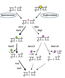
BER – base excision repair
BPS – bathophenanthrolinedisulfonic acid
CDK – cyclin-dependent kinase
CIA – cytosolic iron–sulphur protein assembly
CPD – cyclobutane pyrimidine dimers
CPT – camptothecin
ctDNA – chloroplasts DNA
DDR – DNA damage response
DIG-dUDP – dioxigenin-dUDP
dNTP– deoxyribonucleotide triphosphate
DSB – double strand break
dsDNA – double-stranded DNA
FACS – fluorescence-activated cell sorting
Fe-S – iron sulphur
FRDA – Friedreich ataxia
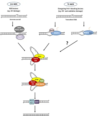
GG-NER – global genomic NER
GPX – glutathione peroxidase
GRX – glutaredoxin
GSH – glutathione
GSSG – oxidized glutathione
GST – glutathione-S-transferase
HR – homologous recombination
HU – hydroxyurea
ICL – interstrand cross-linked repair
ISC – Fe-S cluster
MDA – malondialdehyde
MetO – methionine sulfoxide
MMS – methyl methane sulfonate
NAC – N-acetyl cysteine
NER – nucleotide excision repair
NIF – nitrogen fixation
MMR – mismatch repair
MSR – methionine sulphoxide reductase
mtDNA – mitochondrial DNA
nDNA – nuclear DNA
NHEJ – nonhomologous end join
PAPS – 3′-phosphoadenosine 5′-phosphosulphate
PCR – polymerase chain reaction
PEG – polyethylene glycol
PHGPX – phospholypid hydroperoxide GPXs
PRR – post-replication repair
PRX – thioredoxin peroxidase
RPA – replication protein A
RNAP II – RNA polymerase II
RNR – ribonucleotide reductase
RNS – reactive nitrogen species
ROS – reactive oxygen species
SOD – superoxide dismutase
SUF – sulphur mobilization
ssDNA – single-stranded DNA
UV – ultraviolet
t-BOOH – ter-butyl hydroperoxide
TC-NER – transcription coupled NER
TD – thymine dimer
TLS – translesion synthesis
TRX – thioredoxin
XP – xeroderma pigmentosum
Table 1. Yeast strains employed in this thesis. ... 51
Table 2. Plasmids employed in this study. ... 54
L
L
IS
I
ST
T
O
O
F
F
F
FI
IG
G
UR
U
R
ES
E
S
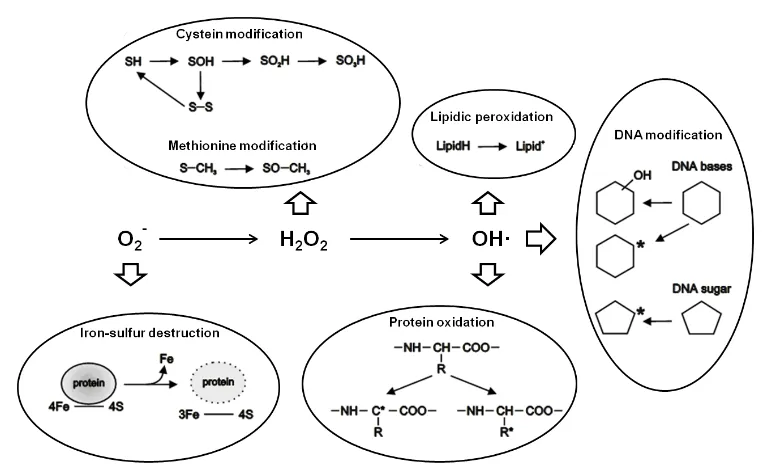
Fig. 1. Scheme of the ROS reactivity with biological molecules ... 2
Fig. 2. TRX-fold domain ... 9
Fig. 3. Reaction mechanisms of the GRX and TRX systems ... 10
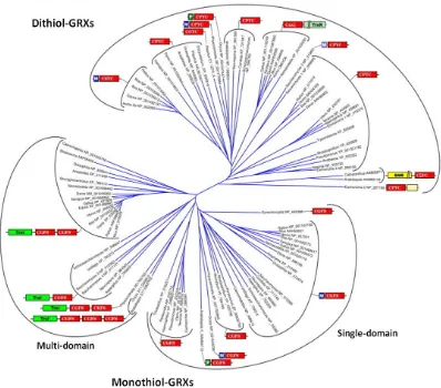
Fig. 4. Classification of GRXs based on phylogeny, active site and domain structure ... 11
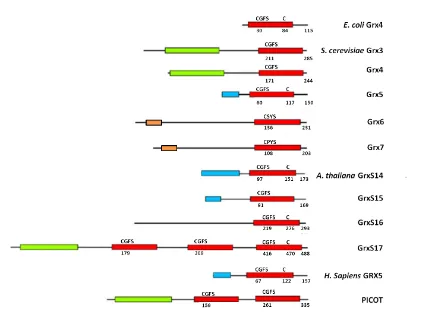
Fig. 5. Domain structure of monothiol GRXs from different organisms ... 14
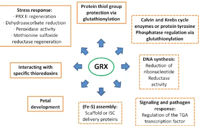
Fig. 6. Confirmed and proposed roles for plant GRXs ... 19
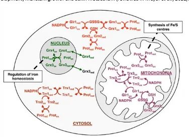
Fig. 7. Components of the S. cerevisiae TRX and GRX systems at the nucleus, mitochondria and cytosol ... 20
Fig. 8. Structures of the most commonly found ISCs ... 21
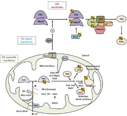
Fig. 9. ISC biogenesis in S. cerevisiae, including ISC assembly, ISC export, and CIA machinery ... 24
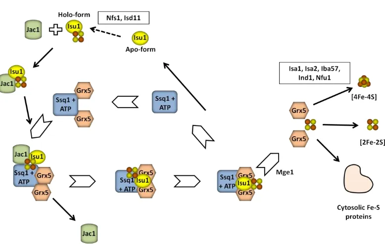
Fig. 10. Working model for the roles of the mitochondrial chaperone Ssq1-Jac1 and the glutaredoxin Grx5 in Fe-S protein maturation in eukaryotes ... 27
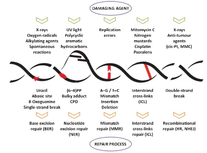
Fig. 11. nDNA repair mechanisms... 34
Fig. 12. The BER pathway ... 35
Fig. 13. The NER pathway including the corresponding subpathways, GG-NER and TC-NER ... 36
Fig. 14. The DNA DSB repair by NHEJ and HR ... 39
Fig. 15. Scheme of MMR ... 41
Fig. 16. Scheme of the ICL repair. 3’ ends are indicated by the arrowheads ... 42
Fig. 17. Scheme of PRR ... 44
Fig. 18. Cell cycle and the DNA damage checkpoints in S. cerevisiae ... 45
Fig. 19. Scheme of Mec1-Rad53-Dun1 checkpoint kinase pathway in S. cerevisiae. ... 46
Fig. 20. Schematic representation of the leu2-k::URA3-ADE2::leu-2K chromosomal recombination system ... 73
Fig. 21. Δgrx5 mutant cells display increased recombination rate ... 74
Fig. 22. Δgrx5 mutant cells display high spontaneous mutation frequency ... 75
Fig. 23. The reduced survival of Δgrx5 mutant cells upon UV radiation is independent of their iron content ... 76
Fig. 27. Δgrx5 mutant cells display hypersensitivity to DNA-damaging agents ... 80
Fig. 28. Δgrx5 and Δssq1 mutant cells display hypersensitivity to DNA damage by UV radiation ... 81
Fig. 29. Sensitivity of Δyfh1 mutant cells to UV irradiation ... 81
Fig. 30. Δgrx5 mutant cells display hypersensitivity to DNA-damaging agents in different genetic backgrounds ... 81
Fig. 31. Δgrx5 mutant cells display hypersensitivity to MMS in anaerobic conditions. ... 82
Fig. 32. A tetO7-GRX5 conditional mutant displays hypersensitivity to MMS ... 83
Fig. 33. Δgrx5Δssq1 mutant cells show additive hypersensitivity to DNA damage by UV radiation, HU and MMS compared to single mutants ... 84
Fig. 34. Δgrx5Δssq1 mutant cells display a hypersensitivity phenotype in the presence of diverse oxidants ... 85
Fig. 35. Overexpression of SSQ1 rescues the sensitivity defects of Δgrx5 mutant cells ... 86
Fig. 36. Scheme of the experimental design for cell cycle synchronization, G1 release and sample collection ... 87
Fig. 37. Cell cycle progression from G1 to G2 is delayed in Δgrx5 and Δssq1 mutants ... 88
Fig. 38. Cell cycle progression from G1 to G2 is delayed in the Δgrx5Δssq1 mutant ... 89
Fig. 39. HU-induced DNA damage causes an increased S-phase delay in the cell cycle progression in the absence of Grx5 ... 90
Fig. 40. The Δgrx5 hypersensitivity to DNA damaging agents is not exacerbated when the NER pathway is compromised ... 91
Fig. 41. TD repair kinetics in wild type, Δgrx5 and Δssq1 mutant cells ... 92
Fig. 42. The Δgrx5 mutant cells display increased hypersensitivity to DNA damaging agents when RAD50 or RAD52 are deleted ... 93
Fig. 43. Both Δrad52Δgrx5 and Δrad50Δgrx5 mutant cells display increased hypersensitivity to antitumor drug CPT. ... 95
Fig. 44. In the absence of Grx5, transcriptional levels of the RNR genes (RNR1-4) do not change. ... 96
Fig. 45. Measures of mRNA relative levels shows no significant differences of any of RNR1-4 mRNAs between both strains. ... 97
T
T
AB
A
B
L
L
E
E
O
O
F
F
C
C
O
O
N
N
T
T
EN
E
N
T
T
S
S
Part I –
Introduction ... 1
1. Oxidative stress ... 1
1.1. General concepts ... 1 1.2. Damage to cellular components ... 1 1.2.1. Lipids ... 2 1.2.2. Proteins ... 2 1.2.3. DNA ... 4 1.3. Cellular effects of ROS in Saccharomyces cerevisiae... 4 1.4. Defence systems against oxidants... 5 1.4.1. Non-enzymatic defence systems ... 5 1.4.2. Enzymatic defence systems ... 7
2. GRX system ... 10
2.1. General aspects ... 10 2.2. Structure of GRXs ... 10 2.3. Classification ... 11 2.4. Dithiol GRXs ... 12 2.5. Monothiol GRXs ... 13 2.5.1. Monothiol GRXs in yeast... 14 2.5.2. Monothiol GRXs in human cells ... 16 2.6. The GRX family in photosynthetic organisms ... 16 2.6.1. Subcellular localization of plant GRXs ... 17 2.6.2. The roles of plant GRXs... 18 2.7. Relation between TRX & GRX systems ... 19
3. Iron-sulphur clusters ... 21
4.1. Nuclear and mitochondrial genomes in S. cerevisiae... 31 4.2. DNA genome instability ... 32 4.3. DNA damage repair systems ... 33 4.3.1. Base Excision Repair ... 34 4.3.2. Nucleotide Excision Repair ... 35 4.3.3. Double strand breaks repair ... 38 4.3.4. Mismatch repair ... 41 4.3.5. Interstrand cross-links repair ... 42 4.3.6. Post-replication repair ... 43 4.4. Cell cycle and Mec1-Rad53-Dun1 dependent DNA damage checkpoint ... 44
Part II –
Objectives ... 49
Part III –
Material and Methods... 51
1. Yeast strains and plasmids ... 51
2. Cell culturing ... 55
2.1. Growth conditions ... 55 2.2. Growth media ... 55 2.2.1. Growth medium for E. coli cells... 55 2.2.2. Growth medium for S. cerevisiae cells ... 55 2.3. Additional compounds ... 56
3. Sensitivity analyses ... 56
4. Extractionof S. cerevisiae genomic DNA ... 57
5. Recombinant DNA methods ... 58
5.1. Gene cloning ... 58 5.2. Plasmid purification from E. coli cultures ... 58 5.2.1. Miniprep technique ... 58 5.2.2. Jet-prep technique ... 59 5.3. Transformation methods for E. coli and S. cerevisiae ... 59 5.3.1. Transformation of E. coli ... 59 5.3.2. Transformation of S. cerevisiae ... 59
6. Construction of null and multiple mutants in S. cerevisiae ... 60
7. Genetic methods ... 61
7.2. Spontaneous mutation frequencies ... 61
8. Analysis of gene expression by Northern blot ... 62
8.1. Solutions... 62 8.2. Sample collection ... 62 8.3. Total RNA extraction ... 63 8.4. Labelling of DNA probes with Dioxigenin-dUDP (DIG-dUDP) ... 63 8.5. Formaldehyde-agarose gel electrophoresis ... 64 8.6. Transfer to Nylon+ membrane and UV crosslinking ... 64 8.7. Hybridization and washing steps... 65 8.8. Chemiluminescent detection ... 65
9. Microscopic techniques ... 65
10. Cell cycle synchronisation and flow cytometry analyses ... 66
10.1. Sample collection ... 66 10.2. Propidium iodide staining ... 66 11. Thymine dimers detection ... 67 11.1. Solutions ... 67 11.2. Protocol ... 67
12. Determination of intracellular iron content ... 70
Part IV –
Results ... 73
1. The absence of the mitochondrial protein Grx5 leads to increased genetic instability .. 73
2. The high recombination and spontaneous mutation rates in the Δgrx5 mutant cells are
not provoked by iron overloading ... 75
3. Formation of Rad52-associated foci is increased in Δgrx5 mutant cells ... 77
4. Δgrx5 mutant cells are hypersensitive to DNA-damaging agents ... 79
5. A GRX5 conditional expression mutant displays the same hypersensitivity as Δgrx5
mutant cells to MMS ... 82
6. Δgrx5Δssq1 mutant cells shown an additive effect of hypersensitivity to DNA-damaging
agents compared to the single mutants ... 83
7. Overexpression of SSQ1 rescues the sensitivity phenotype presented of the Δgrx5
mutant against genotoxics agents ... 85
8. The absence of Grx5 and Ssq1 proteins causes a delay in the cell cycle S-phase
progression ... 86
8.1. The defects in cell cycle progression in Δgrx5 mutant cells are specific of the S-phase . 89
11. Members of the RAD52 epistasis group are affected by the action of the antitumor
drug CPT ... 94
12. Mec1-Rad53-Dun1 checkpoint kinase pathway seems not be affected in Δgrx5 mutant
cells ... 96
13. Overexpression of PRI2, RAD3 or NTG2 cannot rescue the sensitivity defects of cells
lacking GRX5 to DNA damaging agents ... 97
Part V –
Discussion ... 101
Part VI –
Conclusions/ Conclusiones ... 113
Part VII –
References ... 119
P
P
A
A
R
R
T
T
I
I
I
I
I
N
N
N
T
T
T
R
R
R
O
O
O
D
D
D
U
U
U
C
C
C
T
T
T
I
I
I
O
O
O
N
N
N
1.OXIDATIVE STRESS
1.1.General concepts
In an oxidative environment like the one we live in, aerobic life generates oxidants. As a consequence, both photosynthetic and non-photosynthetic organisms are exposed to potential damage by these oxidants to diverse cell components including membrane lipids, proteins and DNA. Various oxidants can be generated under oxidative stress, which occurs when the concentration of these oxidants increases beyond the antioxidant buffering capacity of the cell, as well as during the course of normal metabolism in certain cell compartments (Michelet et al., 2006). There are two main oxidant categories: reactive oxygen species (ROS), and reactive nitrogen species (RNS). Oxygen-cotaining chemical species where that oxygen can assume various oxidation states that include the singlet oxygen, the superoxide anion (O2-), hydrogen peroxide
(H2O2), the highly reactive hydroxyl radical (OH·) and peroxide radicals are considered
ROS (Toledano et al., 2003). In contrast, when oxygen is combined with nitrogen and forms nitric oxide radicals (NO-) and peroxynitrite radicals (ONOO-), these species are described as RNS. To protect their cell components against these oxidants different organisms have evolved several defence mechanisms. Understanding all the processes that are involved in the oxidative stress is of critical importance in biomedical research, since oxidative damage has been related to major human diseases including atherosclerosis, diabetes, cardiovascular diseases, cancer, neurodegenerative disorders (such as Alzheimer, Parkinson, Huntington or Friedreich ataxia), or chronic liver and lung diseases (Ames et al., 1993).
1.2.Damage to cellular components
is associated to its ability to release iron from proteins containing iron-sulphur (Fe-S) clusters (ISCs), like aconitase, which leads to both damage of these proteins and further generation of other ROS through autocatalytic cyclic reactions. Nevertheless, this toxicity might be associated to an iron deprived state due to increased demand for reassembling dissociated ISCs (Toledano et al., 2003).
Fig. 1. Scheme of the ROS reactivity with biological molecules.
1.2.1.Lipids
A lipid peroxidation chain reaction is initiated with the formation of OH· starting with the hydrogen removal from an unsaturated lipid side chain. As a consequence, a first radical is produced which can accept a dioxygen (O2) molecule to form a peroxyl radical
(LOO·) and be transformed to an alkoxyl radical (LO·) to carry on an autocatalytic peroxidation chain reaction. This reaction will be interrupted when two lipid radical molecules combine or in the presence of radical scavengers like alpha-tocopherol (Toledano et al., 2003). The products derived from lipid peroxidation and some of their by-products, such as 4-hydroxynonenal and malondialdehyde (MDA), quickly react with other biological molecules, mainly forming adducts with proteins and DNA bases (Møller et al., 2007).
1.2.2.Proteins
irreversible oxidation of these same cysteine residues and the modulation of protein function (Ghezzi and Bonetto, 2003). Depending on the ROS exposure, aggregation or fragmentation of proteins in vitro can occur. Generally, OH· preferentially promotes protein aggregation, but in the presence of O2, it mainly leads to protein fragmentation.
Under in vitro conditions, OH· initiates protein damage by either attacking the alpha carbon or the amino acid side chain of proteins, forming a carbon radical which initiates an autocatalytic chain reaction that is somewhat similar to that described for lipids. In addition to the generic damage just described, OH· radicals cause the hydroxylation of some amino acids, such as phenylalanine, tyrosine, arginine, histidine and lysine (Toledano et al., 2003). There are several classes of damage that ROS and RNS can cause to protein. These classes are described below.
Carbonylation: Protein carbonylation is an irreversible oxidative damage which leads to a loss of protein function. The oxidation of proteins by ROS generates reactive carbonyl derivatives. These derivates are formed by a variety of oxidative pathways, such as direct oxidation of the protein backbone leading to the formation of protein fragments with an N-terminal α-ketoacyl amino acid residue, oxidation of some amino acid side chains (particularly, histidine, arginine or lysine) into ketone or aldehyde derivatives, reaction with products of lipid peroxidation (such as 4-hydroxy-2-nonenal), or conjugation with reducing sugars (glycation) or their oxidation products (glycoxidation). Protein carbonylation is considered a good indicator of the oxidative damage extent associated with various conditions of oxidative stress, aging and physiological disorders, including neurodegenerative diseases (Ghezzi & Bonetto, 2003; Dalle-Donne et al., 2006).
Nitrosylation: This process can be reversed by protective mechanisms. The covalent attachment of the NO- to cysteine thiol is a post-translational modification that potentially regulates protein functions (Jaffrey et al., 2001). Protein and glutathione thiols can react with NO- derivatives leading to a variety of products including disulfides, sulfenic, sulfinic and sulfonic acids, as well as S-nitrosothiols (Costa et al., 2003). The most common NO- derivative peroxynitrite (ONOO-) is formed by the reaction NO- + O2- →
ONOO-. This NO- derivative can cause depletion of sulphydryl groups and other modifications including lipid oxidation, DNA bases deamination, nitration of aromatic amino acid residues, and oxidation of methionine to its sulfoxide (Møller et al., 2007).
the disulfide stability, forward and reverse reactions kinetics, and the nature and redox state of the environment where the reaction occurs (Gilbert, 1995). The oxidation of thiol to disulfide provoked by different ROS (for example H2O2) is a very important metabolic
redox regulation mechanism. Intra/intermolecular disulfide bonds can be formed between cysteine side chains, and the reduced form can be regenerated with the participation of diverse reductases such as the thioredoxin (TRX) or glutaredoxin (GRX) systems (Møller et al., 2007), which will be explained later in detail.
1.2.3.DNA
DNA molecules do not suffer damage directly by O2- and H2O2 themselves;
instead, damage occurs through Haber Weiss and Fenton-catalyzed OH· production (Toledano et al., 2003). As DNA is negatively charged it binds transition metal cations (such as iron) which catalyze the above reactions. Moreover, other radicals which are formed from protein and lipid peroxidation can also induce DNA damage, such as adducts formed from MDA. At least sixty different DNA modifications by the OH· radical are known (Boiteux, 1997), including single and double DNA strand breaks (DSB), abasic site formation, direct base modifications, and DNA cross-links. It has been estimated that endogenous ROS can cause approximately 50,000 to 200,000 apurinic/apyrimidinic (AP) sites per mammalian cell per day (Martin, 2008). Mitochondrial DNA (mtDNA) holds higher steady-state damage compared with nuclear DNA (nDNA). This accumulation is probably caused by local ROS formation and also by the lack of chromatin protection provided by histones (Szczesny et al., 2003).
1.3. Cellular effects of ROS in Saccharomyces cerevisiae
The studies described in this thesis have been carried out with the model organism S. cerevisiae since this yeast provides the combination and integration of cell biological, biochemical, genetic, and genome-wide experimental approaches that allow us to describe the cellular effects of ROS and analyse the mechanisms of defence against oxidative stress. The cellular tolerance of yeast cells to ROS toxicity differs between growth stages. In stationary phase (when nutrients are exhausted) they show higher resistance to stress, including oxidative and thermal ones, than cells in exponential phase (Toledano et al., 2003). Also, cells grown in non-fermentable medium (glycerol), and therefore forced to respire, are more resistant to H2O2 and menadione (a generator of
the major oxidized targets are mitochondrial proteins, in accordance with the fact that mitochondria become the main source of ROS during respiration (Cabiscol et al., 2000). Menadione and H2O2 have distinctive effects on cells at different cell cycle stages. In
asynchronous cell populations H2O2 arrests cells in G2 phase while menadione does it in
G1 (Flattery-O’Brien, 1998). A detailed study examining the cell death induced by high doses of H2O2 reported the presence of several key features of apoptosis (Madeo et al.,
1999).
1.4.Defence systems against oxidants
To maintain the adequate cellular redox state and minimize the damaging effects of ROS, aerobic organisms evolved diverse non-enzymatic and enzymatic antioxidant defence systems. We will start by describing the former type and then review the later type of antioxidant defences.
1.4.1.Non-enzymatic defence systems
Non-enzymatic defence systems typically consist of small molecules acting as antioxidants, which are soluble in either an aqueous or a lipid environment. These defence systems include for example vitamin E, polyamines, ascorbic acid, lipid-soluble antioxidants, metallothioneins, flavohaemoglobin and glutathione.
Vitamin E: Vitamin E functions as an essential lipid-soluble antioxidant that prevents the propagation of free radicals including lipid peroxyl, alkoxyl and hydroxyl radicals. Yeast cells treated with Trolox, a soluble compound analogue of α-tocopherol (the most biologically active form of vitamin E), revealed decreased intracellular oxidation during normal metabolism (Raspor et al., 2005).
Polyamines: Polyamines are small molecules derived from amino acids implicated in protecting yeast against oxidant stress (by holding some ROS, especially O2-).
As they are positively charged, they can interact with negatively charged molecules such as DNA, RNA and proteins. In S. cerevisiae, the polyamines spermine and spermidine have been found to be essential for aerobic growth and the spe2 mutant (unable to synthesize spermidine) was found to be hypersensitive to oxygen (Balasundaram et al., 1993).
oxidation in the presence of metal ions, to induce apoptosis and also to enhance iron toxicity in hemochromatosis (Krzepiłko et al., 2004). In S. cerevisiae, ascorbate is able to protect cells lacking Cu-Zn-dependent superoxide dismutase (SOD) against toxicity of pure oxygen atmosphere and against shortening of replicative life span, and also to rescue auxotrophy for lysine and methionine (Lewinska et al., 2004).
Lipid-soluble antioxidants: Little is known about lipid-soluble antioxidant molecules in S. cerevisiae. However, it has been observed that the lipid membrane composition of yeast is important in giving resistance to oxidative stress. Cells containing membranes with a higher level of saturated fatty acids are more resistant to oxidative stress than those with a higher level of polyunsaturated fatty acids (Howlett & Avery, 1997).
Metallothioneins: Metallothioneins are a family of cysteine-rich low molecular weight proteins with antioxidant properties. They have the ability to bind a varying number of metal ions (such as iron or cupper). CUP1 and CRS5 are yeast genes for metallothioneins that participate in protecting cells against oxidants (Culotta et al., 1994).
Flavohaemoglobin: Flavohaemoglobin is a metalloprotein that participates in the detection and protection against nitric oxide. Yeast flavohemoglobin, YHb1, has been implicated in both the oxidative and nitrosative stress responses in S. cerevisiae. Mutants deficient in Yhb1 are moderately sensitive to oxidants such as diamide, and expression of the gene YHB1 is induced in the presence of oxygen (Zhao et al., 1996).
GSH, such as the participation in xenobiotic detoxification mediated by glutathione-S-transferases (GSTs), or as a substrate for phytochelatin biosynthesis (Deponte, 2013).
1.4.2.Enzymatic defence systems
This type of defences includes several enzymes which are capable of removing oxygen radicals and their products, and/or repair the damage caused by oxidative stress. Catalase, SOD, glutathione and thioredoxin peroxidases, or methionine reductase are some examples of these systems. The thioredoxin and glutathione reductases will be treated in more detail in following sections.
Catalase: Catalase is a homotetrameric iron-containing enzyme with the capacity to catalyse the breakdown of H2O2 to O2 and H2O. S. cerevisiae contains a
peroxisomal and a cytosolic catalase, catalase A and T respectively, encoded by the CTA1 and CTT1 genes (Cohen et al., 1988). The main physiological role of catalase A seems to be the removal of H2O2 produced by fatty acid β-oxidation, while the physiological role of
catalase T is less clear. Yeast mutants in both genes are hypersensitive to H2O2 in
stationary phase and are unable to assemble an adaptive stress response to H2O2 (Izawa
et al., 1996).
Superoxide dismutase: Practically all aerobic organisms possess SOD activity, which converts O2- into H2O2 and O2. This reaction depends on metal co-factors,
such as iron, copper, or manganese. S. cerevisiae, in common with other eukaryotes, possesses two intracellular SODs: a cytosolic Cu-Zn-SOD, encoded by the SOD1 gene, and a Mn-SOD localized in the mitochondrial matrix, encoded by the SOD2 gene (Toledano et al., 2003). SOD1 is essential for protection against O2 toxicity as shown by the array of
aerobic defects occurring in its absence, including reduced growth rate, poor respiratory growth, and lysine and methionine auxotrophy (Longo et al., 1996). In the case of SOD2, this codes for the primary O2- scavenging enzyme for protecting against O2- generated
during respiration, as suggested by the sensitivity to elevated O2 concentrations of the
sod2 mutants and their inability to grow on a non-fermentable carbon source (van Loon et al., 1986).
reduces lipid hydroperoxides esterified to membranes, in addition to soluble peroxides. Initially three GPXs were described in S. cerevisiae, Gpx1, Gpx2 and Gpx3 (Inoue et al., 1999). Later on, a careful alignment of the sequences of these three proteins with GPXs from higher eukaryotes showed higher sequence identities with PHGPXs than with GPXs, suggesting that all three S. cerevisiae GPXs are in fact PHGPXs (Avery & Avery, 2001). Gpx3 is the most functionally important of the three GPX isoforms based on the studies that demonstrated that the gpx3 mutant is more sensitive to hydroperoxides compared to the other two ones (Inoue et al., 1999; Avery & Avery, 2001). This is because, besides its protective role from peroxides during oxidative stress, Gpx3 also regulates and acts as a peroxide sensor of the major oxidative stress transcription factor Yap1 (Delaunay et al., 2002).
Thioredoxin peroxidase (PRX): PRXs, also known as peroxiredoxins, constitute an evolutionary conserved family of antioxidants, using TRX as the physiological electron donor. They reduce both H2O2 and alkyl hydroperoxides to H2O or
the corresponding alcohols, in combination with thioredoxin reductase, TRX and NADPH. All PRXs have a conserved domain containing an active-site cysteine, and depending on the number of cysteine residues required for their activity, they are classified into 1-Cys and 2-Cys PRXs. S. cerevisiae possesses four 2-Cys PRX, and one 1-Cys PRX (Toledano et al., 2003).
Methionine sulphoxide reductases (MSRs): Due to their importance as antioxidants, these reductases are conserved in almost all organisms from bacteria to humans. ROS can oxidize methionine to methionine sulfoxide (MetO) forming two enantiomers, S-MetO and R-MetO. These modifications can be repaired through the action of MSR, with the participation of TRX (as electron donor), thioredoxin reductase, and NADPH (Moskovitz, 2005). In S. cerevisiae, two genes have been described with MSR activities, MSRA that reduces the S stereoisomer of MetO, and MSRB that reduces the R stereoisomer of MetO. Mutants deficient for MSR activity are hypersensitive to H2O2
(Moskovitz et al., 1997).
NADPH. TRXs participate in the reduction of enzymes that form a disulphide during their catalytic cycle, such as RNR [needed for maintaining deoxyribonucleotide triphosphates (dNTPs) pools for DNA synthesis], 3′-phosphoadenosine 5′-phosphosulphate reductase (PAPS reductase, required for sulphur assimilation) or PRX. In general, they may participate in modulating the redox state of cell protein sulphydryls and therefore in protein folding (Herrero et al., 2008). Structurally the TRX-fold domain contains a four or five-stranded β-sheet flanked by three or more α-helices at either side of the β-sheet (Martin, 1995) (Fig. 2). This structural domain is shared with other protein families such as protein disulphide isomerases (Kamitani et al., 1992), GPXs (Epp et al., 1983), GSTs and GRXs. In spite of this common structure, there are no common biological or catalytic functions between these protein families. S. cerevisiae possesses two cytosolic (Trx1, Trx2) and one mitochondrial (Trx3) TRXs. Deletion of genes for both cytosolic proteins is not lethal in yeast cells, although the double mutant has longer cell cycle S-phase and is auxotrophic for sulphur amino acids. Cytosolic (Trr1) and mitochondrial (Trr2) TRX reductases are present in yeast cells, defining two separate cytosolic and mitochondrial systems. Hypersensitivity to hydroperoxides has been observed in a trr2 mutant, pointing to a role of this redoxin system as antioxidant against ROS generated at mitochondria (Pedrajas et al., 1999).
2.GRX SYSTEM
2.1. General aspects
Almost four decades ago, GRXs were discovered as electron donors for the reduction of E. coli RNR, alternatively to TRXs (Holmgren, 1976). Since then, the role of TRXs and GRXs has been largely extended to the determination of their regulatory functions. In the present time, GRXs are described as thiol-disulfide oxidoreductases that regulate protein redox state, but in contrast to TRXs, they employ GSH as a reducing agent instead of the NADPH. The GRX system also includes NADPH and glutathione reductase, which is responsible for the reduction of GSSG (Fernandes & Holmgren, 2004) (Fig. 3). GRXs participates in a broad spectrum of biological processes such as activation of RNR and PAPS reductase, reduction of ascorbate, GSH-mediated reduction of dihydrolipoamide, regulation of the DNA binding activity of nuclear factors, neuronal protection against dopamine-induced apoptosis and excitotoxic mitochondrial damage, and regulation of signal cascades that protect against oxidative stress (Herrero et al., 2010).
Fig. 3. Reaction mechanisms of the GRX and TRX systems. Adapted from Herrero & de la Torre-Ruiz (2007).
2.2. Structure of GRXs
2.3.Classification
[image:41.595.109.507.282.632.2]Depending on the number of cysteine residues at the active site, a division has been established between dithiol and monothiol GRXs. The dithiol GRXs contain the active site consensus sequence CP/YFC, while the monothiol GRXs have a common active site motif (CXXS) with the presence of only one cysteine residue (Rodríguez-Manzaneque et al., 1999; Herrero et al., 2010). Monothiol GRXs can be further categorized into the single-domain monothiol GRXs, consisting of only one GRX domain, and the multidomain monothiol GRXs that contain an N-terminal TRX-like domain and one to three C-terminal monothiol GRX domains. Dithiol or/and monothiol GRXs are widely present in most living organisms.
Fig. 4. Classification of GRXs based on phylogeny, active site and domain structure. Abbreviations used for non-GRX domains: M- mitochondrial signal peptide (blue boxes), P- plastid targeting sequence (dark green), SNR- sulfonucleotide reductase (yellow), Trxl- thioredoxin like (bright green), TrxR- thioredoxin reductase (green marine) (Lillig et al., 2008).
of dithiol and monothiol GRXs, affecting GSH binding (Lillig et al., 2008). For instance, although the important GRX residues for the interaction with the C-terminal glycine of the GSH molecule are conserved between dithiol and monothiol GRXs, this conservation does not occur in the case of the charged residues of the dithiol GRXs, which are responsible for the interaction with the α-glutamyl residue of GSH. The small but significant differences in GSH binding between dithiol and monothiol GRXs may help to explain the biochemical differences between the two types of proteins. Monothiol GRXs are unable to deglutathionylate the small mixed disulfide between β-mercaptoethanol and a glutathione moiety (Herrero & de la Torre-Ruiz, 2007), an assay which is currently employed to determine the enzyme activity of dithiol GRXs, also named as hydroxyethyl disulfide (HED) assay. From an evolutionary point of view it is interesting to note that the monothiol GRXs show a higher degree of homology compared to the dithiol GRXs (Fig. 4). As an example of this conservation, mitochondrial monothiol GRXs represent a compact phylogenetic unit that evolved from a common bacterial origin. Mitochondrial dithiol GRXs, in contrast, seem to have evolved multiple times separately from each other, for instance, in mammals and fungi (Lillig et al., 2008).
2.4. Dithiol GRXs
As above indicated, these low molecular weight proteins are present in all kingdoms of life. Dithiol GRXs were the first GRXs to be characterized at biochemical and structural levels. The first studies with these proteins were made in bacteria. E. coli contains two classical dithiol GRXs (Grx1 and Grx3) and one unusual dithiol GRX (Grx2) (Fernandes & Holmgren, 2004). Grx1 can serve as electron donor for metabolic enzymes like RNR and PAPS reductase. Grx3 cannot normally compensate the loss of Grx1 and its function in vivo is still unclear. Grx2 contains an N-terminal GRX domain followed by an α-helical domain and is structurally similar to the GST family of proteins (Lillig et al., 2008; Alves et al., 2009).
S. cerevisiae contains two classical dithiol GRXs (Grx1 and Grx2) (Luikenhuis et al., 1998). While Grx1 is exclusively located in the cytosol, Grx2 is located in both, cytosol and mitochondria. Alternative translation initiation from two in-frame ATG sites explains the existence of the two isoforms (Porras et al., 2006). Despite the high degree of homology between Grx1 and Grx2 (64% identity), studies with the respective mutants have demonstrated that the grx1 mutant is sensitive to oxidative stress induced by O2-,
whereas the grx2 mutant is sensitive to H2O2 (Luikenhuis et al., 1998). The same studies
superoxide generator menadione. Both yeast GRXs are also active as GPXs and GSTs (Collinson et al., 2002; Collinson & Grant, 2003).
More recently, another dithiol GRX has been described in S. cerevisiae, Grx8 (Eckers et al., 2009). The amino acid sequence of this molecule supports a TRX-fold structure, displaying more sequence homology with dithiol GRXs than with other GRXs. The mutant lacking Grx8 does not show sensitivity to oxidative stress and a fusion form with a green fluorescence protein (GFP) moiety displays a cytoplasmic location (Huh et al., 2003). Further research is needed to understand better the function of this protein.
Human cells contain two dithiol GRXs. The cytosolic dithiol Grx1 is a functional homologue of E. coli and yeast Grx1 (Lillig et al., 2008). Human Grx1 is an electron donor for RNR and it participates in many different cellular processes such as dehydroascorbate reduction, actin polymerization, protection against oxidative stress, apoptosis after acute cadmium exposure, and cellular differentiation (Johansson et al., 2004). Human Grx2 was characterized as a Fe-S protein. Grx2 has two isoforms (nuclear and mitochondrial) derived from the same gene by alternative exon usage (Gladyshev et al., 2001; Lundberg et al., 2001). Grx2 has an unusual motif (CSYC) at the active site, while Grx1 has a CPYC consensus active site sequence. This subtle modification (Ser for Pro) enables the human Grx2 protein to receive electrons from TRX reductase and to complex an ISC (Fernandes et al., 2005). It was proposed that this ISC serves as redox sensor for the activation of the protein in oxidative stress conditions (Johansson et al., 2004).
2.5.Monothiol GRXs
Fig. 5. Domain structure of monothiol GRXs from different organisms. The position and size of mitochondrial/plastid targeting sequences (blue boxes) and GRX (red), TRX-like domains (green) and the transmembrane domain (orange) is indicated. Numbers correspond to the position of the cysteine residue in the active site motif, the position of a partially conserved second C-terminal cysteine and the total length of the proteins. Adapted from Herrero & de la Torre-Ruiz (2007).
2.5.1.Monothiol GRXs in yeast
[image:44.595.85.509.62.393.2]transcriptional activity is precluded by its temporary cytosolic localization and not by iron availability. In this respect, the primary function of Grx3 and Grx4 appears to be the regulation of Aft1 nuclear localization, by interacting with the transcription factor (Ojeda et al., 2006; Pujol-Carrion et al., 2006).
S. cerevisiae Grx5 was first characterized as a GRX localized at the mitochondrial matrix, which participates in the synthesis of ISCs at this location through the ISC pathway (Rodríguez-Manzaneque et al., 2002). This is one of the several pathways employed for the synthesis of the clusters in living cells (see Section 3.3. of this Introduction). Cells lacking Grx5 are not able to grow on minimal medium or in the presence of non-fermentable carbon sources (Rodríguez-Manzaneque et al., 2002). The lack of this protein also leads to an iron accumulation inside the cells and defects in the activity of enzymes requiring ISCs as cofactors, like aconitase and succinate dehydrogenase (Rodríguez-Manzaneque et al., 2002). Grx5 deficiency has also been related to increased protein oxidative damage in yeast cells (Rodríguez-Manzaneque et al., 1999), maybe because the abnormal iron accumulation inside the cell as a result of the disturbance of ISC synthesis and consequent signalling of the intracellular iron state. In accordance with this, inhibition of Grx5 synthesis in human osteoblasts causes increased ROS formation and apoptosis (Linares et al., 2009). Recently, significant progress on characterization of the role of Grx5 in yeast has been made, showing the interaction between this monothiol GRX and a specialized mitochondrial Hsp70 chaperone Ssq1 during the maturation of all cellular Fe-S proteins (Uzarska et al., 2013). In that work it was shown that this chaperone binds the scaffold protein Isu1 (yeast nomenclature), facilitating dissociation of the newly synthesized ISC. Ssq1 also interacts with Grx5 at a binding site different from that of Isu1. Nevertheless, the detailed role of Grx5 in this synthesis is still not well known. Using the yeast grx5 mutant, it has been shown that bacterial, parasite, plant and vertebrate homologues are able to substitute the function of yeast Grx5 (Cheng et al., 2006; Molina-Navarro et al., 2006; Bandyopadhyay et al., 2008b), pointing to a functional conservation during evolution. Yeast Grx5 and some of its homologues in other organisms contain a second cysteine residue at the C-terminal end (Cys117 in S. cerevisiae) as observed in Fig. 5. On the contrary to the essential role of the cysteine residue at the active site motif, Cys117 is not essential for the biological activity of Grx5 (Bellí et al., 2002).
CSYS and CPYS active site motifs, respectively. Detailed analysis of their primary sequences indicates that Grx6 and Grx7 contain a putative transmembrane domain at their N-terminal ends (Fig. 5), which is responsible for the insertion of the molecules at the membranes of the secretory vesicles, followed by a long linker domain and the GRX module. Consistent with this, it has been shown experimentally that Grx6 is localized at the endoplasmic reticulum and Golgi compartments, while Grx7 is mostly at Golgi (Izquierdo et al., 2008).
2.5.2.Monothiol GRXs in human cells
In human cells two monothiol GRXs exist, one cytoplasmic and the other mitochondrial, Grx3 and Grx5 respectively. Grx3, also named protein kinase C (PKC) interacting cousin of TRX (PICOT), consists of a N-terminal TRX domain that lacks a redox active motif and two monothiol GRX domains each containing the active site motif CGFS (Fig. 5). Grx3 was first characterized as a PKC--binding protein in human T lymphocytes that inhibits PKC--dependent activation of JNK protein kinase and the AP-1 and NF-B transcription factors (Witte et al., 2000). Grx3 is an essential protein with suggested functions in a number of regulatory processes, as are embryonic development, immune response, and cardiac physiology. All of these functions are thought to be mediated by the direct and specific interaction with other proteins, through both the TRX and GRX domains, likely in response to redox signaling by ROS (Haunhorst et al., 2010).
A S. cerevisiae Grx5 homolog protein is also present in human cells (referred as GLRX5). Deficiency of this GRX was previously identified as a cause of anaemia in a zebrafish model (Wingert et al., 2005) and of sideroblastic anaemia in a human patient (Camaschella et al., 2007). More recently, it has been demonstrated at cellular level that human GLRX5 is essential for ISC biosynthesis, for the maintenance of normal mitochondria and for cytosolic iron homeostasis in human cells (Ye et al., 2010).
2.6. The GRX family in photosynthetic organisms
Classes I and II include GRXs with C[P/G/S][Y/F][C/S] and CGFS active sites, respectively, which are conserved from cyanobacteria to higher plants, suggesting that they might have fundamental functions for cellular life. The first class includes homologues to the classical dithiol GRXs such as E. coli Grx1 and Grx3, yeast Grx1 and Grx2, and mammalian Grx1 and Grx2. In the case of the second class this includes homologues to yeast Grx3, Grx4, and Grx5 and to E. coli Grx4. In land plants, gene duplication and fusion events have led to the extension of these two classes, which have been divided into five subclasses (GrxC1, C2, C3, C4 and C5 ⁄ S12) for class I and four subclasses (GrxS14, S15, S16 and S17) for class II (Meyer et al., 2012). The nomenclatures of these GRXs are according to the presence of a cysteine or serine residue at the fourth position of the active site motif (CxxC or CxxS). GrxS14 and GrxS15 are small proteins (approximately 170 amino acids) with a single GRX module, therefore similar to yeast Grx5. GrxS16 is larger (around 290 amino acids) with the GRX module linked to an N-terminal extension, therefore similar to yeast Grx3 and Grx4. GrxS17 possesses a TRX-like module in the N-terminal part followed by two to three GRX domains depending on the organisms (Couturier et al., 2009a).
The other four classes have been identified more recently, in particular because they are restricted to specific organisms, suggesting that they are involved in more specific processes. Class III (CC-type and recently named ROXY GRXs) is composed of 21 members in Arabidopsis thaliana; it includes proteins with CC [M/L][C/S/G/A/I] active sites. Class IV is restricted to land plants and eukaryotic photosynthetic organisms (including many algae), in which proteins with a GRX domain followed by four CxxC repeats that may form a zinc finger are present (Navrot et al., 2006). The last two classes are majoritarily restricted to cyanobacteria. Members of class V are present in some other bacteria, whereas class VI appears to be restricted to cyanobacteria (Couturier et al., 2009a; Meyer et al., 2012).
2.6.1.Subcellular localization of plant GRXs
active site, although in plants two or three of these GRXs (GrxC2, GrxC3, or GrxC4) are predicted to be secreted and might correspond to highly abundant GRXs in the phloem sap (Szederkényi et al., 1997). GrxS17 was predicted to be localized in the cytosol/nucleus. Later this co-localization was demonstrated (Rouhier et al., 2008; Cheng et al., 2011).
2.6.2.The roles of plant GRXs
Fig. 6 summarizes the established roles for plant GRXs and putative functions based on available data in other organisms. Several GRXs (GrxS14, S16, S17 and C1) have the possibility to oscillate between an apomonomer and a holodimer containing an ISC. They could have different and specific in vivo relevant functions. In plastids, the biochemical characterization of GrxS12 suggests that its function, using GSH as an electron donor, is to regulate the protein activity by deglutathionylation, to regenerate antioxidant enzymes, i.e. PRXs or MSR, or to sense the chloroplastic redox/glutathione status (Vieira Dos Santos et al., 2007; Couturier et al., 2009a). GRXs from the second class (GrxS14 and GrxS16) would primarily serve for ISC biogenesis or iron sensing (Couturier et al., 2009a).
Fig. 6. Confirmed and proposed roles for plant GRXs. Established functions are on the left within plain frames. Putative functions are on the right and are represented in dashed frames (these functions are deduced from the known roles of GRXs in other organisms). Adapted from Rouhier et al.(2008).
2.7.Relation between TRX & GRX systems
mitochondrial Glr1 also participates in the maintenance of the redox status of Trx3. Fig. 7 summarizes the TRX and GRX systems present in the different compartments of S. cerevisiae and the respective connections between both.
Another example of the complexity of these systems is the GSSG reduction by the TRX system in some organisms such as Drosophila melanogaster, which lacks glutathione reductase (Kanzok et al., 2001). In the same line, several studies on plant development have shown that the TRX and GRX systems play an important interrelated role in pollen development, meristem growth and auxin metabolism (reviewed in Meyer et al., 2012).
[image:50.595.100.476.221.495.2]3.IRON-SULPHUR CLUSTERS
3.1.General overview
In the modern living world, ISCs are present in all kingdoms of life. Since the discovery of these small inorganic cofactors by Helmut Beinert in the early 60’s, it was found that numerous important biological processes (such as respiration, photosynthesis and nitrogen fixation) require the participation of metalloproteins containing these cofactors. ISCs serve as catalysts in chemical reactions, electron carriers in redox reactions, regulatory sensors, and stabilizers of protein structure (reviewed in Lill, 2009). The most common and simplest of these clusters are the rhombic [2Fe-2S] and cubic [4Fe-4S] types, but also [3Fe-4S] can be found (Fig. 8). All of them contain ferrous (Fe2+) and ferric (Fe3+) iron cations, and inorganic sulphide (S2−) anion (Beinert et al., 1997), derived forms from two of the most versatile and abundant elements in our planet. In most Fe-S proteins, the iron atoms are bound by cysteine residues; however, histidine, serine and aspartic acid residues or backbone amides can also act as ligands (Meyer, 2008). Despite the large number of Fe-S proteins identified, there is no common consensus motif to predict whether a candidate protein can bind an ISC. However, some motifs have been recognized, including the CX4CX2CX≈30C motif in mammalian and plant
[2Fe-2S] ferredoxins and the CX2CX2CX20−40C motif, which was originally defined in
[4Fe-4S] ferredoxins but seems to be present in many other members of the [4Fe-[4Fe-4S] cluster type. Frequently, a proline (or sometimes a glycine) residue is located next to one of the cysteine residues (Lill et al., 2006). Many ISCs are relatively unstable and can be destroyed under oxidative stress conditions, and this is the basis for their role as sensors of oxidant conditions (see next section).
Fig. 8. Structures of the most commonly found ISCs. Iron is shown in orange balls, and sulphur in yellow balls. Adapted from Rouault & Tong (2005).
3.2.Biological functions of Fe-S proteins in eukaryotes
assembled into three groups: electron transfer, catalysis of metabolic reactions and regulatory processes. The first biological function and the most common one is based on the fact that iron ions of the clusters can move between the reduced and oxidised state. For this reason, Fe-S proteins serve as excellent donors and acceptors of electrons in a multitude of biological reactions. Mitochondrial Fe-S proteins from complexes I, II and III of the respiratory chain, or plant ferredoxins, are some of the proteins involved in redox reactions [reviewed in Xu & Møller (2008), and Stehling et al. (2014)]. The most studied example in enzyme catalysis by Fe-S proteins is the mitochondrial protein aconitase of the citric acid cycle. In contrast to the previous proteins functioning as electron carriers, aconitase reacts directly with an enzyme substrate. In this case, aconitase isomerises citrate (substrate) and converts it into isocitrate.
Many of the known Fe-S proteins are located in mitochondria, but along the last decade the number of cytosolic and nuclear Fe-S proteins identified has been increasing. The cytosolic yeast protein isopropylmalate isomerase (Leu1) which participates in the biosynthesis of leucine is one of the well characterised Fe-S proteins. Another example of Fe-S protein located in the cytosol is the yeast sulfite reductase Met5 (also known as Ecm17) involved in the biosynthesis of methionine. The mammalian iron regulatory protein 1 (Irp1) involved in the post-transcriptional control of iron uptake, storage and use (which alternatively can act as cytosolic aconitase) is another example of cytosolic Fe-S protein [reviewed in Lill et al. (2006), and Lill (2009)]. Two yeast Fe-S proteins have cytoplasmic and nuclear co-localization, Rli1 and Nar1. This first one is required for the ribosome biogenesis, translation initiation and termination, and RNA assembling. Nar1 is involved on the maturation of cytosolic and nuclear Fe-S proteins and resistance to oxidative stress (reviewed in Lill et al., 2006).
complexes include Fe-S proteins. Thus, the S. cerevisiae catalytic subunits of the DNA polymerases Polα, Polδ, Polε, and Polζ (Pol1, Pol2, Pol3 and Rev3, respectively) have ISCs (Netz et al., 2011; Jain et al., 2014).
3.3.Fe-S protein biogenesis pathways
export and the CIA machinery are restricted to the maturation of cytosolic and nuclear Fe-S proteins (Stehling & Lill, 2013). Fig. 9 summarizes the ISC biogenesis process in S. cerevisiae.
Fig. 9. ISC biogenesis in S. cerevisiae, including ISC assembly, ISC export, and CIA machinery. Adapted from Lill (2009), and Stehling & Lill (2013). See the text for details.
3.3.1.ISC assembly mechanism
[image:54.595.85.489.127.496.2]