Importancia inmunológica de la proteína N en la infección
por virus de la rabia
Immunologic importance of the N protein in the rabies
virus infection
Recibido el 13 de junio de 2005 y aceptado el 27 de enero de 2006.
*Unidad de Investigación Médica en Inmunología, Hospital de Pediatría del Centro Médico Nacional Siglo XXI, Instituto Mexicano del Seguro Social, AV. Cuauhtémoc 330, Col. Doctores, 06720, México, D.F.
**Laboratorio de Biomembranas, Departamento de Ciencias de la Salud, Universidad Autónoma Metropolitana-Iztapalapa, Av. San Rafael Atlixco N° 186, Col. Vicentina 09340, Iztapalapa, México D.F.
Correspondencia: María Esther Morales-Martínez, Tel./Fax 5627-6943. Correo electrónico: [email protected]
Abstract
Rabies is an infectious disease that attacks animals and humans. It is transmitted through the bite of an infected animal with rabies virus. Although it has lethal consequences, its structure is not very complex, it only has fi ve proteins G, N, P, L, and M2. Due to the necessity of tools to improve the effectiveness of the vaccine against rabies, this work focused on the study of N protein, that is the protein that is fi rst produced and in large quantities when new viral particles replicate. This review presents evidence of the immunologic importance, structure, and maturation of this protein. N protein must be considered to improve the immune response in vaccines against rabies.
Key words: RABIES VIRUS, N PROTEIN, NUCLEOCAPSID, IMMUNE RESPONSE.
Resumen
La rabia es una enfermedad contagiosa que ataca a animales y humanos, se transmite por la mordedura de un animal infectado con el virus de la rabia y aunque causa daño letal, su estructura no es muy compleja, pues sólo presenta cinco proteínas: G, N, P, L y M2. Dada la necesidad de contar con una herramienta para mejorar la efectividad de una vacuna contra la rabia, este trabajo se avocó al estudio de la proteína N, que es la proteína que se produce inicialmente y en mayor cantidad cuando se replican nuevas partículas virales. Esta revisión proporciona evidencia de la importancia inmunológica, estructura y maduración de esta proteína. El trabajo propone que para mejorar la respuesta inmune en las vacunas contra la rabia, se debe considerar a la proteína N.
Palabras clave: VIRUS DE LA RABIA, PROTEÍNA N, NUCLEOCÁPSIDE, RESPUESTA IMMUNE.
Introduction
R
abies is an infectious disease that affects ani-mals and man, it is considered a worldwide health problem (OMS).1 The rabies virus (RV)is transmitted by the bite of dogs, cats, bats, foxes that carry the disease. The infection by bite uses saliva as a vehicle that deposits in striated muscle where it rep-licates itself until it reaches enough concentration to reach a sensorial or motor nerve, where it unites to acetylcholine receptors and enters the central ner-vous system (CNS), infects the neurons and causes a deviant behavior, called “furious rabies”, but when the infection penetrates to the neurocortex the clini-cal profi le changes to “silent rabies”, that can present depression, coma and death by respiratory failure.2
The incubation period between the bite and the presence of CNS signs is between 14 and 90 days and depends on the distance of the bite to the CNS; but occasionally it may last years, this is possible since the virus stays secluded in the striated muscle.2
The rabies is one of the few diseases that can be prevented in humans by vaccination after the expo-sure to the virus, maybe due to its long incubation period. The fast appearance of neutralizing antibod-ies, directed against the glycoprotein G of the virus, seems to be related to the effectiveness of the protec-tion.3,4
The rabies is caused by the replication of a virus that presents a chain of ribonucleic acid in negative side, characteristic which includes it in the order of the Mononegavirals; it belongs to the family of the Rhabdovirus and the Lyssavirus genus that has, as a structural feature, the form of a bullet. There are six different known genotypes (GT): GT 1 includes the viruses of classic rabies and vaccinal strains; GT 2-6, viruses related to the rabies that include the Lagos bat virus (LB: GT2); Mokola virus (Mok: GT3); Duven-hage virus (Duv: GT4); the European bat Lyssavirus 1 (EBL-1: GT5); and of the European bat Lyssavirus 2 (EBL-2: GT6).3 The actual rabies vaccines give
protec-tion against GT2 and GT3.5
The classic rabies (GT1) has universal distribution and attacks the majority of mammals. The other Lys-savirus are more limited in their geographic distribu-tion and the species they affect.
Structure
The dimension of the RV is near 100 to 300 nm of length and 75 nm of width, it presents fi ve structural proteins: G, M2, L, M1 and N (N). The lipid and lipo-protein capsule constitutes the glycolipo-protein G (pro-tein G) and, towards it, neutralizing antibodies are produced. The membrane protein or matrix
(pro-Introducción
L
a rabia es una enfermedad contagiosa que afecta a animales y al hombre, se considera un problema de salud en todo el mundo (OMS).1El virus de la rabia (VR) se transmite por la morde-dura de perros, gatos, murciélagos, zorros que tienen la enfermedad. La infección por la mordedura usa como vehículo a la saliva que se deposita en el mús-culo estriado donde se replica hasta alcanzar una con-centración sufi ciente para llegar a un nervio sensorial o motor, donde se une a receptores de acetilcolina y entra al sistema nervioso central (SNC), infecta a las neuronas y causa un comportamiento aberrante, que se le llama “rabia furiosa”, pero cuando la infección penetra a la neurocorteza el cuadro clínico cambia a “rabia silenciosa”, que puede presentar depresión, coma y muerte por paro respiratorio.2
El periodo de incubación entre la mordedura y la presencia de signos del SNC es entre 14 y 90 días y depende de la distancia de la mordedura al SNC, pero ocasionalmente puede durar años, esto es posible ya que el virus permanece secuestrado en el músculo estriado.2
La rabia es una de las pocas enfermedades que puede ser prevenida en humanos por vacunación des-pués de la exposición al virus, quizá debido a su largo periodo de incubación. La rápida aparición de anti-cuerpos neutralizantes, dirigidos contra la glicopro-teína G del virus, parece relacionarse con la efi cacia de la protección.3,4
La rabia es ocasionada por la replicación de un virus que presenta una cadena de ácido ribonucleico en sentido negativo, característica que lo incluye en el orden de los Mononegavirales; pertenece a la familia de los Rhabdovirus y al género Lyssavirus, que tienen como rasgo estructural la forma de una bala. Se cono-cen seis diferentes genotipos (GT): GT1, incluye a los virus de las rabias clásicas y a las cepas vacunales; GT2-6, virus relacionados con la rabia que incluye el virus Lagos bat (LB: GT2); virus Mokola (Mok: GT3); virus Duvenhage (Duv: GT4); el Lyssavirus de murciélago europeo 1 (EBL-1: GT5); y Lyssavirus de murciélago europeo 2 (EBL-2: GT6).3 Las vacunas actuales de la
rabia dan protección contra GT2 y GT3.5
La rabia clásica (GT1) tiene distribución universal y ataca a la mayoría de los mamíferos. Los otros Lyss-avirus están más limitados en su distribución geográ-fi ca y en cuanto a las especies que afectan.
Estructura
lipo-tein M2) is important as intermediary and catalytic between and inside of the viral cover and the nucle-ocapsid (NC). A transcryptase (protein L) that is formed of 2 142 aminoacids, representing 54% of the genome, is important in the transcription. The phos-phoprotein (protein NS or protein P) formed by 297 aminoacids has two forms with different extensions of phosphorylation, originally considered as compo-nent of the viral capsule. The P protein unites to the N for the synthesis of viral RNA, this union associates with the light chain of the cellular dynein , that is involved in the transport of the viral NC by means of the neuronal axons. Toriumi et al.,6 with the intention
to know the function and conformational changes of the protein P, used a monoclonal antibody (# 402-13) and identifi ed a linear epitope localized in the C- ter-minal region of the protein P, that has a specifi c con-formation to be exposed only when the P protein is associated to the NC.6 The region of the epitope is
essential so that the P protein associates with the NC, but not the formation of free complexes of N-P with new synthesized N protein.
The N protein (nucleoprotein) encapsulates the effi cient viral RNA and specifi cally forms the ribonu-cleoprotein that supplies the mold for the transcrip-tion and replicatranscrip-tion of the RNA by the viral polimerase complex, which includes P protein and L.7 The N
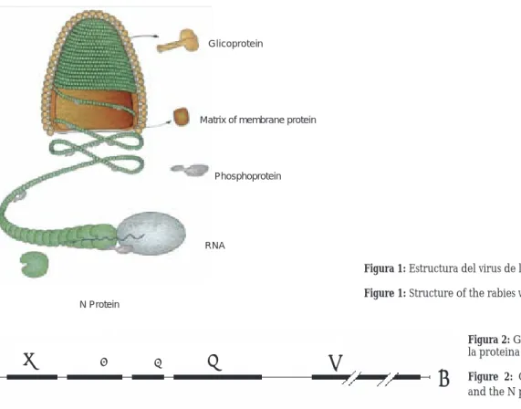
protein constituted by 450 aminoacids intervenes in the humoral and cellular immunity, it is the one with greatest diagnostic importance when its antigen is detected. The internal L, N and P proteins form a complex with the viral RNA to form the nucleocap-sid (RNP or ribonucleicprotein)1,4,8,9 (Figure 1). The
genome of the RV measures almost 12 Kb and codifi es for fi ve RNA monocistronic messengers that traduce for the fi ve described proteins10 (Figure 2).
Immune response
To study the immune response (IR) in the infection by RV, different studies have been done: neutraliz-ing antibodies titer, indexes of cellular proliferation, kinetic of multiplication of the virus, nitric oxide syn-thesis , expression of the gene inductible nitric oxide synthetase (iNOS), cytosines production [interferon
γ (IFN-γ), interleukins 2,4 and 10 ( IL2, IL4, IL10)], cytotoxic T lymphocyte, as well as the expression of the major histocompatibility complex (MHC) class I.
Viruses EBL-1 and EBL-2 have been isolated from European bats and are responsible of human deaths, this is one of the reasons why Herzog et al.3
evalu-ated the response of cells B and T in European bat Lyssavirus 1. They studied a group of healthy patients, without previous vaccination, that were exposed to the bite of a rabid animal, they were immunized with the
proteína constituye la glicoproteína G (proteína G) y hacia ella se producen los anticuerpos neutralizantes. La proteína de membrana o matriz (proteína M2) es importante como intermediario y catalítico entre y dentro de la envoltura viral y la nucleocápside (NC). Una transcriptasa (proteína L) que está formada de 2 142 aminoácidos, representa 54% del genoma, es importante en la transcripción. La fosfoproteína (pro-teína NS o pro(pro-teína M1 o pro(pro-teína P) formada por 297 aminoácidos tiene dos formas con diferentes extensio-nes de fosforilación, originalmente considerada como componente de la envoltura viral. La proteína P se une a la N para la síntesis del ARN viral, esta unión se asocia con la cadena ligera de la dineína celular, que está involucrada en el transporte de la NC viral a través de los axones neuronales. Toriumi et al.,6 con
el propósito de conocer la función y los cambios con-formacionales de la proteína P, usaron un anticuerpo monoclonal (# 402-13) e identifi caron un epítopo lineal localizado en la región C-terminal de la pro-teína P, que tiene una conformación específi ca para estar expuesto sólo cuando la proteína P está asociada a la NC.6 La región del epítopo es esencial para que la
proteína P se asocie con la NC, pero no para la forma-ción de complejos libres de N-P con nueva proteína N sintetizada.
La proteína N (nucleoproteína) encapcida al ARN viral efi ciente y específi camente para formar el com-plejo ribonucleoproteína que proporciona el molde para la transcripción y replicación del ARN por el com-plejo de polimerasa viral, el cual incluye la proteína P y la L.7 La proteína N constituida de 450
aminoá-cidos interviene en la inmunidad humoral y celular, es la de mayor importancia diagnóstica al detectar su antígeno. Las proteínas internas L, N y P forman un complejo con el ARN viral para formar la nucleocáp-side (PRN o ribonucleoproteína)1,4,8,9 (Figura 1). El
genoma del VR mide casi 12Kb y codifi ca para cinco ARN mensajeros monicistrónicos que traducen para las cinco proteínas descritas 10 (Figura 2).
Respuesta inmune
Para estudiar la respuesta inmune (RI) en la infec-ción por el VR se han realizado diferentes estudios: titulación de anticuerpos neutralizantes, índices de proliferación celular, cinética de multiplicación del virus, síntesis de óxido nítrico, expresión del gen óxido nítrico sintetasa inducible (iNOS), producción de citocinas [interferón γ (IFN-γ) interleucina 2, 4 y 10 (IL2, IL4, IL10)], linfocitos T citotóxicos, así como expresión del complejo mayor de histocompatibilidad (MHC) clase I.
muer-tes humanas, ésta es una de las razones por las que Herzog et al.3 evaluaron las respuestas de células B
y T en Lyssavirus de murciélago europeo 1. Estudia-ron a un grupo de pacientes sanos, sin vacunación previa, que estuvieron expuestos a la mordedura de un animal rabioso, se les inmunizó con la cepa PM (Pitman-Moore) posexposición y se les midió in vitro los títulos de anticuerpos neutralizantes e índices de proliferación celular; en este estudio no se encontró correlación entre la respuesta de células B y T en res-puesta a EBL-1.3
La respuesta inmune del portador puede ser impor-tante en el desarrollo de la encefalitis.5,8,11 Se tiene
evidencia de que el VR puede permanecer secues-trado por largos periodos. En humanos hay títulos de anticuerpos sólo cuando están presentes los síntomas neurológicos y en las últimas etapas de la enferme-dad, cuando ocurre la muerte. En 1977, Iwasaki et al.11
estudiaron en ratones el papel de la RI del portador en el desarrollo de la enfermedad encefálica o para-lítica después de la infección por rabia experimental. Trabajaron tres grupos de ratones (testigos normales, inmunosuprimidos y atímicos). Después de adminis-trar la cepa ts2 CVS de VR, se analizó la cinética de multiplicación del virus en SNC y los títulos de anti-cuerpos. En los animales testigo hubo parálisis severa, y causó 80% de muertes, acompañada de infl amación marcada y destrucción del tejido parenquimatoso del SNC, raramente se aisló VR y hubo producción de anti-strain PM (Pitman-Moore) post-exposition and the
titers of neutralizing antibodies and rates of cellular proliferation in vitro were measured. In this study no correlation was found between the response of cells B and T in response to EBL-1.3
The immune response of the carrier can be important in the development of encephalitis.5,8,11
There is evidence that the RV may stay hijacked for long periods. In humans there are titers of antibod-ies only when neurological symptoms are present and in the last stages of the disease, when death occurs. In 1977, Iwasaki et al.11 studied in mice the role of IR
from the carrier in the development of the encephalic or paralytic disease after the infection with experi-mental rabies. They worked with three groups of mice (normal controls, immunosuppressed and athymic). After administering the strain ts2 CVS of RV, the kinetic of the virus’ multiplication in the CNS, and the titers of antibodies were analyzed. In the control animals there was severe paralysis and caused 80% of deaths, accompanied by marked infl ammation and destruction of the parenchymatose tissue of the CNS, RV was rarely isolated and there was produc-tion of antibodies against rabies; while in the immu-nosuppressed (cyclophosphamide) it caused 100% of deaths, there were encephalitis symptoms , in lesser degree paralysis and the histological changes were degeneration and necrosis of neurons, the virus was isolated in all the mice, but no antibody titers were
Glicoprotein
Matrix of membrane protein
Phosphoprotein
RNA
N Protein
3
N
M3
M2
G
L
5
ψ
Figura 1: Estructura del virus de la rabia (P. Le Mercier).51 Figure 1: Structure of the rabies virus (P. Le Mercier).51
Figura 2: Genoma del virus de la rabia y la proteina N.52
cuerpos contra la rabia; mientras que en los inmuno-suprimidos (ciclofosfamida) causó 100% de muertes, hubo síntomas de encefalitis, en menor grado pará-lisis y los cambios histológicos fueron degeneración y necrosis de neuronas, se aisló el virus en todos los ratones, pero no se detectaron niveles de anticuerpos. En los ratones atímicos (inmunodefi cientes) los sínto-mas fueron similares a los inmunosuprimidos, pero con producción de anticuerpos IgM. Se concluyó que la RI del huésped es benéfi ca en la infección por el VR y es importante en la destrucción del tejido parenqui-matoso y ascenso de la parálisis de extremidades.
En los ratones inmunocompetentes (testigos), la destrucción del tejido parenquimatoso quizá se deba a la RI humoral y celular del portador por la generación de anticuerpos citolíticos y a la generación de células citotóxicas en la infección por el VR. La ausencia de destrucción de tejido parenquimatoso en inmunode-fi cientes (atímicos) sugiere dependencia del timo, y la presencia de anticuerpos IgM indica que éstos no par-ticipan en la destrucción del tejido parenquimatoso.11
Sukathida et al.12 evaluaron la síntesis de óxido
nítrico y la expresión del gen de iNOS para estudiar el mecanismo patofi siológico de daño cerebral y funcio-nal de las neuronas durante la infección por VR, con-cluyeron que la inhibición de iNOS retrasa la muerte de los ratones infectados con VR al afectar la replica-ción viral y la apoptosis de las células infectadas.
Respuesta inmune innata
La RI innata constituye un sistema de alerta precoz para detectar y combatir infecciones de cualquier tipo. Al contrario de la inmunidad adaptativa (anti-cuerpos, inmunidad celular específi ca), la innata es de activación rápida y no genera memoria inmuno-lógica. Este sistema de defensa no tiene la exquisita especifi cidad de la inmunidad adaptativa, pero reco-noce de manera muy efi ciente clases genéricas de moléculas producidas por múltiples y variados agen-tes patógenos, incluidos los virus. Cuando detecta alguna de estas moléculas extrañas, el sistema inmune dispara una respuesta de protección; en ella el sistema inmune se esfuerza por detener al agente patógeno. La defensa se inicia mediante los receptores tipo toll (TLR de Toll-like receptors), una familia de proteínas que media la respuesta inmune innata, se encuentran ampliamente distribuidas en la escala zoológica, desde los crustáceos e insectos hasta el ser humano.13 Se ha
determinado que los TLR reconocen moléculas bási-cas para la supervivencia de bacterias, virus, parásitos y otros patógenos. Actualmente se reconocen decenas de distintos TLR que reconocen moléculas específi -cas (el TLR2 se une al ácido lipoproteínico, un com-ponente de la pared bacteriana). Para el caso de los detected. In the athymic mice (immune defi cient) the
symptoms were similar to the immunodepressed, but with production of IgM antibodies. It was concluded that the IR of the carrier is benefi cial in the infec-tion by the RV and is important in the destrucinfec-tion of the parenchymatose tissue and rise of the paralysis of extremities.
In the immunocompetent mice (control), the destruction of the parenchymatose tissue might be due to the humoral and cellular IR of the carrier by the generation of cytolytic antibodies and to the generation of cytoxic cells in the RV infection. The absence of parenchymatose tissue destruction in immunodepressed (athymic) suggests dependence from the thyme, and the presence of IgM antibodies shows that these do not participate in the destruction of the parenchymatose tissue.11
Sukathida et al.12 evaluated the nitric oxide
synthe-sis and the expression of the iNOS gene to study the pathophysiologic mechanism of brain damage and function of the neurons during the infection with RV, and concluded that the inhibition of iNOS retards the death of infected mice with RV by affecting the viral replication and the apoptosis of infected cells.
Innate immune response
The innate IR constitutes an early alert system to detect and fi ght infections of any kind. Contrary to adaptive immunity (antibodies, cellular specifi c immunity), the innate is of fast activation and does not generate immune memory. This defense system does not have the exquisite specifi city of the adaptive immunity, but recognizes in a very effi cient way generic kind of mol-ecules produced by multiple and various pathogen agents, viruses included. When it detects any of these strange molecules, the immune system triggers a pro-tection response; within it, the immune system makes an effort to stop the pathogen agent. The defense initiates by means of receptors e toll (TLR from toll-like receptors), a family of proteins that mediates the innate immune response, that are broadly distributed in the zoological scale, from crustaceous and insects to human beings.13 It has been determined that the
virus y el de la rabia no es la excepción, los TLR más importantes son el TLR3 que reconoce el material genético de los virus y el TLR9 que reconoce secuen-cias repetitivas del genoma propias de los virus y de las bacterias denominadas CpG que son no metiladas.14
En los virus los componentes de éstos presumible-mente activan al sistema inmune innato, pero poco se conoce acerca de su identidad o la ruta involucrada en su reconocimiento.15 En la infección viral, la respuesta
inmune innata también se realiza a través de la induc-ción de interferones tipo I (IFN-α e INF-β), así como por la activación de células NK. La doble cadena de ARN viral que se produce durante el ciclo replicativo puede inducir la expresión de estos IFN-α e IFN-β por la célula infectada. En la respuesta inmune innata las células NK producen citocinas, tienen actividad cito-tóxica y son efectoras contra infecciones virales. Estas células se caracterizan fenotípicamente por la expre-sión de CD56, ausencia de CD3 y producción de IFN-γ, que tiene actividad inmunorreguladora, antiviral y su actividad se puede incrementar por la citocina IL-12, que se produce tempranamente en la respuesta a la infección viral.16,17
Los macrófagos representan parte esencial de la inmunidad innata, cuando estas células detectan una infección viral sintetizan múltiples mediadores infl amatorios, como citocinas (IL-1, IL-6), quimio-cinas, óxido nítrico (NO), proteínas del suero, enzi-mas e interferones. Estos mediadores intervienen en mecanismos antivirales o rutas de acceso intraviral. Los monocitos y fi broblastos también son capaces de sintetizar estas citocinas, pero aún no se conocen los mecanismos que inducen la producción de inter-ferones tipo I. Los IFN-α e INF-β activan la ruta de señalización JAK-SAT y con ello induce la expresión de varios genes. Uno de éstos codifi ca para la enzima 2´-5´oligo-adenilatosintetasa (2-5(A)sintetasa), que activa una ribonucleasa (RNAsa L) y degrada al ARN viral. Otros genes activados por la unión de estos dos interferones inducen una proteína cinasa especí-fi ca llamada dsRNA dependiente de proteína cinasa (PKR), que inactiva la síntesis de proteínas y bloquea la replicación viral en las células infectadas.16,18
Claassen et al.19 mostraron que administrando
antígenos de VR son tomados por los macrófagos y llevados a los nódulos linfoides y al bazo. A pesar de estos datos, la respuesta inmune innata contra VR no está estudiada en su totalidad. Por otra parte, mediante inmunohistoquímica y microscopía electró-nica se localizaron macrófagos reclutados en los sitios de inoculación de VR, en estos sitios de infección los macrófagos indujeron respuestas proinfl amatorias que ayudaron a la eliminación de los virus.20
Los virus se replican mejor cuando las células son activadas, por lo que muchos virus pueden activar deli-probably activate the innate immune system, but
little is known about its identity or the route involved in its recognition.15 In the viral infection, the innate
immune response is achieved through the induction of interferons type I (IFN-α and IFN-β), as well as by the activation of NK cells. The double chain of viral RNA that is produced during the replicated cycle may induce the expression of these IFN-α and IFN-β by the infected cell. In the innate immune response the NK cells produce cytokines , they have cytotoxic activ-ity and are effective against viral infections. These cells are characterized phenotypically by the expres-sion of CD56, absence of CD3 and production of
IFN-γ, that has immune-regulatory and antiviral activity, which can be increased by the cytokine IL-12, which is produced early in the viral infection.16,17
The macrophages represent an essential part in the innate immunity , when these cells detect a viral infection they synthesize multiple infl ammatory mediators, like cytokines (IL-1, IL-6), chemokines, nitric oxide (NO), serum proteins, enzymes and interferons. These mediators intervene in antiviral mechanisms or intra-viral access routes. The mono-cytes and fi broblasts are also capable of synthesizing these cytokines, but the mechanisms that induce the production of interferons type I are not known. The IFN-α and IFN-β activate the signaling route JAK-SAT and with this, induces the expression of several genes. One of these codifi es for the enzyme 2´-5´ oligo ade-nylate synthetase (2-5(A)synthetase, that activates a ribonuclease (L RNAse) and degrades the viral RNA. Other genes activated by union of these two inter-feron induces a specifi c protein kinase called dsRNA dependent of protein kinase (PKR), that inactivates the protein synthesis and blocks the viral replication in the infected cells.16,18
Claassen et al.19 showed that administering RV
antigens, they are captured by the macrophages and taken to the lymphoid nodules and spleen. In spite of these data, the innate immune response has not been studied in its totality. On the other hand, by immune-histochemistry and electronic microscopy, recluse macrophages were localized in the inoculation sites of RV, in these sites of infection, the macrophages induced pro-infl ammatory responses that helped eliminate the viruses.20
The viruses replicate better when the cells are activated; therefore, many viruses can deliberatively activate the cells to increase the viral infection and replication (activation factors, growth factors and superantigens).
by antibodies and complement and manipulate the infl ammatory response produced by the carrier. These strategies include the incorporation of proteins that regulate the complement of the host inside the virion capsule and the expression of viral proteins that regu-late the complement. The vaccinie virus can inhibit the classic and alternative routes to escape the neu-tralization by means of antibodies and protects the offsprings of the virion from the complement’s attack of the host.21
Besides the mediators of the innate immune response against viruses, other molecules are involved, like the nitric oxide (NO) that suppresses the synthe-sis of RNA of the rabies virus, reducing the level of expression of G, L and N proteins, that is of our inter-est.22
Protection
The glycoprotein and the nucleoprotein are the most important inductor antigens to know if there is protec-tion against rabies. Unlike G protein, the N protein is the most preserved through the different geno-types, stimulates the production of TH cells,8 induces
cross protection against intramuscular threats and stimulates the production of neutralizing antibodies induced by the classic vaccines.
The IR at two antigens in humans was studied: NC and glycoprotein in patients with rabies and immu-nized patients (virus grown in Vero cells). The results indicated that the process of immune recognition and antibodies development, occurs early in the pre-clini-cal phase, and the reactivity to N protein is important to the production of neutralizing antibodies.23
The IL-2 increases the protection in experimental rabies when mice are exogenously inoculated , this cytokine exhibited a helper effect in RV vaccines and subunits of these vaccines.24
The NC depends on a physical interaction between N and G proteins to have an adyuvant effect.8 This is
the result of a presentation of products derived from both proteins by one same presenter cell of antigen. Thus, the specifi c TH cells for the N and G protein may act in synergism to stimulate the B lymphocytes and produce neutralizing antibodies.8
T lymphocytes were stimulated from immunized patients with PM vaccinal strain with IL-2; it was found that the T lymphocytes proliferate in response to rabies antigens only when the antigen’s presenter cells express HLA-DR antigen. The stimulation of these T lymphocytes with rabies specifi c antigens induced the production of IFN-γ.25
The N protein from the rabies infl uences over the cytolytic cells or T cells, offering protection against rabies in animals.28 The cytotoxic T lymphocytes are
beradamente a las células para aumentar la infección viral y la replicación (factores de activación, factores de crecimiento y superantígenos).
El complemento también participa en la respuesta inmune innata. Las moléculas que regulan el com-plemento son importantes para evitar la activación de éste. Los virus tienen estrategias para evadir la inmunidad mediada por anticuerpos y complemento y manipulan la respuesta infl amatoria producida por el portador. Estas estrategias incluyen la incorporación de proteínas que regulan el complemento del hués-ped dentro de la envoltura del virión y la expresión de proteínas virales que regulan el complemento. El virus vaccinie puede inhibir las rutas clásica y alterna del complemento y así escapar de la neutralización a través de anticuerpos y protege a la progenie del virión del ataque del complemento del huésped.21
Además de los mediadores de la de la respuesta inmune innata contra los virus, se involucran otras moléculas como el óxido nítrico (NO) que suprime la síntesis de ARN del virus de la rabia, reduciendo el nivel de expresión de las proteínas G, L y N, que es de nuestro interés. 22
Protección
La glicoproteína y la nucleoproteína son los antíge-nos inductores más importantes para conocer si hay protección contra la rabia. A diferencia de la proteína G, la N es la más conservada a través de los diferen-tes genotipos, estimula la producción de células TH,8
induce protección cruzada contra retos intramuscula-res y estimula la producción de anticuerpos neutrali-zantes inducidos por las vacunas clásicas.
Se estudió la RI a dos antígenos en humanos: NC y glicoproteína en pacientes con rabia y pacientes vacu-nados (virus crecido en células Vero). Los resultados indicaron que el proceso de reconocimiento inmune y desarrollo de anticuerpos ocurre tempranamente en la fase preclínica, y que la reactividad a la proteína N es importante para la producción de anticuerpos neutralizantes.23
La IL-2 aumenta la protección en rabia experimen-tal cuando se inyecta exógenamente a ratones, esta citocina exhibió un efecto adyuvante en las vacunas del VR y en las subunidades de estas vacunas.24
La NC depende de una interacción física entre las proteínas N y G para que tenga un efecto adyuvante.8
Esto es resultado de una presentación de productos derivados de ambas proteínas por una misma célula presentadora de antígeno. Así, las células TH especí-fi cas para las proteínas N y G pueden actuar sinérgi-camente para estimular a los linfocitos B y producir anticuerpos neutralizantes.8
inmuniza-dos con la cepa vacunal PM con IL-2, se encontró que los linfocitos T proliferan en respuesta a antígenos de rabia sólo cuando las células presentadoras de antí-geno expresan antíantí-geno HLA-DR. La estimulación de estos linfocitos T con antígenos de rabia específi -cos indujo la producción de IFN-γ.25
La proteína N de la rabia infl uye sobre las células citolíticas o células T, ofreciendo protección contra la rabia en animales.26 Los linfocitos T citotóxicos son
importantes en la eliminación de virus, ya que la PRN del VR es un excelente inductor del complejo mayor de histocompatibilidad (CMH-I). La PRN del VR pro-duce inmunidad protectora sin que se conozcan aún los mecanismos de protección.23 Los antígenos de la
nucleoproteína activan la proliferación de las células B; la proteína N del VR en el complejo PRN induce potente respuesta de células T, ello ocasiona una res-puesta inmune humoral contra el VR. Actualmente algunos investigadores han incorporado genes extra-ños dentro del genoma del VR para producir un virus recombinante que permite examinar así la inmuno-genicidad a antígenos extraños. Se construyó un VR recombinante que expresa la proteína de fusión del VIH-1, N-GFP, que se produjo e incorporó efi ciente-mente a la PRN, lo que indujo en ratones una res-puesta inmune humoral (Koser et al.7) a diferencia
de la proteína GFP sola. Estos datos indican que la nucleoproteína del VR se puede usar como acarrea-dor de antígenos extraños, y puede ser útil para el uso de VR como vector en vacunas muertas contra otras enfermedades infecciosas.7
La glicoproteína de la rabia unida a liposomas in vitro induce síntesis de citocinas específi cas. La producción de IL-2 en células esplénicas de ratones inmunizados con antígenos del virus de la rabia pre-sentados como inmunosomas (liposomas cubiertos de glicoproteína) fueron tan activos como los virus inactivados, mientras que la glicoproteína purifi cada fue inactiva.27
Celis et al.28 informan que la inmunización con
RNP de VR purifi cada no indujo anticuerpos neutra-lizantes, pero protegió a los animales contra un reto viral periférico, aunque no existan anticuerpos neu-tralizantes al momento del reto, la presencia de TH facilita la respuesta inmune rápida de los antígenos presentes en el reto viral.
Mediante péptidos sintéticos se identifi caron in vitro los epítopos inmunodominantes de la NC in vivo por la estimulación de linfocitos T específi cos, ello resultó en incremento de la respuesta de células T y rápida producción de anticuerpos neutralizantes con VR inactivado.8
Perrin et al.29 estudiaron la producción de
citoci-nas de células T en respuesta a varios Lyssavirus (antí-genos de rabia) en ratones BALC/c; se evaluaron important in the elimination of the infectious virus,
since the RNP of the RV is an excellent inductor of the major hystocompatibility complex (MHC-I). The RPN of the RV produces protective immunity, even though the mechanisms of protection are still unknown.23 The antigens of the nucleoprotein
acti-vate the proliferation B cells; the N protein of the RV in the RNP complex induces a powerful response of T cells, this causes a humoral immune response against the RV. At the moment, certain researchers have incorporated strange genes within the genome of the RV to produce a recombinant virus that allows to examine, in this way, the immunogenicity to strange antigens. A recombinant RV was constructed which expresses the fusion protein of the HIV-1, N-GFP, that was effi ciently produced and incorporated to the RNP, which induced an immune humoral response in mice (Koser et al.7), in contrast to the GFP protein
alone. This data indicates that the RV nucleoprotein can be used as a carrier of strange antigens, and may be helpful for the use of RV as vector in dead vaccines against other infectious diseases.7
The glycoprotein of the rabies joined to liposomes in vitro induces the synthesis of specifi c cytokines. The production of IL-2 in splenic cells of immunized mice with antigens of rabies virus presented like immuno-somes (lipoimmuno-somes covered with glycoprotein) were as active as the inactivated viruses, while the purifi ed glycoprotein was inactivated.27
Celis et al. 28 report that the immunization with
RNP of purifi ed RV did not induce neutralized anti-bodies, but protected the animals against a periph-eral viral challenge , even if there are no neutralized antibodies at the moment of the challenge, the pres-ence of TH facilitates rapid immune response of the antigens present in the viral challenge.
By means of synthetic peptides, the immunodo-minant epitopes of the NC in vivo by the stimulation of specifi c T lymphocytes were identifi ed in vitro, this resulted in an increment of the response of T cells and rapid production of neutralized antibodies with inactivated RV.8
Perrin et al.29 studied the production of cytokines
IL-2, IL-4, IFN-γ, así como sus RNAm. Sólo los ratones infectados con virus patógenos perdieron la capaci-dad de producir citocinas in vitro después de la esti-mulación antígeno específi ca. En ratones infectados con virus no patógeno hubo producción de citocinas. Así, la infección con antígenos de rabia patógeno por la ruta periférica induce en los ratones BALB/c pér-dida de respuesta de células T después de la activación antigénica específi ca, pero no después de la activación policlonal (concanavalina A). Concluyeron que en el ratón, la pérdida de la respuesta inmune está relacio-nada con la severidad de la enfermedad. La no res-puesta de células T depende sólo de la severidad de la enfermedad en el ratón.
En otro estudio de Hooper et al.30 se inmunizaron
ratones con PRN por vía oral, se encontró aumento en la respuesta inmune, ello demuestra especifi cidad en respuesta celular y producción de anticuerpos contra la proteína N.
La inmunidad celular y los interferones pueden reducir la carga viral, los anticuerpos virus específi cos y particularmente los anticuerpos neutralizantes al virus son muy importantes en el control de la mayoría de las infecciones virales que afectan el SNC.31 Perrin
et al.24 informaron que la IL-2 aumentó la protección
en rabia experimental; sin embargo, en un estudio previo en que se induce producción de IL-2 y anticuer-pos en células humanas de sangre periférica de indi-viduos vacunados con cepas PM o virus Pasteur, no se encontró correlación entre la producción de IL-2 y los niveles de anticuerpos neutralizantes, se concluyó que la producción de IL-2 podría ser usada para el estudio de la inmunidad celular y células de memoria en la vacunación antirrábica en humanos.32
Superantígenos
Los superantígenos (SAg) pueden activar gran número de linfocitos T, ya que no son internalizados ni procesados por las células presentadoras de antí-geno. Menos de 0.01% de linfocitos T responden a un Sag, entre 5% y 25% de linfocitos T pueden responder a un antígeno de otro tipo. En 1992 se informó que la NC del VR es un SAg específi co para linfocitos T humanos Vβ8, que se une a cadenas HLA clase IIα y es potente activador de linfocitos T en las vacunas contra la rabia.33
En linfocitos de amígdalas humanas se evaluó la capacidad del SAg de NC de VR para activar la proli-feración celular, la producción de citocinas y anticuer-pos. La activación producida por el SAg de la NC se comparó con SAg derivados de Staphylococcus (SEE y TSST-1); a pesar de una débil actividad mitogénica de linfocitos T restringida a células TCD4+, la NC disparó en los linfocitos B la producción de inmunoglobuni-is related with the severity of the dinmunoglobuni-isease . The no
response of T cells only depends on the severity of the disease in the mouse.
In an other work of Hooper et al.30 mice were orally
immunized with RNP, it was found an enhanced immune response, this demonstrates specifi city in cel-lular response and production of antibodies against N protein .
The cellular immunity and interferons may reduce the viral load, the virus-specifi c antibodies and par-ticularly the virus-neutralizing antibodies are very important in the control of the majority of the viral infections that affect the CNS.31 Perrin et al.24 informed
that IL-2 increased the protection in experimental rabies; nevertheless, in a previous study in which IL-2 and antibodies in human cells of peripheral blood of vaccinated individuals with PM strains or Pasteur was induced, there was no correlation between the pro-duction IL-2 and the levels of neutralized antibodies, it was concluded that the production of IL-2 could be used for the study of cellular immunity and memory cells in human anti-rabies vaccine.32
Superantigens
The superantigens (Sag) can activate great number of T lymphocytes, since they are not internalized or processed by the antigen presenter cells. Less than 0.01% of T lymphocytes respond to a Sag, between 5% and 25% of the T lymphocytes may respond to an antigen of other type. In 1992 it was informed that the NC of the RV is a Sag specifi c for human Vβ8 T lymphocytes, that unites to α chain HLA class II and it is a powerful activator of T lymphocytes in vaccines against rabies.33
The capacity of the Sag of NC in RV to activate the cellular proliferation, cytokines and antibodies production was evaluated in lymphocytes of human tonsils. The activation produced by the Sag of the NC was compared to Sag derived from Staphylococcus (SEE and TSST-1); despite a weak T lymphocyte mitogenic activity restricted to CD4+ cells, NC triggered B lym-phocytes to produce immunoglobuline G (IgG) (in quantities similar to those produced by Sag of SEE and TSST-1), and did not triggered the production of IgM. The cytokines produced by the activation of the NC were IL-4 and IL-10, this suggests that Sag of the NC induces a TH2. The cytokines produced by
nas G (IgG) (en cantidades similares a las producidas por los SAg de SEE y TSST-1), y no disparó la produc-ción de IgM. Las citocinas producidas por activaproduc-ción de la NC fueron IL-4 e IL-10, ello sugiere que el SAg de la NC induce una respuesta TH2. Las citocinas
pro-ducidas por activación de TSST-1 fueron IL2 e IFN-γ lo que sugiere que TSST-1 induce una respuesta TH1. El patrón TH2 inducido por el SAg de la NC podría
explicar la capacidad para incrementar la respuesta de anticuerpos in vivo (a un antígeno inyectado simul-táneamente). La NC de la rabia puede disparar la acti-vación de células B policlonales, como otros Sag.34
Astoul et al.35 informan que la NC del VR es un SAg
exógeno específi co Vβ8 en humanos y Vβ6 en rato-nes. Estos autores estudiaron el efecto del SAg de la rabia, en respuesta a un antígeno no relacionado, el virus de la infl uenza, y compararon la respuesta en dos cepas de ratones congénitas: BALB/c y BALB/d2. Los primeros responden al Sag de rabia, mientras que los segundos no responden por carecer del receptor de células T Vβ6. En ratones BALB/c, la coinyección del SAg de rabia con el virus de infl uenza inactivado produce incremento rápido y de larga duración en los títulos de anticuerpos IgG e IgM específi cos contra el virus, incluyendo anticuerpos protectores, inhibidores y hemoaglutinantes, también se incrementó la prolife-ración antígeno específi ca, así como la secreción de IL-2 e IL-4 por linfocitos de nódulo linfoide, cuando se compararon con los ratones que sólo recibieron el virus de la infl uenza. Sin embargo, en ratones BALB/ d2 durante el establecimiento de la respuesta prima-ria, el aumento en las células T estimuladas estuvo restringida a células TCR Vβ6. Estos datos establecen que el SAg de la rabia estimula respuestas T y B espe-cífi cas a antígenos no relacionados, esta propiedad es la responsable de la capacidad adyuvante de la NC.
Análisis estructural de la proteína N
El anticuerpo monoclonal contra el epítopo lineal 5-2-26 dependiente de fosforilación es útil para estu-diar procesos de fosforilación de la proteína N, Kawai et al.36 investigaron la maduración antigénica de la
proteína N del VR con anticuerpos monoclonales, encontraron que esta proteína no se fosforila inme-diatamente en la serina 389, primero se asocia con la proteína P para formar la nucleocápside y luego se presenta la fosforilación.
Goto et al.37 estudiaron los sitios antigénicos de
la proteína N del VR, pero ahora al clasifi carlos del I al IV, se observó que estos sitios están compuestos de epítopos que dependen de linearidad y conforma-ción. Por medio de anticuerpos monoclonales y con el uso de octapéptidos sintéticos determinaron que los del sitio I y del sitio IV constituyen una pequeña Astoul et al.35 report that NC of the RV is a Vβ8
spe-cifi c exogenous SAg in humans and Vβ6 mice. These authors studied the effect of rabies SAg, in response to unrelated antigen , the infl uenza virus, and compared the response in two congenic strains of mice: BALB/ c and BALB/d2. The fi rst are rabies SAg responsive, whereas the second ones are not responsive because they lack the Vβ6 T cell receptor. In BALB/c mice, co-injection of rabies SAg with inactivated infl uenza virus resulted in a rapid and long term increase in the titers of infl uenza virus specifi c antibodies IgG and IgM against the virus, including protective hemagglu-tination-inhibiting antibodies, also the specifi c prolif-eration was incremented, as well as the IL-2 and IL-4 secretion by lymph node lymphocytes, when compared to mice that received infl uenza virus only. In contrast, in BALB/d2 mice, during the establishment of the primary response, the increase in infl uenza-primed T cells was mainly restricted to the Vβ6 TCR. These data establish that rabies SAg can stimulate both T and B cell-specifi c responses to an unrelated antigen, this quality is the responsible of the adjuvant capacity of the NC.
Structural analysis of the N protein
The monoclonal antibody against the lineal 5-2-26 epitope dependent on phosphorylation is useful for the study of N protein phosphorylation, Kawai et al.36
investigated the antigenic maturation of the N protein of the RV with monoclonal antibodies, and found that this protein does not immediately phosphorylate in the serine 389, initially it associates with the protein P of RV to form the nucleocapsid and then presents phosphorylation.
Goto et al.37 studied the antigens’ sites of the N
pro-tein of RV, but now, as classifying them from I to IV, it was observed that these sites are composed of epitopes that depend on linearity and conformation. By means of monoclonal antibodies and with the use of syn-thetic octapeptides they determined that the ones of site I and IV conform a small region that covers the aminoacids 357-387, which are the most conserved and common in the Lyssavirus. It was found that the epitopes in site I express themselves in the immature way of N protein and the epitopes of site IV express themselves in the mature N protein.
Phosphorylation
phos-región que cubre los aminoácidos de 357-387, que son los más conservados y comunes en los Lyssavirus. Se encontró que los epítopos en el sitio I se expresan en la forma inmadura de la proteína N y los epítopos del sitio IV se expresan en la proteína N madura.
Fosforilación
La fosforilación de las proteínas en residuos como serina, treonina y tirosina son de las formas más fre-cuentes de modifi cación postraduccional en células eucarióticas, que están ligadas al control de funciones celulares. Muchas proteínas virales son fosforiladas y su fosforilación es importante en el ciclo viral infec-cioso. La fosforilación de algunas de las proteínas virales se realiza por cinasas asociadas con el virus, y otras por cinasas celulares. La fosforilación por una cinasa celular es un prerrequisito para lograr mayor fosforilación por cinasas asociadas con virus.9
La proteína N del virus de la rabia se fosforila no sólo en células infectadas con virus sino también cuando se expresa sola en células de insecto y de mamíferos, ello sugiere que la cinasa celular más que la cinasa asociada al virus quizá está involucrada en la fosforilación de la proteína N del virus de la rabia. En un estudio se probó que la casein-cinasa II (CK-II) de origen celular es capaz de fosforilar a la proteína N del virus de la rabia in vivo e in vitro.38 La fosforilación
de la proteína N es sobre un residuo de serina en la posición 389.11
Una función importante de la proteína N a nivel estructural es encapsidar y proteger al genoma. Tam-bién está involucrada en el cambio entre la transcrip-ción y replicatranscrip-ción viral9 como se demuestra en estudios
con el virus de la estomatitis vesicular (VSV), donde se observa que la replicación no puede iniciarse en ausencia de sufi ciente proteína N para la encapsida-ción del molde de crecimiento. Cuando las cantida-des de proteína N permanecen bajas, la transcripción reconoce una señal de paro, y cuando la transcripción es sufi ciente participa en el ensamble y crecimiento del molde y no responde a la señal de paro, este evento corresponde a la transición entre la transcripción y la replicación. Esta encapsidación específi ca inicia en el 5´ terminal de los ARNs.39
Para demostrar los dominios de la proteína N que gobiernan la especifi cidad de unión, Kouznetzoff et al.40 probaron in vitro la habilidad de la proteína N en
ambas formas: con longitud completa y truncada para interactuar con una sonda ARN sintética correspon-diente a la 5´ terminal del antisentido. Mostraron que en la proteína N completa y en la NH2 terminal de 376
aminoácidos están todos los determinantes para la interacción específi ca. Un péptido cercano al COOH terminal de t42 (posición 298-352) localizado en la phorylation is important in the infectious viral cycle.
The phosphoryrilation of some of the viral proteins is performed by kinases associated with the virus, and others by cellular kinases. The phosphorylation for one cellular kinase is a pre-requisite to obtain larger phosphorylation by kinases associated with virus.9
The protein N of rabies virus is phosphorylated not only in infected cells with viruses, but also when it expresses itself in insect and mammal cells; this sug-gests that cellular kinase, better than the associated kinase to the virus, may be involved in the phospho-rylation of N protein of rabies virus. In a study, it was proven that the casein-kinase II (CK-II) of cellular origin is capable of phosphorylating the N protein of the rabies virus in vivo and in vitro. The phosphoryla-tion of N protein is above a serine residue in the 389 position.11
An important function of the N protein at struc-tural level is to encapside and protect the genome. It is also involved in the change between viral transcrip-tion and replicatranscrip-tion as shown in studies the vesicular stomatitis virus (VSV), where it is observed that repli-cation can not be initiated in the absence of suffi cient N protein to encapside the growth mold. When the quantities of N protein remain low, the transcription recognizes a stop signal, and when the transcription is suffi cient, participates in the growth and fi t of the mold and does not respond to the stop signal, this event corresponds to the transition between the tran-scription and the replication. This specifi c encapsida-tion initiates in 5´ terminal of the RNAs.39
To demonstrate the domains of the N protein that govern binding specifi city, Kouznetzoff et al.40 tested
in vitro the ability of N protein in both forms: full-length and truncated to interact with a synthetic RNA probe corresponding to the 5´ end of the antigenome. They showed that the entire N protein and the NH2
terminal of 376 aminoacids are all of the determinants for specifi c interaction. A peptide near the COOH terminal of t42 (position298-352), which is located in the most conserved region of the Rabdoviridae N proteins, bound directly to the viral RNA. Both N pro-teins may possess a new type of RNA binding motif and folding proteins that contribute to the architec-ture of the RNA binding site.
phosphoryla-región más conservada de las proteínas N de los Rha-bdoviridae se une directamente al ARN viral. Ambas proteínas N pueden poseer un nuevo tipo de motif conformacional de unión a ARN y proteínas de dobla-miento que contribuyen a la arquitectura del sitio de unión al ARN.
Al mutar la serina por cualquiera de los siguien-tes aminoácidos: glicina, ácido aspártico, asparagina, ácido glutámico o glutamina en la posición 389, y exa-minar los efectos de estas mutaciones sobre la trans-cripción y replicación en el genoma, produjeron una síntesis de proteína N no fosforilada, reducción de la transcripción y replicación viral. Las curvas de creci-miento indican que la producción del virus mutado en serina por alanina fue diez mil veces menor que la del virus silvestre. Los resultados indican que la fos-forilación del VR es necesaria para la transcripción y replicación.9
La proteína N del VR y la del VSV no tienen alto grado de homología en su secuencia primaria de nucleótidos; sin embargo, ellas han conservado regio-nes y proteínas características similares. La proteína N del VR tiene cuatro aminoácidos conservados con homología a los de VSV. Ambas proteínas tienen estructura helicoidal similar.38 La defosforilación de la
proteína N purifi cada con fosfatasa aumentó su habi-lidad para encapsidar en la síntesis de ARN in vitro. Cuando la proteína N no está fosforilada, disminuyen las tasas de transcripción y replicación en el genoma igual que en el virus completo.
El molde para la transcripción y replicación de virus ARN de cadena negativa es una estructura N-ARN; es decir, el ARN está asociado con la nucleoproteína viral. Las señales de transcripción serán reconocidas sólo cuando el ARN viral está unido a la proteína N. Cuando la proteína N se expresa sola en células euca-rióticas, se une inespecífi camente a ARN celulares. En células infectadas por rhabdovirus toda la proteína N se une al ARN genómico y no a ARNm o celulares, esto es porque la fosfoproteína se une como una cha-perona a la nucleoproteína y previene así la unión de N a ARNm celular.
Cuando la nucleoproteína N de virus ARN expre-sada en células de insecto se une a ARN celular forma un complejo N-ARN como una nucleocápside viral; sin embargo, en células infectadas por virus, la pro-teína N es prevenida de uniones ARN celular por la formación de un complejo soluble entre la proteína N y la fosfoproteína viral, llamado complejo N°-P. La proteína N sólo es liberada de este complejo por unión a un nuevo ARN o un ARN complementario. En un estudio realizado por Mavrakis et al.41 se observó la
coexpresión de las proteínas N y P virales de rabia en células de insecto y se purifi có el complejo N°-P que se encontró en forma soluble como N°-P2.
tion of the RV is necessary for the transcription and replication.9
N Protein of the RV and the VSV do not have a high degree of homology in primary nucleotide and protein sequences; nevertheless, they have conserved regions and similar protein characteristics. The N protein of the VR has four conserved amino acid stretches homologous with those of VSV. Both pro-teins have similar helical structure.38 The
dephospho-rylation of the N protein purifi ed with phosphatase increases its ability to encapsid in the RNA synthesis in vitro. When N protein is not phosphorylated, the rate of transcription and replication are diminished in the genome as well as in the virus.
The mold for the transcription and replication of the negative short chain RNA is a N-RNA structure; that is to say, the RNA is associated to the viral nucle-oprotein. The transcription signs will only be recog-nized when the viral RNA is joined to the N protein. When the N protein expresses alone in eukaryotic cells, it nonspecifi cally joins to RNA cells. In infected cells by rabdovirus all the protein joins to the genomic RNA and not to RNAm or cellular, this is because the phosphoprotein joins as a chaperone to the nucleopro-tein and prevents the union of N to cellular RNAm.
When the nucleoprotein N of RNA virus, expressed in insect cells, joins to a cellular RNA it forms a N-RNA complex like a viral nucleocapsid; nevertheless, in infected cells by virus, the N protein is prevented from joining to cellular RNA by the formation of a soluble complex between the N protein and the viral phosphoprotein, called N°-P complex. The N protein is only liberated from this complex by the union to a new RNA or a complementary RNA. In a study done by Mavrakis et al.,41 the co-expression of the viral N
and P proteins of rabies in insect cells was observed and the soluble complex N°-P2 was purifi ed.
Recombinant N protein
Within the methods that permit the obtainment of RNP is the baculovirus system [pathogens that attack insects and other arthropods].* Utilizing molecular biology techniques, the gene that codifi es for the N protein of RV can be obtained, which inserts in the genome of the baculovirus Autographa californica nuclear polyhedrosis. The expression of the recom-binant gene is controlled by the gene promoter of the polyhedrin. In order to obtain the recombinant pro-tein, the baculovirus infects in insect cells [Spodoptera frungiperda (Sf9)].4,42,43 The N protein expressed in
The RV as a viral vector in the use of vaccines has many advantages: it is not pathogenic for a great number of animal species if it administered orally or intradermically, its genome organization allows easy genetic modifi cations compared to other complex genomes of DNA and RNA virus. The uncommon genes that are expressed are stable. The rhabdovirus have a cytoplasmic replication cycle and there is no evidence for recombination and integration of the genome of the host cell. A vector based on RV can be inductor of immunity for mucous membranes against HIV-1.
The RV grows with high titers in many lines of cells without killing them, which allows the expression of HIV-1 genes compared with cytoplasmic vectors.44
Schnell et al.44 produced a recombinant RV which
expresses the protein gp-160 of the HIV, in a stable and functional way in cellular lines of human T lym-phocytes. The infection of the mouse with this virus that expresses this protein gave a strong humoral response, directed against the protein gp-160 of HIV-1 cover after a unique challenge against protein gp120. The titers of neutralized antibodies detected in mouse serum against HIV-1 were superior to 1:800, these results indicate that the live recombinant RV that expresses the protein HIV-1 gp-160 can be used as an effective vector for the HIV-1 vaccine.
The RV can be used as a vector of expression to construct a recombinant virus that expresses cyto-chrome C, which is an essential pro-apoptotic protein for the proteolytic activity of Apaf-1 and for the acti-vation of caspases. Pulmanausahakul et al.45
demon-strated in vitro that the expression of cytochrome C in the recombinant RV is associated with the accelerated cellular death, and in vivo increases the immunogenic-ity and attenuates the pathogenicimmunogenic-ity.
Thirty nine activated genes are known which inter-vene in the infection, including involved genes in the regulation of the cellular metabolism, protein synthe-sis, synaptic activity, growth and cellular differentia-tion. Prosniak et al.48 demonstrated the effects of the
RV infection in different patterns of gene expression in the brain of mice. An early response pattern (three days after infection) and a late response (6-7 days after infection) associated with peak RV replication. The results suggest that a number of host genes may be involved in the replication and spread of RV in the brain.
In a study by Loza-Rubio et al.47 N protein
recom-binant was used to detect the mRNA of IL-2 in lym-phocytes of immunized chicken, for these animals were inoculated with an inactive vaccine against avian infl uenza with N protein added. The results showed increment of expression of IL-2 in immunized ani-mals when compared to control aniani-mals.
Proteína N recombinante
Dentro de los métodos que permiten la obtención de PRN se encuentra el sistema de baculovirus [patóge-nos que atacan a los insectos y otros artrópodos].* Empleando técnicas de biología molecular se puede obtener el gen que codifi ca para la proteína N del VR, el cual se inserta en el genoma del baculovirus Autographa californica nuclear de la polyhedrosis. La expresión del gen recombinante es controlada por el promotor del gen de la polyhedrina. Para obtener la proteína recombinante, el baculovirus se infecta en células de insecto [Spodoptera frungiperda (Sf9)].4,42,43
La proteína N expresada en células de insecto es anti-génica e inmunoanti-génicamente comparable a la PRN del VR y representa una fuente potencial para una vacuna contra la rabia efectiva y económica para la inmunización en humanos y animales.42,43
El VR como vector viral en el uso de vacunas tiene varias ventajas: no es patógeno para un amplio número de especies animales si se administra vía oral o intra-dérmica, su organización genómica permite modifi ca-ciones genéticas fáciles a diferencia de otros genomas complejos de virus ADN y ARN. Los genes extraños que se expresan son estables. Los rhabdovirus tienen un ciclo de replicación citoplásmico y no hay eviden-cia para recombinación e integración en el genoma de la célula portadora. Un vector basado en VR puede ser inductor en inmunidad a mucosas contra VIH-1.
El VR crece con títulos altos en varias líneas de células sin matarlas, lo cual permite la expresión de genes VIH-1 comparados con vectores citoplásmicos.44
Schnell et al.44 produjeron un VR recombinante que
expresa la proteína gp-160 del VIH, de forma estable y funcional en líneas celulares de linfocitos T humanos. La infección del ratón con este virus que expresa esta proteína dio una respuesta humoral fuerte, dirigida contra la proteína gp-160 de envoltura VIH-1 después de un reto único con proteína gp120. Los títulos de anticuerpos neutralizantes detectados en el suero del ratón contra VIH-1 fueron superiores a 1:800, estos resultados indican que el VR recombinante vivo que expresa la proteína VIH-1 gp-160 puede servir como un vector efectivo para una vacuna VIH-1.
El VR puede ser usado como vector de expresión para construir un virus recombinante que exprese citocromo C, que es una proteína proapoptótica esen-cial para la actividad proteolítica de Apaf-1 y para la activación de caspasas. Pulmanausahakul et al.45
demostraron in vitro que la expresión del citocromo C en el VR recombinante se asocia con la muerte celu-lar acelerada, e in vivo aumenta la inmunogenicidad y atenúa la patogenicidad.
The N protein in rabies’ diagnosis
The N protein is primarily produced and in large quantities, that is why it is of importance for the detec-tion of the rabies antigen. The CDC (Center for Dis-ease Control and Prevention, Atlanta, Georgia) for the diagnosis of rabies utilizes the gene of N protein for tests of PCR and formation of oligodendograms.48,49
All the determinations and the genetic typifi ca-tion of the rabies virus isolated in America and other parts of the world are made based on a 320 pb seg-ment localized in N protein, it is there where different isolates of distinct species , vectors and geographical sites are based.49
The fl uorescent antibody test developed by Coons and Kaplan in 1950,50 is very useful for the rabies´
diag-nosis. The utilized material can be fresh or frozen. A marked antibody with fl uorochrome (fl uorescein isothiocyanate), is incubated with the tissue so that reacts with specifi c antigen (N protein), there will be fl uorescent particles if the antigen is present.
Other test for the detection of rabies antigen is by enzymatic immunodiagnosis, called RREID. It is a highly sensible and specifi c method. The nucleocap-sid antigen is extracted from the infected cells with RV and it is purifi ed by a cesium chloride gradient. The viral antigens are emulsifi ed with Freud adjuvant and they are inoculated IM in rabbits. The antinu-cleocapsid IgG antibodies of rabies are purifi ed by chromatography and adsorbed in the solid phase, the incubation of a positive sample with the IgGs is revealed with a conjugate to rabbit peroxidase, appear-ing a yellow color after addappear-ing the substrate and cro-magene (o-phenylenediamine); fi nally evaluated with a spectrometer.
The N protein that encapsides and protects the genome is involved in the transcription change and viral replication, it favors the cellular and humoral immune response, since it increases the production of neutralizing antibodies, for which it is said that has a coadjuvant effect, it has been considered as a superantigen when compared to conventional super-antigens, it has a similar response, it also favors the TH2 response (increases the IL-4 and IL-10). It is a
exogenous specifi c superantigen of Vβ8 in humans and Vβ6 in mice.
The antigenic maturity depends on the N protein when it is phophorylated in a serine residue on posi-tion 389 and by the presence of a cellular caseine.
It is the antigen of greater diagnosis importance on infected tissue.
The gene that codifi es for the N protein is used in molecular epidemiology of rabies and other Lyssavi-rus.
It can be used as a strange protein carrier;
there-Se conocen 39 genes activados que intervienen en la infección, incluyen genes involucrados en la regula-ción del metabolismo celular, síntesis proteínica, acti-vidad sináptica, crecimiento y diferenciación celular. Prosniak et al.46 demostraron los efectos de la infección
del VR en diferentes patrones de expresión de genes en el cerebro de ratones. Un patrón de fase temprana (tres días después de la infección) y un patrón de fase tardía (6-7 días de la infección) que se asocia con el pico de replicación de VR. Los resultados sugieren que genes del huésped pueden estar involucrados en la replicación y amplifi cación del VR en el cerebro.
En un estudio de Loza-Rubio et al.47 se usó proteína
N recombinante para detectar el mARN de IL-2 en lin-focitos de pollos inmunizados, para ello se vacunaron animales con una vacuna inactiva contra la infl uenza aviar adicionada con proteína N recombinante del VR. Los resultados mostraron incremento de la expresión de IL-2 en animales inmunizados cuando se compara-ron con animales testigo.
La proteína N en el diagnóstico
de la rabia
La proteína N se produce primero y en grandes can-tidades, por ello es importante para la detección del antígeno de la rabia. El CDC (Centers for Disease Control and Prevention, Atlanta, Georgia) para el diagnóstico de la rabia utiliza el gen de la proteína N para pruebas de PCR y formación de oligodendogra-mas.48,49
Todas las determinaciones y la tipifi cación gené-tica de los virus de la rabia aislados en América y otras partes del mundo se hacen con base en un segmento de 320 pb localizado en la proteína N, en él se basan los diferentes aislados de las distintas especies, vecto-res y sitios geográfi cos.49
La prueba de anticuerpos fl uorescentes desarro-llada por Coons y Kaplan en 1950,50 es muy útil en el
diagnóstico de la rabia. El material utilizado puede ser fresco o congelado. Un anticuerpo marcado con el fl uorocromo (isotiocianato de fl uoresceína), se incuba con el tejido para que reaccione con el antígeno espe-cífi co (proteína N), si el antígeno está presente habrá partículas fl uorescentes.
fore, is useful for an increased protective response in the use of vaccines.
Acknowledgments
Special thanks to the National Council of Science and Technology of Mexico, for its support in study 34214B, as well as scholarship 169991 granted to Maria Esther Morales in the doctorate program on biological sci-ences of the Metropolitan Autonomous University.
Referencias
IgGs se revela con un conjugado a peroxidasa antico-nejo, aparece un color amarillo después de adicionar el sustrato y el cromógeno (o-fenilendiamino); fi nal-mente se evalúa cuantitativanal-mente en un espectróme-tro.
La proteína N que encapsida y protege al genoma está involucrada en el cambio de la transcripción y replicación viral, favorece la respuesta inmune celular y humoral, ya que aumenta la producción de anticuer-pos neutralizantes por lo que se dice que tiene efecto adyuvante, se ha considerado como un superantígeno debido a que si se compara con superantígenos con-vencionales, su respuesta es similar, también favorece la respuesta TH2 (aumenta la IL-4 e IL-10). Es un
superantígeno exógeno específi co de Vβ8 en huma-nos y Vβ6 en ratones.
De la proteína N depende la maduración antigé-nica cuando se fosforila en el residuo de serina de la posición 389 y por la presencia de una caseína celu-lar.
Es el antígeno de mayor importancia diagnóstica en tejidos infectados.
El gen que codifi ca para la proteína N se usa en la epidemiología molecular de la rabia y otros Lyssa-virus.
Puede ser utilizada como acarreador de proteínas extrañas; por tanto, es útil para el aumento de la res-puesta protectora en el uso de vacunas.
Agradecimientos
Se agradece al Consejo Nacional de Ciencia y Tecnolo-gía, de México, por el apoyo a la investigación 34214B, así como por la beca 169991 otorgada a María Esther Morales en el programa de doctorado en ciencias bio-lógicas de la Universidad Autónoma Metropolitana.
1.
2.
3.
4.
5.
6.
7.
8.
9.
10.
11.
12.
Hostnik P, Bidovec A. Modern rabies extermination methods-ten years of rabies control by orally vacci-nating foxes in Slovenia. Res Rep -Univ Ljubl Vet Fac 1999;36:223-230.
Murphy FA, Gibbs EPJ, Horzinek MC , Studdert MJ, editors. Veterinary Virology. San Diego: Academic Press, 1999.
Herzog M, Fritzell C, Lafage M, Montaño HJA, Scott-Algara D, Lafon M. T and B cell human responses to European bat lyssavirus after post-exposure rabies vac-cination. Clin Exp Immunol 1991;85:224-230.
Wiktor TJ, György E, Dieter Schlumberger H, Sokol F, Koprowski H. Antigenic properties of rabies virus com-ponents. J Immunol 1973;110:269-276.
Drings A, Jallet C, Chambert B, Tordo N, Perrin P. Is there an advantage to including the nucleoprotein in a rabies glycoprotein subunit vaccine? Vaccine 1999;17:1549-1557.
Toriumi H, Honda Y, Morimoto K, Tochikura TS, Kawai A. Structural relationship between nucleocap-sid-binding activity o the rabies virus phosphoprotein (P) and exposure of epitope 402-13 located at the C terminus. J Gen Virol 2002;83:3035-3043.
Koser LM, McGettigan JP, Tan GS, Smith ME, Koprowsky H, Dietzschold B, et al. Rabies virus nucleoprotein as a carrier for foreign antigens. Proc Natl Acad Sci USA 2004;102:9405-9410.
Ertl HCJ, Dietzschold B, Gore M, Otvos Jr L, Larson JK, Wunner WH, et al. Induction of rabies virus-specifi c T-helper cells by synthetic peptides that carry dominant T-helper cell epitopes of the viral ribonucleoprotein. J Virol 1989;63:2885-2892.
Wu X, Gong X, Foley HD, Schnell M J, Fu Z. Both Viral Transcription are reduced when the rabies virus nucleoprotein is not phosphorylated. J Virol 2002;76:4153-4161.
Conzelmann KK, Cox JH, Schneider LG, Thiel HJ. Molecular cloning and complete nucleotide sequence of the attenuated rabies virus SAD B19. Virology 1990;175:485-499.
Iwasaki Y, Gerhard W, Clark HF. Role of host immune response in the development of either encephalitic or paralytic disease after experimental rabies infection in mice. Infect Immun 1977;18:220-225.
Sukathida U, Chareeporn S, Yaowapa M. Inducible nitric oxide synthase inhibition delays death of rabies
13.
14.
15.
16.
17.
18.
19.
virus-infected mice. J Med Microbiol 2001;50:238-242. Beutler B. Inferences, questions and possibilities in toll-like receptor signaling. Nature 2004;430:257-263. Hemmi H, Takeuchi O, Kawai T, Kaisho T, Sato S, Sanjo H, et al. A toll-like receptor recognizes bacterial DNA. Nature 2000;408:740-745.
Diebold SS, Kaisho T, Hemmi H, Akira S, Sousa CR. Innate antiviral responses by means of TLR7-media-ted recognition of single-stranded RNA. Science 2004;303:1529-1531.
Goldsby RA, Kindt TJ, Osborne BA. Kuby Immunology. 4th ed. New York: W.H. Freeman and Company, 2000. He X-S, Dragh M, Mahmood K, Holmes TH, Kemble GW, Dekker CL, et al. T cell-dependent production of IFN-γ by NK cells in response to infl uenza A virus. J Clin Invest 2004;114:1812.