1 2 3 4 5 6 7 8 9 10 11 12 13 14 15 16 17 18 19 20 21 22 23 24 25 26 27 28 29 30 31 32 33 34 35 36 37 38 39 40 41 42 43 44 45 46 47 48 49 50 51 52 53 54 55 56 57 58 59 60
López-Miguel et al. -1
Clinical and Molecular Inflammatory Response in Sjögren Syndrome-Associated Dry Eye Patients under Desiccating Stress
Alberto López-Miguel;1,2 Marisa Tesón;1 Vicente Martín-Montañez;1 Amalia Enríquez-de-Salamanca;1,3 Michael E. Stern;4, María J. González-García.1,3 Margarita Calonge.1,3
1
Instituto de Oftalmobiología Aplicada, Universidad de Valladolid, Valladolid,
Spain.
2
Visión I+D, SL; Valladolid, Spain.
3
Biomedical Research Networking Center in Bioengineering, Biomaterials and
Nanomedicine, Valladolid, Spain.
4
Allergan Inc, Irvine, CA, USA.
Short title: Adverse environments elicit inflammation in severe dry eye.
"Supplemental Material available at AJO.com"
Corresponding author:
Margarita Calonge. Instituto de Oftalmobiología Aplicada, Universidad de
Valladolid, Valladolid, Spain. Telephone: +34983184750 Fax: +34983184763.
1 2 3 4 5 6 7 8 9 10 11 12 13 14 15 16 17 18 19 20 21 22 23 24 25 26 27 28 29 30 31 32 33 34 35 36 37 38 39 40 41 42 43 44 45 46 47 48 49 50 51 52 53 54 55 56 57 58 59 60 61
INTRODUCTION
Dry eye disease is characterized by ocular discomfort, visual disturbance, tear film instability, as well as increased tear film osmolarity and lacrimal functional unit inflammation.1,2 It is considered one of most common eye disorders among adults worldwide. Its prevalence has been estimated to be 4.34% in men3 and 7.8% in women in the United States of America (USA).4 It is also one of the main reasons for seeking eye care among older people because its overall risk raises by 1.06 for each increased decade.5 Sjögren syndrome (SS) is a chronic systemic autoimmune disease characterized by lymphocytic infiltration of the exocrine glands and mucosal epithelia, resulting in dry eye as well as dry
mouth.6 This condition causes one of the most severe types of dry eye disease; the prevalence of its primary type varies greatly, from 0.09% to 2.7% depending on the diagnostic criteria used.6 However, what is really evident is that SS has a strong female propensity, with ratios (female to male) as high as 20:1 reported in some populations.7
Ocular discomfort (including dry eye feeling) is the second most reported symptom in artificially-created environments, where people living in urban areas spend most of their time.8 These indoor environments tend to alter the tear film because of their low humidity, and high air flow9 occurring inside conventional buildings, airplanes, and vehicles also tends to produce dry eye. The use of visual display terminals has grown exponentially worldwide, which further impacts dry eye prevalence negatively. The percentage of office workers using these devices and being diagnosed with dry eye has increased up to 10% and 21% in male and female Japanese office workers, respectively.10 Adverse environmental conditions could trigger the exacerbation of properly managed dry eye patients or borderline subjects. Therefore, our research group and others have studied the clinical and tear changes occurring in dry eye patients after exposing them to several desiccating conditions to evaluate how the lacrimal functional unit responds to a controlled adverse environment.11-15 However, we were not able to find in the literature reports addressing the variation of tear molecules occurring only in SS-associated dry eye patients when being exposed to controlled adverse conditions in spite of the following two well-known facts: first, inflammation plays a major role in dry eye disease,2 and second, concentrations of some tear molecules in severe dry eye patients are consistently different from healthy subjects.16-19
The analysis of the changes occurring in tear molecules of SS dry eye patients could be interesting from a therapeutic viewpoint,20 because this
biochemical assessment could provide information regarding the mechanism of the acute exacerbations that these severe dry eye patients frequently suffer. Consequently, the purpose of the present study was to analyze how the lacrimal functional unit of SS dry eye patients is affected, from a clinical and biochemical standpoint, after exposing them to desiccating conditions while performing daily living tasks within an environmental chamber.
1 2 3 4 5 6 7 8 9 10 11 12 13 14 15 16 17 18 19 20 21 22 23 24 25 26 27 28 29 30 31 32 33 34 35 36 37 38 39 40 41 42 43 44 45 46 47 48 49 50 51 52 53 54 55 56 57 58 59 60
This prospective cross-over pilot study adhered to the tenets of the Declaration of Helsinki and was approved by the University of Valladolid Ethics Committee. All candidates provided informed consent. The study was double-masked and the same examiner always performed the clinical tests. Dry eye patients were recruited among level 3, as classified by the International Dry Eye Workshop dry eye severity grading scheme.21
During a preliminary visit, the recruited SS dry eye patients were screened for the following inclusion criteria: ocular surface disease index22 score ≥ 33 and a corneal fluorescein staining ≥ grade 2 (Oxford Scale).3
Patients showing a corneal fluorescein staining grade 2 needed a tear break-up time < 3 seconds to be included in the study following severe dry eye diagnosing criteria recently established by Baudouin et al.23 The recruited SS dry eye patients had been previously diagnosed of SS following the American European Consensus Group criteria.24 We included only female patients to prevent misinterpretation of data because of the female-to-male prevalence ratio reported in SS.7 Exclusion criteria were pregnancy or nursing, contact lens wear, any ocular surgery within the last 6 months, any acute or chronic ocular disease other than dry eye, concomitant allergies (even if mild), and any systemic anomaly (except SS) that contraindicated being subjected to any environmental controlled condition. Patients were not recruited if they were using topical cyclosporine A eyedrops within 3 months prior to the screening visit and/or topical corticosteroids within 1 month prior to that preliminary visit. Any other topical treatment was an
exclusion criteria. Only artificial tears and lubricants were allowed to be used as needed. All participants were instructed not to instill any eyedrop within the 4 hours prior to any evaluation. The eye having more severe corneal fluorescein staining was selected.
Environmental Conditions
All patients were exposed for 2 hours to two different environmental conditions in two different sessions while performing near vision tasks (reading, playing cards, etc), within an environmental chamber located inside the Controlled Environmental Research Laboratory (University of Valladolid, Valladolid, Spain).13 The environmental conditions were the following: (1) Simulated adverse condition of 23ºC, 5% relative humidity, and a low air flow of 0.10 m/s; and (2) simulated normal condition of 23ºC, 45% relative humidity and 0.10 m/s air flow.25 The order of the exposure to each condition was randomized and separated between 2 to 5 days.
Examination Procedure
All individuals were evaluated twice: immediately before and immediately after the 2-hour exposure. The examinations were performed in the sequence outlined below, with a 2-to-5-minute interval between tests.
1 2 3 4 5 6 7 8 9 10 11 12 13 14 15 16 17 18 19 20 21 22 23 24 25 26 27 28 29 30 31 32 33 34 35 36 37 38 39 40 41 42 43 44 45 46 47 48 49 50 51 52 53 54 55 56 57 58 59 60 61
dryness, grittiness, stinging, pain, itching, sensitivity to light, and blurred vision. We averaged all items to compute the total modified Single-Item Score Dry Eye Questionnaire score. Thus, the maximum possible score was 10.
Tear osmolarity. This was assessed using the TearLab osmometer (TearLab Corporation, San Diego, CA, USA).
Phenol red thread test. (Zone Quick Test; Menicon Company Ltd., Nagoya, Japan) The thread was placed over the external canthus, and the length of the wetted thread was read 15 seconds later.
Conjunctival hyperemia. Nasal and temporal bulbar areas were assessed independently based on the Efron scale.26 The final score was the average of the values.
Tear sample collection. A 2-µl tear sample was collected from the external ocular canthus using a glass capillary tube (Drummond Scientific, Broomall, PA, USA), avoiding reflex tearing as much as possible. Samples were diluted 1/10 in a 0.5 ml microtube (Sarstedt AG&Co, Nümbrecht, Germany) containing ice-cold Cytokine Assay Buffer (Merck Millipore, Millipore Iberica, Madrid, Spain) and frozen at -80°C until analysis.
Tear break-up time. This was defined as the time between the last of 3 blinks and the appearance of the first dry spot after instillation of 5 µl of 2% sodium fluorescein. This procedure was repeated 3 times, and the final tear break-up time value was the average.
Corneal fluorescein staining. This was evaluated using a cobalt-blue filter over the light source slit-lamp biomicroscope (SL-8Z, Topcon Corp., Tokyo, Japan) and a yellow Wratten #12 filter (Eastman Kodak, Rochester, NY, USA) 2 minutes after instillation of 5 µl of 2% sodium fluorescein. The Oxford (0-5 score)27 and the Baylor28 schemes were used for assessment.
Conjunctival lissamine green staining. Lissamine green strips (GreenGlo, HUB Pharmaceuticals, LLC, Rancho Cucamonga, CA, USA) were wetted with 25 µl sodium chloride and then gently applied into the inferior fornix. One minute later, staining was evaluated following the Oxford scheme.27
Schirmer test without topical anesthesia. One Schirmer sterile strip (Tearflo, HUB Pharmaceuticals, LLC, CA, USA) was placed in the lateral canthus of the inferior lid margin. The length of wetting was measured after 5 minutes, with eyes closed.
Analysis of tear molecules
Two commercial immune-bead based arrays were used to analyze 16 molecules in tear samples with Luminex IS-100 equipment (Luminex
Corporation, Austin, TX, USA). The concentrations of epidermal growth factor (EGF), chemokine (C-X3-C motif) ligand 1 (CX3CL1)/Fractalkine, interferon (IFN)-γ, interleukin (IL)-1β, IL-2, IL-6, chemokine (C-X-C motif) ligand 8 (CXCL8)/IL-8, IL-10, IL-12p70, IL-17A, IL-1 receptor antagonist (IL-1RA),
1 2 3 4 5 6 7 8 9 10 11 12 13 14 15 16 17 18 19 20 21 22 23 24 25 26 27 28 29 30 31 32 33 34 35 36 37 38 39 40 41 42 43 44 45 46 47 48 49 50 51 52 53 54 55 56 57 58 59 60
simultaneously with a 15-plex assay (HCYTO-60K 15X-Milliplex Millipore Iberica, Spain). The matrix metalloproteinase-9 (MMP-9) concentration was measured in a separate assay with a MMP-9 single-plex assay (HMMP2-55K Panel 2, Milliplex, Millipore Iberica, Spain), which recognized both the MMP-9 inactive zymogen and MMP-9 active forms. The samples were analyzed following the manufacturer's protocol, as previously described.13,25 The
minimum detectable concentrations (in pg/ml) for molecules analyzed were as follows: IFN-γ and TNF-α, 0.1; CXCL8/ 8 and 17A, 0.2; 2, 6, and IL-10, 0.3; IL-1β and IL-12p70, 0.4; CCL5/RANTES, 1; CXCL10/IP-IL-10, 1.2; EGF, 2.7; IL-1RA, 2.9; VEGF, 5.8; CX3CL1/fractalkine, 6; and MMP-9, 10. Data were stored and analyzed with the "Bead View Software" (Upstate-Millipore
Corporation, Watford, UK). In some samples, the assayed molecule was undetectable. To include those samples in the statistical analysis, we assigned each the minimum detectable value provided by the assay manufacturer as previously reported.13,25 However, molecules that were detected in less than 50% of the samples were not statistically analyzed any further.
Data Analysis
Data were expressed as the mean ± standard error of the mean (SEM) regardless of data normality. Statistical analyses were performed using the Statistical Package for the Social Sciences software (SPSS 19.0 for Windows; SPSS Inc., Chicago, IL, USA) and R software by a licensed statistician. For comparisons between clinical dry eye tests performed before and after the environmental exposure, as well as before the exposure during both conditions, the Wilcoxon test was used. For tear molecule concentrations, we performed the same comparisons, but we used a parametric t-test after performing a logarithmic transformation (log 2) as previously performed.29 Spearman
correlation coefficients between parameters were calculated to determine what variables could predict the inflammatory response and to explore relationships between the changes in tear molecule concentrations and clinical variables. Two-sided P-values ≤ .05 were considered statistically significant.
RESULTS Screening Visit
A total of 14 SS dry eye females (58.9±2.8 years-old; range, 40-75) were recruited. Mean SS disease duration was 8.1±3.8 years. At inclusion visit, the mean ocular surface disease index, corneal fluorescein staining (Oxford
scheme) and tear break-up time values were 57.0±5.1 units, 2.7±0.3 units, and 1.6± 0.2 seconds, respectively. Four out of the 14 SS patients recruited were also diagnosed of meibomiam gland disease.
Clinical Tests
1 2 3 4 5 6 7 8 9 10 11 12 13 14 15 16 17 18 19 20 21 22 23 24 25 26 27 28 29 30 31 32 33 34 35 36 37 38 39 40 41 42 43 44 45 46 47 48 49 50 51 52 53 54 55 56 57 58 59 60 61
4.5±0.5, P= .04) and temporal (P= .01) areas (Table 1 & Figure 1). After 2-hour exposure to the normal condition, SS dry eye patients showed only a significant increase of corneal fluorescein staining in the nasal area (P= .03) (Table 1). No patients suffered any adverse event throughout the whole study.
Tear Cytokines/Chemokines and MMP-9 levels
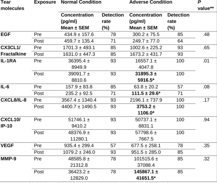
Among the 16 molecules analyzed in tears, 8 (EGF, Fractalkine, IL-1RA, IL-6, IL-8, IP-10, VEGF, and MMP-9) had a detection rate above 50% for both conditions. Consequently, these 8 were statistically analyzed; concentrations are summarized in Table 2. Mean concentration values of molecules with a detection rate <50% are also provided (Supplemental Material at AJO.com).
Prior to undergoing both exposures, only IL-1RA showed significantly different values (Table 2). There were no significant (P≥ .05) changes in the tear
molecule concentrations before and after exposure to the normal condition. On the contrary, after the adverse condition there was a significant increase in concentrations of IL-1RA (P= .01), IL-6 (P= .02), IL-8 (P= .03), and MMP-9 (P= .03) (Table 2).
Variables Predicting Inflammatory Response of the Lacrimal Functional Unit
A significant (P ≤ .04) inverse relationship was found between nasal (rs = -0.62) and temporal (rs = -0.55) baseline corneal staining and the worsening observed in these corneal areas after the adverse exposure. Additionally, a near
significant (P = .07) inverse (rs = -0.49) association was also observed between baseline inferior corneal staining and the increase showed in this parameter. Regarding tear molecules, we also found a significant inverse (rs = -0.55, P = .05) association between MMP-9 baseline values and its increase observed after the adverse exposure. The same occurred for IP-10 tear values (rs = -0.77,
P = .001).
Relationship between Changes in Tear Molecules and Clinical Variables
We found a consistent relationship between the increase in IL-1RA and the worsening of corneal integrity in terms of corneal staining scores for the central (rs= 0.62, P = .02), temporal (rs= 0.56, P = .04), and inferior (rs= 0.58, P = .03) areas. We also observed a significant inverse association between the increase in IL-6 (rs= -0.55, P = .04), IL-8 (rs= -0.52, P = .05) and MMP-9 (rs= -0.66, P = .02) tear concentration levels and the decrease in Schirmer test scores after the adverse exposure.
DISCUSSION
Although the exact etiopathogenesis of dry eye disease is not yet completely understood, there is global agreement regarding its inflammatory nature.1,2 With the aim of better understanding the disease and better showing dry eye
therapeutics efficacy, customized instruments and facilities where
1 2 3 4 5 6 7 8 9 10 11 12 13 14 15 16 17 18 19 20 21 22 23 24 25 26 27 28 29 30 31 32 33 34 35 36 37 38 39 40 41 42 43 44 45 46 47 48 49 50 51 52 53 54 55 56 57 58 59 60
eye patients worsened after undergoing short-term desiccating conditions. This clinical deterioration was accompanied by significant changes in the activity of several tear molecules (IL-1RA, IL-6, IL-8, and MMP-9) previously reported to be elevated in patients with dry eye as compared to healthy subjects.16-19
Differences in dry eye test scores between the measurements obtained before each exposure were negligible (Table 1). Likewise, when looking at the changes observed in the clinical dry eye tests after exposure to the normal condition, as expected, there were no significant variations in all tests performed, except for a slight increase in fluorescein staining in the nasal cornea. It has been previously demonstrated that spontaneous blink rate while reading is lower compared to other actions (i.e., conversation),30 which could explain this mild worsening observed in our SS dry eye patients in terms of nasal corneal staining increase.
Concerning the adverse condition, our SS patients showed a worsening of the lacrimal functional unit (Table 1). This finding agrees with previous studies 12-15
reporting that dry eye patients having diverse etiology can suffer ocular surface deterioration when exposed to desiccating conditions, which also
occurs in healthy subjects.14,30 Most of the dry eye tests tended to worsen in our study, showing significant deterioration in tear osmolarity, conjunctival
hyperemia, and corneal staining (Table 1). The increase in tear osmolarity could be associated to tear evaporation increase (low relative humidity environment31) and to unconscious blink rate reduction (volunteers were reading30). The sum of both factors could have produced a slight decrease in tear volume that would have yielded an increase in the concentration of solutes, leading to tear hyperosmolarity.32 In our study, we did not observe a significant reduction of tear volume as measured with phenol red thread tear and Schirmer test (Table 1). Nonetheless, these tests might be useful to support diagnosis of dry eye when the scores obtained are below a cut-off value;33 this does not mean that they are useful to detect minimal changes in tear volume, as has been widely reported, due to their lack of consistency.34,35
In our study, the exposure to the adverse condition also produced an
increase in corneal fluorescein staining, not only from a global viewpoint (Oxford scheme), but also for each area analyzed (Baylor scheme). However, significant changes were only observed for the nasal and temporal areas (Table 1 & Figure 1). Under desiccating environments, epithelial cells are more likely to be
exposed because of tear imbalance, which in our SS patients might have been even higher as a result of the meibomiam gland dysfunction that the vast
majority suffered. Hyperosmolarity stimulates the death of the epithelial surface even further.32 Consequently, a reduction in ocular surface integrity is to be expected, which in our case was also accompanied by an increase in conjunctival hyperemia (Table 1).
In our SS patients, we found an inverse association between the severity of corneal integrity prior to the adverse exposure and the change observed in corneal staining. The same inverse relationship occurred for MMP-9 and IP-10 tear levels. These findings might indicate that those SS patients whose lacrimal functional unit is already highly compromised might not show so much
1 2 3 4 5 6 7 8 9 10 11 12 13 14 15 16 17 18 19 20 21 22 23 24 25 26 27 28 29 30 31 32 33 34 35 36 37 38 39 40 41 42 43 44 45 46 47 48 49 50 51 52 53 54 55 56 57 58 59 60 61
impaired. Alex et al.14 previously observed just the opposite findings, those individuals having worse dry eye test scores were the ones showing higher worsening after exposing them to adverse conditions using goggles. This discrepancy might arise from the type of participants recruited in each study. They included 15 normal and 10 dry eye patients (only 4 were SS patients) in their study, while all of our individuals were SS patients who showed poorer corneal integrity than the dry eye patients recruited by Alex et al.14
We were unable to find previous reports addressing the initial changes in tear molecules occurring only in severe dry eye patients when suffering an acute exacerbation. Consequently, we evaluated the tear concentrations of 16 inflammatory molecules before and after both exposures. In our study, we did not observe significant differences in tear molecule concentrations before each exposure, except for IL-1RA (Table 2). Inter-day differences can be assigned to the inter-day variability reported in these tear molecules.36 Despite this fact, tear molecule levels could be more consistent than in some clinical dry eye tests.34 We did not observe any significant changes in tear molecule concentrations after the normal condition exposure. However, there was a significant increase in IL-1RA, IL-6, IL-8, and MMP-9 levels after 2-hour exposure to the adverse condition (Table 2).
IL-1RA is an endogenous inhibitor of IL-1 that impedes the activities of the pro-inflammatory forms of IL-1 by competitively binding to the type 1 IL-1 receptor. Huang et al.36 have reported that the concentration of this tear molecule is proportional to the severity of the dry eye, correlating with corneal staining. Moreover, our group37 found that there was an inverse relationship between the levels of IL-1RA and Schirmer and tear break-up time values. Consequently, it seems that this tear molecule is highly involved in the
inflammatory process occurring in dry eye patients. In fact, Amparo et al.38 have reported that Anakinra, a topical IL-1 receptor antagonist, reduced symptoms and corneal epitheliopathy in patients with dry eye after 12-week treatment; they suggested that the administration of an IL-1 antagonist might be a novel
therapeutic option in dry eye.
In our study, we also found an increase in IL-6 tear levels after undergoing desiccating conditions (Table 2). The IL-6 molecule is a pro-inflammatory cytokine frequently elevated in tears of dry eye patients,39,40 moreover, its concentration is increased in SS patients in comparison with non-SS
patients.17,18 It has also been suggested that it is one of the most important tear molecules in dry eye, because it has been found to be one of the factors
involved in desiccation-induced cell death in experimental settings.41
Additionally, increased IL-6 tear levels might be one of the earliest observable changes in patients with dry eye,16 as we also showed after exposing our SS dry eye patients to adverse conditions. However, its moderate percentage of detection as shown in this study and previous ones,13,25,37 as well as its moderate inter-day36 and intra-day42 variability, might still prevent this tear molecule from being the gold-standard biomarker for dry eye disease.
1 2 3 4 5 6 7 8 9 10 11 12 13 14 15 16 17 18 19 20 21 22 23 24 25 26 27 28 29 30 31 32 33 34 35 36 37 38 39 40 41 42 43 44 45 46 47 48 49 50 51 52 53 54 55 56 57 58 59 60
term desiccation,41 which might also have occurred in our SS dry eye patients. The increase of IL-8 tear levels might be a signal to recruit inflammatory cells to the ocular surface, which might be responsible for the deterioration of the
corneal and conjunctival integrity, as previous authors have hypothesized.43 In fact, Lam et al.39 reported a direct relationship between IL-8 concentration and corneal and conjunctival stainings.
We also observed that MMP-9 tear concentration (active and inactive forms), which is usually already elevated in dry eye patients44 especially in those having SS,19 also increased after the adverse condition. This finding agrees with the response observed in experimental settings when exposing mice to desiccating stress.45 Such increased MMP-9 tear levels were also observed in moderate dry eye patients under different adverse conditions.13,25 This tear molecule has gained popularity in relation to dry eye, as some authors have indicated its potential utility as a biomarker to diagnose dry eye disease using a MMP-9 based point-of-care device.46 Additionally, other authors have shown in-vivo benefits of using MMP-9 inhibitors for restoring tear production.47
In the present study we also found consistent associations between the increase in IL-1RA tear levels and the worsening of the corneal staining, in addition to the relationship between the decrease in Schirmer test and the increase of IL-6, IL-8 and MMP-9 tear levels. These findings provide more evidence (at least in SS patients) to support the relationship existing between the response of the lacrimal functional unit in terms of tear molecule secretion and the clinical signs commonly observed in the slit lamp examination. Thus, it enhances the relevance of performing further research to address the exact mechanism underlying the clinical worsening occurring not only SS patients but in all dry eye patients.
Our research group13,25 and others15 have previously showed that not only mild to moderate dry eye patients suffer ocular surface worsening under diverse adverse conditions, but also non-symptomatic subjects. Moreover, this dry eye exacerbation was accompanied by a significant variation in some tear
molecules (IL-6 and MMP-9)13,25 that were also increased in the present study. This means that the acute inflammatory response might be driven by similar mechanisms regardless of the etiological causes already provoking dry eye. Nonetheless, in this study our research group observed for the first time that IL-1RA might play a major role when dealing with SS patients not only based on its increased tear concentration, but also because of its significant association with higher corneal staining. Therefore, the response of SS patients might differ from that of other patients having evaporative-type dry eye (mild to moderate),
especially taking into account that SS is a chronic systemic autoimmune disease usually causing severe dry eye, while the origin of mild to moderate evaporative dry eye is not usually systemic.
1 2 3 4 5 6 7 8 9 10 11 12 13 14 15 16 17 18 19 20 21 22 23 24 25 26 27 28 29 30 31 32 33 34 35 36 37 38 39 40 41 42 43 44 45 46 47 48 49 50 51 52 53 54 55 56 57 58 59 60 61
this population also suffers worsening of the ocular surface when exposed to desiccating stress.14,25,31 Another limitation is that our results do not strictly apply to severe non-SS dry eye patients exposed to adverse conditions, as the origin of the inflammatory process causing dry eye can be different from the one present in SS.17 However, a similar worsening of the lacrimal functional unit to the one described here or the one previously reported by our group13,25 and others14,31 should be expected in terms of ocular surface worsening and increased inflammatory status, Finally, the number of patients included in the present study, although not high, has proved to be enough to show adequate evidence of the increase in the basal inflammatory state occurring in severe dry eye patients subjected to short-term desiccating conditions.
In conclusion, we showed that low humidity conditions can produce an increase in key clinical dry eye signs such as corneal staining or tear osmolarity in female SS dry eye patients. In addition, adverse environments that people are exposed to daily can produce significant changes in tear levels of some inflammatory molecules (IL-1RA, IL-6, IL-8, and MMP-9) in these severe dry eye patients, suggesting an increase in the basal inflammatory state of their lacrimal functional unit. Our study outcomes provide further evidence supporting current industry efforts to develop new therapeutics for ameliorating
1 2 3 4 5 6 7 8 9 10 11 12 13 14 15 16 17 18 19 20 21 22 23 24 25 26 27 28 29 30 31 32 33 34 35 36 37 38 39 40 41 42 43 44 45 46 47 48 49 50 51 52 53 54 55 56 57 58 59 60
ACKNOWLEDGEMENTS / DISCLOSURE
FUNDING/SUPPORT: Supported in part by Grant SAF2010-15361 from the Ministry of Economy and Competitiveness, Madrid, Spain: Programa de Proyectos de Investigación Fundamental no orientada. And by Grant VA174U14 from the Junta de Castilla y León (Consejería de Educación), Valladolid, Spain.
No funding organizations had a role in the design or conduct of this research.
FINANCIAL DISCLOSURES: The authors report the following conflicts of interest: Margarita Calonge was a consultant to Allergan and has received speaker/adviser honoraria from Xoma (Berkeley, CA, USA), Servier
Laboratories Ltd (Suresnes, France), and Allergan (Irvine, CA, USA). Michael E. Stern was an employee of Allergan, Inc (Irvine, CA, USA). Alberto López-Miguel was an employee of VISIÓN I+D, SL (Valladolid, Spain).
CONTRIBUTIONS OF AUTHORS: Design of the study (M.C., M.J.G.G., A.E.S., M.E.S.); conduct of the study (M.T., A.L.M., V.M.M.); sample collection (M.T., A.L.M., V.M.M.); management (A.E.S., M.J.G.G., M.C.); analysis (A.E.S., M.J.G.G., M.C.); interpretation of the data (A.E.S., M.E.S., M.J.G.G., M.C); manuscript preparation (M.T., A.L.M., A.E.S., M.J.G.G.); manuscript review (M.E.S., M.C.), and final approval of the manuscript (M.J.G.G., A.E.S., M.C., M.E.S.).
1 2 3 4 5 6 7 8 9 10 11 12 13 14 15 16 17 18 19 20 21 22 23 24 25 26 27 28 29 30 31 32 33 34 35 36 37 38 39 40 41 42 43 44 45 46 47 48 49 50 51 52 53 54 55 56 57 58 59 60 61
REFERENCES
1. Stern ME, Beuerman RW, Fox RI, Gao J, Mircheff AK, Pflugfelder SC. The pathology of dry eye: the interaction between the ocular surface and lacrimal glands. Cornea 1998;17(6):584-589.
2. Lemp AM, Baudouin C, Baum J, et al. The definition and classification of dry eye disease: report of the Definition and Classification
Subcommittee of the International Dry Eye WorkShop (2007). Ocul Surf
2007;5(2):75-92.
3. Schaumberg DA, Dana R, Buring JE, Sullivan DA. Prevalence of dry eye disease among US men: estimates from the Physicians' Health Studies.
Arch Ophthalmol 2009;127(6):763-768.
4. Schaumberg DA, Sullivan DA, Buring JE, Dana MR. Prevalence of dry eye syndrome among US women. Am J Ophthalmol 2003;136(2):318-326.
5. Galor A, Feuer W, Lee DJ, et al. Prevalence and risk factors of dry eye syndrome in a United States veterans affairs population. Am J
Ophthalmol 2011;152(3):377-384.
6. Patel R, Shahane A. The epidemiology of Sjögren's syndrome. Clin Epidemiol 30;6:247-255.
7. García-Carrasco M, Ramos-Casals M, Rosas J, et al. Primary Sjögren syndrome: clinical and immunologic disease patterns in a cohort of 400 patients. Medicine (Baltimore) 2002;81(4):270-80.
8. Brightman HS, Milton DK, Wypij D, Burge HA, Spengler JD. Evaluating building-related symptoms using the US EPA BASE study results.
Indoor Air 2008;18(4):335-345.
9. Wolkoff P. Ocular discomfort by environmental and personal risk factors altering the precorneal tear film. Toxicol Lett 2010;199(3):203-212. 10. Uchino M, Schaumberg DA, Dogru M, et al. Prevalence of dry eye
disease among Japanese visual display terminal users. Ophthalmology
2008;115(11):1982-1988.
11. Sheppard JD, Torkildsen GL, Lonsdale JD, et al. Lifitegrast ophthalmic solution 5.0% for treatment of dry eye disease: results of the OPUS-1 phase 3 study. Ophthalmology 2014;121(2):475-483.
12. Madden LC, Tomlinson A, Simmons PA. Effect of humidity variations in a controlled environment chamber on tear evaporation after dry eye therapy. Eye Contact Lens 2013;39(2):169-174.
13. Tesón M, González-García MJ, López-Miguel A, et al. Influence of a controlled environment simulating an in-flight airplane cabin on dry eye disease. Invest Ophthalmol Vis Sci 2013;54(3):2093-2099.
14. Alex A, Edwards A, Hays JD, et al. Factors predicting the ocular surface response to desiccating environmental stress. Invest Ophthalmol Vis Sci
2013;54(5):3325-3332.
15. Moore QL, De Paiva CS, Pflugfelder SC. Effects of dry eye therapies on environmentally induced ocular surface disease. Am J Ophthalmol
2015;160(1):135-142.
1 2 3 4 5 6 7 8 9 10 11 12 13 14 15 16 17 18 19 20 21 22 23 24 25 26 27 28 29 30 31 32 33 34 35 36 37 38 39 40 41 42 43 44 45 46 47 48 49 50 51 52 53 54 55 56 57 58 59 60
17. Lee SY, Han SJ, Nam SM, et al. Analysis of tear cytokines and clinical correlations in Sjögren syndrome dry eye patients and non-Sjögren syndrome dry eye patients. Am J Ophthalmol 2013;156(2):247-253. 18. Yoon KC, Jeong IY, Park YG, Yang SY. Interleukin-6 and tumor
necrosis factor-alpha levels in tears of patients with dry eye syndrome.
Cornea 2007;26(4):431-437.
19. Solomon A, Dursun D, Liu Z, Xie Y, Macri A, Pflugfelder SC. Pro- and anti-inflammatory forms of interleukin-1 in the tear fluid and conjunctiva of patients with dry eye disease. Invest Ophthalmol Vis Sci
2001;42(10):2283–2292.
20. Sullivan DA, Hammitt KM, Schaumberg DA, et al. Report of the
TFOS/ARVO Symposium on global treatments for dry eye disease: an unmet need. Ocul Surf 2012;10(2):108-116.
21. Pflugfelder SC, Geerling G, Kinoshita S et al. Management and therapy of dry eye disease: report of the Management and Therapy
Subcommittee of the International Dry Eye WorkShop (2007). Ocul Surf
2007;5(2):163-178.
22. Schiffman RM, Christianson MD, Jacobsen G, Hirsch JD, Reis BL. Reliability and validity of the Ocular Surface Disease Index. Arch Ophthalmol 2000;118(5):615-621.
23. Baudouin C, Aragona P, Van Setten G, et al. Diagnosing the severity of dry eye: a clear and practical algorithm. Br J Ophthalmol
2014;98(9):1168-1176.
24. Vitali C, Bombardieri S, Jonsson R, et al. Classification criteria for Sjogren’s Syndrome: a revised version of the European criteria proposed by the American-European Consensus Group. Ann Rheum Dis 2002;61(6):554-558.
25. López-Miguel A, Tesón M, Martín-Montañez V, et al. Dry eye
exacerbation in patients exposed to desiccating stress under controlled environmental conditions. Am J Ophthalmol 2014;157(4):788-798. 26. Efron N. Grading scales for contact lens complications. Ophthalmic
Physiol Opt 1998;18(2):182-186.
27. Bron AJ. The Doyne Lecture. Reflections on the tears. Eye
1997;11(5):583-602.
28. Pflugfelder SC, Beurerman RW, Stern ME. Dry Eye and Ocular Surface Disorders. New York: Marcel Decker; 2004.
29. Huang JF, Yafawi R, Zhang M, McDowell M, Rittenhouse KD, Sace F, Liew SH, Cooper SR, Pickering EH. Immunomodulatory effect of the topical ophthalmic Janus kinase inhibitor tofacitinib (CP-690,550) in patients with dry eye disease. Ophthalmology 2012;119(7):43-50. 30. Doughty MJ. Consideration of three types of spontaneous eyeblink
activity in normal humans: during reading and video display terminal use, in primary gaze, and while in conversation. Optom Vis Sci
2001;78(10):712-725.
31. Abusharha AA, Pearce EI. The effect of low humidity on the human tear film. Cornea 2013;32(4):429-434.
1 2 3 4 5 6 7 8 9 10 11 12 13 14 15 16 17 18 19 20 21 22 23 24 25 26 27 28 29 30 31 32 33 34 35 36 37 38 39 40 41 42 43 44 45 46 47 48 49 50 51 52 53 54 55 56 57 58 59 60 61
33. Bron AJ, Abelson MB, Ousler G, et al. Methodologies to diagnose and monitor dry eye disease: report of the Diagnostic Methodology
Subcommittee of the International Dry Eye WorkShop (2007). Ocul Surf
2007;5(2):108-152.
34. Nichols KK, Mitchell GL, Zadnik K. The repeatability of clinical measurements of dry eye. Cornea 2004;23(3):272-285.
35. Sullivan BD, Crews LA, Sönmez B, et al. Clinical utility of objective tests for dry eye disease: variability over time and implications for clinical trials and disease management. Cornea 2012;31(9):1000-1008.
36. Huang JF, Zhang Y, Rittenhouse KD, Pickering EH, McDowell MT. Evaluations of tear protein markers in dry eye disease: repeatability of measurement and correlation with disease. Invest Ophthalmol Vis Sci
2012;53(8):4556-4564.
37. Enriquez-de-Salamanca A, Castellanos E, Stern ME, et al. Tear cytokine and chemokine analysis and clinical correlations in evaporative-type dry eye disease. Mol Vis. 2010;16:862-873.
38. Amparo F, Dastjerdi MH, Okanobo A, et al. Topical interleukin 1 receptor antagonist for treatment of dry eye disease: a randomized clinical trial.
JAMA Ophthalmol 2013;131(6):715-723.
39. Lam H, Bleiden L, de Paiva CS, Farley W, Stern ME, Pflugfelder SC. Tear cytokine profiles in dysfunctional tear syndrome. Am J Ophthalmol
2009;147(2):198-205.
40. Na KS, Mok JW, Kim JY, Rho CR, Joo CK. Correlations between tear cytokines, chemokines, and soluble receptors and clinical severity of dry eye disease. Invest Ophthalmol Vis Sci 2012;53(9):5443-5450.
41. Higuchi A, Kawakita T, Tsubota K. IL-6 induction in desiccated corneal epithelium in vitro and in vivo. Mol Vis 2011;17:2400-2406.
42. Benito MJ, González-García MJ, Tesón M, et al. Intra- and inter-day variation of cytokines and chemokines in tears of healthy subjects. Exp Eye Res 2014;120:43-49.
43. Massingale ML, Li X, Vallabhajosyula M, Chen D, Wei Y, Asbell PA. Analysis of inflammatory cytokines in the tears of dry eye patients.
Cornea 2009;28(9):1023-1027.
44. Chotikavanich S, De Paiva CS. Li de Q, et al. Production and activity of matrix metalloproteinase-9 on the ocular surface increase in
dysfunctional tear syndrome. Invest Ophthalmol Vis Sci
2009;50(7):3203-3209
45. Corrales RM, Stern ME, De Paiva CS, Welch J, Li DQ, Pflugfelder SC. Desiccating stress stimulates expression of matrix metalloproteinases by the corneal epithelium. Invest Ophthalmol Vis Sci 2006;47(8):3293-3302.
46. Sambursky R, Davitt WF 3rd, Latkany R, et al. Sensitivity and specificity of a point-of-care matrix metalloproteinase 9 immunoassay for
diagnosing inflammation related to dry eye. JAMA Ophthalmol
2013;131(1):24-28.
1 2 3 4 5 6 7 8 9 10 11 12 13 14 15 16 17 18 19 20 21 22 23 24 25 26 27 28 29 30 31 32 33 34 35 36 37 38 39 40 41 42 43 44 45 46 47 48 49 50 51 52 53 54 55 56 57 58 59 60
48. Ding Y, Fu Q, Tian Z, Li M, Zhu N. Influence of indoor design air parameters on energy consumption of heating and air conditioning.
Energy and Buildings 2013;56:78–84.
1 2 3 4 5 6 7 8 9 10 11 12 13 14 15 16 17 18 19 20 21 22 23 24 25 26 27 28 29 30 31 32 33 34 35 36 37 38 39 40 41 42 43 44 45 46 47 48 49 50 51 52 53 54 55 56 57 58 59 60 61
FIGURE CAPTIONS
Figure 1. Images showing corneal fluorescein staining in a Sjögren
López-Miguel et al. -t1
TABLES
Table 1. Clinical test outcomes (mean ± standard error of the mean) in Sjögren syndrome-dry eye patients before and after 2-hour exposure to each controlled environment.
SIDEQ: Single-item score dry dye questionnaire; T-BUT: Tear break up time; SEM: standard error of the mean.
*P≤ .05: Comparison by Wilcoxon test before (pre) and after (post) 2-hour exposure. **P value corresponding to comparisons between dry eye disease test scores obtained prior to undergoing both exposures.
Dry Eye Disease Test Exposure Normal
Condition
Mean ± SEM
Adverse Condition
Mean ± SEM
P value**
Modified SIDEQ Pre 4.5 ± 0.5 4.8 ± 0.8 .67
Post 3.1 ± 0.6 3.6 ± 0.6
Tear osmolarity Pre 320.9 ± 4.7 315.7 ± 3.0 .24
Post 317.5 ± 5.1 327.7 ± 5.1*
Phenol red thread test Pre 13.6 ± 1.8 13.4 ± 1.6 .93
Post 13.7 ± 2.0 12.3 ± 1.3
Conjunctival hyperemia (mean)
Pre 1.5 ± 0.1 1.3 ± 0.1 .25
Post 1.6 ± 0.1 1.6 ± 0.1*
T-BUT Pre 1.3 ± 0.1 1.3 ± 0.1 1.0
Post 1.5 ± 0.2 1.2 ± 0.1
Corneal fluorescein staining (Oxford scheme)
Pre 2.4 ± 0.2 2.5 ± 0.2 .16
Post 2.3 ± 0.2 2.7 ± 0.2
Corneal fluorescein staining (Baylor scheme)
Central Pre 2.1 ± 0.4 2.1 ± 0.5 1.0
Post 2.1 ± 0.4 2.6 ± 0.6
Nasal Pre 2.9 ± 0.5 3.6 ± 0.5 .29
Post 3.6 ± 0.5* 4.5 ± 0.5*
Temporal Pre 3.4 ± 0.4 3.5 ± 0.5 .83
Post 3.5 ± 0.5 4.7 ± 0.4*
Superior Pre 0.9 ± 0.4 1.2 ± 0.4 .39
Post 1.0 ± 0.5 1.6 ± 0.4
Inferior Pre 5.1 ± 0.3 4.7 ± 0.4 .30
Post 4.9 ± 0.5 5.4 ± 0.4
Lissamine green conjunctival staining
Pre 2.0 ± 0.3 1.9 ± 0.3 .83
Post 2.1 ± 0.3 1.9 ± 0.3
Schirmer test (no anesthesia)
Pre 4.1 ± 0.7 4.1 ± 0.6 .90
Table 2. Tear molecule detection rates and concentrations in Sjögren syndrome-dry eye patients pre- and post-exposure (2 hours) to simulated environments. Only the 8 molecules that had detection rate ≥50% out of the 16 measured are shown.
EGF: epidermal growth factor; CX3CL1: chemokine (C-X3-C motif) ligand 1; IL-1RA: interleukin 1 Receptor antagonist; IL: interleukin; CXCL8: chemokine (C-X-C motif) ligand 8; CXCL10: chemokine (C-X-C motif) ligand 10; IP-10: Interferon γ-induced protein 10; VEGF: vascular endothelial growth factor; MMP-9: matrix
metalloproteinase-9; SEM: standard error of the mean.
*P≤ .05: Comparison by t-test before (pre) and after (post) 2-hour exposure to each environmental condition. **P value corresponding to comparisons by t-test between tear molecule concentrations obtained prior to undergoing both exposures.
Tear molecules
Exposure Normal Condition Adverse Condition P
value** Concentration
(pg/ml) Mean ± SEM
Detection rate (%)
Concentration (pg/ml)
Mean ± SEM
Detection rate (%)
EGF Pre 434.9 ± 157.6 78 300.2 ± 75.5 85 .48
Post 459.7 ± 135.4 71 249.7 ± 77.0 64 CX3CL1/
Fractalkine
Pre 1701.3 ± 493.1 85 1002.6 ± 225.2 93 .65 Post 1631.0 ± 447.3 85 1673.2 ± 431.7 93
IL-1RA Pre 36395.4 ±
8949.9
93 16557.1 ± 4047.8
100 .01
Post 39091.7 ± 8810.6
93 31895.3 ±
5916.5*
100
IL-6 Pre 157.9 ± 83.8 85 63.8 ± 20.2 57 .08
Post 235.2 ± 92.5 71 111.5 ± 29.6* 71
CXCL8/IL-8 Pre 3567.4 ± 1340.4 93 2196.1 ± 737.9 100 .17
Post 4400.7 ± 1490.5 93 3753.2 ± 1106.0*
100
CXCL10/ IP-10
Pre 51746.1 ± 9410.2
93 50737.1 ± 8831.1
100 .94
Post 48376.9 ± 11280.1
93 57798.6 ± 7667.5
100
VEGF Pre 935.4 ± 299.4 57 677.5 ± 258.1 78 .35
Post 1079.2 ± 246.0 93 951.5 ± 285.0 85
MMP-9 Pre 48585.8 ±
21312.8
78 101515.6 ± 37088.4
85 .32
Post 36423.2 ± 12829.0
78 145867.1 ±
41651.5*
TABLE OF CONTENTS STATEMENT
TITLE:
Clinical and Molecular Inflammatory Response in Sjögren Syndrome-Associated Dry Eye Patients under Desiccating Stress
MANUSCRIPT NUMBER: AJO-15-870
Sjögren syndrome patients suffer from a chronic systemic autoimmune disease that causes dry eye disease. These patients usually experience exacerbations characterized by a reduction of the corneal integrity as well as an increase in some tear inflammatory mediators (interleukin-1 receptor antagonist, interleukin-6, interleukin-8, matrix metallopeptidase-9) when they are exposed to adverse conditions (desiccating stress). This study provides more evidence to target certain inflammatory molecules when designing new therapeutics and also shows that adverse environments can be tightly
López-Miguel et al. -St1
SUPPLEMENTAL TABLE
Table 1. Tear molecule detection rates and concentrations of molecules whose detection levels were below the limit in less than 50% of the total number of samples (n=14). Sjögren syndrome-dry eye patients were exposed for 2 hours to 2 simulated environments of 23ºC and 0.10 m/s air flow having different relative humidity: normal condition 45%, and adverse condition 5%.
IFN-γ: interferon-γ, IL: interleukin; CCL5/RANTES: chemokine (C-C motif) ligand 5 (CCL5)/regulated on activation, normal T-cell expressed and secreted; TNF-α: tumor necrosis factor-α.
*Mean obtained after computing only the real values detected for each molecule. Minimum detectable values were not computed to obtain the mean value for each molecule.
Tear molecules
Exposure Normal Condition Adverse Condition
Concentration (pg/ml)
Mean*
Detection rate N out of 14 (%)
Concentration (pg/ml)
Mean*
Detection rate N out of 14 (%)
IFN-γ Pre 31.2 4 (28) 105.0 1 (7)
Post 70.9 3 (21) - 0 (0)
IL -1β Pre 30.7 2 (14) 31.7 3 (21)
Post 42.8 6 (43) 19.4 6 (43)
IL -2 Pre - 0 (0) 57.0 1 (7)
Post - 0 (0) - 0 (0)
IL -10 Pre 53.2 2 (14) 38.4 2 (14)
Post 152.0 2 (14) 17.0 2 (14)
IL -12p70 Pre 49.5 5 (36) 31.0 1 (7)
Post 63.4 4 (28) 104.6 3 (21)
IL -17A Pre - 0 (0) 18.0 1 (7)
Post 79.3 1 (7) - 0 (0)
CCL5/RANTES Pre 225.5 6 (43) 64.3 6 (43)
Post 216.4 6 (43) 187.9 5 (36)
TNF-α Pre 34.3 4 (28) 42.9 1 (7)