Serum level of soluble vascular cell adhesion
molecule in patients with hepatocellular carcinoma and
its association with severity of liver disease
Antonio Diaz-Sanchez,* Ana Matilla,** Oscar Nuñez,*** Diego Rincon,** Raquel Lorente,** Oreste Lo Iacono,**** Beatriz Merino,** Ana Hernando,** Rocio Campos,* Gerardo Clemente,** Rafael Bañares**
* Gastroenterology Unit, Hospital Universitario del Sureste, Arganda del Rey, Madrid, Spain. ** Liver Unit-CIBEREHD, Hospital General Universitario Gregorio Marañon, Madrid, Spain. *** Gastroenterology Unit, Hospital Universitario Infanta Sofia, San Sebastian de los Reyes, Madrid, Spain.
**** Gastroenterology Unit, Hospital del Tajo. Aranjuez, Madrid, Spain.
ABSTRACT
Background. VCAM-1 (soluble vascular cell adhesion molecule-1) plays a role in liver angiogenesis. Hepato-cellular carcinoma (HCC) has important angiogenic activity, so expression of VCAM-1 may be pathogenic. Aim. To assess the association between serum VCAM-1 (sVCAM-1) levels and features of tumour and liver disease in patients with and without HCC, and to study the influence of HCC treatment on sVCAM-1 levels. Material and methods. Concentrations in peripheral (sVCAM-1-P) and hepatic (sVCAM-1-H) veins were analysed using ELISA in 134 consecutive patients with chronic liver disease between May 2004 and February 2006, who underwent a splanchnic haemodynamic study. Of these patients, 58 had HCC. Results. sVCAM-1-P and sVCAM-1-H were well correlated in both groups. No association was found between sVCAM-1-H and tumour features. No differences were observed in sVCAM-1-H between HCC and non-HCC cirrhotic patients. There was a significant linear association between Child-Pugh stage and sVCAM-1-H in HCC-patients (Child-Pugh A [2,485 ± 1,294 ng/mL] vs. Child-Pugh B [3,408 ± 1,338 ng/mL] vs. Child-Pugh C [4,096 ± 862 ng/ mL]; p = 0.007). Seven non-cirrhotic HCC patients had a significantly lower sVCAM-1-H than cirrhotic HCC patients. Treatment of HCC leads to an increase in sVCAM-1-H levels although this was not associated with the necrosis response to treatment. Conclusions. sVCAM-1 levels are more closely associated with the se-verity of underlying liver disease than with the presence of HCC. sVCAM-1 levels are not associated with tumour features or invasiveness; therefore, sVCAM-1 does not seem to play an important role in the angio-genic processes of HCC.
Key words. Cirrhosis. Hyaluronic Acid. Vascular cell adhesion molecule-1.
Correspondence and reprint request: Antonio Diaz-Sanchez, M.D. Gastroenterology Unit, Hospital Universitario del Sureste Ronda del Sur 10, 28500 Arganda del Rey, Madrid, Spain. Tel.: +0034 699871075
E-mail: [email protected]
Manuscript received: June 19, 2012. Manuscript accepted: August 30, 2012.
INTRODUCTION
Soluble vascular cell adhesion molecule-1 (VCAM-1), a member of the immunoglobulin superfamily, is expressed on various types of cells, including endo-thelial cells. This form is released into the bloods-tream following proteolytic cleavage by vascular
endothelial activation mediated by proinflammatory cytokines.1,2 This molecule plays an important
role in providing attachment to the developing en-dothelium during angiogenesis.3,4 thus promoting
adhesion of leucocytes to the endothelium.
Due to its angiogenic potency, elevated serum levels of VCAM-1 have been described in patients with gastric cancer,5,6 colorectal cancer,7 melanoma,8
leukaemia9 and breast cancer.10 VCAM-1 has been
significantly correlated with tumour stage and the development of metastasis in neoplasms such as those presented above.
molecule (sVCAM-1) have been associated with in-creased fibrosis and angiogenesis in different aetio-logies of liver disease.11-13 Furthermore, sVCAM-1
has recently been observed to act as a potential marker of hyperdynamic circulation, which is closely related to the different stages of liver cirrhosis.14
However, information about the influence of sVCAM-1 in patients with hepatocellular carcinoma (HCC) is scarce.15,16 Chronic hepatitis and cirrhosis
are the underlying liver conditions in most patients with HCC; therefore sVCAM-1 could be involved in the progression of both the tumour and chronic liver disease.
Owing to the fact that sVCAM-1 is associated with increasing fibrosis and angiogenesis in liver disease and its direct relation with tumour burden in several neoplastic diseases, we hypothesized that this molecule may act as a marker of advanced HCC in cirrhotic patients.
Therefore, this study was designed to assess the correlation between sVCAM-1 levels and the clinical and tumour features of patients with HCC. We also evaluated the influence of locoregional treatment of HCC on sVCAM-1 levels and the response to treat-ment.
MATERIAL AND METHODS
We performed a prospective single-centre cohort study between May 2004 and February 2006 in which 58 consecutive patients with HCC underwent a hepatic haemodynamic study to assess the hepatic venous pressure gradient (HVPG) as a prognostic criterion in candidates for treatment of HCC. Ten of these patients proceeded from a previous published cohort designed to assess the correlation between sVCAM-1 and splanchnic and systemic haemodynamic measurements.14 Due to the difficult for the
per-formance of an analysis of association between sVCAM-1 with HCC features due to the small sam-ple size in that previous study, we collected for the present study 48 patients with HCC between Octo-ber 2004 and the final date of inclusion, in February 2006. In this period of time, 25 patients were evalua-ted for liver transplantation, and 20 finally received an organ. Seven patients underwent surgical resec-tion of their tumour, 11 patients underwent radio-frequency ablation and 8 patients underwent transarterial chemoembolisation (TACE). Seven patients were treated with the best supportive care due to severe comorbidity or impaired liver function in cases with extensive HCC (in these cases the patients received a hepatic haemodynamic study in
the context of a clinical trial). A further 76 consecu-tive patients analysed in the previously published co-hort with cirrhosis but no radiological evidence of HCC and normal alpha-fetoprotein serve as compa-rative group.14 They also underwent a hepatic
hae-modynamic study as part of the routine clinical work-up. Patients with a previous liver transplant, HIV infection, or aetiology of liver disease due to Budd-Chiari syndrome or sinusoidal obstruction syndrome were excluded. The diagnosis of cirrhosis was established by liver biopsy or by a combination of clinical, biological and ultrasound findings. No patient had ongoing bacterial infection at the time of the haemodynamic measurements.
Demographic data and data on the aetiology of li-ver disease were collected. Biochemical and clinical variables were recorded to determine the model for end-stage liver disease (MELD) score17 and
Child-Pugh score.18 The maximum permissible interval
between the haemodynamic study and blood tests was 1 week; the maximum interval since the last dy-namic imaging technique (computed tomography scan or magnetic resonance) was 1 month.
Diagnosis of HCC was established following the algorithm of the American Association for the Study of Liver Diseases (AASLD).19 The Barcelona Clinic
Liver Cancer (BCLC) classification was used to stage HCC.20
Treatment performance and definition of response
Of the whole HCC cohort, 8 patients underwent TACE and 11 patients underwent radiofrequency ablation. Of these, sVCAM-1-P levels were deter-mined in 9 patients treated with radiofrequency ablation and in 5 patients treated with TACE, at days 1, 3 and 7 after treatment was performed. The selection of these patients was due to the inclusion of them in another prospective study.
Radiofrequency ablation was performed percuta-neously except when the nodule was in a difficult ac-cess location or in the proximity of other organs or blood vessels, performing in thoses cases by laparos-copy or laparotomy accesses. The electrode used for performing the procedure was a Berchtold Medical Electronics with an Elektrotom 106 HF-Thermo ge-nerator (Berchtold Medizin-ElektroniK Tuttlingen) at 350 KHz, with saline solution perfussion of 60-80 mL/h, and with a temperature threshold of 70 oC to
100 oC.
of lesions with less size the mixture consists in 40 mg of adriamicin with 8 cc of lipiodol. After the deli-bery of the mixture the vessels were embolized with gelfoam.
The response to the treatment was evaluated 6 weeks after treatment performance by dynamic ima-ging techniques. All the imaima-ging files were reviewed subsequently for this purpose in the present study, using the modified Response Evaluation Criteria in Solid Tumors (RECIST) for HCC.21 Complete
Res-ponse was considered in the case of disappearance of any intratumoral arterial enhancement in all target lesions after treatment. Fail of treatment was consi-dered in the rest of cases.
Haemodynamic measurements
After an overnight fast, the patient was prepared for the study in the supine position. Under local anaesthesia, a vascular introducer sheath (Medikit Co Ltd, Tokyo, Japan) was placed into the right in-ternal jugular vein. Then, under fluoroscopy, a 7F balloon catheter (Cordis SA, Miami, FL, USA) was conducted to the right hepatic vein for the measure-ment of free and wedged hepatic venous pressure (FHVP and WHVP) as described previously.22 FHVP
was measured in the hepatic vein with the tip of the catheter just beyond the junction with the inferior vena cava. HVPG was calculated as WHVP minus FHVP. Portal hypertension was defined as the pre-sence of an HVPG > 5 mmHg. All haemodynamic measurements were performed using a previously calibrated strain-gauge transducer and recorded at least in duplicate.
The tracings of the haemodynamic studies were evaluated by two independent investigators who were unaware of the identity and diagnosis of the patient.
Laboratory determinations
Blood samples were collected from peripheral and hepatic veins during the haemodynamic studies. sVCAM-1 levels were quantified using an enzyme-linked immunosorbent assay (ELISA) kit designed to measure human soluble VCAM-1 concentration in serum (Human VCAM-1 Immunoassay, R&D Sys-tem Minneapolis, MN, USA). sVCAM was detected by a monoclonal murine antibody to human VCAM-1. Each measurement was made in duplicate and serum VCAM-1 levels were determined by extrapolation from a standard curve generated for each set of samples assayed. The test has a sensitivity threshold
of < 2.0 ng/mL, an intra-assay variability of 4.3%, an interassay variability of 8.5%, and a mean refe-rence value of 553 ng/mL (range 395-714 ng/mL) on a panel of 105 blood donors.
Serum hyaluronic acid and serum vascular endo-thelial growth factor (VEGF) levels were measured by ELISA according to the manufacturer’s instruc-tions (Corgenix Inc., Denver, Colorado, USA and R&D System Minneapolis, Minnesota, USA).
Statistical analysis
Quantitative variables were expressed as the mean (Standard Deviation) or median (Interquartile Range), and qualitative variables were expressed as frequencies. Categorical variables were compared using the χ2; continuous variables were
compa-red using the t test or the Mann-Whitney U test when appropriate. One-way analysis of variance (ANOVA) with polynomial contrasts was applied. Pearson or Spearman correlations were used to eva-luate the association between continuous variables as appropriate. In the case of performing multiple comparations between variables the Bonferroni correction was applied as appropiate. The differences in the mean values of sVCAM after treatment per-formance were evaluated by a general linear model comparing the variables by the Bonferroni method. Those variables with a p value of 0.1 or less were chosen for enter in a multivariate analysis with a multiple linear regression model. Statistical signifi-cance was set at a p value of 0.05 (2-tailed). Statisti-cal analysis was performed using SPSS 15.0 (SPSS® 15.0; SPSS Inc., Chicago, Illinois, USA).
Ethical considerations
The study protocol was approved by the local Ethics Committee and the Institutional Review Board, and was in accordance with the Helsinki De-claration. All the samples were collected after a signed informed consent was obtained from each patient before the hepatic haemodynamic study.
RESULTS
Patient population
abuse (15.5%). Five patients were infected by hepati-tis B virus and all but one of them had data of cir-rhosis. One of these patients was received antiviral treatment with lamivudin and two with lamivudin and adefovir. Most patients were Child-Pugh grade A (62.1%). Compared with the non-HCC patients, patients with HCC were older, showed a better MELD score and liver function parameters, and had less episodes of hepatic decompensation. Seven pa-tients with HCC did not have established cirrhosis as their underlying liver disease.
Tumour characteristics are shown in table 2. BCLC stage A was predominant (55.2%), mean tu-mour size was 4.8 ± 3.7 cm, the lesion was solitary in 63.2% of patients, both lobes were involved in 19.2% of cases, and vascular invasion was present in 6.9% of patients.
Correlation of sVCAM-1
between peripheral and hepatic veins
A strong correlation was observed in levels of sVCAM-1 between samples collected from peripheral veins (sVCAM-1-P) and those from hepatic veins
(sVCAM-1-H) in the whole cohort (r = 0.80; p = 0.0001) (Figure 1). This correlation was also very high in patients with HCC (r = 0.85; p = 0.0001) and without HCC (r = 0.77; p = 0.0001).
Association between sVCAM-1-H and features of HCC
Table 2 shows the relationship between sVCAM-1-H levels and the features of HCC. An almost signi-ficant statistically inverse correlation was found bet-ween sVCAM-1-H concentration and total tumour size in centimetres in both Child-Pugh A and B-C patients (p = 0.08). There was no significant asso-ciation in either Child-Pugh group with the fol-lowing: alpha-fetoprotein levels, lobular tumour extension, vascular thrombosis, lymph node invol-vement, fulfilment of the Milan criteria, number of nodules or infiltrating tumour pattern. The differences with respect to BCLC stage were significant through-out the HCC cohort, although these differences were lost when we performed a subgroup analysis based on Child-Pugh stage.
Table 1. Epidemiological and clinical characteristics of patients with and without hepatocellular carcinoma.
HCC patients (n = 58) Non-HCC patients (n = 76) p
N (%)
Sex (Male/Female) 47 (81)/11 (19) 53 (70) / 23 (30) 0.13
Aetiology 0.25
Hepatitis C virus 40 (69) 42 (55)
Alcohol 9 (15) 31 (41)
Hepatitis B virus 5 (9) 3 (4)
Other 4 (7)
-Cirrhosis criteria fulfilled 51 (88) 76 (100) 0.002
Child-Pugh (A/B/C) 36 (62) / 16 (28) / 6 (10) 26 (34) / 37 (49) / 13 (17) 0.005
Ascites 16 (28) 39 (51) 0.007
Hepatic encephalopathy 2 (4) 4 (5) 0.99
Presence of oesophageal varices 32 (60) 61 (80) 0.007
Previous variceal bleeding 5 (9) 13 (17) 0.17
Mean ± SD
Age (years) 60.3 ± 11.5 50.3 ± 7.6 0.001
Platelets (cells/µL) 131,569 ± 89,646 100,921 ± 60,713 0.03
INR 1.19 ± 0.38 1.51 ± 0.55 0.001
Bilirubin (mg/dL) 1.9 ± 1.6 3.9 ± 4.9 0.003
Albumin (g/dL) 3.6 ± 0.6 3.2 ± 0.6 0.001
Creatinine (mg/dL) 1 ± 0.7 0.9 ± 0.7 0.66
MELD score 10 ± 5 15 ± 6 0.001
In addition to this, a model of linear regression was performed adjusting the model with the BCLC, observing that this variable only explained the 6.7% of the variability of sVCAM-1-H with absence of statistical significance (p = 0.059) (Table 3). Therefore, the influence of liver function impairment was predominant.
Correlation between
sVCAM-1-H levels and severity of liver disease
In HCC patients, sVCAM-1-H levels had no signi-ficant relationship with sex, age, aetiology or
pre-Table 2. Tumour characteristics of HCC patients and association with sVCAM-1-H levels.
Mean ± SD
N (%) sVCAM-1-H (ng/mL) p
Total tumour size 0.08
≤ 3 cm 22 (38) 3,424 ± 1,477
3-5 cm 16 (28) 2,407 ± 1,153
≥ 5 cm 20 (34) 2,704 ± 1,314
Number of nodules† 0.23
Single nodule 37 (69) 2,989 ± 1,455
Multinodular 17 (31) 2,476 ± 160
Infiltrating pattern 0.11
Present 4 (7) 3,991 ± 814
Absent 54 (93) 2,835 ± 1,382
Lobar tumour extension 0.88
One lobe 46 (79) 2,936 ± 1,446
Bilobar 12 (21) 2,865 ± 1,156
Vascular thrombosis 0.24
Present 4 (7) 3,700 ± 1,069
Absent 54 (93) 2,858 ± 1,389
Lymph node involvement 0.91
Present 17 (29) 2,951 ± 1,357
Absent 41 (71) 2,908 ± 1,407
Milan criteria fulfilled 0.16
Yes 42 (72) 3,089 ± 1,355
No 16 (28) 2,521 ± 1,390
Alpha-fetoprotein > 20 ng/mL 0.36
Yes 32 (55) 2,760 ± 1,309
No 26 (45) 3,107 ± 1,457
Alpha-fetoprotein > 200 ng/mL 0.38
Yes 50 (86) 2,851 ± 1,387
No 8 (14) 3,321 ± 1,335
BCLC stage‡ 0.02
0 very early 8 (14) 2,818 ± 1,273
A early 32 (55) 3,005 ± 1,364
B intermediate 7 (12) 1,577 ± 587
C advanced 5 (9) 3,042 ± 1,703
D terminal 6 (10) 4,097 ± 862
BCLC: Barcelona Clinic Liver Cancer. †Excluding infiltrating pattern. ‡The differences were lost when the Child-Pugh classification was applied.
vious alcohol consumption. There was a significant linear association between Child-Pugh stage and sV-CAM-1-H (Child-Pugh A [2,485 ± 1,294 ng/mL] vs.
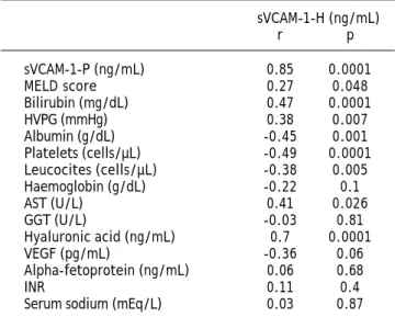
Table 3. Correlation of continuous variables with sVCAM-1-H in HCC patients.
sVCAM-1-H (ng/mL)
r p
sVCAM-1-P (ng/mL) 0.85 0.0001
MELD score 0.27 0.048
Bilirubin (mg/dL) 0.47 0.0001
HVPG (mmHg) 0.38 0.007
Albumin (g/dL) -0.45 0.001
Platelets (cells/µL) -0.49 0.0001
Leucocites (cells/µL) -0.38 0.005
Haemoglobin (g/dL) -0.22 0.1
AST (U/L) 0.41 0.026
GGT (U/L) -0.03 0.81
Hyaluronic acid (ng/mL) 0.7 0.0001
VEGF (pg/mL) -0.36 0.06
Alpha-fetoprotein (ng/mL) 0.06 0.68
INR 0.11 0.4
Serum sodium (mEq/L) 0.03 0.87
MELD: model for end stage liver disease. HVPG: hepatic venous pressure gradient. AST: aspartate aminotransferase. GGT: gamma-glutamil trans-peptidase. VEGF: vascular endothelium growth factor. INR: international normalized ratio.
Figure 1. Correlation between sVCAM-1-P and sVCAM-1-H in the whole cohort.
sVCAM-1-H (ng/mL)
7,000
6,000
5,000
4,000
3,000
2,000
1,000
0
0 1,000 2,000 3,000 4,000 5,000 6,000 7,000 sVCAM-1-P (ng/mL)
r = 0.80 p = 0.0001
Figure 2. Association between sVCAM-1-H levels and different features in HCC patients. A. Levels of sVCAM-1-H (ng/mL) according to the Child-Pugh classification. B. Levels of sVCAM-1-H (ng/mL) according to the model for end-stage liver disease (MELD) score. C. Correlation of sVCAM-1-H levels (ng/mL) with hyaluronic acid concentration (ng/mL). sVCAM-1-H: soluble vascular cell adhesion molecule-1 in hepatic veins.
non-HCC patients. The correlations between sVCAM-1-H and continuous variables are expressed in table 3.
Although without reaching significant
associa-7,000
6,000
5,000
4,000
3,000
2,000
1,000
0
sVCAM-1-H (ng/mL)
A B
C
A B C
Child-Pugh
p = 0.007
r = 0.27; p = 0.048 7,000
6,000
5,000
4,000
3,000
2,000
1,000
0
sVCAM-1-H (ng/mL)
5 10 15 20 25 30 35
MELD score 5,000
4,000
3,000
2,000
1,000
0
sVCAM-1-H (ng/mL)
0 100 200 300 400 500 600 Hyaluronic acid (ng/mL)
tion, in the HCC group the relationship between sVCAM-1-H and complications of cirrhosis demon-strated higher levels of this molecule in patients with previous history of hepatic decompensation: ascites (3,092 ± 958 vs. 2,854 ± 1,534 ng/mL), presence of esophageal varices (3,272 ± 1,302 vs. 2,719 ± 1,398 ng/mL) or variceal bleeding (3,444 ± 1,584 vs. 2,867 ± 1,391 ng/mL). In agreement with these data, sVCAM-1-H levels were different among the clinical stages proposed in the Baveno IV consensus confer-ence, showing an increase in the levels compared with patients without decompensation (stage 1: 2,489 ± 1,495 vs. stage 2: 3,493 ± 1,432 vs. stage 3: 3080 ± 970 vs. stage 4: 2,552 ± 1,387; p = 0.16).
A multivariate linear regression model was per-formed with the entire cohort to investigate the in-fluence of sVCAM-1-H in the severity of liver disease. Finally the model contained the following variables: serum bilirubin, MELD score, HVPG and Baveno IV stages (1-2-3 vs. 4), explaining the 42.8% of the variability of the influence of sVCAM-1-H in this setting, with a high significance (p < 0.001) (Table 4).
Seven HCC patients had no diagnosis of cirrhosis. The mean level of sVCAM-1-H in these patients was 1,460 ± 1,030 ng/mL, which was significantly lower than that observed in the HCC patients with cirrho-sis (3,099 ± 1,301 ng/mL; p = 0.003) (Figure 3).
Association between sVCAM-1-H and levels of hyaluronic acid and VEGF
Serum levels of hyaluronic acid in peripheral and hepatic veins were measured in 21 patients as a marker of liver fibrosis. sVCAM-1-H was highly correlated with both measurements (r = 0.72 and r = 0.70, respectively; p = 0.0001) (Figure 2C).
Furthermore, levels of VEGF were analysed in the peripheral and hepatic veins of 28 patients, and no robust association was found with sVCAM-1-H levels (r = -0.33 and r = -0.36, respectively; p = 0.06).
Association between sVCAM-1-H and cirrhotic patients with and without HCC
The distribution of sVCAM-1-H levels in cirrhotic and non-cirrhotic patients with and without HCC is shown in figure 3. The 7 non-cirrhotic patients with HCC had a significantly lower level of sVCAM-1-H than cirrhotic patients (1,460 ± 1,030 vs. 2,900 ± 1,424 ng/mL; p = 0.003) (Figure 3A).
Table 4.
Multivariate linear regression models for tumoral features and severity of liver disease explaining sVCAM-1-H.
RR
2
B (SE)
Inferior CI95%
Superior CI95%
p
Model for tumoral features
BCLC (A-B
vs.
C-D)
0.258
0.067
-875.87 (453.87)
-1,785.94
35.57
0.059
Model for severity of liver disease features
Final model
0.654
0.428
2,052.15 (463.35)
1,117.72
2,986.58
<
0.001
Bilirubin
-932.08 (226.14)
476.03
1,388.12
< 0.001
MELD
--143.13
(56.88)
-257.84
-28.43
0.016
HVPG
-53.72 (26.97)
-0.68
108.11
0.053
Baveno IV (1-2-3
vs.
4)
--1,365.63 (696.69)
-2,770.64
39.37
0.056
The mean sVCAM-1-H levels were 3,099 ± 1,301 ng/mL in the HCC group and 2,900 ± 1,424 ng/mL in the non-HCC group with no significant differences between the groups (Figure 3B). These results were maintained when we analysed the differences between both groups stratified by Child-Pugh stage (A vs. B-C) (Figure 3C) or MELD score (MELD > 15 vs. < 15).
In the follow-up 8 patients without HCC in the moment of inclusion in the study, developed HCC. The median time for the detection of the tu-mor after the performance oh the basal haemody-namic study was 11 (3.25-30.75) months. Three of these patients received a liver transplant in the follow-up because of HCC. Other two patients without HCC in the inclusion time who were transplanted in the follow-up, developed an HCC that was discovered incidentally in the liver ex-planted, without correlation with the previous dynamic imaging techniques. There were no diffe-rences in sVCAM-1-H concentration between the patients who developed (8/76) and not developed
the tumor (2,595 ± 1,248 vs. 2,976 ± 1,446 ng/ mL; p = 0.48).
HCC treatment and sVCAM-1 concentration
We measured sVCAM-1-P levels in 14 patients who received treatment for HCC during follow-up, at days 1, 3 and 7, and 6 weeks after treatment was performed. Radiofrequency ablation was performed in 9 patients and TACE was chosen in 5 patients. Complete response was achieved in 22% of patients received radiofrequency ablation compared with 80% of those received TACE. The median number of ses-sions in the radiofrequency ablation group was 1.4. This could cause a suboptimal complete response in the Radiofrequency group. We observed a progressive increase in levels of sVCAM-1-P over time with a maximum level in the seventh day, but without sta-tistically significant differences between the measures at 1st, 3rd or 7th day after the treatment performance. Levels then decreased to their baseline values (Figure 4). Although higher levels of sVCAM-1-P Figure 3. Association between sVCAM-1-H levels and the severity of liver disease. A. Differences in sVCAM-1-H levels (ng/mL) between HCC patients with and without cirrhosis. B. Association between sVCAM-1-H levels (ng/mL) and the presence of hepa-tocellular carcinoma in the whole cohort. C. Levels of sVCAM-1-H (ng/mL) according to the Child-Pugh classification in patients with and without HCC. sVCAM-1-H: soluble vascular cell adhesion molecule-1 in hepatic veins.
7,000
6,000
5,000
4,000
3,000
2,000
1,000
0
sVCAM-1-H (ng/mL)
Yes No
Cirrhosis
p = 0.003
7,000
6,000
5,000
4,000
3,000
2,000
1,000
0
sVCAM-1-H (ng/mL)
Yes No
Hepatocellular carcinoma
p = ns 7,000
6,000
5,000
4,000
3,000
2,000
1,000
0
sVCAM-1-H (ng/mL)
A B-C
Child-Pugh p = 0.37
Hepatocellular carcinoma
were associated with failure of achievement of com-plete response of HCC in both kind of treatments, this difference was not statistically significant (com-plete response 56.5% [2,985 ± 1,486 ng/mL] vs. non-complete response 43.5% [3,786 ± 1,498 ng/mL]; p = 0.21).
DISCUSSION
Although the importance of VCAM-1 as a circula-ting angiogenesis-related marker and its role in the development and prognosis of several types of can-cer are well established, its effect on HCC is uncan-cer- uncer-tain and literature reports are scarce.15,16 One
important difference between our study and previous reports is the measurement of VCAM-1 from hepatic and peripheral veins. This allowed us to determine VCAM-1 titers directly at the drainage site of those molecules released by the tumour and the non-can-cerous liver tissue. In this sense we have demons-trated a significant correlation between both sites; therefore VCAM-1 maintains its levels once it has been released from intrahepatic circulation. This find-ing will make it easier to measure the “real” level of VCAM-1 in future studies. One advantage of the strong correlation observed for sVCAM-1 concentra-tions in the peripheral and hepatic veins was the op-portunity to perform an analysis using sVCAM-1-H, because the most reliable levels were associated with VCAM-1 released directly by both the tumour and non-cancerous liver tissue. The results of the analy-sis using sVCAM-1-P are conanaly-sistent with those shown for sVCAM-1-H in the present study.
It has been reported that sVCAM-1 is overexpres-sed in chronic liver disease and that overexpression closely reflects the presence of liver cirrhosis23,24 and
portal hypertension.25
So, the presence of an underlying chronic liver disease in patients with HCC can make it difficult to analyse sVCAM-1 levels due to the fact that chronic hepatitis or cirrhosis in the non-cancerous liver tis-sue could also contribute to the release of VCAM-1. This is seen clearly in our study with the observa-tion that high sVCAM-1 levels in HCC patients correlate positively with the MELD score and total serum bilirubin levels and inversely with platelet and white cell count and serum albumin levels, sug-gesting that sVCAM-1 levels are associated with the severity of underlying liver disease and progressive impairment of liver function. Likewise, sVCAM-1 levels increase with a poorer Child-Pugh grade in patients with HCC, and patients without cirrhosis have a significantly lower sVCAM-1 concentration.
Due to the differences in liver function between patients with and without HCC, we have performed our analysis based on Child-Pugh subgroups to avoid possible bias in the interpretation of the results. These differences are well explained because the most of the patients in the HCC group were received the haemodynamic study in the context of the evaluation for liver transplant and surgical resection of HCC in whom the measurement of HVPG is systematically performed. So, in the HCC group the 69% of patients have a BCLC stage of 0 or A, usually associated with a well preserved liver function.
In a recent study14 multivariate regression
analy-sis showed sVCAM-1 to be associated with systemic vascular resistance in patients with cirrhosis, and patients who died or underwent transplantation during follow-up had significantly greater values of sVCAM-1 at baseline than those who did not. These findings may lead to a better understanding of fibro-genesis, and identifying related biological markers could prove helpful during clinical follow-up, when assessing response to current antiviral or antifibro-tic therapy, and, in patients with end-stage liver disease, for prioritizing transplant candidates.
Serum hyaluronic acid is a well-known marker of liver fibrosis, and its levels are increased in patients with severe fibrosis or cirrhosis.26 In our study,
sVCAM-1 levels are directly correlated with the con-centration of serum hyaluronic acid, thus suppor-ting the relationship between sVCAM-1 and the presence of cirrhosis in patients with HCC.
In our study, sVCAM-1 are not associated with the clinical features of HCC, contrary to the finding Figure 4. sVCAM-1 concentration across the time in
pa-tients who received locoregional treatment of HCC. 6,000
5,000
4,000
3,000
2,000
sVCAM-1-H (ng/mL)
of higher sVCAM-1 levels associated with more advanced-stage tumours in other cancers. Indeed, sVCAM-1 is reduced in patients with larger tu-mours. These observations are consistent with those of Ho, et al.15 and Hyodo, et al.16
Our results show how the release of VCAM-1 in hepatic circulation seems to depend to a greater ex-tent on the non-cancerous tissue than the tumour, since there are no significant differences between sVCAM-1 levels in patients with and without HCC, so these levels increase in line with the degree of liver function impairment. Although a small number of patients in each group could be criticized as a pos-sible bias in this association, we think that the analysis with this cohort of patients is correct due to the little data about this aspect in the literature and the far apart of the result of significance.
The reason why expression of sVCAM-1 is down-regulated in patients with HCC is unknown, mostly due to the lack of data on this molecule in the con-text of HCC. In fact, the concentration of VEGF, an important pro-angiogenic molecule whose levels are high in many tumours,27-30 including HCC,31-33 is
in-versely correlated with sVCAM-1 levels in our study. Although there are no robust conclusions, this may lead to think that sVCAM-1 is not a basic angiogenic factor in the development of HCC. In this sense, using immunoperoxidase staining of VCAM-1 in tis-sue from patients with chronic hepatitis, cirrhosis and HCC, Hyodo, et al.16 demonstrated that VCAM-1
was distributed on the endothelial cells of the vessels and the dendritic cells at lymphocyte aggre-gations in the portal areas, and on Kupffer cells and sinusoidal endothelial cells in the liver tissue of pa-tients with chronic hepatitis and cirrhosis, but that it was not expressed by HCC cells. Further studies analysing the implication of VCAM-1 in the angiogenic pathways of HCC and its correlation between cancerous liver tissue and serum may help clarify the real role of this molecule in HCC.
The influence of loco-regional treatment of HCC in the release of VCAM-1 and its subsequent serum measurement is not well known. Only one study has evaluated this premise but did not find changes in serum concentrations of VCAM-1 during the first 24 h after TACE.34 In our study, we observe an
increa-se in sVCAM-1 levels over time, with a peak on the seventh day in response to tumour necrosis, but non-cancerous tissue necrosis may also have a signif-icant influence on the release of VCAM-1. This in-crease in sVCAM-1 levels due to ischemic injury has been observed in patients after hepatectomy.35 The
concentration of post-treatment sVCAM-1 was not
associated with tumour response. Although the per-formance of survival analysis and disease free recur-rence rate in our work could be provided interesting data, the heterogeneity of the treatments applied to the HCC patients and the small sample size of trea-ted patients would made impossible to express a so-lid conclusion, so our study was not designed to evaluate survival, but Ho, et al.15 demonstrated that
low preoperative serum levels of VCAM-1 in patients undergoing liver resection was directly associated with better disease-free survival and overall survi-val. Therefore, future studies must elucidate the po-tential influence of sVCAM-1 as a prognostic marker in patients with HCC who have undergone treat-ment.
On the other hand, although the results of the study are negatives to respect the influence of sVCAM-1 in HCC, we think that our results are important in that they allow to discard this molecule in future investigations about the development of new antiangiogenic drugs in the treatment of HCC.
In conclusion, our study demonstrates that relea-se of sVCAM-1 arirelea-ses more from the relea-severity of underlying liver disease and the existence of cirrho-sis, rather than from the presence of HCC. sVCAM-1 levels are not associated with the tumour or inva-siveness features in HCC patients, and there are no differences with patients who do not have HCC; therefore, sVCAM-1 does not seem to play an important role in the angiogenic processes of HCC. The ische-mic stroke caused by loco-regional treatment of HCC stimulates the release of VCAM-1, although this is not correlated with the response to treatment or necrosis. Further studies with a well-defined pri-mary objective and adequate sample size are needed before drawing significant conclusions about this marker.
ABBREVIATIONS
• VCAM-1: soluble cell adhesion molecule-1.
• HCC: hepatocellular carcinoma.
• sVCAM-1: serum soluble cell adhesion
mo-lecule-1.
• sVCAM-1-P: serum soluble cell adhesion
mo-lecule-1 in peripheral veins.
• sVCAM-1-H: serum soluble cell adhesion
mo-lecule-1 in hepatic veins.
• ELISA: enzyme-linked immunosorbent assay.
• HVPG: hepatic venous pressure gradient.
• TACE: transarterial chemoembolisation.
• MELD: model for end-stage liver disease.
Liver Diseases.
• BCLC: Barcelona Clinic Liver Cancer.
• RECIST: Response Evaluation Criteria in Solid
Tumors.
• FHVP: free hepatic venous pressure.
• WHVP: wedged hepatic venous pressure.
• VEGF: vascular endothelial growth factor.
• INR: international normalized ratio.
CONFLICT OF INTEREST STATEMENT
All the authors disclosure no financial relationship that might lead to a conflict of interest in relation to the manuscript.
ACKNOWLEDGEMENTS
We are indebted to Tom O’Boyle, Virginia Sebas-tian and Cristina Fernandez for their expert mana-gement in the English correction and statistical analysis of the present manuscript.
REFERENCES
1. Pigott R, Dillon LP, Hemingway IH, Gearing AJ. Soluble forms of E-selectin, ICAM-1 and VCAM-1 are presented in the su-pernatants of cytokine activated cultured endothelial cells. Biochem Biophys Res Commun 1992; 187: 584-9. 2. Osborn L, Hession C, Tizard R, Vassallo C, Luhowskyj S,
Chi-Rosso G, Lobb R. Direct expression cloning of vascular cell adhesion molecule 1, a cytokine-induced endothe-lial protein that binds to lymphocytes. Cell 1989; 59: 1203-11.
3. Byrne GJ, Ghellal A, Iddon J, Blann AD, Venizelos V, Kumar S, Howell A, et al. Serum soluble vascular cell adhesion mo-lecule-1: role as a surrogate marker of angiogenesis. J Natl Cancer Inst 2000; 92: 1329-36.
4. Koch AE, Halloran MM, Haskell CJ, Shah MR, Polverini PJ. Angiogenesis mediated by soluble forms of E-selectin and vascular cell adhesion molecule-1. Nature 1995; 376: 517-9.
5. Alexiou D, Karayiannakis AJ, Syrigos KN, Zbar A, Sekara E, Michail P, Rosenberg T, et al. Clinical significance of serum levels of E-selectin, intercellular adhesion molecule-1, vascular adhesion molecule-1 in gastric cancer patients.
Am J Gastroenterol 2003; 98: 478-85.
6. Nakata B, Hori T, Sunami T, Ogawa Y, Yashiro M, Maeda K, Sawada T, et al. Clinical significance of serum soluble in-tercellular adhesion molecule 1 in gastric cancer. Clin Can-cer Res 2000; 6: 1175-9.
7. Alexiou D, Karayiannakis AJ, Syrigos KN, Zbar A, Kremmyda A, Bramis I, Tsigris C. Serum levels of E-selectin, ICAM-1 and VCAM-1 in colorectal cancer patients: correlation with clinicopathological features, patient survival and tu-mor surgery. Eur J Cancer 2001; 37; 2392-7.
8. Langley RR, Carlisle R, Ma L, Specian RD, Gerritsen ME, Granger DN. Endothelial expression of vascular cell adhe-sion molecule-1 correlates with metastatic pattern in spontaneous melanoma. Microcirculation 2001; 8: 335-45. 9. Chistiansen I, Sundström C, Tötterman TH. Elevated serum
levels of soluble cell adhesion molecule-1 (sVCAM-1) closely
reflects tumour burden in chronic B-lymphocytic leukae-mia. Br J Hematol 1998; 103: 1129-37.
10. O’Hanlon DM, Fitzsimons H, Lynch J, Tormey S, Malone C, Given HF. Soluble adhesion molecules (E-selectin, ICAM-1 and VCAM-1) in breast carcinoma. Eur J Cancer 2002; 38: 2252-7.
11. Pirisi M, Fabris C, Falleti E, Soardo G, Toniutto P, Vitulli D, Gonano F, et al. Serum soluble vascular-cell adhesion molecule-1 (VCAM-1) in patients with acute and chronic liver diseases. Dis Markers 1996; 13: 11-7.
12. Lo Iacono O, Garcia-Monzon C, Almasio P, García-Buey L, Craxí A, Moreno-Otero R. Soluble adhesion molecules co-rrelate with liver inflammation and fibrosis in chronic he-patitis C treated with interferon-alpha. Aliment Pharmacol Ther 1998; 12: 1091-9.
13. Giron-Gonzalez JA, Martinez-Sierra C, Rodriguez-Ramos C, Rendón P, Macías MA, Fernández-Gutiérrez C, Díaz F, et al. Adhesion molecules as a prognostic marker of liver cirr-hosis. Scand J Gastroenterol 2005; 40: 217-24.
14. Lo Iacono O, Rincon D, Hernando A, Ripoll C, Catalina MV, Salcedo M, Clemente G, et al. Serum levels of soluble vascu-lar cell adhesion molecule are related to hyperdynamic circulation in patients with liver cirrhosis. Liver Int 2008; 28: 1129-35.
15. Ho JW, Poon RT, Tong CS, Fan ST. Clinical significance of serum vascular cell adhesion molecule-1 levels in patients with hepatocellular carcinoma. World J Gastroenterol
2004; 10: 2014-8.
16. Hyodo I, Jinno K, Tanimizu M, Doi T, Nishikawa Y, Ho-sokawa Y, Moriwaki S. Intercellular adhesion molecule-1 release from human hepatocellular carcinoma. Cancer De-tect Prev 1996; 20: 308-15.
17. Kamath PS, Wiesner RH, Malinchoc M, Kremers W, Ther-neau TM, Kosberg CL, D’Amico G, et al. A model to predict survival in patients with end-stage liver disease. Hepato-logy 2001; 33: 464-70.
18. Child CG, Turcotte JC. Surgery and portal hypertension.
Major Probl Clin Surg 1964; 1: 1-85.
19. Bruix J, Sherman M. Management of hepatocellular carci-noma. Hepatology 2005; 42: 1208-36.
20. Llovet JM, Bru C, Bruix J. Prognosis of hepatocellular car-cinoma: The BCLC staging classification. Semin Liver Dis
1999; 19: 329-38.
21. Lencioni R, Llovet JM. Modified RECIST (mRECIST) Assess-ment for Hepatocellular Carcinoma. Semin Liver Dis 2010; 30: 52-60.
22. Groszmann RJ, Wongcharatrawee S. The hepatic venous pressure gradient anything worth doing should be done right. Hepatology 2004; 39: 280-2.
23. Adams DH, Burra P, Hubscher SG, Elias E, Newman W. En-dothelial activation and circulating vascular adhesion mo-lecules in alcoholic liver disease. Hepatology 1994; 19: 588-94.
24. Lim AG, Jazrawi RP, Levy JH, Petroni ML, Douds AC, Maxwell JD, Northfield TC. Soluble E-selectin and vascular cell adhesion molecule-1 (VCAM-1) in primary biliary cirrho-sis. J Hepatol 1995; 22: 416-22.
25. Yamaguchi N, Tokushige K, Haruta I, Yamahuchi K, Hayas-hi N. Analysis of adhesion molecules in patients with idio-pathic portal hypertension. J Gastroenterol Hepatol
1999; 14: 364-9.
27. Fontanini G, Vignati S, Boldrini L, Chinè S, Silvestri V, Lucchi M, Mussi A, et al. Vascular endothelial growth fac-tor is associated with neovascularization and influences progression of non-small cell lung carcinoma. Clin Cancer Res 1997; 3: 861-5.
28. Yoshiji H, Gomez DE, Shibuya M, Thorgeirsson UP. Ex-pression of vascular endothelial growth factor, its re-ceptor, and other angiogenic factors in human breast cancer. Cancer Res 1996; 56: 2013-6.
29. Samoto K, Ikezaki K, Ono M, Shono T, Kohno K, Kuwano M, Fukui M. Expression of vascular endothelial growth factor and its possible relation with neovascularization in human brains tumors. Cancer Res 1995; 55: 1189-93. 30. Brown LF, Berse B, Jackman RW, Tognazzi K, Manseau
EJ, Senger DR, Dvorak HF. Expression of vascular per-meability factor (vascular endothelial growth factor) and its receptors in adenocarcinomas of the gastroin-testinal tract. Cancer Res 1993; 53: 4727-35.
31. Park YN, Kim YB, Yang KM, Park C. Increased expression of vascular endothelial growth factor and angiogenesis in the early stage of multistep hepatocarcinogenesis. Arch Pathol Lab Med 2000; 124: 1061-5.
32. Iavarone M, Lampertico P, Iannuzzi F, Manenti E, Donato MF, Arosio E, Bertolini F, et al. Increased expression of vascular endothelial growth factor in small hepatocellular carcinoma. J Viral Hepat 2007; 14: 133-9.
33. Poon RTP, Lau CPY, Cheung ST, Yu WC, Fan ST. Quantitati-ve correlation of serum leQuantitati-vels and tumor expression of vascular endothelial growth factor in patients with hepato-cellular carcinoma. Cancer Res 2003; 63: 3121-6.
34. Spahr L, Becker C, Pugin J, Majno PE, Hadengue A. Acute portal hemodynamics and cytokine changes following selecti-ve transarterial chemoembolization in patients with cirrhosis and hepatocellular carcinoma. Med Sci Monit 2003; 9: 383-8. 35. Shimada MJ, Kajiyama K, Hasegawa H, Gion T, Ikeda Y,