Cirrhosis in Hemochromatosis:
Independent Risk Factors in 368 HFE
p.C282Y Homozygotes
James C. Barton,*,† Christine E. McLaren,‡ Wen-pin Chen,§ Grant A. Ramm,||
Gregory J. Anderson,¶,** Lawrie W. Powell,¶,**,†† V. Nathan Subramaniam,||,‡‡ Paul C. Adams,§§
Pradyumna D. Phatak,|||| Lyle C. Gurrin,¶¶ John D. Phillips,*** Charles J. Parker,††† Mary J. Emond,‡‡‡ Gordon D. McLaren§§§,||||||
* Southern Iron Disorders Center, Birmingham, AL, USA.
† Department of Medicine, University of Alabama at Birmingham, Birmingham, AL, USA. ‡ Department of Epidemiology, University of California, Irvine, CA, USA. § Chao Family Comprehensive Cancer Center, Irvine, CA, USA. || QIMR Berghofer Medical Research Institute, Brisbane, QLD, Australia. ¶ Faculty of Medicine, The University of Queensland, Herston, QLD, Australia.
** School of Chemistry and Molecular Bioscience, University of Queensland, Brisbane St. Lucia, QLD, Australia.
†† Royal Brisbane & Women’s Hospital, Herston, QLD, Australia. ‡‡ Institute of Health and Biomedical Innovation and School of Biomedical Sciences, Queensland University of Technology, Brisbane, QLD, Australia. §§ Department of Medicine, London Health Sciences Centre, London, ONT, Canada. |||| Rochester General Hospital, Rochester, NY, USA. ¶¶ Melbourne School of Population and Global Health, The University of Melbourne, Victoria, Australia.
*** Departments of Medicine and Pathology, University of Utah School of Medicine, Salt Lake City, UT, USA.
††† Division of Hematology and Hematologic Malignancies, University of Utah School of Medicine, Salt Lake City, UT, USA. ‡‡‡ Department of Biostatistics, University of Washington, Seattle, WA, USA. §§§ Department of Veterans Affairs Long Beach Healthcare System, Long Beach, CA, USA. |||||| Division of Hematology/Oncology, Department of Medicine, University of California, Irvine, CA, USA.
September-October, Vol. 17 No. 5, 2018: 871-879
The Official Journal of the Mexican Association of Hepatology, the Latin-American Association for Study of the Liver and
the Canadian Association for the Study of the Liver
Manuscript received: Manuscript received:Manuscript received:
Manuscript received:Manuscript received: March 26, 2018. Manuscript accepted:Manuscript accepted:Manuscript accepted: June 21, 2018.Manuscript accepted:Manuscript accepted:
DOI:10.5604/01.3001.0012.3169
A B S T R A C T A B S T R A C T A B S T R A C T A B S T R A C T A B S T R A C T
Introduction and aim. Introduction and aim.Introduction and aim.
Introduction and aim.Introduction and aim. We sought to identify independent risk factors for cirrhosis in HFE p.C282Y homozygotes in a cross-sec-tional study. Material and methods.Material and methods.Material and methods.Material and methods.Material and methods. We evaluated 368 p.C282Y homozygotes who underwent liver biopsy and compared char-acteristics of those with and without cirrhosis. We performed multivariable logistic regression on cirrhosis with: age; sex; race/ ethnicity; diabetes; blood pints/units donated voluntarily; erythrocyte pints/units received; iron supplement use; alcohol intake, g/d; body mass index, kg/m2; swollen/tender 2nd/3rd metacarpophalangeal joints; elevated alanine aminotransferase; elevated aspartate aminotransferase; steatosis/fatty liver; iron removed by phlebotomy, g; and GNPAT p.D519G positivity. Results.Results.Results.Results.Results. Mean age of 368 participants (73.6% men) was 47 ± 13 (standard deviation) y. Cirrhosis was diagnosed in 86 participants (23.4%). Participants with cirrhosis had significantly greater mean age, proportion of men, diabetes prevalence, mean daily alcohol intake, prevalence of swol-len/tender 2nd/3rd metacarpophalangeal joints, mean serum ferritin, elevated alanine aminotransferase, elevated aspartate ami-notransferase, and mean iron removed; and significantly fewer mean blood pints/units donated. GNPAT p.D519G positivity was detected in 82 of 188 participants (43.6%). In a multivariable model for cirrhosis, there were four significant positive associations: age (10-y intervals) (odds ratio 2.2 [95% confidence interval 1.5, 3.3]); diabetes (3.3; [1.1, 9.7]); alcohol intake (14 g alcohol drinks/ d) (1.5 [1.2, 1.8]); and iron removed, g (1.3 [1.2, 1.4]). There was no statistical evidence of two-way interactions between these var-iables. Conclusion.Conclusion.Conclusion.Conclusion.Conclusion. In conclusion, cirrhosis in HFE p.C282Y homozygotes is significantly associated with age, diabetes, daily al-cohol intake, and iron removed by phlebotomy, taking into account the effect of other variables.
Key words. Key words.Key words.
INTRODUCTION
Hemochromatosis is an autosomal recessive condition that is usually caused by homozygosity for the p.C282Y mutation
(c.845G>A; rs1800562) of the HFE (high iron) gene
(chro-mosome 6p21.3).1 HFE p.C282Y homozygosity occurs in
0.3%-0.6% of persons of European descent and accounts for ~90% of hemochromatosis-related iron loading in
Cauca-sians.1 Decreased hepcidin expression in p.C282Y
homozy-gotes increases iron absorption.2 Although clinical penetrance
of iron overload is mild in some p.C282Y homozygotes, se-vere iron overload occurs in other p.C282Y homozygotes and is complicated by arthropathy, diabetes, other endocrinopathy,
cirrhosis, primary liver cancer, and cardiomyopathy.1
Cirrhosis is the major complication of iron overload in
persons with hemochromatosis.1 Cirrhosis risk in persons
with hemochromatosis and HFE p.C282Y homozygosity is
greater with age,3,4 male sex,4,5 diabetes,6 alcohol consumption,7
and severe iron overload.4,8 Iron loading in p.C282Y
homozy-gotes may also be modified by blood donation9,10 or oral iron
supplements.11 No study of p.C282Y homozygotes has
report-ed joint evaluation of these and other factors in a multivariable statistical model of cirrhosis risk.
We sought to evaluate a large cohort of HFE p.C282Y
ho-mozygotes who underwent liver biopsy (n = 368), to compare characteristics of those with (n = 86) and without (n = 282) cirrhosis, and to identify independent factors that influence cirrhosis risk. This sample size provides statistical power to estimate independent joint effects in a multivariable model. Available observations included: age at diagnosis; sex; race/ ethnicity; diabetes; lifetime whole blood pints/units donated; lifetime erythrocyte pints/units received as transfusion; use of iron supplements; daily alcohol intake; body mass index,
kg/m2; swollen/tender 2nd/3rd metacarpophalangeal joints;
serum ferritin; elevated alanine aminotransferase (ALT) activity; elevated aspartate aminotransferase (AST) activity; steatosis/ fatty liver; cirrhosis; and iron removed by phlebotomy to achieve iron depletion (QFe, g). We also determined the as-sociation of cirrhosis with positivity for glyceronephosphate
O-acyltransferase (GNPAT) p.D519G (c.1556A>G;
rs11558492; chromosome 1q42.2), a polymorphism associated
with increased iron loading in p.C282Y homozygotes.12,13
We discuss the present results in the context of other reports of cirrhosis risk, iron phenotypes, and non-iron conditions in persons with hemochromatosis and p.C282Y homozygosity.
MATERIALS AND METHODS
Study performance
This study was performed in accordance with the Dec-laration of Helsinki. Approval was obtained from institu-tional review boards at: University of California, Irvine;
University of Western Ontario; QIMR Berghofer Medi-cal Research Institute; Rochester General Health System; Cancer Council Victoria, Australia; University of Utah; and Department of Veterans Affairs Long Beach Health-care System. All participants provided written informed
consent. Consortium study sites identified HFE p.C282Y
homozygotes in clinical practice settings associated with the authors or from population studies in which one or more of the authors are/were chief or principal investiga-tors: 1) the Hemochromatosis and Iron Overload Screen-ing (HEIRS) Study that recruited from five centers in North America; 2) the HealthIron study of genetic and en-vironmental modifiers of hemochromatosis phenotypes in Melbourne, Victoria, Australia; 3) a screening study of American Red Cross blood donors conducted at the Uni-versity of Utah; and 4) a study of the Prevalence of Iron Overload and Frequency of the Hemochromatosis Gene conducted at the Department of Veterans Affairs Long Beach Healthcare System.
Clinical and laboratory data collection
Information was obtained from medical records of par-ticipants identified through clinical practices, from the National Institutes of Health BioLincc biorepository for
HEIRS Study participants,12 the Melbourne Collaborative
Cohort Study data and sample repository at the Cancer Council Victoria and University of Melbourne in Austral-ia for the HealthIron study, and the University of Utah. A 72-question case report form was created and approved by study investigators before data collection commenced. Observations recorded at diagnosis of hemochromatosis included: age; sex; race/ethnicity; diabetes; lifetime pints/ units of whole blood voluntarily donated before diagnosis of hemochromatosis; lifetime pints/units of erythrocytes received as transfusion; use of iron supplements; daily
al-cohol intake, g; body mass index, kg/m2; swollen/tender
2nd/3rd metacarpophalangeal joints; serum ferritin; serum ALT; serum AST; steatosis/fatty liver; and cirrhosis. After participants achieved iron depletion by phlebotomy, QFe
as defined previously12,14 was also recorded.
HFE p.C282Y and GNPAT p.D519G genotyping
HFE p.C282Y homozygosity was diagnosed at referring
institutions or in screening programs. Positivity for GNPAT p.D519G (heterozygosity or homozygosity) was
detected with exome sequencing12 or single-nucleotide
polymorphism analysis.15
Participant selection
Inclusion criteria included: 1) HFE p.C282Y
homozy-gosity; 2) liver biopsy data; and 3) consent to perform DNA analysis.
Diagnosis of hemochromatosis in p.C282Y ho-mozygotes does not depend on the availability of liver biopsy specimens. Thus, recommendations that partic-ipants undergo liver biopsy to investigate possible cir-rhosis or non-iron liver conditions were made on an individual basis. A total of 1,014 participants with p.C282Y homozygosity were enrolled in the present study, among whom were 418 participants whose cir-rhosis status (Yes/No) was available. Of the 418 par-ticipants, a liver biopsy report was not available in 50 participants. After excluding these 50 participants, the analysis cohort consisted of 368 participants. Of these, 99 participants were diagnosed or treated by the present physician co-authors in five consortium cent-ers and 269 other participants were diagnosed in screening programs. The 368 participants resided in the USA, Canada, and Australia.
Diagnosis of liver conditions
Steatosis/fatty liver was defined as a combination of three variables: steatosis or steatohepatitis reported by pa-thologists; history of fatty liver (yes, no); estimated daily alcohol consumption. We defined alcoholic steatosis/fatty liver as the combination of steatosis/fatty liver and con-sumption of > 60 g alcohol daily. To identify other liver conditions, we reviewed text comments for REDCap liver biopsy entries (available for 63 participants) and clinical practice documentation (all 368 participants). Liver biopsy specimens were interpreted by clinical pa-thologists at the respective participating institutions. Cirrhosis was defined as the histological occurrence of regenerating nodules of hepatocytes surrounded by
bands of fibrous connective tissue.16
Statistics
We used available observations for 368 participants. For participants who reported making voluntary blood
dona-tions of unknown number, we imputed a value of one pint/unit. For participants who reported that they received erythrocyte transfusions of unknown number, we imput-ed a value of one pint/unit. For participants without trans-fusion data, we imputed a value of zero pints/units. For some computations, we defined age as 10-y intervals and
14 g of pure ethanol as one alcohol drink-equivalent.17 We
performed log10 transformation of serum ferritin values to
determine geometric means.
Descriptive data are displayed as enumeration (n), per-cent (%), mean ± 1 standard deviation (SD), or odds ratio (OR) with 95% confidence interval [95% CI] obtained by
inverting the Wald χ2 Test of the null hypothesis of no
as-sociation. We compared characteristics of participants with and without cirrhosis using Fisher’s exact test or the Wilcoxon Rank-Sum test, and applied univariate logistic regression models. We used multivariable backward step-wise logistic regression to estimate the magnitude of the association between the diagnosis of cirrhosis (dichoto-mous dependent variable) and variables that possibly con-tribute to cirrhosis risk. For each predictor variable, observations with non-valid or missing responses were ex-cluded from analyses. Quantitative variables inex-cluded age at diagnosis; lifetime whole blood pints/units donated vol-untarily; lifetime erythrocyte pints/units received as trans-fusion; mean daily alcohol intake, g; body mass index, kg/
m2; and QFe, g. Qualitative variables included sex; race/
ethnicity; diagnosis of diabetes; use of iron supplements (dichotomous); swollen/tender 2nd/3rd metacarpophalan-geal joints; steatosis/fatty liver; elevated ALT activity;
ele-vated AST activity; and GNPAT p.D519G positivity
(homozygosity or heterozygosity). Serum ferritin and he-patic iron concentration, surrogates for storage iron, were not included in multivariable analyses because their posi-tive correlations with QFe are significant. Elevated ALT activity and elevated AST activity were additional con-founding variables and were also excluded from final mul-tivariable logistic regression analyses.
Models were examined for the presence of interactions between age at presentation, diabetes, daily alcohol intake, and QFe. For continuously-valued exposure variables, we assumed that there is a linear relationship between the magnitude of the exposure and the log-odds of cirrhosis. Although this assumption indicates the correct direction of the association (increased or decreased cirrhosis risk), it does not exclude the possibility that the true relation-ship between exposure and cirrhosis risk is non-linear, in-vestigation of which would require additional data. Other sources of potential bias include lack of available data for some variables in some participants and lack of participant
recall of some data. We used SAS® v.9.4 (Statistical
RESULTS
Characteristics of participants at diagnosis
Of 368 participants (73.6% men), 98.6% reported Cau-casian race/ethnicity (Table 1). Mean age at hemochroma-tosis diagnosis was 47 ± 13 (SD) y. Forty-six of 356 participants (12.9%) were diagnosed to have diabetes. Mean pints/units of blood donated voluntarily was 3.1 ± 11.4 (range 0-118; 345 participants). Mean pints/units of erythrocytes received as transfusion was 0.3 ± 3.2 (range 0 - 54; 345 participants). Twenty-six of 230 participants (11.3%) reported using iron supplements. Mean daily alcohol intake was 1.9 ± 2.5 drink-equivalents (range 0-14.3; 333 participants).
Mean body mass index was 26.8 ± 4.8 kg/m2 (range
17.3-48.5; 235 participants). Swelling and/or tenderness of the 2nd/3rd metacarpophalangeal joints was reported in 146 of 266 participants (54.9%). ALT was elevated in 147 of 241 participants (61.0%). AST was elevated in 128 of 269 participants (47.6%). Mean serum ferritin was 4,047 ± 3,319 pmol/L (22-20,794; 359 participants).
Steatosis/fatty liver occurred in 88 of 287 participants (30.7%). One or more of the three variables we used to di-agnose steatosis/fatty liver was missing in 91 participants. The prevalence of missing steatosis/fatty liver data did not differ significantly between participants with cirrhosis and participants without cirrhosis (27.9% (24/86) vs. 23.8% (67/282); p = 0.4757).
Cirrhosis was diagnosed in 86 of 368 participants (23.4%). Cirrhosis prevalence was greater in men than
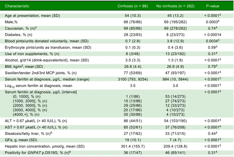
Table 1. Characteristics of 368 HFE C282Y homozygotes.1
Characteristic Cirrhosis (n = 86) No cirrhosis (n = 282) P-value
Age at presentation, mean (SD) 54 (10.3) 45 (13.2) < 0.00013
Male,% 88 (76/86) 69 (195/282) 0.00034
Caucasian, % (n)2 99 (85/86) 99 (278/282) 0.744
Diabetes, % (n) 28 (23/83) 8 (23/273) < 0.00014
Blood pints/units donated voluntarily, mean (SD) 0.7 (2.8) 3.8 (12.9) 0.00343
Erythrocyte pints/units as transfusion, mean (SD) 0.1 (0.3) 0.4 (3.6) 0.593
Use of iron supplements, % (n) 6 (3/48) 13 (23/182) 0.314
Alcohol, g/d/14 (drink-equivalents/d), mean (SD) 3.5 (3.3) 1.3 (1.8) < 0.00013
BMI, kg/m2, mean (SD) 26.8 (4.4) 26.9 (4.9) 0.793
Swollen/tender 2nd/3rd MCP joints, % (n) 77 (53/69) 47 (93/197) < 0.00014
Serum ferritin at diagnosis, μg/L, median (range) 3100 (793, 9254) 984 (10, 5844) < 0.00013
Log10 serum ferritin at diagnosis, mean 3.5 3.0 < 0.00013
Serum ferritin at diagnosis, μg/L (interval] < 0.00014
(0, 1000], % (n) 1 (1/86) 53 (14/273)
(1000, 2000], % (n) 15 (13/86) 27 (74/273)
(2000, 3000], % (n) 29 (25/86) 12 (33/273)
(3000, 4000], % (n) 20 (17/86) 4 (10/273)
(4000,+], % (n) 35 (30/86) 4 (10/273)
ALT > 0.67 μkat/L (> 40 IU/L), % (n) 86 (44/51) 54 (103/190) < 0.00014
AST > 0.67 μkat/L (> 40 IU/L), % (n) 85 (52/61) 37 (76/208) < 0.00014
Steatosis/fatty liver, % (n)5 27 (17/62) 33 (71/215) 0.444
QFe, g, mean (SD) 19 (10.1) 7 (4.7) < 0.00013
Hepatic iron concentration, μmol/g, mean (SD) 351.4 (155.7) 209.4 (128.8) < 0.00013
Positivity for GNPAT p.D519G, % (n)6 36 (17/47) 46 (65/141) 0.314
women (28.0% vs. 10.3%, respectively; p = 0.0004). Mean
liver iron concentration was 238 ± 146 μmol/g dry weight
(range 9-847; n = 262). Mean liver iron concentration was significantly greater in participants with cirrhosis than in those without cirrhosis (Table 1). The prevalence of alco-holic steatosis/fatty liver was greater in participants with cirrhosis than participants without cirrhosis (21.0%
(13/62) vs. 7.0% (15/215); p = 0.0031). Mean QFe was
10.1 ± 8.2 g (range 0.5-50; n = 307). One participant with-out cirrhosis was diagnosed to have hepatitis C. Another participant without cirrhosis was diagnosed to have “acute hepatitis.” A participant with cirrhosis was diagnosed to have extensive hepatic sarcoidosis.
GNPAT p.D519G
GNPAT p.D519G was detected in 82 of 188 partici-pants (43.6%; 13 homozygotes, 69 heterozygotes; allele frequency 0.25).
Participants with cirrhosis
Mean age at diagnosis, proportion of men, prevalence of diabetes, mean daily alcohol intake, prevalence of swol-len/tender 2nd/3rd metacarpophalangeal joints, mean se-rum ferritin, elevated ALT, elevated AST, and mean QFe were significantly greater in participants with cirrhosis.
Figure 1. Figure 1.Figure 1.
Figure 1.Figure 1. Odds ratios (black dots) and 95% confidence intervals for prediction of cirrhosis. The odds ratios for the continuous predictor variables including age increments since diagnosis (10 y intervals), incremental alcohol intake (g/14 (drink/d)), and additional iron removed to achieve iron depletion (g) represent the increased risks of biopsy-proven cirrhosis for the indicated increase in each individual predictor variable, adjusted for the presence of the remaining predictors. The diagnosis of diabetes also increases cirrhosis risk, adjusted for the presence of the remaining predictors.
-5 5 15 25 35 45 55 65 75 85 95 105 115 125 135 ≥145
Odds ratio Age increments
50 y 40 y 30 y 20 y 10 y
Incremental alcohol Intake, g/14 (drink/d) 5 drinks/d 4 drinks/d 3 drinks/d 2 drinks/d 1 drink/d
Diabetes Yes vs. no
Fe removed 20 g 15 g 10 g 5 g 1
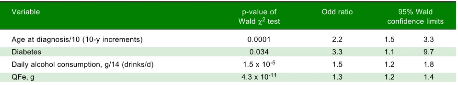
Table 2. Independent variables in a multivariable logistic regression on cirrhosis in 274 HFE p.C282Y homozygotes.*
Variable p-value of Odd ratio 95% Wald
Wald χ2 test confidence limits
Age at diagnosis/10 (10-y increments) 0.0001 2.2 1.5 3.3
Diabetes 0.034 3.3 1.1 9.7
Daily alcohol consumption, g/14 (drinks/d) 1.5 x 10⁻⁵ 1.5 1.2 1.8
QFe, g 4.3 x 10⁻¹¹ 1.3 1.2 1.4
Mean blood pints/units donated voluntarily was signifi-cantly lower in participants with cirrhosis (Table 1).
Multivariable logistic regression on cirrhosis
Regression on cirrhosis revealed four significant posi-tive associations: age; diabetes; alcohol drink-equivalents/d; and QFe (Table 2). There was no significant association of
cirrhosis with other variables, including sex and GNPAT
p.D519G positivity, after inclusion of these significant four variables. There was no statistical evidence of two-way interactions between age, diabetes, alcohol drink-equivalents/d, and QFe.
The OR for continuous predictor variables age at diag-nosis (10-y intervals), daily alcohol intake (g), and QFe (g) represents the increased odds of cirrhosis for each one unit increase in each individual predictor variable, adjust-ed for the presence of the remaining pradjust-edictors (Table 2). As shown in Figure 1, the odds of cirrhosis is 4.9-fold higher [95% CI 2.4, 10.0] with daily alcohol intake of 4 drinks/d than that of no alcohol intake, adjusted for age at diagnosis, diabetes, and QFe. For an estimated daily alco-hol intake of 5 drinks/d, the odds of cirrhosis increases 7.3-fold [95% CI 3.0, 17.8], adjusted for the additional pre-dictors. In participants whose QFe was 10 g, the odds of cirrhosis increases 11.8-fold [95% CI 5.7, 24.5]. In partici-pants whose QFe was 15 g, the odds of cirrhosis increases by 40.5-fold [95% CI 13.5, 121.6], adjusted for age at diag-nosis, daily alcohol intake, and diabetes. The occurrence of diabetes increases the odds of cirrhosis 3.3-fold [95% CI 1.1, 9.7], adjusted for the presence of the remaining predictors (Table 2).
DISCUSSION
Cirrhosis in this cohort of 368 HFE p.C282Y
homozy-gotes was associated with four significant independent var-iables: age; diabetes; alcohol drink-equivalents/d; and QFe. There was no statistical evidence of two-way inter-actions between these four variables. Progressive incre-ments of each of the three continuous variables were associated with progressive increments in cirrhosis risk, taking into account the effect of diabetes. The diagnosis of diabetes also increases cirrhosis risk, taking into account the effect of the three continuous variables. Altogether, the large sample size of the present study permitted the ascer-tainment of significant independent variables that contrib-ute to cirrhosis risk and pathogenesis that were previously
suggested by analyses of smaller cohorts.3,8
Greater age at diagnosis of hemochromatosis was sig-nificantly associated with greater cirrhosis risk in the present multivariable logistic regressions. In other
stud-ies of HFE p.C282Y homozygotes, cirrhosis was
diag-nosed only those aged > 40 y.3,8 In a retrospective study
of 60 patients with hemochromatosis, risk of high-grade
hepatic fibrosis increased with greater age at diagnosis.18
In 182 patients with hemochromatosis phenotypes, ad-vanced hepatic fibrosis was significantly associated with
age, after adjustment for HFE genotype and other
varia-bles.5 In a study of patients with hemochromatosis
phe-notypes reported before the discovery of HFE, those
with cirrhosis were significantly older than those with-out cirrhosis.19
The prevalence of cirrhosis in the present men with
HFE p.C282Y homozygosity was more than two-fold
greater than that of the women in univariate analyses. In 182 patients with hemochromatosis phenotypes, ad-vanced hepatic fibrosis was significantly associated with
male sex, after adjustment for HFE genotype and other
variables.5 In two studies of persons with
hemochroma-tosis phenotypes reported before the discovery of HFE,
the prevalence of cirrhosis in men was also significantly
greater than that in women.19,20 Heavy alcohol
consump-tion did not account for the difference between men and
women.19,20 The unexpected finding that male sex was
not independently associated with cirrhosis in the present multivariable analyses could be explained in part by either a greater prevalence of presenting manifesta-tions in women, suggesting that liver biopsy was indicat-ed, or a greater prevalence of previously unsuspected cirrhosis in women than men.
The prevalence of diabetes was more than three-fold higher in the present participants with cirrhosis than in
those without cirrhosis. In a report of 291 other HFE
p.C282Y homozygotes selected because they underwent liver biopsy, diabetes was associated with increasing he-patic fibrosis stage independent of male sex, alcohol
con-sumption, and iron loading.6 In a study of 159 consecutive
referred hemochromatosis probands with p.C282Y ho-mozygosity, cirrhosis was not significantly associated with
diabetes in multivariable regressions.14
The mean number of donated blood pints/units was significantly lower in the present participants with cir-rhosis than without circir-rhosis, although the mean number was relatively low in both participant subgroups. The numbers of blood pints/units voluntarily donated before diagnosis of hemochromatosis were not significantly as-sociated with cirrhosis in multivariable logistic
regres-sions. In a previous study of HFE p.C282Y homozygotes,
numbers of blood units donated before diagnosis of he-mochromatosis were not significantly related to num-bers of therapeutic phlebotomy units required to achieve
iron depletion.9 In another study, there was no
not volunteer blood donors before diagnosis.21 In the
present and previous studies,9,21 the proportions of
per-sons who voluntarily donated blood before hemochro-matosis diagnosis were small.
Use of iron supplements was reported by 11% of the present participants, consistent with a survey of a large
number of hemochromatosis patients.22 The present
analy-ses did not detect a significant association of cirrhosis with reports of supplemental iron use. In a previous study of 213
screening study participants with HFE p.C282Y
homozy-gosity, there was no significant relationship between serum
ferritin levels and reports of supplemental iron use.23
Greater daily alcohol intake was significantly associated with greater cirrhosis risk in the present univariate and
multivariable analyses. In 224 HFE p.C282Y homozygotes
without viral hepatitis or non-alcoholic steatohepatitis reported previously, the prevalence of cirrhosis was
nine-fold greater in those who consumed ≥ 60 g of alcohol
daily than in those who consumed less than this amount.7
In a study of 291 p.C282Y homozygotes, there was a sig-nificant positive association of heavy alcohol intake with
hepatic fibrosis stage.24 In two reports published before
the discovery of HFE, heavy alcohol intake by persons
with hemochromatosis was associated with greater cirrhosis risk.19,25
The prevalence of either elevated ALT or elevated AST was significantly greater in participants with cirrhosis than without, although neither elevated ALT nor elevated AST was significantly associated with cirrhosis in multivariable logistic regressions. Elevated AST in patients with hemo-chromatosis and iron overload is typically associated with
hepatic injury, especially cirrhosis.26 In a study of 197
pa-tients with HFE p.C282Y homozygosity, the absence of
severe hepatic fibrosis was associated with the
combina-tion of serum ferritin ≤ 2,247 pmol/L, lack of
hepatomega-ly, and normal serum AST.8
Steatosis/fatty liver was not significantly associated with cirrhosis in this study, although the prevalence of cir-rhosis was three-fold greater in participants with steatosis/ fatty liver who also consumed > 60 g alcohol daily than in participants with steatosis/fatty liver whose daily alcohol
consumption was ≤ 60 g. It is possible that morphologic
evidence of hepatic steatosis disappeared in some partici-pants before their respective liver biopsy specimens re-vealed cirrhosis. In another study, 41% of 214 patients with
hemochromatosis and HFE p.C282Y homozygosity had
biopsy-proven steatosis that was independently associated with male sex, excess alcohol intake, hepatic iron content,
and fibrosis.27 In a study of 291 p.C282Y homozygotes,
in-creasing fibrosis stage revealed a significant positive asso-ciation with “any steatosis.”24
Cirrhosis was significantly associated with QFe in the present multivariable regressions. Previous studies
con-cluded that persons with cirrhosis and hemochromatosis
phenotypes with or without demonstration of HFE
p.C282Y homozygosity typically have severe hepatic si-derosis.3,5,8,19,28
The proportion of the present participants with GNPAT p.D519G positivity did not differ significantly between those with and without cirrhosis. Similarly, allele frequencies of p.D519G in Italian hemochromatosis patients with absent/mild fibrosis and severe fibrosis/ cirrhosis did not differ significantly.29 In contrast, cirrhosis was significantly associated with the patatin-like
phospholipase domain-containing-3 (PNPLA3) I148M
polymorphism (rs738409) in Italian patients with hemo-chromatosis and p.C282Y homozygosity whose body
mass index was < 25 kg/m2.30PCSK7 (proprotein
conver-tase subtilisin/kexin type 7) rs236918 C allele was a risk factor for cirrhosis in 244 German and Austrian/
Swiss31and 187 Italian32 patients with hemochromatosis
and p.C282Y homozygosity, but not 112 Swedish patients
with hemochromatosis and p.C282Y homozygosity.31
A strength of the present study is that this is the largest cohort for cirrhosis in hemochromatosis in which all
participants were demonstrated to be HFE p.C282Y
homozygotes and underwent liver biopsy. Limitations of the present data include missing responses, non-valid data, and lack of observations. Our review of REDCap liver biopsy text entries and clinical practice documentation indicates that the proportion of p.C282Y homozygotes who had hepatitis C and other non-iron liver conditions unrelated to alcohol consumption and non-alcoholic stea-tosis/fatty liver that may increase cirrhosis risk is small. The present results cannot confirm or exclude the possi-bility that the independent variables we identified also in-fluence lesser degrees of hepatic injury than cirrhosis interpreted as fibrosis stages 1, 2, or 3. The present study was not designed to evaluate the association of cirrhosis
with alleles other than GNPAT p.D519G. Longitudinal
evaluation of survival and risk of primary liver cancer in the present cohort was beyond the scope of this study.
CONCLUSIONS
Cirrhosis in HFE p.C282Y homozygotes is
significant-ly associated with age, diabetes, daisignificant-ly alcohol intake, and iron removed by phlebotomy, taking into account the ef-fect of other variables.
ABBREVIATIONS
• ALT: alanine aminotransferase.
• AST: aspartate aminotransferase.
• CI: confidence interval.
• HFE: high iron gene.
• OR: odds ratio.
• QFe: quantity of iron (g) removed by phlebotomy to
achieve iron depletion.
• SD: standard deviation.
GRANT SUPPORT
The authors recognize and appreciate financial support in part by grant 1R24DK093433-01 from the National In-stitute of Diabetes and Digestive and Kidney Diseases, grant P30 CA-62203 from the National Cancer Institute, and funds from the Department of Veterans Affairs. V.N.S. and G.A.R. are supported by Senior Research Fellowships from the NHMRC of Australia. J.C.B. is supported in part by Southern Iron Disorders Center.
AUTHOR CONTRIBUTIONS
All authors contributed equally to this work. J.C.B. conceived the study and drafted the manuscript. J.C.B., P.D.P., P.C.A., L.W.P., V.N.S., J.D.P., C.J.P., and G.D.M. contributed samples and data. C.E.M. and M.J.E. provided design and analysis oversight. C.E.M., W.P.C., G.D.M., J.C.B., and M.J.E. reviewed and approved the final dataset for analysis. W.P.C., C.E.M., and M.J.E. tabulated out-comes and performed statistical analyses. C.E.M., M.J.E., G.A.R., G.J.A., L.W.P., V.N.S., P.C.A, P.D.P., L.C.G., J.D.P., C.J.P., and G.D.M. contributed to the manuscript.
DISCLOSURES
None of the authors has a conflict of interest to report.
REFERENCES
1. Edwards CQ, Barton JC. Hemochromatosis. In: Greer JP, Ar-ber DA, Glader B, List AF, Means Jr. RT, Paraskevas F, Rodgers GM, eds. Wintrobe’s Clinical Hematology. Philadel-phia: Wolters Kluwer/Lippincott Williams & Wilkins; 2014, pp. 662-81.
2. Bridle KR, Frazer DM, Wilkins SJ, Dixon JL, Purdie DM, Craw-ford DH, Subramaniam VN, et al. Disrupted hepcidin regula-tion in HFE-associated haemochromatosis and the liver as a regulator of body iron homoeostasis. Lancet 2003; 361: 669-73.
3. Bacon BR, Olynyk JK, Brunt EM, Britton RS, Wolff RK. HFE
genotype in patients with hemochromatosis and other liver diseases. Ann Intern Med 1999; 130: 953-62.
4. Barton JC, Barton JC, Acton RT, So J, Chan S, Adams PC. In-creased risk of death from iron overload among 422 treated probands with HFE hemochromatosis and serum levels of ferritin greater than 1000 μg/L at diagnosis. Clin Gastroen-terol Hepatol 2012; 10: 412-16.
5. Cheng R, Barton JC, Morrison ED, Phatak PD, Krawitt EL, Gordon SC, Kowdley KV. Differences in hepatic phenotype between hemochromatosis patients with HFE C282Y
ho-mozygosity and other HFE genotypes. J Clin Gastroenterol
2009; 43: 569-73.
6. Wood MJ, Powell LW, Dixon JL, Ramm GA. Clinical cofactors and hepatic fibrosis in hereditary hemochromatosis: the role of diabetes mellitus. Hepatology 2012; 56: 904-11.
7. Fletcher LM, Dixon JL, Purdie DM, Powell LW, Crawford DH. Excess alcohol greatly increases the prevalence of cirrhosis in hereditary hemochromatosis. Gastroenterology 2002; 122: 281-89.
8. Guyader D, Jacquelinet C, Moirand R, Turlin B, Mendler MH, Chaperon J, David V, et al. Noninvasive prediction of fibrosis in C282Y homozygous hemochromatosis. Gastroenterology
1998; 115: 929-36.
9. Barton JC, Preston BL, McDonnell SM, Rothenberg BE. Se-verity of iron overload in hemochromatosis: effect of volun-teer blood donation before diagnosis. Transfusion 2001; 41:
123-9.
10. Wood MJ, Powell LW, Ramm GA. Environmental and genetic modifiers of the progression to fibrosis and cirrhosis in he-mochromatosis. Blood 2008; 111: 4456-62.
11. Barton JC, Lee PL, West C, Bottomley SS. Iron overload and prolonged ingestion of iron supplements: clinical features and mutation analysis of hemochromatosis-associated genes in four cases. Am J Hematol 2006; 81: 760-7. 12. McLaren CE, Emond MJ, Subramaniam VN, Phatak PD, Barton
JC, Adams PC, Goh JB, et al. Exome sequencing in HFE
C282Y homozygous men with extreme phenotypes identi-fies a GNPAT variant associated with severe iron overload.
Hepatology 2015; 62: 429-39.
13. Besson-Fournier C, Martinez M, Vinel JP, Aguilar-Martinez P, Coppin H, Roth MP. Further support for the association of
GNPAT variant rs11558492 with severe iron overload in he-mochromatosis. Hepatology 2016; 63: 2054-5.
14. Barton JC, Barton JC, Acton RT. Diabetes in first-degree family members: a predictor of type 2 diabetes in 159 non-screening Alabama hemochromatosis probands with HFE
C282Y homozygosity. Diabetes Care 2014; 37: 259-66. 15. Farrell CP, Overbey JR, Naik H, Nance D, McLaren GD,
McLaren CE, Zhou L, et al. The D519G polymorphism of glyceronephosphate O-acyltransferase is a risk factor for familial porphyria cutanea tarda. PLoS One 2016; 11:
e0163322.
16. McCormick PA. Hepatic Cirrhosis. In: Dooley JS, Lok ASF, Burroughs AK, Heathcote EJ, eds. Sherlock’s Diseases of the Liver and Biliary System. Chichester: Wiley-Blackwell; 2012, pp. 103-20.
17. Bowman SA, Clemens JC, Friday JE, Thoreig RC, Moshfegh AJ. Food Patterns Equivalents Database 2011-12: Methodol-ogy and User Guide [Online]. Food Surveys Research Group, Beltsville Human Nutrition Research Center, Agricul-tural Research Service, U S Department of Agriculture, Beltsville, Maryland. 2014.
18. Olynyk JK, St Pierre TG, Britton RS, Brunt EM, Bacon BR. Du-ration of hepatic iron exposure increases the risk of signifi-cant fibrosis in hereditary hemochromatosis: a new role for magnetic resonance imaging. Am J Gastroenterol 2005; 100:
837-41.
19. Loréal O, Deugnier Y, Moirand R, Lauvin L, Guyader D, Jouanolle H, Turlin B, et al. Liver fibrosis in genetic hemo-chromatosis. Respective roles of iron and non-iron-related factors in 127 homozygous patients. J Hepatol 1992; 16:
122-7.
21. Power TE, Adams PC. Hemochromatosis patients as volun-tary blood donors. Can J Gastroenterol 2004; 18: 393-6. 22. McDonnell SM, Grindon AJ, Preston BL, Barton JC, Edwards
CQ, Adams PC. A survey of phlebotomy among persons with hemochromatosis. Transfusion 1999; 39: 651-6. 23. Gordeuk VR, Lovato L, Barton J, Vitolins M, McLaren G,
Acton R, McLaren C, et al. Dietary iron intake and serum ferritin concentration in 213 patients homozygous for the
HFE C282Y hemochromatosis mutation. Can J Gastroenter-ol 2012; 26: 345-9.
24. Wood MJ, Crawford DH, Wockner LF, Powell LW, Ramm GA. Serum ferritin concentration predicts hepatic fibrosis better than hepatic iron concentration in human HFE hemochroma-tosis. Liver Int 2017; 37: 1382-8.
25. Powell LW. The role of alcoholism in hepatic iron storage disease. Ann N Y Acad Sci 1975; 252: 124-34.
26. Lin E, Adams PC. Biochemical liver profile in hemochromato-sis. A survey of 100 patients. J Clin Gastroenterol 1991; 13: 316-20.
27. Powell EE, Ali A, Clouston AD, Dixon JL, Lincoln DJ, Purdie DM, Fletcher LM, et al. Steatosis is a cofactor in liver injury in hemochromatosis. Gastroenterology 2005; 129: 1937-43. 28. Bassett ML, Halliday JW, Powell LW. Value of hepatic iron
measurements in early hemochromatosis and determination of the critical iron level associated with fibrosis. Hepatology
1986; 6: 24-9.
29. Greni F, Valenti L, Mariani R, Pelloni I, Rametta R, Busti F, Ravasi G, et al. GNPAT rs11558492 is not a major modifier
of iron status: study of Italian hemochromatosis patients and blood donors. Ann Hepatol 2017; 16: 451-56.
30. Valenti L, Maggioni P, Piperno A, Rametta R, Pelucchi S, Mar-iani R, Dongiovanni P, et al. Patatin-like phospholipase do-main containing-3 gene I148M polymorphism, steatosis, and liver damage in hereditary hemochromatosis. World J Gas-troenterol 2012; 18: 2813-20.
31. Stickel F, Buch S, Zoller H, Hultcrantz R, Gallati S, Osterre-icher C, Finkenstedt A, et al. Evaluation of genome-wide loci of iron metabolism in hereditary hemochromatosis identifies
PCSK7 as a host risk factor of liver cirrhosis. Hum Mol Genet 2014; 23: 3883-90.
32. Pelucchi S, Galimberti S, Greni F, Rametta R, Mariani R, Pello-ni I, Girelli D, et al. Proprotein convertase 7 rs236918 associ-ated with liver fibrosis in Italian patients with HFE-related hemochromatosis. J Gastroenterol Hepatol 2016; 31:
1342-8.
Correspondence and reprint request:
James C. Barton, M.D.
Southern Iron Disorders Center, 2022 Brookwood Medical Center Drive, Suite 626, Birmingham, AL 35209 USA.