An Overview of Liver Transplant Pathology:
Data from a Tertiary Referral
Centre in Western India
Fiona Fonseca,* Bijal Kulkarni,* Meenal Hastak,* Vinay Kumaran,** Vibha Varma,** Sorabh Kapoor**
* Department of Histopathology and Laboratory Medicine, Kokilaben Dhirubhai Ambani Hospital and Medical Research Centre, Mumbai, Maharashtra, India. ** Department of Hepatobiliary surgery and Liver Transplant, Kokilaben Dhirubhai Ambani Hospital and Medical Research Centre, Mumbai, Maharashtra, India.
May-June, Vol. 17 No. 3, 2018: 426-436
INTRODUCTION
Chronic liver disease (CLD) contributes significantly to the burden of disease worldwide. Failure to detect and treat these diseases early, may cause a number of them to progress to an irreversible stage, which may not be amena-ble to treatment and often lead to fatal complications. Spe-cial attention should be given to the evaluation of ESCLD in infants and children, as many of the etiologies are dis-tinct from the conditions affecting the adult population. Liver transplantation (LT) has proven to be an effective modality to prolonging a better quality of life in many pa-tients with end stage CLD.
While the history of orthotopic LT in India is relatively short, the program has steadily progressed, in no small part due to the efforts to increase awareness among the general population about liver disease and the scarcity of donors.
The aim of our study is threefold: 1) To attempt an eti-ological categorization of cases of end stage CLD and acute liver failure separately in adults and children ≤ 12 years, through an extensive pre-LT clinical, laboratory and radiological examination and thorough examination of the explant livers. 2) To assess the donor liver for prevalence of steatosis and unexpected incidental findings, like granu-lomas. 3) Analysis of post-transplant liver biopsies to es-tablish the cause of graft dysfunction.
The Official Journal of the Mexican Association of Hepatology, the Latin-American Association for Study of the Liver and
the Canadian Association for the Study of the Liver
Manuscript received: Manuscript received: Manuscript received: Manuscript received:
Manuscript received: April 14, 2017. Manuscript accepted:Manuscript accepted:Manuscript accepted:Manuscript accepted:Manuscript accepted: July 31, 2017.
DOI:10.5604/01.3001.0011.7387
A B S T R A C T A B S T R A C T A B S T R A C T A B S T R A C T A B S T R A C T
Introduction and aim. Introduction and aim. Introduction and aim. Introduction and aim.
Introduction and aim. 1. Study of liver explants - Etiologic types of end-stage chronic liver disease (ESCLD) and acute liver fail-ure (ALF) in adults and children. 2. Assessment of donor steatosis and incidental granulomas. 3. Post-transplant liver biopsies. Ma-Ma-Ma-Ma- Ma-terial and methods.
terial and methods. terial and methods. terial and methods.
terial and methods. Specimens of 180 explant hepatectomies, 173 donor wedge and 30 core liver biopsies, and 58 post transplant liver biopsies received in our department from April 2013 to March 2017. Results.Results.Results.Results.Results. 1. Most common causes of ESCLD in adults were: alcohol related (30.32%), hepatitis virus related (18.71%) and non-alcoholic steatohepatitis related (18.06%); and in children ≤ 12 years were: biliary atresia (27.27%), autoimmune disease (18.18%) and Wilson's disease (18.18%). Most common causes of ALF in adults and children were anti-tubercular therapy induced and idiopathic, respectively. 2. Prevalence rate of moderate steatosis (be-tween 30-60%) was 4.28%. Incidental granulomas were seen in 5 cases. 3. Most common diagnoses of post-transplant biopsies in adults included acute cellular rejection (ACR) (36.17%), recurrence of viral disease (8.51%) and moderate non-specific portal triaditis (8.51%). Among children ≤ 12 years, most common diagnoses included unremarkable liver parenchyma, ACR and ischemia/reper-fusion injury. Conclusion. Conclusion. Conclusion. Conclusion. Conclusion. 1. Alcohol- and hepatitis- virus related ESCLD, and biliary atresia are leading indications for liver trans-plantation in adults and children, respectively. 2. Prevalence of 4.28% of moderate steatosis, is much lower than that documented in western literature. Only 5 cases of incidental granulomas is unexpectedly low in a country endemic for tuberculosis. 3. Most com-mon diagnoses of post-transplant liver biopsies in adults has been acute rejection, which is similar to the findings from much larger published series.
Key words. Key words.Key words. Key words.
MATERIAL AND METHODS
During 4 year period from April 2013 to March 2017, 180 patients underwent LT in our institute.
Patients were selected based on the Child-Turcotte-Pugh classification, Model for End Stage Liver Disease score and Pediatric End-Stage liver disease score for CLD and the King's College criteria for hepatic failure.1-4
Explant livers
Pre-LT categorization of the etiologic type of CLD and acute liver failure
Was done on the basis of clinical and investigational data. These included a history of alcohol consumption, drug intake, blood transfusion and intake of medication. Serology for hepatitis B virus (HBV), hepatitis C virus (HCV) and human immunodeficiency virus (HIV) infec-tion, an autoantibody profile including antinuclear, anti-smooth muscle and anti-mitochondrial antibodies, and tests for serum levels of iron, ferritin and ceruloplasmin were carried out. Imaging studies were performed to de-tect neoplastic lesions.
In pediatric patients, a detailed family history was elicited and a thorough physical examination was per-formed, in addition to laboratory tests. Imaging modali-ties were helpful in cholestatic liver disease to ascertain the etiology.
Morphological evaluation
Was carried out after serially sectioning the explant liv-ers at 1 cm intervals, adequate fixation in 10% neutral buff-ered formalin and representative sampling. Four sections from the right lobe, 2 sections from the left lobe and 1 sec-tion from the hilum were studied in each case. Paraffin sections were stained by haematoxylin and eosin (H & E), and when required, by special stains for collagen, reticu-lin, iron and copper.
The microscopic evaluation included the type of nod-ules (macro vs. micro), fibrosis, inflammation and the type of inflammatory cells, steatosis, Mallory hyaline bodies, necrosis, bile ductular proliferation, deposition of iron or copper pigment and liver tumors.
In cases of acute liver failure, emphasis was laid upon the zone of necrosis and the integrity of the reticulin framework.
Donor biopsies
While no randomised controlled trials have been con-ducted to determine the tests required to evaluate a living
donor, we follow the general medical practice of existing transplant programs5 (Table 1). All living donors fell into
the age group of 18-60 years and were required to be relat-ed to the recipient, vide the Transplantation of Human Organs and Tissues Rules, 2013 published by the Ministry of Health and Family welfare, Government of India.
173 intra-operative wedge and 30 preoperative core liv-er biopsies from donors wliv-ere analysed for the prevalence of steatosis. The indications for pre-LT core liver biopsies included steatosis on Plain CT scan, with a liver attenua-tion index (LAI) < 5, abnormal liver funcattenua-tion tests or a body mass index (BMI) > 30.
The samples were adequately fixed, stained with H & E stain and the percentage of steatosis was calculated by two consultant pathologists (Dr. MH and Dr. BK).
Also incidentally detected were epithelioid granulomas in 5 wedge biopsies from asymptomatic donors.
Post-transplant liver biopsies
58 post-transplant liver biopsies from both adult and pediatric recipients were studied after appropriate processing & staining with H & E. Special stains for colla-gen, reticulin, hemosiderin, etc. and immunohistochemis-try for C4d and CK 7 were done when indicated.
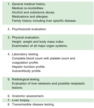
Table 1. Evaluation of a living donor.
1. General medical history. Medical co-morbidities. Alcohol and substance abuse. Medications and allergies.
Family history including liver specific disease.
2. Psychosocial evaluation.
3. Physical evaluation.
Height, weight and body mass index. Examination of all major organ systems.
4. Laboratory testing.
Complete blood count with platelet count and coagulation profile.
Hepatic function profile. Autoantibody profile.
5. Radiological testing.
Evaluation of liver steatosis and possible neoplastic lesions.
6. Anatomic assessment. 7. Liver biopsy.
Table 2. Etiologic categorisation of end stage CLD and acute liv-er failure in adults.
Etiologic category Cases (n)
End stage CLD 155*
Alcoholic liver disease 47 (30.32%)
Hepatitis virus related 29
HBV 10 (6.45%)
HCV 14 (9.03%)
HBV + HCV 1 (0.65%)
HBV + Alcoholic liver disease 4 (2.58%)
Autoimmune liver disease 12 (7.74%) Non-alcoholic steatohepatitis 28 (18.06%)
Biliary tract disease 6 (3.87%)
Metabolic disease 7 (4.52%)
Autoimmune liver disease + 1 (0.65%)
alcoholic liver disease
Cryptogenic 25 (16.13%)
Acute liver failure 11†
Antitubercular therapy related 3 (27.27%) Hepatitis E virus induced 3 (27.27%)
Vascular cause 2 (18.18%)
Indeterminate 1 (9.09%)
Metastatic malignancy 1 (9.09%)
Rattol poisoning 1 (9.09%)
Retransplantation
Graft loss due to chronic rejection 1
* Figures in parentheses are proportions among all cases of end stage CLD.
† Figures in parentheses are proportions among all cases of acute liver
failure. HBV: hepatitis B virus. HCV: hepatitis C virus.
Table 4. Etiologic association with hepatocellular carcinoma.
Etiologic category Cases of HCC* (n)
Hepatitis virus related 9
HBV 6 (40%)
HCV 2 (13.33%)
HBV + HCV 1 (6.67%)
Alcoholic liver disease 1 (6.67%)
Autoimmune liver disease 1 (6.67%)
NASH related 4 (26.67%)
* Figures in parentheses are proportions among all cases of hepatocellular carcinoma. HBV: hepatitis B virus. HCV: hepatitis C virus. NASH: nonalcoholic steatohepatitis.
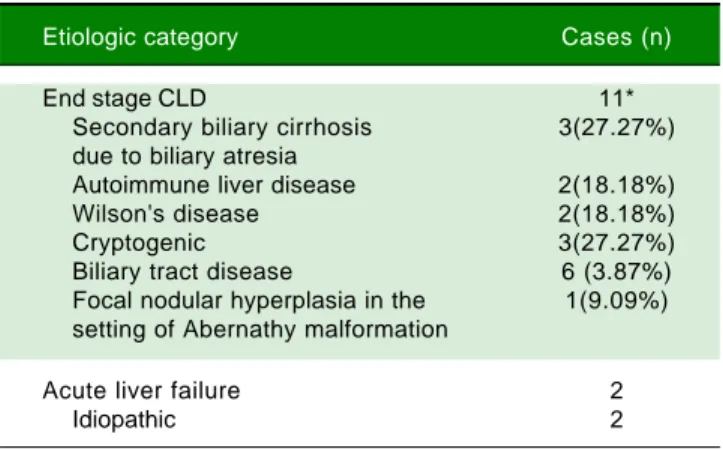
Table 3. Etiologic categorisation of end stage CLD and acute liv-er failure in children ≤ 12 years.
Etiologic category Cases (n)
End stage CLD 11*
Secondary biliary cirrhosis 3(27.27%) due to biliary atresia
Autoimmune liver disease 2(18.18%)
Wilson's disease 2(18.18%)
Cryptogenic 3(27.27%)
Biliary tract disease 6 (3.87%)
Focal nodular hyperplasia in the 1(9.09%) setting of Abernathy malformation
Acute liver failure 2
Idiopathic 2
* Figures in parentheses are proportions among all cases of end stage CLD. The following features were evaluated in the liver
biop-sies:
• Adequacy of the biopsy, based on the number of portal tracts (biopsies with at least 5 portal tracts were con-sidered adequate).
• Liver architecture.
• Presence of features of acute or chronic rejection, re-currence of original disease, biliary obstruction, infec-tion, malignancy, etc.
Grading in cases of rejection was according to the Banff schema.6
RESULTS
Explant livers
The 180 patients in this study included 167 adults and 13 children ≤ 12 years of age. Among the adult patients, there were 134 males and 33 females, with the oldest re-cipient being 70 years old. Among the pediatric patients,
there were 5 males and 8 females, ranging in age from 8 months to 12 years.
Of the 167 adult patients, 28 received whole livers from cadaveric donors, while the remaining 139 underwent liv-ing donor LTs. All the pediatric patients underwent livliv-ing donor LTs.
The etiologic categorisation of end stage CLD and acute liver failure in our study for the adult and pediatric cohorts has been summarised in tables 2 and 3.
In 15 out of 155 cases of end stage CLD in adults, the CLD was associated with hepatocellular carcinoma (HCC) (Table 4). Eight out of the 15 cases of HCC were treated with trans-arterial chemo-embolisation (TACE) prior to LT. Histopathological examination of these tu-mors revealed a range of responses, from complete tumor necrosis to completely viable tumor.
Donor biopsies
Twelve of the donors underwent both pre-operative core and intra-operative wedge biopsies, 1 donor underwent a core biopsy, followed by a wedge biopsy 2 weeks later, as well as a repeat core biopsy 2 months after that. Two do-nors underwent repeat core biopsies 20 days and 45 days following their first biopsies.
The acceptable degree of hepatic steatosis for living do-nor LT has not been firmly defined, but there is a general consensus that macro and/microvesicular steatosis of more than 30% has been associated with a risk of sub-opti-mal graft function.7 In our program, we have excluded all potential donors revealing steatosis of more than 20%.
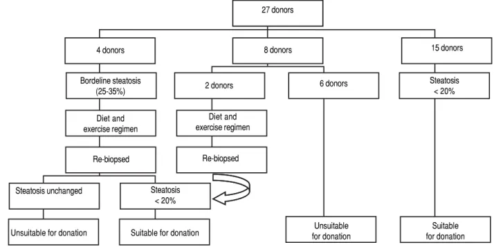
Figure 1 demonstrates the results of the pre-operative core biopsy examination.
The prevalence of mild steatosis (< 30%) was 34.22%, moderate steatosis (30%-60%) was 4.28% and severe stea-tosis (> 60%) was 1.07% in our series. 60.43% of donor bi-opsies did not reveal any steatosis.
Incidental epithelioid granulomas were noted in only 5 donor wedge biopsies. These granulomas were non-necr-otising, and special stains were carried out to detect acid fast bacilli, fungal organisms and parasites, with negative results.
Post-transplant liver biopsies
During the study period of 4 years, a total of 58 core bi-opsies were obtained from 42 patients. Eight of these pa-tients were pediatric papa-tients, who were biopsied a total of 11 times. Seven adults and 3 children were biopsied twice,
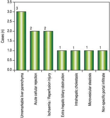
and 3 adult patients were biopsied 3 times. Acute cellular rejection was the most common diagnosis in adults (Figure 2). In the pediatric patients, the most common diagnosis was essentially unremarkable liver parenchyma, indicating that the rise in liver enzymes and/ bilirubin was not caused by graft dysfunction, but systemic causes (Figure 3).
DISCUSSION
Explant livers
End stage chronic liver disease
We have attempted to analyse the prevalence of the etio-logic types of end stage CLD (ESCLD) and acute liver failure (ALF) separately for adults and children ≤ 12 years from a combination of clinical, investigational and repre-sentative morphologic data. However, owing to a small sample size and selection of patients from a hospital-based living donor transplant program from a relatively high in-come group, our data cannot be taken as representative of the prevalence of ESCLD or ALF in the general popula-tion of western India. Nevertheless, we believe that as the LT program progresses, we may have a wider representa-tion of patients across the cross-secrepresenta-tion of the popularepresenta-tion.
Adults
Out of 155 cases of ESCLD, 47 were due to alcoholic liver disease (ALD). The morphologic features included
Figure 1. Figure 1. Figure 1.
Figure 1. Figure 1. Pre-operative needle core biopsies. 4 donors
Bordeline steatosis (25-35%)
Diet and exercise regimen
Re-biopsed
8 donors 27 donors
Steatosis unchanged Steatosis < 20%
Unsuitable for donation Suitable for donation 2 donors
Diet and exercise regimen
Re-biopsed
15 donors
Steatosis < 20% 6 donors
Unsuitable for donation
Figure 2. Figure 2. Figure 2. Figure 2.
Figure 2. Case wise distribution of post LT core biopsies in adults.
micronodular and mixed nodular cirrhosis, Mallory hya-line bodies and variable degrees of steatosis. While CLD due to alcohol abuse has long been reported from devel-oped countries, there is a paucity of data on its prevalence in India. A landmark study by Nayak N, et al. (2012) of 372 cases of CLD from North India,8 has put the prevalence of
alcohol-related cirrhosis at 23.1%, which is slightly lower than the prevalence in our patients of 30.32%.
Close to a fifth of our ESCLD cases (29 out of 155) were associated with Hepatitis B and C virus (HBV & HCV) infections. While the reported global prevalence of cirrhosis due to Hepatitis virus infections is about 60%,9
the reduced percentage in our study can probably be at-tributed to a small sample size and lack of a representative patient population from the lower socio-economic strata, in whom the incidence is expected to be higher due to lack of awareness of and inaccessibility to hepatitis B vac-cination. However, within the Hepatitis virus group, HCV and HBV are responsible for an equal number of cases of ESCLD, with 1 patient having a co-infection of both vi-ruses. In the developed countries, the stringent imple-mentation of HBV infection control measures including vaccination of all live births, screening of all blood and blood products prior to transfusion, etc have resulted in a reduction in the prevalence of HBV related CLD in re-cent years.10
The etiology in 12 out of 155 cases was autoimmune liv-er disease which showed morphologic features of septal lymphoplasmacytic infiltrate and bile ductular prolifera-tion along with serological evidence of autoantibody posi-tivity in all the cases. Non-Alcoholic Fatty Liver Disease (NAFLD) related cirrhosis was diagnosed in 28 cases, af-ter a careful correlation with clinical findings, investiga-tions and the histomorphology which revealed variable grades of inflammation, ductular proliferation and an ab-sence of steatosis. Our prevalence of 18.06% of NAFLD-related cirrhosis is similar to the findings of Nayak, et al., who reported a prevalence of 16.7%.8 Six cases of
second-ary bilisecond-ary cirrhosis were seen which included Caroli’s disease and portal cavernoma with extra hepatic portal vein obstruction, 7 cases of cirrhosis were due to metabol-ic disease whmetabol-ich included Wilson’s disease and primary hemochromatosis and 25 cases were classified as cryp-togenic since there were no specific morphologic features related to any known entity. Our incidence of 16.13% is similar to the finding of 14.8% cases of cryptogenic cirrho-sis reported by Ayata, et al.11
HCC was detected in 15 cases of ESCLD, i.e. 9.68%. The frequency of HCC in other studies based on larger cohorts ranges from 8% to 38%.12 The strong association of
HCC with cirrhosis is explained by 2 factors: the repeti-tive cycles of hepatocyte regeneration provide a milieu for the accumulation of mutations, and secondly the common 3.5
3.0 2.5 2.0 1.5 1.0 0.5 0.0
Figure 3. Figure 3. Figure 3. Figure 3.
Figure 3. Case wise distribution of post LT core biopsies in children.
Acute cellular rejection Recurrent viral disease
Non-specific moderate portal infiltrate
Ischaemic causes
Unremarkable liver parenchyma
Chronic rejection
Intrahepatocytic and canalicular stasis
Acute humoral rejection
Lobular hepatocyte drop-out
Ischaemia / Reperfusion injury Extra-hepatic biliary obstruction
Macro and microvesicular steatosis
Ascending cholangitis and cholestasis
20
15
10
5
0
Unremarkable liver parenchyma
Acute cellular rejection
Ischaemia / Reperfusion injury Extra-hepatic biliary obstruction
Intrahepatic cholestasis Microvesicular steatosis
Non-specific portal infiltrate
2 2
1 1 1 1
3 17
4 4 4 4
3 3
2 2
1 1 1 1
Cases (n)
etiologic agents for cirrhosis, the Hepatitis viruses B and C, play a direct carcinogenic role in the liver.9,13 Two
cas-es were HCV related, 6 were HBV related and 1 was a co-infection of HBV and HCV. Though it doesn’t reflect in our present study, trends show that HCV, rather than HBV is fast becoming a leading cause of HCC, likely due to a rapid increase in blood transfusions, injectibles, intrave-nous drug abuse and lack of an HCV vaccine.14
Acute liver failure
Establishing the cause of fulminant hepatitis (FH) is an important step in the management of acute liver fail-ure. Fulminant hepatic failure is characterized by a dete-rioration in liver function and hepatic encephalopathy. The main causes of FH are viral infections, drugs, and in-determinate causes.15 A survey carried out in the United
States between 1998 and 2008 in 1147 patients, showed that the major etiologies of ALF were paracetamol over-dose (46%) followed by indeterminate causes (14%), drug-related (11%), HBV (7%), other causes (7%), au-toimmune hepatitis (5%), ischemic hepatitis (4%), hepa-titis A virus (3%), and Wilson’s disease (2%).16 From the
European Liver Transplantation Registry, the most com-mon causes of transplanted FH between 1972 and 2007 were viral causes (HAV: 1%; HBV: 15%), paracetamol overdose (8%), non-paracetamol drug overdose (antibi-otics > ecstasy > anti-tubercular treatment > Non-ster-oidal anti-inflammatory drugs) (11%), indeterminate causes (48%), and other causes (17%).15 In our series, the
etiologies of ALF leading to LT were: anti-tubercular treatment (ATT) related (27.27%), hepatitis E virus (HEV) related (27.27%) and vascular/ischaemic related (18.18%), indeterminate (9.09%), Rattol poisoning relat-ed (9.09%) and due to extensive metastatic malignancy (9.09%).
However, despite intensive investigations and the use of the most modern techniques [polymerase chain reac-tion (PCR) methods, molecular techniques, genomic DNA, and biochemical methods], the causes of most cases of FH or ALF may remain unknown.15
Acute liver failure secondary to ATT is rare in the developed world. These cases represented only 2.47% of the total number of patients admitted with ALF be-tween 1986 and 2008 in a study from France.17 In a
ret-rospective study of patients from the United Network for Organ Sharing registry (1987-2006), only 0.07% of LT procedures for ALF were due to ATT (50 of 73,977 transplant patients).18 However, tuberculosis (TB)
con-tinues to remain a significant infectious disease across much of the developing world. ATT is a leading cause of drug-induced liver injury (DILI) in India and of drug-induced acute liver failure leading to death
(DI-ALF).19 In contrast, non-TB antibiotics and paracetamol
are the commonest causes of DILI and drug-induced ALF in western countries. In a large series of acute liver failure cases in New Delhi, anti-TB drugs contributed to 5.7% patients with ALF(70/ 1223), with 67% mortali-ty.20-22
HEV is an enterically transmitted RNA virus responsi-ble for large waterborne epidemics of ALF or FH in en-demic regions such as India, Central America, Africa, and Asia.23 The overall mortality due to fulminant liver failure
in endemic regions varies from 0.5% to 4%, but in preg-nant women with HEV FH, mortality is higher (20%). Our patients were females who contracted the infection during the second half of their pregnancies.
Children ≤≤≤≤≤ 12 years
ESCLD
Biliary atresia is the most common cause of chronic cholestasis and LT in children, with the mean age of LT for patients being 11 months.24,25 Out of 11 children with
ESCLD, 3 patients i.e. 27.27% had secondary biliary cir-rhosis due to biliary atresia- 2 females and 1 male, ranging in age from 8 months to 7 years at the time of LT. In 2 of the older patients within the cohort, i.e. 11 and 12 years, the etiology was autoimmune, which is in keeping with the common causes of ESCLD in older children and adolescents.24
In 3 children it was not possible to ascertain the cause of cirrhosis and hence were labelled cryptogenic. Cryp-togenic cirrhosis in children is hypothesised to result from the progression of fatty liver disease or from the ef-fects of complex metabolic syndromes, such as mitochon-driopathies.26
The etiology in 2 of the children was Wilson’s disease, and both patients had elevated levels of dry weight of cop-per in the explant livers.
One patient presented with hepatopulmonary syn-drome in the setting of an Abernathy malformation, and fo-cal nodular hyperplasia was incidentally noted within the explant liver.
Acute liver failure
In a large prospective study in Europe and North America, by the Pediatric Acute Liver Failure Study Group, in 49% of patients, the cause of ALF could not be determined.27 In our admittedly small series also, we were
Donor biopsies
The new millennium has seen a rapid rise in the preva-lence of obesity, diabetes mellitus and the metabolic syn-drome.28 The sequelae of these states on the liver is the
development of NAFLD, with deposition of triglycerides in the liver.29 In the scenario of liver transplantation,
se-vere steatosis i.e. > 60%, of the donor liver is a risk factor for primary non-function of the engrafted liver, while moderate steatosis i.e. 30%-60% has also been associated with a risk of suboptimal graft function.7,29
Although many LT programs advocate protocol liver core biopsy as part of donor evaluation, in our centre bi-opsies have been performed selectively for the evaluation of steatosis when an imaging modality shows findings of increased fat content in the liver or the BMI exceeds 30.
The prevalence of moderate steatosis in our series of both living and cadaveric donors is 4.28%. After excluding the 28 cadaveric donors, the prevalence of moderate stea-tosis in living donors is 4.40%, which is significantly lower than the value of 25% reported by Selzner, et al. in the liv-ing donor population in the West.30 This lower prevalence
may be attributed to the lower mean age of the living liver donor population i.e. 36.20 years and a preponderance of women i.e. 93 women compared to 60 men.
The presence of incidental granulomas in donor biop-sies may have a large number of etiologic possibilities, in-cluding infective, drug-induced and autoimmune, to name a few.31 In the Indian scenario, TB continues to be the
leading culprit of granulomatous inflammation.19 In such
an eventuality of proven tubercular infection in the donor, the immunocompromised recipient should be closely monitored for any manifestation of active tubercular infec-tion. The decision to institute first line ATT will then
have to be weighed against the risk of their hepatotoxic ef-fects on the transplanted liver.
In our study, the recipients were closely followed up, but did not manifest any signs of active TB. The donors also did not develop signs or symptoms of active pulmo-nary or extra-pulmopulmo-nary tubercular disease.
Post-transplant liver biopsies
Liver allograft pathology plays a crucial role in the diag-nosis and management of patients post LT. Liver needle biopsy is a convenient method for monitoring allograft function. Interpretation of post-transplant biopsies re-quires knowledge of the original disease, time after trans-plantation and the immunosuppressive drug levels. The histopathological features of acute and chronic rejection are well established and liver biopsy continues to be the “gold standard” for diagnosing these two conditions.
Adults
In our study, acute cellular rejection (ACR) was the most common finding in post-transplant biopsies, occur-ring in 12 patients, 2 of whom were biopsied on 3 separate occasions, and 3 of whom were biopsied on 2 separate oc-casions. One of the patients who was biopsied thrice had moderate, severe and mild acute rejection in sequential bi-opsies carried out on days 55, 143 and 390 post-transplant (Figures 4 and 5); while the other patient to be biopsied thrice, had mild, moderate and mild rejection in sequen-tial biopsies carried out on days 503, 796 and 820 post-transplant. Among the 3 patients who were biopsied twice each, 2 were reported as ACR in one of their 2 biopsies, with the second biopsies showing non-specific portal
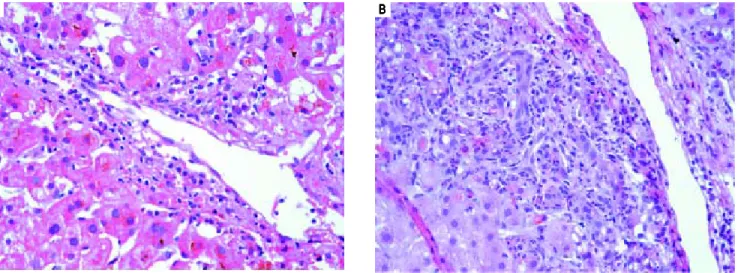
in-Figure 4. Figure 4. Figure 4. Figure 4.
Figure 4. Acute cellular rejection A.A.A.A.A. Banff criteria, mild. The above microphotograph (400x) depicts venulitis. B. B. B. B. B. Banff criteria, severe. The above micro-photograph (400x) depicts ductulitis, expansion of the portal tract and spillover of the inflammation into the hepatic parenchyma.
A AA
Figure 6. Figure 6. Figure 6.
Figure 6. Figure 6. Recurrence of HCV infection. Lymphoid aggregate within the portal tract.
Figure 5. Figure 5.Figure 5.
Figure 5.Figure 5. Acute cellular rejection. A. A. A. A. A. Banff grade - moderate. B. B. B. B. B. Banff grade - severe.
flammation. The 3rd patient had severe ACR in the 1st bi-opsy and mild ACR in the 2nd bibi-opsy. The commonest cause for most of these episodes of rejection was non-compliance with immunosuppressant drugs. ACR was also the commonest finding in larger series of post-trans-plant liver biopsies published from Iran and China.32,33
The next major histopathologic findings were recur-rence of hepatitis C in the allograft liver which was noted in 3 patients (Figure 6) Re-infection of the liver allograft with the hepatitis C virus after transplantation is practical-ly a universal occurrence.34,35 Hepatocyte expression of
HCV antigens has been demonstrated as early as 10 days after LT.36 In our study, the post-transplant biopsies were
performed between 87 to 207 days post LT. Two of the
bi-opsies showed early changes of recurrence i.e. lym-phocytic infiltrate in the portal tract associated with lobu-lar inflammation. The other 2 biopsies showed chronic changes of reccurrence with METAVIR grades for fibrosis ranging from 2-3.37 All the cases had high serum viral
loads. Our findings reflect those in other studies which show that acute hepatitis affects the graft between 2-5 months post LT while chronic hepatitis is established about 6-12 months post LT.38
We reported 4 biopsies as non-specific moderate portal infiltrates without evidence of rejection, accompanied by cholestasis, which did not warrant any active intervention, and 4 biopsies as unremarkable liver parenchyma. The in-dications for biopsy in these patients were elevated liver enzymes and/ bilirubin levels, which were probably sec-ondary to systemic infections and underlying co-morbidi-ties.
The incidence of chronic rejection has been reported to be between 2 and 20% in literature.32 We have reported
3 biopsies as chronic rejection during our study period (Figure 7).
Three patients had extensive intrahepatocytic and ca-nalicular bile stasis, and were found to have biliary stric-tures on imaging. Four patients showed sinusoidal and pericentral zone congestion and hemorrhage, probably secondary to ischaemic causes.
One patient who had uncontrolled diabetes mellitus and had been transplanted for NASH-related CLD, showed macro-vesicular steatosis involving about 20% hepatocytes. The biopsy from the lone patient transplant-ed for Caroli’s disease showtransplant-ed ascending cholangitis, cholestasis and portal fibrosis in a biopsy taken 1002 days post-transplant.
Two biopsies showed lobular hepatocyte dropout, without any accompanying inflammation or collapse of A
A A A
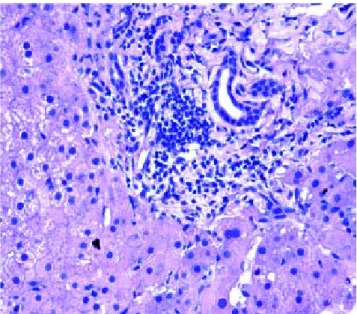
Figure 7. Figure 7. Figure 7. Figure 7.
Figure 7. Chronic rejection. A. Fibrosis of the portal tract is seen. B. B. B. B. Bile duct senescence and portal fibrosis is seen in the above microphotograph (400x).B.
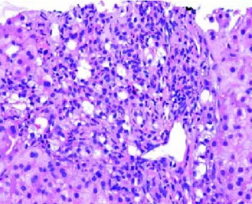
Figure 8. Figure 8. Figure 8. Figure 8.
Figure 8. Extrahepatic biliary obstruction. Florid bile ductular proliferation accompanied by a neutrophilic infiltrate.
the reticulin framework, which could not be explained, even after extensive investigations.
We had 1 case of reperfusion injury, 2 cases of acute hu-moral rejection and 1 case of extrahepatic biliary obstruc-tion (Figure 8), which were lower than the documented percentages in other studies.32,33 We have not reported any
cases of cytomegalovirus infection, post-transplant lym-phoproliferative disorder or post liver transplant cirrhosis in the present series.
Children ≤≤≤≤≤ 12 years
Among the pediatric patients who underwent LT, 8 pa-tients were biopsied for rising liver enzymes and / serum bilirubin levels (Figure 3). ACR was seen in 2 patients aged 11 years and 19 months (at the time of biopsy), on days 518 and 53 post transplant respectively. The ACR was of a severe grade in the former, and of a mild grade in the latter. This low rate of ACR in our series is similar to the findings of Shepherd et al, who studied more than 2,000 pediatric patients < 18 years who have undergone LT dur-ing an 11 year period, and reported the overall rejection rate as 0.29 episodes per patient year (0.20 in infants and 0.44 in adolescents).39
The biopsies of 2 patients revealed changes of ischaemia/reperfusion injury. The biopsies were obtained on days 16 and 15 post transplant respectively, and both re-cipients underwent reduced left lateral segment living donor liver transplants.
A biopsy from a 12 year old patient on day 39 post trans-plant revealed features of extra hepatic biliary obstruction. Two biopsies were performed on a 10 year old patient who was transplanted for Wilson’s disease and presented with high grade fever and abdominal pain, on days 115 and 147 post transplant. The 1st biopsy revealed non-specific portal inflammation and sinusoidal congestion, and the pa-tient was started on antibiotics and appeared to improve. However the patient was re-admitted 20 days following discharge, with deranged liver enzymes, and the biopsy re-vealed cholestatic liver injury and spotty necrosis. Biliary complications occur in approximately 10%-30% of pediat-A
AA
ric liver transplant recipients40 and is noted in 2 biopsies in
our series over a 4 year duration.
Two patients were biopsied after presenting with high grade fever, decreased appetite and slightly elevated liver enzymes, to rule out rejection or cytomegalovirus infec-tion. The viral loads were within normal limits and the bi-opsies did not reveal any abnormalities. One of these patients, a 3 and a half year old male, again presented with symptoms of acute gastroenteritis and elevated liver en-zymes, about 200 days following his prior admission, and was re-biopsied. His biopsy revealed prominent micro-vesicular steatosis, following which the administration of Methylprednisolone was stopped.
We have not reported any cases of post-transplant viral or fungal infection, post-transplant lymphoproliferative disorders, recurrent or de novo autoimmune hepatitis or chronic rejection necessitating re-transplantation among our cohort of pediatric recipients.
CONCLUSIONS
• In our series of explant hepatectomies in adults, alcohol-and hepatitis- virus related CLD are leading indi-cations for liver transplantation. HCV, more than HBV, is now the major cause of both CLD and HCC. • Biliary atresia is the most common cause of ESCLD
and LT in children, which is mirrored in the results of our study.
• Steatosis in donor livers is a risk factor for inferior graft function after orthotopic liver transplantation. The prevalence of 4.28% of moderate steatosis (be-tween 30-60% steatosis) in our series is much lower than that documented in western literature.
• The finding of only 5 cases of non caseating granulo-mas in our case series of donors was unusual for a country like India, which has the highest world-wide burden of tuberculosis.
• In our analysis of post-transplant liver biopsies in adults, the most common pathological diagnoses have been acute cellular rejection, followed by recurrence of the primary viral disease and non-specific moderate portal inflamma-tion. Early and accurate diagnosis aids the clinicians in ti-trating the dosage of immunosuppressants and anti-viral medications, thus optimising graft function.
• Among the pediatric cohort, the most common diag-noses have been essentially unremarkable liver paren-chyma due to non-hepatic etiologies, followed by ACR and ischaemia/reperfusion injury.
• As we share our experience 4 years into our LT pro-gramme, we expect that our data on explant hepatecto-mies, donor biopsies and post-transplant liver biopsies will more closely mirror the global trends in the years to come.
ABBREVIATIONS
• ACR: acute cellular rejection. • ALD: alcoholic liver disease. • ALF: acute liver failure. • BMI: body mass index. • CLD: chronic liver disease. • CT: computerised tomography.
• ESCLD: end stage chronic liver disease. • FH: fulminant hepatitis.
• H & E: haematoxylin and eosin. • HBV: hepatitis B virus.
• HCV: hepatitis C virus. • HEV: hepatitis E virus.
• HIV: human immunodeficiency virus. • LAI: liver attenuation index.
• LT: liver transplantation.
• NAFLD: non alcoholic fatty liver disease. • PCR: polymerase chain reaction.
• TACE: trans:arterial chemo:embolization.
REFERENCES
1. Child III CG, Turcotte JG. Surgery & portal hypertension. In: The liver and portal hypertension, Child III CG (Ed.). Saun-ders, Philadelphia; 1964, p. 50.
2. Kamath PS, Wiesner RH, Malinchoc M, Kremers W, Therneau TM, Kosberg CL, D'Amico G, et al. A model to predict survival in patients with end stage liver disease. Hepatology 2001; 33(2): 464-70.
3. McDiarmid SV, Anand R, Lindblad AS. Principal investigators and institutions of the studies of Pediatric liver transplanta-tion (SPLIT) Research group. Development of a pediatric end-stage liver disease score to predict poor outcome in children awaiting liver transplantation. Transplantation 2002; 74: 173-81.
4. O'Grady J, Alexander G, Hayllar KM, Williams R. Early indica-tors of prognosis in fulminant hepatic failure. Gastroenterol-ogy 1989; 97(2): 439-45.
5. James Trotter. Selection of donors for living donor liver transplantation. Liv Transplant 2003; 9(Suppl. 2): S2-S7. 6. Demetris AJ, Batts KP, Dhillon AP, Ferrell L, Fung J, Geller
SA, Hart J, et al. Banff schema for grading liver allograft re-jection: an international consensus document. Hepatology 1997; 25: 658-63.
7. Strasberg SM, Howard TK, Molmenti EP, Herti M. Selecting the donor liver: risk factors for poor function after orthotopic liver transplantation. Hepatology 1994; 20(4, Pt. 1): 829-38. 8. Nayak NC, Jain D, Vasdev N, Gulvani H, Saigal S, Soin A.
Eti-ologic types of end-stage chronic liver disease in adults: analysis of prevalence and their temporal changes from a study on native liver explants. Eur J of Gastroenterol and Hepatol 2012; 24: 1199-208.
9. Perz JF, Armstrong GL, Farrington LA, Hutin YJ, Bell BP. The contributions of hepatitis B virus and Hepatitis C virus infec-tions to cirrhosis and primary liver cancer worldwide. J Hepatol 2006; 45(4): 529-38.
Ita-ly): results of a pilot vaccination project. Res Virol 1998; 149(5): 263-70.
11. Ayata G, Gordon FD, Lewis WD, Pomfret E, Pomposelli JJ, Jenkins RL, Khettry U. Cryptogenic cirrhosis: clinicopatho-logic findings at and after liver transplantation. Hum Pathol 2002; 33(11): 1098-104.
12. Daniel Dutra Romualdo da Silva, Rios Leite VH. Cirrhotic liver explants: regenerative nodule, dysplasia and hepatocellular carcinoma occurrence. J Bras Med Lab 2005: 41:437-42. 13. Nayak NC. Hepatocellular carcinoma- a model of human
can-cer: clinicopathological features, etiology and pathogenesis. Indian J Pathol Microbiol 2003; 46: 1-16.
14. Alter MJ. Epidemiology of hepatitis C virus infection. World J Gastroenterol 2007; 13: 2436-41.
15. Phillipe Ichai, Didier Samuel. Etiology and prognosis of fulmi-nant hepatitis in adults. Liver Transpl 2008; 14(Suppl. 2): S67-S69.
16. Lee WM, Squires RH Jr, Nyberg SL, Doo E, Hoofnagle JH. Acute liver failure: summary of a workshop. Hepatology 2008; 47: 1401-15.
17. Ichai P, Saliba F, Antoun F, Azoulay D, Sebagh M, Antonini TM, Escaut L, et al. Acute liver failure due to antitubercular therapy: Strategy for antitubercular treatment before and af-ter liver transplantation. Liver Transpl 2010; 16: 1136-46. 18. Mindikoglu AL, Magder LS, Regev A. Outcome of liver
trans-plantation for drug-induced acute liver failure in the United States: analysis of the United Network for Organ Sharing database. Liver Transpl 2009; 15: 719-29.
19. Harshad Devarbhavi. Antituberculous drug-induced liver in-jury: current perspective. Tropical Gastroenterology 2011; 32: 167-74.
20. Kumar R, Shalimar, Bhatia V, Khanal S, Sreenivas V, Gupta SD, Panda SK, et al. Antituberculosis therapy-induced acute liver failure: magnitude, profile, prognosis, and predictors of outcome. Hepatology 2010; 51: 1665-74.
21. Devarbhavi H, Dierkhising R, Kremers WK. Antituberculosis therapy drug-induced liver injury and acute liver failure. Hepatology 2010; 52: 798-9.
22. Devarbhavi H, Dierkhising R, Kremers WK, Sandeep MS, Ka-ranth D, Adarsh CK. Single-center experience with drug-in-duced liver injury from India: causes, outcome, prognosis, and predictors of mortality. Am J Gastroenterol 2010; 105: 2396-404.
23. Balayan MS. Epidemiology of hepatitis E virus infection. J Vi-ral Hepat 1997; 4: 155-65.
24. Pinto RB, Schneider ACR, Silveira TR. Cirrhosis in children and adolescents: an overview. World J Hepatol 2015; 7: 392-405.
25. Leonis MA, Balistreri WF. Evaluation and management of End-stage liver disease in children. Gastroenterology 2008; 134: 1741-51.
26. Molleston JP, Sokol RJ, Karnsakul W, Miethke A, Horslen S, Magee JC, Romero R, et al. Evaluation of the child with sus-pected mitochondrial liver disease. J Pediatr Gastroenterol Nutr 2013; 57: 269-76.
27. Squires RH Jr., Schneider BL, Bucuvalas J, Alonso E, Sokol RJ, Narkewicz MR, Dhawan A, et al. Acute liver failure in
children: the first 348 patients in the pediatric acute liver fail-ure study group. J Pediatr 2006; 148: 652-8.
28. Mokdad AH, Ford ES, Bowman BA, Dietz WH, Vinicor F, Bales VS, Marks JS. Prevalence of obesity, diabetes and obesity-related health risk factors, 2001. JAMA 2003; 289(1): 76-9.
29. Dare AJ, Phillips ARJ, Chu M, Hickey AJR, Bartlett A. Ap-praisal of donor steatosis in liver transplantation: a survey of current practice in Australia and New Zealand. Trans-plant Research and Risk Management 2102; 4: 31-7. 30. Selzner M, Clavien PA. Fatty liver in liver transplantation and
surgery. Semin Liver Dis 2001; 21(1): 105-13.
31. Pungpagong S, Krishna M, Abraham SC, Keaveny AP, Dick-son RC, Nakhleh RE. Clinicopathologic findings and outcomes of liver transplantation using grafts from donors with unrec-ognised and unusual diseases. Liv Transplant 2006; 12: 310-5.
32. Geramidazeh B, Motevalli D, Nikeghbalian S, Seyed Ali Malek Hosseini. Histopathology of Post-transplant liver biopsies, the first report from Iran. Hepat Mon 2013; 13: e9389. 33. Cong WM, Zhang SY, Wang ZL, Xue L, Liu YS, Zhang SH.
Pathologic diagnosis of 1123 post-transplant liver biopsies from 665 transplant patients. Zhonghua Bing Li Xue Za Zhi 2005; 34: 716-9.
34. Feray C, Samuel D, Thiers V, Gigou M, Pichon F, Bismuth A, Reynes M, et al. Reinfection of liver graft by hepatitis C virus after liver transplantation. Clin Invest 1992; 89(4): 1361-5. 35. Arnold JC, Tox U, Goeser T, Otto G, Muller HM, Hofmann
WJ, Stremmel W, et al. Recurrent hepatitis C virus infection after liver transplantation-long term follow-up with respect to the HCV genotypes/subtypes. Z Gastroenterol 1997; 35: 255-61.
36. Rodriguez-Luna H, Vargas HE. Management of hepatitis C vi-rus infection in the setting of liver transplantation. Liver transplantation 2005; 11: 479-89.
37. Bedossa P, Poynard T. An algorithm for the grading of activi-ty in chronic hepatitis C. The METAVIR cooperative study group. Hepatology 1996; 24: 289-93.
38. Shih-Hsien Hsu, Ming-Lun Yeh, Shen-Nien Wang. New In-sights in Recurrent HCV Infection after Liver Transplanta-tion. Clinical and Developmental Immunology 2013. Article ID 890517.
39. Shepherd RW, Turmelle Y, Nadler M, Lowell JA, Narkewicz MR, McDiarmid SV, Anand R, et al. Risk factors for rejection and infection in Pediatric liver transplantation. Am J Trans-plant 2008; 8: 396-403.
40. Spada M, Riva S, Maggiori G, Cintorino D, Gridelli B. Pediatric liver transplantation. World J Gastroenterol 2009; 15: 648-74.
Correspondence and reprint request:
Fiona Fonseca, M.B.B.S, MD. (Pathology) 303, Golf view Apartments, Next to St. James Church,
Fatehgunj, Baroda-390002, Gujarat, India. Tel.: +919819512790