Evaluation of tumor markers for the
differential diagnosis of benign and malignant ascites
Fang Liu,*, Xinjuan Kong,**, Qian Dou,** Jin Ye,*** Dong Xu,**** Haitao Shang,*** Keshu Xu,*** Yuhu Song***
* Institute of Hematology, Union Hospital, Tongji Medical College, Huazhong University of Science and Technology, Wuhan, 430022 P. R. China. ** Department of Gastroenterology, the Affiliated Hospital of Medical College, Qingdao University, Qingdao, P.R. China.
*** Department of Gastroenterology, Union Hospital, Tongji Medical College, Huazhong University of Science and Technology, Wuhan, 430022 P. R. China. **** Department of Infectious Diseases, Tongji Hospital, Tongji Medical College, Huazhong University of Science and Technology, Wuhan, 430030 P. R. China.
Liu F and Kong XJ contributed equally to this work.
ABSTRACT
Introduction. The diagnosis of malignant ascites is a challenging problem in clinical practice, non-invasive
techniques should be developed to improve diagnostic accuracy. The diagnostic performances of tumor markers in malignant ascites remained unsettled. Our aim was to evaluate diagnostic performance of tumor markers in differential diagnosis of benign and malignant ascites. Material and methods. A total of 437 patients were enrolled, and the relevant parameters of the patients were analyzed for the diffe-rentiation of benign ascites from malignant ascites. Results. At the predetermined cutoff values of tumor makers, tumor markers in ascitic fluid showed better diagnostic performance than those in serum. Combined use of tumor markers and the cytology increased the diagnostic yield of the latter by 37%. In cytologically negative malignant ascites, tumor markers provided assistance in differentiating malignant ascites from benign ascites, and the combination of ascitic tumor markers yielded 86% sensitivity, 97% specificity.
Conclusion. Use of a panel of tumor markers exhibited excellent diagnostic performance in diagnosing
malignant ascites, which indicated the detection of tumor markers may represent a beneficial adjunct to cytology, thus guiding the selection of patients who might benefit from further invasive procedures.
Key words. CEA. CA19-9. CA15-3. Citology.
Correspondence and reprint request: Yuhu Song, M.D., Ph.D.
Department of Gastroenterology, Union Hospital, Tongji Medical College, Huazhong University of Science and Technology Wuhan, 430022, P.R. China. Tel.: 0086-27-85726678
E-mail: [email protected], [email protected]
Manuscript received: October 30, 2013. Manuscript accepted: December 26, 2013. INTRODUCTION
Ascites is the pathological accumulation of fluid within abdominal cavity, which is commonly associat-ed with chronic hepatic diseases, malignant neopla-sia, cardiac insufficiency, tuberculous peritonitis, renal diseases and etc.1,2 Ascites presents a challenging
di-agnostic problem for gastroenterologists because of the lack of specific and sensitive differential diagnos-tic strategies. Malignant ascites accounts for about 10% of all cases of ascites and commonly associated with gastric, colorectal, pancreatic, hepatobiliary, ovarian, endometrial, primary peritoneal
carcino-mas.3,4 In clinical practice, complete separation
be-tween malignant ascites and benign ascites has not been always possible. Cytology for malignant ascites is an important diagnostic tool due to its high spe-cificity, but positive results are obtained only in 40-60% of cases.5-7 Peritoneal biopsy upon laparoscope
provides an alternative diagnostic strategy for ma-lignant ascites.8,9 But its application is limited in
clinical practice because this procedure is not avail-able at all facilities and may be too invasive for the patients. Therefore, other non-invasive techniques should be developed for the differentiation between malignant ascites and benign ascites.
Tumor markers are produced directly by the tumor or by non-tumor cells as a response to the presence of a tumor, which offers a putative clinical use in the screening, diagnosis and treatment of various cancers. Thus, numerous studies demonstrated the detection of serum or ascitic tumor markers was helpful in differ-entiating malignant ascites from benign ones;5,7,10-18
mark-ers involved confined their clinical value. Therefore, the diagnostic value of tumor marker in malignant as-cites remained unsettled.
In the present study, the aims of our study were:
• To evaluate diagnostic value of a panel of tumor markers such as carcinoembryonic antigen (CEA), carbohydrate antigen 15-3 (CA 15-3), car-bohydrate antigen 19-9 (CA19-9) and CA-125 in diagnosing malignant ascites.
• To evaluate diagnostic value of tumor markers in cytologically negative malignant scites.
• To assess their usefulness as differential diagnos-tic markers among different types of cancers.
MATERIAL AND METHODS
Study population
In this study, we collected 437 patients with the ascites at Union Hospital and Tongji Hospital affili-ated to Tongji Medical College, Huazhong Universi-ty of Science and Technology, the Affiliated Hospital of Medical College, Qingdao University from 2007 to 2012. All the patients had undergone diagnostic pa-racentesis with the measurement of several parame-ters. The parameters included cytological examination and a panel of tumor markers. The study was approved by the hospital ethics committee of the participant centers.
Diagnostic criteria
The causes of ascites were determined by well-es-tablished clinical criterion. The patients were classi-fied into two groups named as benign and malignant ascites according to the etiology. The benign group consisted of two groups: portal hypertension and non-portal hypertension. Portal hypertension was defined by clinical manifestation of ascites, esopha-geal and gastric varices, and splenomegaly, which were confirmed by using clinical assessment, labora-tory findings, imaging data and/or histological ex-amination. In this study, the causes of ascites resulted from portal hypertension included liver cirrhosis, cardiac insufficiency, Budd-Chiari syn-drome, veno-occlusive disease. The etiology for be-nign ascites without portal hypertension included tuberculous peritonitis, pancreatic ascites, nephrotic syndrome, connective tissue disease, secondary bac-terial peritonitis. Malignant ascites was defined in the patients with positive cytology of ascitic fluid; in addition, the patients with negative ascitic cytology
finding were diagnosed as malignant ascites when the patients had a known primary malignancy after ruling out benign causes of the ascites. The diagno-sis of malignant ascites caused by primary liver can-cer was based on tumor localization as confirmed by CT, MRI or liver biopsy after ruling out the metas-tasis of gastrointestinal malignancies. In addition, low SAAG (defined as less than 1.1 g/dL) should be required for eliminating the ascites resulted from portal hypertension.
Pancreatic ascites was diagnosed when the ascitic fluid amylase level was at least twice that of the up-per limit of normal (ULN) for serum amylase; in ad-dition, pancreatic cancer should be excluded.19
Tuberculous peritonitis was defined in patients with granulomatous inflammation on peritoneal biopsy, or complete clinical and laboratory response after antituberculous therapy when exudative ascites and the exclusion of other causes. Secondary bacterial peritonitis with gastrointestinal perforation was de-fined by:
• The presence of ascitic fluid neutrophil count of greater than 250/mm3, and
• Extravasation of contrast material or peritoneal free air on radiography or computerized tomogra-phy, and/or perforation of the intestinal wall demonstrated at surgery.20
Tumor marker assay, and cholesterol assay cytological assay
Ascites samples obtained by paracentesis were col-lected in tubes, and then sent for biochemical assay and cytological examination. Carcinoembryonic an-tigen (CEA), cancer anan-tigen 12-5 (CA12-5), CA19-9, CA15-3, alpha-feto-protein (AFP) and prostate specific antigen (PSA) were measured in serum and ascitic fluid according to instruction manual using chemiluminescence microparticle immuno assay (Fu-jirebio Diagnostics Inc, Malvern, PA 19355,USA). Serum CEA, CA 12-5, CA15-3, and CA199 levels of 5 ng/mL, 35 U/mL, 31 U/mL, and 37 U/mL, respec-tively, were adopted as the upper limits of normal in healthy subjects.
Statistical analysis
the two groups. Differences were considered signifi-cant for P < 0.05.
RESULTS
Etiology of ascites
In this study, the study population consisted of 437 patients was divided into two groups: benign as-cites and malignant asas-cites. Two-hundred twenty-four patients had a benign ascites, 160 patients had ascites associated portal hypertension, 64 patients with ascites of non-portal hypertensive etiology. Among malignant group, there were 121 patients with positive fluid cytology findings, and 92 patients with cytology-negative ascites. The specific etiologies and the histological subtypes of tumors were pre-sented in Table 1. For cytologically-positive malig-nant ascites, maligmalig-nant ascites with unkown primary carcinoma (Table 1) referred to the patients with unspecified origin. For cytologically-negative malignant ascites, a diagnosis of unspecified prima-ry was made in the patients with a known
malig-nancy which involved two or more organs after rul-ing out other causes. It was confirmed by radiologi-cal finding and/or histologiradiologi-cal examination.
Diagnostic performance of tumor markers
Tumor markers are the molecules, which are used in the screening, diagnosis, and treatment of vari-ous cancers. In this study, we evaluated the diag-nostic performance of a panel of tumor markers in distinguishing malignant ascites from benign as-cites. In order to detect the correlation of tumor markers in ascitic fluid and serum, the concentra-tion of tumor markers in serum and ascitic fluid was detected simultaneously. Results of tumor markers in the whole population were detailed in table 2. In the study, the cutoff points for tumor markers were defined; the cutoff points were: 50 ng/mL for CEA, 200 U/mL for CA19-9, 350 U/mL for CA12-5, 75 U/mL for CA15-3. The cutoff values of tumor markers were defined according to diagnostic effica-cy of and receiver operating characteristic plot
Table 1. Etiology of benign and malignant ascites.
Malignant ascites (213)
Stomach Colorectal Pancreas Liver Gynecological Cholangio- Unknown Other carcinoma primary cancers
Positive cytology (121) 26 11 7 6 24 1 42 4
Negative cytology (92) 28 7 7 4 4 4 32 6
Benign 224
Portal hypertension 160
Liver cirrhosis 151
HBV HCV NAFLD Alcohol Schistosome Autoimmune Unknown
liver diseases
105 5 1 21 8 7 4
Veno-occlusive diseases 2
Budd-chiari syndrome 6
Cardiac ascites 1
Non-portal hypertension (64)
Secondary bacterial peritonitis 2
Tuberculous peritonitis 58
Nephrotic syndrome 2
Connective tissue disease 1
Acute pancreatitis 1
(ROC) of tumor markers.22 Significant difference
was noted in the percentage of serum CEA, serum CA 19-9, serum CA15-3, ascitic CEA, ascitic CA 19-9 and CA15-3; but not in serum CA 12-5 and ascitic CA12-5 between benign and malignant group (Table 2). Serum CEA and CA 153 higher than the cut-off points were specifically found in the patients with malignant ascites (Table 2). Thirty-nine percent of malignant ascites yielded a positive result in serum CA199 concentration higher than the cut-off value. Raised serum CA199 (3.17%) was also detected in a few cases of benign ascites, most of those cases had suffered from biliary calculi; fortunately, ascitic CA19-9 higher than cut-off point was not detected in those cases. As shown in table 3, the sensitivities of serum CEA, CA 19-9 and CA15-3 taken separately were 9, 39, and 12%, respectively; the sensitivities of ascitic CEA, CA 125, and CA153 were superior to
that of serum tumor makers; but the specificities of ascitic tumor makers were similar to those of serum markers in malignant ascites (Table 3). It indicated that ascitic tumor markers exhibited better diagnos-tic performance than serum markers. In addition, we observed that diagnostic performance of CEA and CA19-9 was superior to that of CA15-3 due to high-er sensitivity. In ordhigh-er to furthhigh-er evaluate diagnos-tic performance, the sensitivity, specificity, positive predictive value (PPV), and negative predictive value (NPV) of cytology and oftumor markers, alone or in combination, were calculated (Table 3). As shown in table 3, ascitic tumor markers showed better diagnostic advantage than serum markers through the sensitivity and predictive value. Taken together, ascitic CEA, CA199 and CA153 yielded a positive result in 85% of the malignant ascites with 97% spe-cificity. Then we evaluated the diagnostic efficacy
Table 2. Positive rate of tumor markers at the predetermined cutoff points in benign and malignant ascites.
Benign ascites Malignant ascites
Portal Non-portal Total Cytological Cytological Total
hypertension hypertension positivity negativity
Serum CEA 0/160 0/64 0/224 4.13% (5/121) 15.22% (14/92) 8.92% (19/213)
Ascitic CEA 0/160 1.85% (1/64) 1/224 (0.45%) 46.28% (56/121) 72.83% (67/92) 57.75% (123/213) Serum CA199 4.43% (7/158) 0/63 7/221 (3.17%) 33.33% (39/117) 46.67% (42/90) 39.13% (81/207) Ascitic CA199 0.63% (1/158) 1.59% (1/63) 2/221 (0.90%) 55.56% (65/117) 62.22% (56/90) 58.45% (121/207) Serum CA125 65.56% (99/151) 52.46% (32/61) 131/212 (61.79%) 57.66% (64/111) 32.56% (28/86) 46.70% (92/197) Ascitic CA125 82.12% (124/151) 81.97% (50/61) 174/212 (82.08%) 93.69% (104/111) 93.02% (80/86) 93.40% (184/197)
Serum CA153 0/149 0/59 0/208 19.81% (21/106) 2.44% (2/82) 12.23% (23/188)
Ascitic CA153 0/149 5.08% (3/59) 3/208 (1.44%) 35.85% (38/106) 7.32% (6/82) 23.40% (44/188)
Cut off points for CEA, CA19-9, CA12-5 and CA15-3 were 50 ng/mL, 200 U/mL, 350 U/mL, 75 U/mL respectively.
Table 3. Sensitivity, specificity, PPV, and NPV of cytology and tumor markers in the whole population of ascites.
Variables Sensitivity (%) Specificity (%) PPV (%) NPV (%)
Cytology 56.81 100 100 51.26
CEA
Serum 8.92 100 100 53.59
Ascites 57.75 99.55 99.19 71.25
CA199
Serum 39.13 96.83 92.05 62.94
Ascites 58.45 99.10 98.37 71.80
CA153
Serum 12.23 100 100 55.76
Ascites 23.40 98.56 93.62 58.74
CEA+CA199+CA153
Serum 51.64 96.88 94.02 67.81
Ascites 85.45 97.32 96.81 87.55
CEA+CA199+CA153+cytology
Serum 80.28 96.88 96.07 83.78
of cytology, the results (Table 3) showed its efficacy was similar to that of ascitic CEA or CA199 alone. Finally, we evaluated the diagnostic performance of combination of cytology and tumor makers in the whole population of ascites. The combined use of cytological examination and serum markers allowed to confirm the tumoral diagnosis in 80% of malignant ascites with 97% specificity, 96% PPV, and 84% NPV; whereas the combination of cytology and as-citic markers yielded a sensitivity of 94% with a spe-cificity of 97% in diagnosing malignant ascites. The PPV and NPV were 97% and 94%, respectively (Table 4). All these indicated that a panel of tumor markers used represented a helpful adjunct to cytol-ogy for the diagnosis of malignant ascites.
Diagnostic value of tumor markers in cytologically negative malignant ascites
We evaluated the additional value of ascitic tu-mor makers in cytologically- negative malignant as-cites. The positivity, specificity and predictive value
of tumor markers, either individually or in combi-nation were reported in table 4. CA 15-3, a tumor maker of gynaecological cancer, showed an inferior diagnostic performance because of low s sensitivity. The diagnostic yield of the combined three tumor markers in cytologically negative malignant effu-sions was 86%, with a specificity of 97% and a PPV of 92% and a NPV of 94%. The combination of three tumor markers exhibited a convincing diagnostic performance in cytologically-negative ascites.
Tumor markers and tumor types
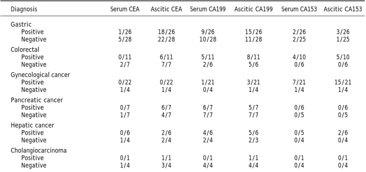
It is well-known that some tumor markers are specific for a particular type of cancers. Therefore, we evaluated diagnostic performance of tumor mark-ers according to the histologic type of cancer. As shown in table 5, CEA was quite effective in stomach, colorectal cancer, pancreatic cancer, cholangi-ocarcinoma, CA19-9 showed high sensitivity in pan-creatic cancer and cholangiocarcinoma; whereas for gynecological cancer, CA 15-3 was specific and
sensi-Table 5. Cases showing tumor marker values higher than the cutoff value among different types of cancer with ascites.
Diagnosis Serum CEA Ascitic CEA Serum CA199 Ascitic CA199 Serum CA153 Ascitic CA153
Gastric
Positive 1/26 18/26 9/26 15/26 2/26 3/26
Negative 5/28 22/28 10/28 11/28 2/25 1/25
Colorectal
Positive 0/11 6/11 5/11 8/11 4/10 5/10
Negative 2/7 7/7 2/6 5/6 0/6 0/6
Gynecological cancer
Positive 0/22 0/22 1/21 3/21 7/21 15/21
Negative 1/4 1/4 0/4 1/4 1/4 1/4
Pancreatic cancer
Positive 0/7 6/7 6/7 5/7 0/6 0/6
Negative 1/7 4/7 7/7 7/7 0/5 0/5
Hepatic cancer
Positive 0/6 2/6 4/6 5/6 0/5 2/6
Negative 1/4 2/4 2/4 2/3 0/4 0/4
Cholangiocarcinoma
Positive 0/1 1/1 0/1 1/1 0/1 0/1
Negative 1/4 3/4 4/4 4/4 0/4 0/4
Table 4. Sensitivity, specificity, PPV, and NPV of ascitic tumor markers in cytologically negative malignant ascites.
Variables Sensitivity (%) Specificity (%) PPV (%) NPV (%)
CEA 72.82 99.55 98.52 89.92
CA199 62.22 99.10 96.55 86.56
CA153 7.31 98.56 66.67 72.95
tive. On the other hand, we also detected the con-centration of serum and ascitic tumor markers in order to investigate the correlations of tumor mark-ers in ascitic fluid and serum. The patients with se-rum tumor markers higher than cutoff points always yielded a positive result in ascitic concentra-tion, whereas some of the patients with positive re-sult in ascitic tumor markers did not show elevated concentration in serum. These indicated the exami-nation of tumor markers in ascitic fluid provided advantage over the examination of serum mark-ers in the diagnosis of malignant ascites.
DISCUSSION
Malignancy is an important cause of ascites; to date, cytological examination is an important tech-nique for malignant ascites. But cytological exami-nation using classic technique is often difficult either because of its scarceness or its delicate dis-tinction with reactive mesothelial or inflammatory cells in effusion.21 Although invasive procedures
im-prove diagnostic accuracy, their application is re-stricted because of the invasiveness and limited diagnostic efficacy. Therefore, we collected a rela-tively large sample consisted of 437 patients and then evaluated diagnostic performance of tumor markers for distinguishing malignant ascites from benign ascites.
Our study revealed that CEA, CA19-9 and CA15-3 were proven to be valuable in distinguishing benign ascites from malignant ascites. Among tumor mak-ers, ascitic CEA showed the best efficacy through ROC curve (data no shown). More than half of pa-tients with the ascites had an elevated CA12-5 high-er than cut-off value. These results whigh-ere consistent with previous report.22-25 It indicate that testing
se-rum or ascite for CA12-5 is not helpful in the differ-ential diagnosis of ascites. Therefore, we did not recommend the detection of CA12-5 in clinical practice, which is consistent with AASLD guide-line.2
Tuzun, et al.16 found that tumor markers were
highly correlated in serum and ascitic fluid in the malignant group and the detection of tumor mark-ers in ascitic fluid does not have any advantage over serum analysis. Our results (Table 3) showed that diagnostic performance of tumor markers in as-citic fluid was superior to that in serum. As report-ed by Tuzun16 and Chen,10 mean levels of CEA were
significantly higher in ascitic fluid than that in se-rum in the malignant group. All these demonstrated that the measurement of these markers in ascitic
fluid should substitute the detection of serum mark-ers due to better diagnostic performance of ascitic tumor markers. Therefore, the measurement of as-citic tumor markers should be recommended in clin-ical practice when the patients are suspected to be malignant ascites. As shown in table 3, the perform-ance of ascitic CEA or CA199 individually was com-parable to that of cytological examination; while the combination of CEA, CA19-9 and CA15-3 yielded bet-ter sensitivity (85%) and specificity (97%) compared with CEA, CA199 and CA153 alone. The addition of tumor markers to cytological analysis resulted in a 37% increase of diagnostic rate with little effect on its specificity. On the other hand, ascitic PSA and AFP were also evaluated, and the results (data not shown) showed poor performance in the diagnosis of malignant ascites because of weak positive rate and/ or the lack of specificity. Thus, a panel of tumor markers consisted of CEA, CA19-9 and CA15-3 should be determined when malignant ascites were suspected.
We also evaluated the role of ascitic tumor mark-ers in diagnosing malignant ascites with a negative cytology. We found that the combined three tumor markers in cytologically negative malignant yielded 86% sensitivity, 97% specificity, 92% PPV of and 94% NPV. These revealed the determination of the three tumor markers exhibited a convincing diag-nostic performance in cytologically-negative ascites. Therefore, we recommend to use a panel of tumor makers in the patiets who have clinical data sugges-tive of malignant ascites, but negasugges-tive cytologic find-ings. Two patients of the secondary peritonitis were included in this study, one case had elevated CEA (58.4 ng/mL) in ascitic fluid. This phenomenon was reported by previous research, in which the elevations of ascitic fluid CEA or alkaline phosphatase had been shown to be accurate for detecting perforation-related secondary peritonitis.20
In conclusion, the study demonstrated that use of a panel of tumor markers exhibited excellent diag-nostic performance in diagnosing malignant ascites, combined use of tumor markers and the cytology in-creased the diagnostic yield. In cytologically negative malignant ascites, tumor markers provided assistance in differentiating malignant ascites from benign ascites.
Thus, these indicated that detection of tumor markers may represent a beneficial adjunct to cytol-ogy in diagnosing malignant ascites.
ACKNOWLEDGMENTS
The work was supported by Chinese Nation-al ClinicNation-al Key Discipline (2011-2013) and NationNation-al Natural Science Foundation of China (No. 81072003 and 81270506).
REFERENCES
1. Runyon BA. Management of adult patients with ascites due to cirrhosis. Hepatology 2004; 39: 841-56.
2. Runyon BA. Management of adult patients with ascites due to cirrhosis: an update. Hepatology 2009; 49: 2087-107. 3. Becker G, Galandi D, Blum HE. Malignant ascites:
systema-tic review and guideline for treatment. Eur J Cancer 2006; 42: 589-97.
4. Sangisetty SL, Miner TJ. Malignant ascites: A review of prognostic factors, pathophysiology and therapeutic mea-sures. World J Gastrointest Surg 2012; 4: 87-95.
5. Pinto MM. CA-15.3 assay in effusions: comparison with carcinoembryonic antigen and CA-125 assay and cytologic diagnosis. Acta Cytol 1996; 40: 437-42.
6. Cascinu S, Del Ferro E, Barbanti I, Ligi M, Fedeli A, Catala-no G. Tumor markers in the diagCatala-nosis of malignant serous effusions. Am J Clin Oncol 1997; 20: 247-50.
7. Ammon A, Eiffert H, Reil S, Beyer JH, Droese M, Hiddemann W. Tumor-associated antigens in effusions of malignant and benign origin. Clin Investig 1993; 71: 437-44.
8. Jenkins PF, Ward MJ. The role of peritoneal biopsy in the diagnosis of ascites. Postgrad Med J 1980; 56: 702-3. 9. Luck NH, Khan AA, Alam A, Butt AK, Shafquat F. Role of
la-paroscopy in the diagnosis of low serum ascites albumin gradient. J Pak Med Assoc 2007; 57: 33-4.
10. Chen SJ, Wang SS, Lu CW, Chao Y, Lee FY, Lee SD, Wu SL, et al. Clinical value of tumour markers and serum-ascites albumin gradient in the diagnosis of malignancy-related ascites. J Gastroenterol Hepatol 1994; 9: 396-400.
11. Loewenstein MS, Rittgers RA, Feinerman AE, Kupchik HZ, Marcel BR, Koff RS, Zamcheck N. Carcinoembryonic anti-gen assay of ascites and detection of malignancy. Ann
In-tern Med 1978; 88: 635-8.
12. Passebosc-Faure K, Li G, Lambert C, Cottier M, Gentil-Pe-rret A, Fournel P, Perol M, et al. Evaluation of a panel of molecular markers for the diagnosis of malignant serous effusions. Clin Cancer Res 2005; 11: 6862-7.
13. Porcel JM, Vives M, Esquerda A, Salud A, Perez B, Rodri-guez-Panadero F. Use of a panel of tumor markers (carci-noembryonic antigen, cancer antigen 125, carbohydrate antigen 15-3, and cytokeratin 19 fragments) in pleural fluid for the differential diagnosis of benign and malignant effusions. Chest 2004; 126: 1757-63.
14. Shahab N. Tumor markers in malignant ascites. Arch
In-tern Med 2002; 162: 949-50.
15. Singer G, Rebmann V, Chen YC, Liu HT, Ali SZ, Reinsberg J, McMaster MT, et al. HLA-G is a potential tumor marker in malignant ascites. Clin Cancer Res 2003; 9: 4460-4. 16. Tuzun Y, Celik Y, Bayan K, Yilmaz S, Dursun M, Canoruc F.
Correlation of tumour markers in ascitic fluid and serum: are measurements of ascitic tumour markers a futile at-tempt? J Int Med Res 2009; 37: 79-86.
17. Tuzun Y, Yilmaz S, Dursun M, Canoruc F, Celik Y, Cil T, Bo-yraz T. How to increase the diagnostic value of malignan-cy-related ascites: discriminative ability of the ascitic tumour markers. J Int Med Res 2009; 37: 87-95.
18. Wang T, Qian X, Wang Z, Wang L, Yu L, Ding Y, Liu B. Detection of cell-free BIRC5 mRNA in effusions and its potential diagnostic value for differentiating malig-nant and benign effusions. Int J Cancer 2009; 125: 1921-5.
19. Runyon BA, Hoefs JC, Morgan TR. Ascitic fluid analysis in malignancy-related ascites. Hepatology 1988; 8: 1104-9.
20. Wu SS, Lin OS, Chen YY, Hwang KL, Soon MS, Keeffe EB. Ascitic fluid carcinoembryonic antigen and alkaline phos-phatase levels for the differentiation of primary from se-condary bacterial peritonitis with intestinal perforation. J
Hepatol 2001; 34: 215-21.
21. Bedrossian CW. Diagnostic problems in serous effusions.
Diagn Cytopathol 1998; 19: 131-7.
22. Nguyen HN. Extreme CA125 elevation from hepatitis C and ascites. Gynecol Oncol 1998; 70: 441-2.
23. Runyon BA. Malignancy-related ascites and ascitic fluid “humoral tests of malignancy”. J Clin Gastroenterol 1994; 18: 94-8.
24. Sevinc A, Sari R, Buyukberber S. Cancer antigen 125: tu-mor or serosal marker in case of ascites? Arch Intern Med 2001; 161: 2507-8.
25. Zuckerman E, Lanir A, Sabo E, Rosenvald-Zuckerman T, Matter I, Yeshurun D, Eldar S. Cancer antigen 125: a sen-sitive marker of ascites in patients with liver cirrhosis.