Natural history of hepatitis C virus infection in
a cohort of asymptomatic post-transfused subjects
María Virginia Reggiardo,* Fabián Fay,† Mario Tanno,* Gabriela García-Camacho,** Oscar Bottaso,‡ Sebastián Ferretti,* Alicia Godoy,§ Claudio Guerrita,* Mauro Paez,* Federico Tanno,* Orlando Ruffinengo,* Silvina Benetti,† Silvia E García Borrás,|| M Celina Rossi,|| Julio Vorobioff,* Fernando Bessone,* Hugo Tanno*
*Gastroenterology and Hepatology Department, Hospital Provincial del Centenario, School of Medicine, University of Rosario, Rosario, Argentina. †CIBIC, Rosario, Argentina.
‡Immunology Institute, School of Medicine, University of Rosario, Rosario, Argentina. §Department of Pathology, School of Medicine, University of Rosario, Rosario, Argentina.
||Histocompatibility and Immunogenetics Laboratory, School of Biochemistry and Pharmaceutical Science, University of Rosario, Rosario, Argentina.
ABSTRACT
Background & aims. Studies about the natural history of hepatitis C virus (HCV) infection report variable progression to cirrhosis depending on study design. Retrospective cross-sectional liver clinic studies over-estimate the rate of fibrosis progression due to inclusion of patients with more severe disease leaving mild and asymptomatic patients underrepresented. We evaluated fibrosis progression in a group of “healthy” asymptomatic subjects, attending to a voluntary campaign for the detection of HCV infection. Material and methods. A detection campaign was launched on subjects transfused before 1993. Of 1699 volun-teers, 61(3.6%) had HCV infection. A liver biopsy was performed in 40 (65%). Assessed risk factors for liver fi-brosis were: sex, body mass index, alcohol consumption (> 20 g/d - >40g/d ), genotype, HLA-DRB1 alleles, present age, age at infection and duration of infection. Results. 25 (62.5%) were women with a me-dian age of 52.5 years. The meme-dian duration of infection was 21.5 years with a meme-dian age at infection of 27 years. As regards fibrosis, 25 (62.5%) had a Low Stage (F0-F1), 8 patients, 20%, had severe fibrosis, one patient (2.5%) had cirrhosis. Alcohol consumption was the only risk factor associated with fibrosis progres-sion. Conclusions. The low progression to cirrhosis may be explained by the clinical characteristics of our population: asymptomatic middle-aged “healthy” subjects infected at young age. The progression to seve-re fibrosis was noticeable; hence a longer follow-up might demonstrate changes in this outcome. Signifi-cant alcohol consumption clearly worsens the natural history of HCV infection; this is no so evident for occasional or mild alcohol consumers.
Key words. Alcohol consumption. Fibrosis progression. Fibrosis risks factors.
Correspondence and reprint request: María Virginia Reggiardo
Gastroenterology & Hepatology Department, Hospital Provincial del Centenario, School of Medicine, University of Rosario
Urquiza, 3101. Rosario (2000), Argentina Tel.: ++54 341-4393511
Fax: ++54 341-4387014
E mail: [email protected]
Manuscript received: September 09,2011. Manuscript accepted: December 20, 2011.
INTRODUCTION
The natural history of chronic hepatitis C virus (HCV) infection is benign in most patients, principa-lly due to a slow fibrosis progression.1 Nevertheless some patients have a poor outcome progressing to cirrhosis and related complications. The high global incidence of HCV infection in the world makes HCV
short term by which the HCV infection is certainly diagnosed. Also, the study population has to be a large community- or population- based one avoiding the referral bias. Furthermore, different population representing the whole spectrum of the disease should be included. For instance, the outcome seems to be very different between men transfused at middle age10 and young pregnant women.11-12
Given this constraints, studies employing other designs should be taken into account when at-tempting to expand our knowledge on the natural history of chronic HCV infection.
Our study is a retrospective-prospective cohort study where subjects with a recognized source of in-fection in the past were followed up prospectively. The population consists of a group of “healthy” sub-jects, attending to a voluntary campaign for detec-ting HCV infection. Participants were transfused before 1993, had no antecedents of hemodialysis or intravenous drug use.
In these subjects, we sought to evaluate the hepa-tic fibrosis progression and some factors associated with the development of this event.
MATERIAL AND METHODS
Patients
A voluntary detection campaign was launched in Rosario, a 1,000,000 inhabitant city of Argenti-na, between August and October 2005. Subjects transfused before 1993 were called through a TV and radio advertisement campaign to which 1,699 subjects attended. Year 1993 was chosen as a cut off because the ministerial resolution that regula-tes the screening for HCV in blood banks in Ar-gentina was promulgated in December 1992. The oldest date of transfusion in our population was in 1960.
In the first visit patients were informed of the study purposes, completed an epidemiological and clinical form and a blood sample was collected to as-ses HCV infection. Informed consent was obtained from each subject. The study protocol conforms to the ethical guidelines of the 1975 Declaration of Hel-sinki as reflected in a priori approval by the institution’s Human Research Committee.
Subjects with antecedents of hemodialysis or in-travenous drug use were excluded at the time of fi-lling an epidemiological/clinical form that was administered by trained personnel.
HCV antibodies (anti-HCV) were evaluated by third generation enzyme immunoassay
(ELISA-Wienner) and HCV-RNA were detected using Amplicor Monitor HCV assay Roche (detection limit = 50 IU/ mL). HCV genotype was determined by using res-triction fragment length polymorphism (RFLP).
All anti-HCV(+)/HCV-RNA(+) patients where offered to continue their follow up at the Gastroen-terology and Hepatology Department, Hospital Pro-vincial del Centenario, Rosario, Argentina.
Assessments during follow-up included routine haematological, biochemical tests, and liver function tests together with clinical, physical and ultrasound examination.
Hepatitis B surface antigen (HBsAg), antibodies against hepatitis B core antigen (anti-HBc) and Hu-man Immunodeficiency Virus (HIV) antibodies whe-re also evaluated in all cases.
A liver biopsy was offered to all patients and HLA-DRB1 (Human Leukocyte Antigen DRB1) alle-les where evaluated in biopsied patients. The allealle-les were tested using polymerase chain reaction and se-quence-specific primers technique (PCR-SSP).
Among the 1,699 attendees, 85 (5.1%) were anti-HCV(+), of these, 61 (71%) were anti-HCV(+)/HCV-RNA(+) and were included in the study. The anti-HCV(+)/HCV-RNA (-) subjects, were regarded as having a resolved infection and not included. All Anti-HCV(+) (ELISA) patients were confirmed by Line Immunoassay (LIA-Innogenetics) so false posi-tives were discarded.
A liver biopsy could be performed in 40 of them (65 %). Liver biopsy was not performed in 21 remai-ning patients for the following reasons: 7 patients never attended after the first visit, 8 patients had an incomplete follow-up (4 of them refused to have a li-ver biopsy performed). Three patients under a regu-lar follow up, with no evidence of severe liver disease, have conditions precluding liver biopsy: ab-normal fibrinolysis, non Hodgkin lymphoma, and anticoagulant treatment. Another patient had a pro-longed prothrombin time related to his hepatic di-sease. Two patients received HCV treatment without liver biopsy.
Histopathology
Table 1. Patient characteristics.
Biopsied Non biopsied P
N 40 21
-Females (%, n) 62.5% (25) 47.6% (10) 0.264 †
Present age, years* 52.5 (20-73) 51 (20-77) 0.873 ‡
Age at infection, years* 27 (0-45) 29 (0-55) 0.533 ‡
Duration of infection, years* 21.5 (14-46) 21 (14-39) 0.767 ‡
Genotype (%, n) 0.536 †
1 50.0% (20) 38.1% (8)
2 37.5% (15) 52.4% (11)
3 12.5% (5) 9.5% (2)
*Data represent median (range). †††††χ2 test. ‡‡‡‡‡Mann Whitney U test.
Table 2. Presence of risk factors for fibrosis progression according to histological stage.
Low stage (0-1) Intermediate/High stage (2-3-4) p Median (range) Median (range)
N 25 15
-Actual age (years) 52 (20-73) 55 (39-62) 0.679*
Age at infection(years) 27 (0-45) 29 (1-41) 0.783*
Duration of infection(years) 23 (14-40) 21 (14-46) 0.761*
*Mann Whitney U test.
F3-numerous septa without cirrhosis; and F4-cirrhosis.13
Risk factors for fibrosis progression
The following risk factors for progression of liver fibrosis were evaluated: sex, body mass index (BMI): (< 25 kg/m2 or ≥ 25 kg/m2), alcohol consumption (> 20 g/d - > 40 g/d ), genotype 1 and no 1 HLA-DRB1 alleles, present age, age at infection and dura-tion of infecdura-tion. Duradura-tion of infecdura-tion was analyzed as a continue variable and also categorized in < 20 years or ≥ 20 years of infection. Date of transfusion was considered the date of infection. Duration of fection was considered the time between date of in-fection (transfusion) and liver biopsy.
Alcohol consumption: Data were thoroughly re-corded during the follow up by the same researcher. Consumption was graded according to a scale ta-king multiples from a basal 20 g/day value.
BMI was collected at the first visit of the follow up and at the time of the liver biopsy. There was no sig-nificant difference between these values. We consider BMI at the time of liver biopsy for the analysis.
Alcohol consumption and BMI could not be eva-luated in all the subjects in whom liver biopsy was not performed. Smoking habit was not assessed.
Viral coinfection (HIV and HVB) were evaluated in all. The patients included never received treat-ment before liver biopsy.
Evaluation of fibrosis progression
Fibrosis progression was evaluated in two ways. First, all biopsied patients where classified into two groups: low stage (F0-F1) and intermediate/high stage (F2- F3- F4). Second, the rate of fibrosis pro-gression (Fibrosis Stage in METAVIR units/Dura-tion of infecunits/Dura-tion in years) was calculated according transfusion date. In both cases the association with different factors for fibrosis progression was analyzed.
Statistical analysis
Pearson’s χ2 test with Yates correction, if applica-ble. Odds Ratios (OR) were calculated; with p values being corrected according to the number of compari-sons (α/n). The significance was established at p < 0.05.
RESULTS
Liver biopsy was performed in 40 patients, repre-senting 65% of the anti-HCV(+)/HCV-RNA(+) 61 patients detected in this voluntary campaign. Cases had a median present age of 52.5 years, median age at infection of 27 years and median duration of in-fection of 21.5 years; 25 of them (62.5%) were wo-men (Table 1). Of the 1699 subjects who attended 1326 (78%) were females and 373 (22%) were males. No significant difference was found between all the attendees and infected subjects regarding age. There was a predominance of females either among atten-dees or those that finally were recruited into the stu-dy, more evident in the former group.
Data about genotype, alcohol consumption and BMI are shown in table 1 and 3. No significant di-fference was found between biopsied and non biop-sied patients regarding sex, present age, age at infection, duration of infection and genotype (Ta-ble 1). All patients had undetecta(Ta-ble HIV
antibo-dies and HBsAg. Two patients were positive for anti-HBc.
Fibrosis stage was as follows:
• Low stage 25 (62.5%) F0 = 8, F1 = 17.
• Intermediate/High stage 15 (37.5%) F2 = 6; F3 = 8; F4 = 1.
The median rate of fibrosis progression was 0.0520 units of fibrosis/year (range: 0.000-0.214).
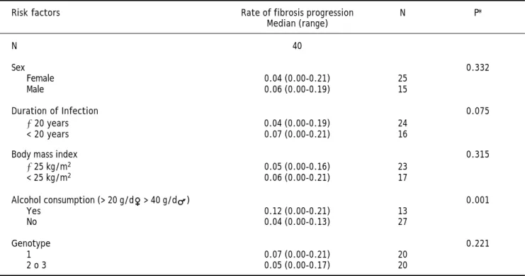
No association between severity of fibrosis and, sex, BMI, present age, age at infection and duration of infection was found (Tables 2, 3 and 4). The same was true when analyzing HLA-DRB1 alleles (data not shown). As regards the viral genotype distribution, the initial statistical difference seen in the univariate analysis became insignificant when adjusting for al-cohol consumption (Table 3). Analysis by employing the exact logistic regression showed that alcohol was the only factor being significantly associated with fibrosis [OR = 13.38; 95% CI 2.38-105.64; intercept estimation -1.4816 ± 0.494 (SE), p = 0.0015; regres-sion coefficient 2.5942 ± 0.8027, 0 = 0.0012]. The median rate of fibrosis progression was signifi-cantly higher in patients with alcohol consumption (0.12 units of fibrosis/year) respect to patients without it (0.04, p = 0.001) (Table 4).
Table 3. Presence of risk factors for fibrosis progression according to histological stage.
Risk factors All biopsied patients Low stage (0-1) Intermediate/ P* High stage (2-3-4)
N 40 25 15
Sex 0.109
Female 25 18 7
Male 15 7 8
Duration of infection 0.999
≥ 20 years 24 15 9
< 20 years 16 10 6
Body mass index 0.083
≥ 25 kg/m2 23 17 6
< 25 kg/m2 17 8 9
Alcohol consumption (> 20 g/d > 40 g/d ) 0.001
Yes 13 3 10
No 27 22 5
Genotype † 0.022
1 20 9 11
2 o 3 20 16 4
Table 4. Rate of fibrosis progression according to risk factors.
Risk factors Rate of fibrosis progression N P*
Median (range)
N 40
Sex 0.332
Female 0.04 (0.00-0.21) 25
Male 0.06 (0.00-0.19) 15
Duration of Infection 0.075
≥ 20 years 0.04 (0.00-0.19) 24
< 20 years 0.07 (0.00-0.21) 16
Body mass index 0.315
≥ 25 kg/m2 0.05 (0.00-0.16) 23
< 25 kg/m2 0.06 (0.00-0.21) 17
Alcohol consumption (> 20 g/d > 40 g/d ) 0.001
Yes 0.12 (0.00-0.21) 13
No 0.04 (0.00-0.13) 27
Genotype 0.221
1 0.07 (0.00-0.21) 20
2 o 3 0.05 (0.00-0.17) 20
*Mann Whitney U test. Rate of fibrosis progression was calculated as follows: fibrosis stage in METAVIR units/Duration of infection in years.
Mean follow up after the first visit (time between diagnosis and liver biopsy) was 9.9 months, median: 7 months (range: 1-60 months). Most of the subjects were biopsied during the first year of the diagnosis and more than a half received HCV treatment after that.
DISCUSSION
Our findings show a low occurrence of cirrhosis in an asymptomatic community-based population. While our results are in line with previous reports showing that significant alcohol consumption is as-sociated with more severe fibrosis, mild alcohol in-take did not worsen the natural history of HCV infection in this series patients.
The risk of developing cirrhosis is quite variable among studies analyzing the natural history of chro-nic HCV infection.7
Retrospective-prospective studies in women infec-ted at a young age report the lowest rates of cirrho-sis, 1% to 2%,11,12 whereas higher rates, 20% to 50%, were seen in cross-sectional retrospective stu-dies from referral centres.14-19 In the latter studies, individuals attend for better diagnosis and treat-ment, requiring complementary studies and specific therapeutic procedures. Hence, subjects with more severe disease are overrepresented for which fibrosis
progression could be overestimated (referral bias). On the other hand, in community-based studies in nonclinical settings, asymptomatic subjects with mild disease (too “healthy” to be recognized in for-mer designs) are overrepresented. It follows that some grade of ascertainment bias (systematic failure to represent equally all classes of subjects supposed to be represented in a sample) is almost unavoidable due to the heterogeneity of the population included in different studies with quite variable rates of fibro-sis progression. A global estimation of progression to cirrhosis is very difficult given the wide variation of studies including subjects with quite different ra-tes of fibrosis progression.
into consideration subjects “too healthy” to be cognized in other designs (e.g. cross-sectional re-trospective liver clinic studies) and whose evolution is also part of the wide variable scenario of the na-tural history of HCV infection.
Our sample derives from a large population of healthy persons that voluntarily attended to a detec-tion campaign, composed of well-diagnosed asympto-matic individuals, unlikely to be included in cross-sectional liver clinic studies. Furthermore, people included in our study ignored that had been infected with HCV for many years until the time they where included. This is particularly important regarding alcohol consumption, keeping in mind that in other large retrospective-prospective cohorts including young women,11,12 alcohol abstinence was advised and probably the natural history of the di-sease could have been modified with this sole action. It is worth commenting that present study deals with a retrospective-prospective cohort where sub-jects with a recognized source of infection in the past were followed-up prospectively. Previous re-trospective-prospective studies report a lower preva-lence of cirrhosis than retrospective ones.10-12,20-24
In our cohort, the follow up after the first visit (time between diagnosis and liver biopsy) was short: mean: 9.9 months, median: 7 months (range: 1-60 months). Many patients received treatment after li-ver biopsy was performed. Hence, although the eva-luation and staging of all the patients were prospective, the prospective follow-up was short compared with the retrospective follow-up, for ethi-cal reasons.
The patients were categorized in two groups ac-cording to stage of fibrosis, the decision to consider F2 Stage as the limit to intermediate/high stage was based on the fact that the presence of fibrous septa in a liver biopsy is a marker of progressive fibrosis. It is also well known, that persons with no or mini-mal fibrosis (METAVIR stage 0-1) have a low risk for liver-related complications and liver-related dea-th, over the next 10 to 20 years.25 Although only 65% of the subjects could be biopsied, we consider that the histological findings represent the entire se-ries, as no significant difference was found between biopsied and non biopsied patients (Table 1).
For transfusion-acquired infection, the date of transfusion is conventionally used as the time of HCV infection. This allows calculating the rate of fi-brosis progression (Fifi-brosis Stage in METAVIR units/Duration of infection). In previous studies the estimated fibrosis progression calculated indirectly according the date of infection and stage of fibrosis
in a single biopsy was similar to the estimated ob-served in paired biopsy samples.1,26
This approach assumed that progression rates are constant and linear across fibrosis stages. However it has been strongly suggested that fibrosis progres-sion is non linear and that the rate of fibrosis varies between stages,7,27 and accelerates with patient age-ing. For these reasons, using fibrosis progression ratio for estimating de velocity of fibrosis has clear limitations; but it is useful when comparing with another series.
Our sample was a homogeneous population of transfused people, of whom 60% had minimal or no fibrosis after a reasonably long follow-up (median 21.5 years). The median global rate of fibrosis pro-gression was 0.0520 units of fibrosis/year, the half of published in another series,1,28 and coincident with the low incidence of cirrhosis (2.5%), although the occurrence of severe fibrosis (F3) was 20%. We considered that liver biopsy was enough to evaluate disease severity in biopsied patients. Nevertheless other lab/clinical data which may reflect disease se-verity might have been useful in subjects without li-ver biopsy but could not be assessed in them.
Age, age at infection, duration of infection and sex are recognized factors associated with fibrosis progression but no relationship was found in our sample between these factors and fibrosis. Previous studies showed that age of infection was the main risk factor for fibrosis even after controlling for du-ration of infection.1,28 In our series median age at in-fection was 27 years (0-45); only two patients where over 40 years, approximately the age of infection at which the fibrosis progression begin to increase.1 The fact that most of our subjects where less than 40 years at the time of infection, might have acted as a “protective” factor rendering the other factors, i.e., sex and duration of infection unable to reach a statistically significant association with more rapid fibrosis. Also, another study of the same group showed that fibrosis progression begins to accelera-te at 50 years of age independently of duration of in-fection;29 in our series median present age was 52.5 years. Hence, the low fibrosis progression in our study may have several explanations: young age at infection and young present age, along with the fact that patients derived from an asymptomatic “heal-thy” population like subjects included in nonclinical/ community settings.
small sample size. In line with the demonstration of an inconsistent association between a particular HLA DRB1 allele and fibrosis,31,32 presence of fibro-sis was not related with a distinct frequency of some HLA DRB1 gene.
In parallel, increased BMI was not associated to fibrosis. Nevertheless, the relative contribution of obesity cannot be fully discarded in light of its well-known coexistence with insulin resistance, steato-sis, and steatohepatitis, factors that were not assessed in the present study.33
Cumulated evidence has established that alcohol consumption has a deleterious effect on hepatitis C severity and our results are in line with this evidence as significant alcohol consumption (> 20 g/d -> 40 g/d ) was strongly associated with fibrosis progression. Several studies have shown that alco-hol intake with levels of consumption greater than 50 g/day is associated with an increased rate of fi-brosis progression.34,35,37 Nevertheless, no agree-ment exists about the potentially dangerous threshold, for which the impact of lower levels of al-cohol consumption remains unclear.34,37-40
In the United States, the National Institute on Alcohol Abuse and Alcoholism (NIAAA) has esta-blished age- and sex-specific recommended con-sumption thresholds.41 We consider that a different range for men and women was appropriate and a cut off of > 20 g/d for women and > 40g/d for men was simply, reliable and near the NIAAA recom-mendations. In our study, abstainers (n: 17) and mild drinkers (n: 10) where analyzed together. More stratification into different grades of alcohol consumption could not be done due to the small sample size. In larger studies it would be very use-ful to assess differences among abstainers, mild, moderate and heavy drinkers. Our data seem to su-pport the hypothesis that mild consumption would not be associated with more rapid fibrosis in HCV infected patients.36,40
Features of present study provide a different and valuable look within the quite variable scenario on natural history of hepatitis C. On one hand, cohort studies in post-transfusional cases included patients with a history of post-transfusion hepatitis, most of them after cardiac surgery or haematological disor-ders.10,42-45 In our study, instead, patients were apparently healthy, asymptomatic subjects who at-tended a screening campaign. On the other hand our patients were not individuals referred for further as-sessment of their HCV infection status, conversely, they attended to a referral centre without evidence, until then, of their liver disease. These facts partly
explain that the percentage of progression to cirrhosis of 2.5% in 21 years resembles the figures reported, in community-based and blood donor studies;5 and in retrospective-prospective as well as nonclinical studies.7
In line with other reports, our study points out that the natural history of many patients with HCV infections seems to be benign and non progressive for a long time. As above stated many studies sug-gest that fibrosis begins to accelerate with patient ageing, hence, a longer follow up might demonstrate changes in this otherwise benign course. This is su-pported by the fact that while the occurrence of cirr-hosis in our series was low, presence of severe fibrosis was noticeable (20%).
Given the low rate of progression observed in po-pulation studies, young patients detected in this set-ting, with minimal or no alcohol consumption could be assessed with non-invasive techniques (e.g. tran-sient elastography). Treatment could be delayed wai-ting for better options, in those without significant fibrosis according with predictors of treatment res-ponse like viral genotype and IL28B haplotype.
Although large studies are needed, it seems that occasional or mild alcohol consumers behave similar to abstainers in terms of fibrosis progression. A “safe” threshold for alcohol consumption in HCV in-fected patients is quite difficult to define, however, as others authors have suggested mild alcohol con-sumption is not associated with increased fibro-sis36,40 and physicians must keep these data in mind when counselling these patients.
ACKNOWLEDGEMENTS AND DISCLOSURES
The authors declared that they not have anything to disclose regarding funding or conflict of interest with respect to this manuscript.
ABBREVIATIONS
• HCV: hepatitis C virus.
• HLA: human leukocyte antigen.
• Anti-HCV: hepatitis C virus antibodies.
• RFLP: restriction fragment length polymorphism. • HBsAg: hepatitis B surface antigen.
• anti-HBc: hepatitis B core antigen antibodies. • HIV: human immunodeficiency virus.
REFERENCES
1. Poynard T, Bedossa P, Opolon P. Natural history of liver fi-brosis progression in patients with chronic hepatitis C.
Lancet 1997; 349: 825-32.
2. Seeff LB, Hoofnagle JH. Appendix: The National Institutes of Health Consensus Development Conference Manage-ment of Hepatitis C 2002. Clin Liver Dis 2003; 7: 261-87. 3. Prieto M, Berenguer M, Rimola A, Loinaz C, Barrios C,
Cle-mente G, et al. Liver transplantation in hepatitis C. A Spa-nish multicentre experience. Eur J Gastroenterol Hepatol
1998; 10: 771-5.
4. Davila JA, Morgan RO, Shaib Y, McGlynn KA, El-Serag HB. Hepatitis C infection and the increasing incidence of he-patocellular carcinoma: a population-based study. Gas-troenterology 2004; 127: 1372-80.
5. Freeman AJ, Dore GJ, Law MG, Thorpe M, Von Overbeck J, Lloyd AR, Marinos G, et al. Estimating progression to cirr-hosis in chronic hepatitis C virus infection. Hepatology
2001; 34: 809-16.
6. Missiha SB, Ostrowski M, Heathcote EJ. Disease progres-sion in chronic hepatitis C: modifiable and nonmodifiable factors. Gastroenterology 2008; 134: 1699-714.
7. Thein HH, Yi Q, Dore GJ, Krahn MD. Estimation of stage-specific fibrosis progression rates in chronic hepatitis C virus infection: a meta-analysis and meta-regression. He-patology 2008; 48: 418-31.
8. Fu B, Tom BD, Delahooke T, Alexander GJ, Bird SM. Event-biased referral can distort estimation of hepatitis C virus progression rate to cirrhosis, and of prognostic influen-ces. J Clin Epidemiol 2007; 60: 1140-8.
9. Seeff LB. Natural history of chronic hepatitis C. Hepatolo-gy 2002; 36: S35-S46.
10. Seeff LB, Hollinger FB, Alter HJ, Wright EC, Cain CM, Bus-kell ZJ, et al. Long-term mortality and morbidity of trans-fusion-associated non-A, non-B and type C hepatitis; a National Heart, Lung and Blood Institute collaborative stu-dy. Hepatology 2001; 33: 455-63.
11. Wiese M, Grüngreiff K, Güthoff W, Lafrenz M, Oesen U, Porst H; East German Hepatitis C Study Group. Outcome in a hepatitis C (genotype 1b) single source outbreak in Germany-a 25-year multicenter study. J Hepatol 2005; 43: 590-8.
12. Kenny-Walsh E for the Irish Hepatology Research Group. Clinical outcomes after hepatitis C infection from contami-nated anti-D immune globulin. N Eng J Med 1999; 340: 1228-33.
13. Bedossa P, Poynard T. An algorithm for the grading of ac-tivity in chronic hepatitis C. The METAVIR Cooperative Study Group. Hepatology 1996; 24: 289-93.
14. Kiyosawa K, Sodeyama T, Tanaka E, Gibo Y, Yoshizawa K, Nakano Y, et al. Interrelationship of blood transfusion, NANB hepatitis and hepatocellular carcinoma: analysis by detection of antibody to hepatitis C. Hepatology 1990; 12: 671-5.
15. Tong MJ, el-Farra NS, Reikes AR, Co RL, et al. Clinical out-comes after transfusion-associated hepatitis C. N Engl J Med 1995; 332: 1463-6.
16. Niederau C, Lange S, Heintges T, Erhardt A, Buschkamp M, Hürter D, et al. Prognosis of chronic hepatitis C: results of a large, prospective cohort study. Hepatology 1998; 28: 1687-95.
17. Gordon SC, Elloway RS, Long JC, Dmuchowski CF. The pa-thology of hepatitis C as a function of mode of transmis-sion: blood transfusion versus intravenous drug abuse.
Hepatology 1993; 18: 1338-43.
18. Ferenci P, Ferenci S, Datz C, Rezman I, Oberaigner W, Strauss R. Morbidity and mortality in paid Austrian plasma donors infected with hepatitis C at plasma donation in the 1970s. J Hepatol 2007; 47: 31-6.
19. Forns X, Ampurdanès S, Sanchez-Tapias JM, Guilera M, Sans M, Sánchez-Fueyo A, et al. Long-term follow-up of chronic hepatitis C in patients diagnosed at a tertiary-care center. J Hepato 2001; 35: 265-71.
20. Vogt M, Lang T, Frösner G, Klingler C, Sendl AF, Zeller A, et al. Prevalence and clinical outcomes of hepatitis C infec-tion in children who underwent cardiac surgery before the implementation of blooddonor screening. N Engl J Med
1999; 341: 866-70.
21. Thomas DL, Astemborski J, Rai RM, Anania FA, Schaeffer M, Galai N, et al. The natural history of hepatitis C virus in-fection: host, viral, and environmental factors. JAMA
2000; 284: 450-6.
22. Casiraghi MA, De Paschale M, Romanò L, Biffi R, Assi A, Bi-nelli G, Zanetti AR. Long-term outcome (35 years) of hepa-titis C after acquisition of infection through mini transfusions of blood given at birth. Hepatology 2004; 39: 90-6.
23. Rodger AJ, Roberts S, Lanigan A, Bowden S, Brown T, Crofts N. Assessment of long-term outcomes of communi-ty-acquired hepatitis C infection in a cohort with sera stored from 1971–1975. Hepatology 2000; 32: 582-7. 24. Seeff LB, Miller RN, Rabkin CS, Buskell-Bales Z,
Straley-Ea-son KD, Smoak BL, et al. 45-year follow-up of hepatitis C virus infection in healthy young adults. Ann Intern Med
2000; 132: 105-11.
25. National Institutes of Health Consensus Development Con-ference Statement: Management of hepatitis C: 2002-June 10-12, 2002. Hepatology 2002; 36: S3-S20.
26. Sobesky R, Mathurin P, Charlotte F, Meussalli J, Olivi M, Vi-duud M, et al. Modeling the impact of interferon alfa treat-ment on liver fibrosis progression in chronic hepatitis C: a dynamic view. Gastroenterology 1999; 116: 378-86. 27. Yi Q, Wang PP, Krahn M. Improving the accuracy of
long-term prognostic estimates in hepatitis C virus infection. J Viral Hepat 2004; 11: 166-74.
28. Pradat P, Voirin N, Tillmann HL, Chevallier M, Trépo C. Progression to cirrhosis in hepatitis C patients: an age-dependent process. Liver Int 2007; 27: 335-9.
29. Poynard T, Ratziu V, Charlotte F, Goodman Z, McHutchi-son J, Albrecht J. Rates and risk factors of liver fibrosis progression in patients with chronic hepatitis C. J Hepa-tol 2001; 34: 730-9.
30. Bochud PY, Cai T, Overbeck K, Bochud M, Dufour JF, Müll-haupt B, et al. Genotype 3 is associated with accelerated fibrosis progression in chronic hepatitis C. J Hepatol
2009; 51: 655-66.
31. Tillmann HL, Chen DF, Trautwein C, Kliem V, Grundey A, Berning-Haag A, et al. Low frequency of HLA-DRB1*11 in hepatitis C virus induced end stage liver disease. Gut
2001; 48: 714-8.
32. Thursz M, Yallop R, Goldin R, Trepo C, Thomas HC. Influen-ce of MHC class II genotype on outcome of infection with hepatitis C virus. Lancet 1999; 354: 2119-24.
33. Monto A, Alonzo J, Watson JJ, Grunfeld C, Wright TL. Steatosis in chronic hepatitis C: relative contributions of obesity, diabetes mellitus, and alcohol. Hepatology 2002; 36: 729-36.
34. Peters MG, Terrault NA. Alcohol use and hepatitis C. Hepa-tology 2002; 36: S220-S225.
36. Monto A, Patel K, Bostrom A, Pianko S, Pockros P, McHut-chison JG, Wright TL. Risks of a range of alcohol intake on hepatitis C-related fibrosis. Hepatology 2004; 39: 826-34. 37. Ostapowicz G, Watson KJ, Locarnini SA, Desmond PV. Role
of alcohol in the progression of liver disease caused by he-patitis C virus. Hepatology 1998; 27: 1730-5.
38. Hutchison SJ, Bird SM, Goldberg DJ. Influence of alcohol on the progression of hepatitis C virus infection: a meta-analysis. Clin Gastroenterol Hepatol 2005; 3: 1150-9. 39. Westin J, Lagging LM, Spak F, Aires N, Svensson E, Lindh
M, et al. Moderate alcohol intake increases fibrosis pro-gression in untreated patients with hepatitis C virus in-fection. J Viral Hepat 2002; 9: 235-41.
40. Cheung O, Sterling RK, Salvatori J, Williams K, Hubbard S, Luketic VA, et al. Mild alcohol consumption is not associa-ted with increased fibrosis in patients with chronic hepa-titis C. J Clin Gastroenterol 2011; 45: 76-82.
41. The Physician’s Guide to Helping Patients with Alcohol Pro-blems. Rockville, MD: National Institute on Alcohol Abuse and Alcoholism (NIAAA); 1995. NIH publication no. 95-3769. 42. Tremolada F, Casarin C, Alberti A, Drago C, Tagger A, Ribe-ro ML, Realdi G. Long-term follow-up of non-A, non-B (type C) post-transfusion hepatitis. J Hepatol 1992; 16: 273-81. 43. Gruber A, Norder H, Magnius L, Rotzen M, Rubio C,
Grill-ner L, Bjorkholm M. Late seroconversion and high chroni-city rate of hepatitis C virus infection in patients with hematologic disorders. Ann Oncol 1993; 4: 229-34. 44. Di Bisceglie AM, Goodman ZD, Ishak KG, Hoofnagle JH,
Mel-polder JJ, Alter HJ. Long-term clinical and histopathologi-cal follow-up of chronic posttransfusion hepatitis.
Hepatology 1991; 14: 969-74.