Xu D, et al. , 2019; 18 (1): 58-67
58
Performance of Serum Glypican
3 in Diagnosis of Hepatocellular Carcinoma:
A meta-analysis
Dahai Xu,*,** Chang Su,*** Liang Sun,* Yuanyuan Gao,* Youjun Li* * Department of Human Anatomy, College of Basic Medical Sciences, Jilin University, Changchun, Jilin, China. ** Department of Emergency, The First Hospital of Jilin University, Changchun, Jilin, China. *** Department of Thyroid Surgery, The First Hospital of Jilin University, Changchun, Jilin, China.
January-February, Vol. 18 No. 1, 2019: 58-67
ORIGINAL ARTICLE
The Official Journal of the Mexican Association of Hepatology, the Latin-American Association for Study of the Liver and
the Canadian Association for the Study of the Liver
Manuscript received: Manuscript received: Manuscript received: Manuscript received:
Manuscript received: September 08, 2017. Manuscript accepted:Manuscript accepted:Manuscript accepted: December 11, 2017.Manuscript accepted:Manuscript accepted:
DOI:10.5604/01.3001.0012.7863 A B S T R A C T A B S T R A C T A B S T R A C T A B S T R A C T A B S T R A C T
Introduction and aim. Introduction and aim. Introduction and aim. Introduction and aim.
Introduction and aim. Serum glypican-3 (GPC3) has been explored as a non-invasive biomarker of hepatocellular carcinoma (HCC). However, controversy remains on its diagnostic accuracy. Therefore, we aimed to conduct a systematic review and meta-analysis to evaluate the differential diagnostic accuracy of serum GPC3 between HCC and liver cirrhosis (LC) cases. MaterialMaterialMaterialMaterialMaterial and methods.
and methods.and methods. and methods.
and methods. After the strict filtering and screening of studies from NCBI, PUBMED, Clinical Trials, Cochrane library, Embase, Prospero and Web of Science databases, 11 studies were selected. All studies provided the sensitivity and specificity of GPC3 and the alpha-fetoprotein (AFP) in the HCC and LC diagnosis. The sensitivity and specificity, and the area under the receiver operating characteristic curve (AUC) were determined and compared between GPC3 and AFP, which was set as a positive control. Re-Re-Re-Re- Re-sults.
sults.sults. sults.
sults. Pooled sensitivity (95% CI) and specificity (95% CI) were 0.55 (0.52-0.58) and 0.58 (0.54-0.61) for GPC3, 0.54 (0.51-0.57) and 0.83 (0.80-0.85) for AFP, and 0.85 (0.81-0.89) and 0.79 (0.73-0.84) for GPC3 + AFP, respectively. The AUCs of GPC3, AFP and GPC3 + AFP were 0.7793, 0.7867 and 0.9366, respectively. GPC3 had a nearly similar sensitivity as AFP, while the specificity and AUC of GPC3 was lower than that of AFP. The combination of GPC3 and AFP yielded a better sensitivity and AUC than GPC3 or AFP. Conclusion.Conclusion.Conclusion.Conclusion.Conclusion. Serum GPC3 is inferior to AFP in the differential diagnosis between HCC and LC. However, the combination of GPC3 and AFP exhibited a much better performance.
Key words. Key words.Key words. Key words.
Key words. Alpha fetoprotein. Biomarker. Liver cirrhosis. Sensitivity. Specificity.
INTRODUCTION
Hepatocellular carcinoma (HCC) is the fifth most common cancer worldwide, and the second leading cause of site-specific cancer-related death. Usually, HCC is asymptomatic at the early stage. However, it is always de-tected at the advanced stage when diagnosed, which limits treatment options. Thus, the early diagnosis of HCC is of great significance to enable early therapeutic intervention and prolong the survival period.1 In this context, the sero-logical level of alpha-fetoprotein (AFP) has been used as a classical marker of HCC. However, AFP has modest sen-sitivity and specificity for HCC diagnosis,2,3 which makes its application questionable. Indeed, AFP has been exclud-ed from the present guidelines of the American Associa-tion for the Study of Liver Disease and the European Association for the Study of the Liver due to its modest
accuracy.4 Therefore, there is an urgent need to explore a surrogate serological marker with higher sensitivity and specificity for HCC diagnosis.
GPC3 as a noninvasive marker for HCC diagnosis, many investigators have explored this issue in comprehensive settings with respect to the diagnostic efficiency of GPC3, compared to AFP.
Several liver diseases, such as viral hepatitis, autoim-mune hepatitis, alcoholic hepatitis and primary biliary cir-rhosis, have progressed to liver cirrhosis (LC), which is a major predisposing factor of HCC risk. It is noteworthy that although the vast majority of HCC cases developed from cirrhotic livers, not all cirrhotic livers always end with HCC. In previous meta-analysis studies,13-18 no meta-analysis study employed cirrhotic patients as con-trols. Therefore, the novelty of the present systemic re-view and meta-analysis over all previous studies originates from the use of LC cases as the control for the study. Through this approach, we were able to precisely and comprehensively assess the accuracy of GPC3 in the dif-ferential diagnosis between HCC and LC, compared to AFP.
MATERIAL AND METHODS
Literature search strategy
Two independent investigators (Chang Su and Dahai Xu) conducted an electronic literature search on seven da-tabases, which include the NCBI, PUBMED, Clinical Trials, Cochrane library, Embase, Prospero and Web of Science databases. The search was updated as of Novem-ber 10, 2017. The entry terms used for the literature search were as follows:
• HCC. Liver neoplasm, hepatic neoplasm, hepatocellu-lar cancer, hepatic cancer, and liver cancer.
• GPC3. Glypican, glypican 3, glypican3, and glypican-3. • AFP. Alpha-Fetoprotein and alpha Fetoprotein. No
limit was set on publication time, study design and publishing format, and only publications in the Eng-lish language were searched.
Inclusion and exclusion criteria
Inclusion criteria:
• Studies that accurately diagnosed the experimental group with HCC
• Studies that measured serum GPC3 and AFP protein. • Studies that determined the HCC diagnostic
sensitivi-ty and specificisensitivi-ty of GPC3 and AFP. Exclusion criteria:
• Letters, reviews, case reports, abstracts, editorials, and expert opinions.
• Studies that lack sufficient data to obtain the sensitivity and specificity of GPC3 and AFP in HCC and LC. • Studies on experimental models, such as laboratory
an-imals and cultured cells.
• Studies that considered specimens other than blood. • Studies that evaluated serum maker levels by
messen-ger RNA, DNA, or DNA polymorphisms.
• Studies that focused on diseases other than primary hepatocellular carcinoma.
Different articles with the same authors and data were checked to avoid duplicates, and the most recent or most complete study was selected.
Data extraction
After selecting all the eligible studies, two investigators (Liang Sun and Yuanyuan Gao) independently extracted the data. The collected data included the first author’s name, the country of origin of the patients, publication year, name of the journal, study design, number of patients, age and gender, assay type, cut-off value, and raw data (True Positive, TP; False Positive, FP; False Negative, FN; True Negative, TN). For disagreements, a third in-vestigator (Youjun Li) was consulted to make the judg-ment.
Assessment of
quality of the selected articles
Two independent investigators (Chang Su and Dahai Xu) assessed the quality of the included studies according to the Quality Assessment of Studies of Diagnostic Accu-racy II (QUADAS-2).14 Signaling questions on the risk of bias in the assessment check list were labeled as ‘‘yes’’, ‘‘no’’, or ‘‘unclear’’. Items that assessed applicability risk were labeled as ‘‘high’’, ‘‘low’’, or ‘‘unclear’’. If the study design was cross-sectional, the risk of bias of the patient selection domain was labeled as ‘‘high risk’’. A third inves-tigator (Youjun Li) was consulted for disagreements.
Statistical analysis
Xu D, et al. , 2019; 18 (1): 58-67
60
P-value > 0.1 was considered with insignificant heteroge-neity. If the heterogeneity was not identified, the fixed-ef-fects model was used for the meta-analysis, and if the heterogeneity was identified, the random-effects model (DerSimonian-Laird) was used. Publication bias was measured by Egger’s test using Stata 12 (StataCorp LP, College Station, TX, USA). P < 0.05 was considered sta-tistically significant. In order to analyze the source of het-erogeneity, threshold analysis was conducted using Meta-Disc 1.4 and meta-regression was conducted using Stata 12.
RESULTS
Study selection
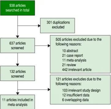
Initially, 938 potentially relevant articles were retrieved from the databases mentioned above. Then, due to dupli-cation, 301 articles were excluded. Next, after reviewing the titles and abstracts, 10 abstracts, 21 case reports, 11 meta-analyses, 21 reviews and 442 irrelevant articles (sub-jects were not human and the specimens were not serum, or GPC3 was determined based of the expression level of its mRNA or DNA) were excluded. Subsequently, by reading the full text, 121 studies were excluded due irrele-vant design or insufficient data to calculate the sensitivity and specificity of GPC3. Finally, a total of 11 studies were eligible8-11,19-25 (Figure 1). All included studies were ap-proved by the Ethics Committee, and an informed con-sent was obtained from all subjects.
Features and methodology
The clinical features and methodology of these eligible studies are summarized in table 1.
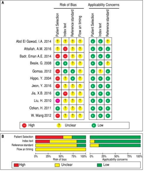
Quality assessment of the studies
QUADAS-2 quality assessment was conducted to eval-uate the quality of the included studies (Figure 2). All studies were retrospectively designed, and none of these studies were randomized controlled trials (RCTs). Mere-ly two studies were consecutive investigations.8,10 Thus, the patient selection domain of other studies was labeled as ‘high risk’. Since there was no fixed diagnostic standard in each study, the flow and timing domain was labeled as ‘unclear’, while most of the studies included the cut-off levels of either serum GPC3 or AFP.8-11,19,20,22,23,25 Two studies21,24 did not include the cutoff value for GPC3, while two studies21,24 did not include the cutoff value for AFP. Notably, the baseline-pretreatment level of serum GPC3 was determined in four studies,8,9,19,24 and the re-maining seven studies did not mention whether the
sam-ple was collected before therapy.10,11,20-23,25 On the other hand, some studies indicated that their thresholds were not pre-specific (the cut-off point was fixed before the test), because their cut-off values were determined based on the ROC analysis.8-10,19 Hence, the index test was set as ‘high risk’. Since the temperature of the serum sample storage is critical for assay accuracy, three different tem-peratures (-20°C, -70°C and -80°C) were consid-ered.8,9,11,19,21,24 Studies with high risk of bias were not excluded to investigate the heterogeneity in the following step.8-10,19,21-23,25,26
Diagnostic accuracy of serum GPC3 and AFP for HCC diagnosis
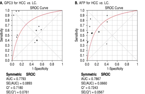
Sensitivity, specificity, LR+, LR-, DOR and I2 were calculated for all the included studies (Table 2 and Figure 3). The sensitivity of GPC3 was almost the same as that of AFP, but the specificity of GPC3 was lower than that of AFP. Moreover, the AUC of GPC3 (0.7793) was lower than that of AFP (0.7867) and GPC3 + AFP (0.9366) (Figure 4).
Investigation for heterogeneity
Based on Egger’s test, the included studies did not have publication bias (Coefficient = 3.00, P = 0.052 for GPC3; Coefficient = 2.56, P = 0.065 for AFP) (Figure 5). Fur-thermore, the diagnostic odds ratio revealed significant
Figure 1. Figure 1.Figure 1.
Figure 1.Figure 1. Study selection process.
505 articles excluded due to the following reasons:
10 abstract 21 case report 11 meta-analysis 21 review
442 irrelevant article 121 articles excludes due to the following reasons:
103 irrelevant study design 12 insufficient data 6 overlapping data
938 articles searched in total
637 articles screened
132 articles screened
11 articles included in meta-analysis
61
, 2019; 18 (1): 58-67
Author Year Country HCC/LC Gender (M/F) Age (year) Etilolgy GPC3 AFP
HCC LC HCC LC HCC LC Control Cut-off Assay Cut-off Assay
value type value type
Jia, X. B. 2016 China 283/267 2.41 5.07 59 ± 9.6 52.9 ± 11.1 HBV 80.6% HBV 80.9% 0.002 ELISA 20 ELISA
ng/mL ng/mL
Jeon, Y. 2016 South 157/156 4.23 1.36 60.8 ± 11.8 56.7 ± 10.8 HBV 67.5%, HBV 74.4%, 0.61 ELISA 20 ELICA
Korea HCV 10.8%, HCV 23.7%, ng/mL ng/mL
ALD 10.8% ALD 1.9%
Attallah, A. M. 2016 Egypt 138/56 NA NA 54.9 ± 12.1 51.89 ± 8.9 HCV 100% HCV 100% 6 ELISA 400
Chemil-ng/mL U/L
umines-ence
Badr, Eman A.E. 2014 Egypt 30/30 5.00 NA NA NA NA HCV 100% 240 ELISA 200 ELISA
μg/mL ng/mL
Abd, El Gawad, I.A. 2014 Egypt 40/10 4.00 4 59 (44-77) 57 (44-72) HBV 17.5%, NA 4.9 ELISA 20 MEIA
HCV 65% ng/mL ng/mL
Gomaa, A H.O. 2012 Egypt 31/30 4.17 3.29 43-65 42-65 HBV 12.9%, HBV 16.7%, 5.41 ELISA 42.32 NA
HCV 87.1% HCV 83.3% ng/mL ng/mL
Wang, W. 2012 China 78/97 1.90 1.6 53.6 (46-66) 50.2 (38-68) HBV 100% HBV 100% NA ELISA NA ELISA
HBV 52%, HBV 29.1%,
Ozkan, H. 2011 Turkey 75/55 2.57 1.5 63 ± 9.9 58 ± 12.7 HCV 22.7%, HCV 16.4%, 3.9 NA 13
Chemil-ALD 2.7% ALD 10.9% pg/mL ng/mL
umines-cence
Liu, H. 2010 China 75/32 2.75 3 55.4 ± 9.91 53.3 ± 5.81 HBV 84%, HBV 78.1%, 300 ELISA 400
Chemil-HCV 16% HCV 21.9% ng/mL ng/mL
umines-cence
Beale, G. 2008 England 50/41 4.00 2.15 67.5 ± 12.02 54.3 ± 9.62 ALD 60%, ALD 80.5%, NA ELISA NA NA
NAFLD 40% NAFLD 19.5%
Hippo, Y. 2004 Japan 69/38 NA NA NA NA NA NA NA ELISA NA ELISA
Xu D, et al. , 2019; 18 (1): 58-67
62
heterogeneity (I2 = 85.3%, P < 0.001 for GPC3; I2 = 86.8%, P < 0.001 for AFP). The threshold effect test (Spearman’s correlation coefficient was -0.209, P = 0.537, for GPC3; Spearman’s correlation coefficient was 0.392, P = 0.233, for AFP) indicated that the cut-off point was not the source of heterogeneity. In addition, the meta-regres-sion analysis indicated that publication year, country, sam-ple size, clinical characteristics methodology and the quality of articles were not correlated with heterogeneity. The sensitivity analysis indicated that the pooled estimates were stable and not influenced by a single study (Figure 6).
A subgroup analysis was conducted by publication year, country, methodology, clinical features, and study quality. However, subject size was the only possible source of het-erogeneity found (whether the number of patients in both HCC and LC groups were ³ 40 or not). For GPC3, in the subgroup that had a larger sample size was 66.5%, while the heterogeneity in another subgroup was 77.2%. The present meta-analysis did not include any RCTs. Other covariates were also potential sources of heteroge-neity, such as the differences in operating protocol, enrollment period, pathological grades and tumor burden.
Table 2. Diagnostic values of GPC3 and/or AFP for HCC vs. LC
Serum Sen. (95%CI) Spe. (95%CI) LR+ (95%CI) LR- (95%CI) DOR (95%CI) biomarker
GPC3 0.55 (0.52-0.58) 0.58 (0.54-0.61) 1.69 (1.20-2.39) 0.67 (0.50-0.90) 3.64 (1.74-7.60)
AFP 0.54 (0.51-0.57) 0.83 (0.80-0.85) 4.40 (2.39-8.08) 0.51 (0.42-0.63) 11.83 (5.02-27.88)
GPC3 + AFP 0.85 (0.81-0.89) 0.79 (0.73-0.84) 5.23 (1.75-15.65) 0.18 (0.07-0.47) 31.95 (5.75-177.65)
Sen.: Sensitivity. Spe.: Specificity. LR+: Positive likelihood Ratio. LR-: Negative likelihood Ratio. DOR: Diagnostic odds ratio.
Figure 2. Figure 2. Figure 2. Figure 2.
Figure 2. Quality evaluation of the included studies by QUADAS-2. A.A.A.A. Risk of bias and con-A. cerns graph by QUADAS-2. B.B.B.B. Risk of bias andB. concerns summary by QUADAS-2.
0% 25% 50% 75% 100%
Risk of bias
0% 25% 50% 75% 100%
Applicability concerns Patient Selection
Index text Reference standard Flow an timing
High Unclear Low Patient Selection Index text Reference standard Flow an timing Patient Selection Index text Reference standard Risk of Bias Applicability Concerns
Abd El Gawad, I.A. 2014 ? ? ? ? + + ? Attallah, A.M. 2016 ? – ? ? + + +
Badr, Eman A.E. 2014 – – ? ? + + +
Beale, G. 2008 + + ? ? + + ?
Gomaa. 2012 – + + ? + ? +
Hippo, Y. 2004 – ? + ? + + +
Jeon, Y. 2016 – ? ? ? + + +
Jia, X.B. 2016 + – ? ? + + +
Liu, H. 2010 – ? ? ? + + +
Ozkan, H. 2011 + ? ? ? + + +
W. Wang 2012 – ? ? ? + + + – High ? Unclear + Low A
A A A A
DISCUSSION
In the present study, we found that the sensitivity of GPC3 was nearly the same as that of AFP, while the spe-cificity of GPC3 was lower than that of AFP. However, the combination of GPC3 and AFP can significantly
in-Figure 3. Figure 3.Figure 3.
Figure 3.Figure 3. Sensitivity and specificity of GPC3 and/or AFP for HCC vs. LC.
crease the sensitivity and AUC in the differential diagnosis between HCC and LC. However, the specificity of GPC3+AFP was lower than that of AFP.
Recently, GPC3 has become a focus in HCC studies. Many studies have explored the performance of GPC3 in liver tissues for HCC diagnosis,27-29 and some clinical
tri-0.0 0.2 0.4 0.6 0.8 1 Sensitivity
Sensitivity (95% CI) Sensitivity (95% CI)Sensitivity (95% CI) Sensitivity (95% CI) Sensitivity (95% CI) Jia X. B 2016 0.40 (0.35-0.46) Jeon Y 2016 0.60 (0.52-0.68) Attallah A.M. 2016 0.60 (0.51-0.68) Badr Eman A.E. 2014 1.00 (0.88-1.00) Abd El Gawad I.A. 2014 1.00 (0.91-1.00) Gomaa 2012 0.90 (0.74-0.98) W. Wang 2012 0.36 (0.25-0.48) Ozkan H. 2011 0.61 (0.49-0.72) Liu H. 2010 0.47 (0.35-0.59) Beale G. 2008 0.68 (0.53-0.80) Hippo Y. 2004 0.51 (0.38-0.83)
Pooled sensitivity = 0.55 (0.52 to 0.58) Chi-square = 148.71; df = 10 (p = 0.0000) Inconsistency (I-square) = 93.3% A.
A. A. A.
A. GPC3 for HCC vs. LC.
0.0 0.2 0.4 0.6 0.8 1 Sensitivity
Specificity (95% CI) Specificity (95% CI) Specificity (95% CI) Specificity (95% CI) Specificity (95% CI) Jia X. B 2016 0.51 (0.44-0.57) Jeon Y 2016 0.52 (0.44-0.60) Attallah A.M. 2016 0.52 (0.38-0.65) Badr Eman A.E. 2014 0.93 (0.78-0.99) Abd El Gawad I.A. 2014 0.90 (0.55-1.00) Gomaa 2012 0.97 (0.83-1.00) W. Wang 2012 0.55 (0.44-0.65) Ozkan H. 2011 0.42 (0.29-0.56) Liu H. 2010 0.91 (0.75-0.98) Beale G. 2008 0.46 (0.31-0.63) Hippo Y. 2004 0.89 (0.75-0.97)
Pooled sensitivity = 0.58 (0.54 to 0.61) Chi-square = 101.87; df = 10 (p = 0.0000) Inconsistency (I-square) = 90.2%
0.0 0.2 0.4 0.6 0.8 1 Sensitivity
Sensitivity (95% CI) Sensitivity (95% CI)Sensitivity (95% CI) Sensitivity (95% CI) Sensitivity (95% CI) Jia X. B 2016 0.57 (0.51-0.63) Jeon Y 2016 0.55 (0.47-0.63) Attallah A.M. 2016 0.36 (0.28-0.45) Badr Eman A.E. 2014 0.83 (0.65-0.94) Abd El Gawad I.A. 2014 0.90 (0.76-0.97) Gomaa 2012 0.77 (0.59-0.90) W. Wang 2012 0.33 (0.23-0.45) Ozkan H. 2011 0.68 (0.56-0.78) Liu H. 2010 0.37 (0.26-0.49) Beale G. 2008 0.56 (0.41-0.70) Hippo Y. 2004 0.55 (0.43-0.67)
Pooled sensitivity = 0.54 (0.51 to 0.57) Chi-square = 90.37; df = 10 (p = 0.0000) Inconsistency (I-square) = 89.9% B.
B. B. B.
B. AFP for HCC vs. LC.
0.0 0.2 0.4 0.6 0.8 1 Sensitivity
Specificity (95% CI) Specificity (95% CI) Specificity (95% CI) Specificity (95% CI) Specificity (95% CI) Jia X. B 2016 0.76 (0.71-0.81) Jeon Y 2016 0.91 (0.85-0.95) Attallah A.M. 2016 1.00 (0.94-1.00) Badr Eman A.E. 2014 0.93 (0.78-0.99) Abd El Gawad I.A. 2014 0.60 (0.26-0.88) Gomaa 2012 0.60 (0.41-0.77 W. Wang 2012 0.61 (0.50-0.71) Ozkan H. 2011 0.95 (0.85-0.99) Liu H. 2010 1.00 (0.89-1.00) Beale G. 2008 1.00 (0.91-1.00) Hippo Y. 2004 0.89 (0.75-0.97)
Pooled sensitivity = 0.83 (0.80 to 0.85) Chi-square = 113.29; df = 10 (p = 0.0000) Inconsistency (I-square) = 91.2%
Sensitivity (95% CI) Sensitivity (95% CI)Sensitivity (95% CI) Sensitivity (95% CI) Sensitivity (95% CI) Attallah A.M. 2016 0.93 (0.87-0.96) Badr Eman A.E. 2014 1.00 (0.88-1.00) Gomaa 2012 0.84 (0.66-0.95) Ozkan H. 2011 0.67 (0.55-0.77) Beale G. 2008 0.82 (0.69-0.91)
Pooled sensitivity = 0.85 (0.52 to 0.89) Chi-square = 33.54; df = 4 (p = 0.0000) Inconsistency (I-square) = 88.1% C.
C. C. C.
C. GPC3 + AFP for HCC vs. LC. Specificity (95% CI)Specificity (95% CI)Specificity (95% CI)Specificity (95% CI)Specificity (95% CI) Attallah A.M. 2016 0.91 (0.80-0.97) Badr Eman A.E. 2014 1.00 (0.88-1.00) Gomaa 2012 0.90 (0.73-0.98) Ozkan H. 2011 0.73 (0.59-0.84) Beale G. 2008 0.46 (0.31-0.63)
Pooled sensitivity = 0.79 (0.73 to 0.84) Chi-square = 44.91; df = 4 (p = 0.0000) Inconsistency (I-square) = 91.1% 0.0 0.2 0.4 0.6 0.8 1
Sensitivity
Xu D, et al. , 2019; 18 (1): 58-67 64 Figure 4. Figure 4. Figure 4. Figure 4.
Figure 4. ROC curves of GPC3 and/or AFP for the diagnosis between HCC and LC.
Figure 6. Figure 6. Figure 6. Figure 6.
Figure 6. Sensitivity analysis plots.
A. A. A. A.
A. GPC3 for HCC vs. LC.
0.0 0.2 0.4 0.6 0.8 1 1-Specificity 1.0 0.9 0.8 0.7 0.6 0.5 0.4 0.3 0.2 0.1 0.0 SROC Curve Sensitivity Symmetric SROC Symmetric SROC Symmetric SROC Symmetric SROC Symmetric SROC AUC = 0.7793 SE(AUC) = 0.0893 Q* = 0.7180 SE(Q*) = 0.0761
B. B. B. B.
B. AFP for HCC vs. LC.
0.0 0.2 0.4 0.6 0.8 1 1-Specificity 1.0 0.9 0.8 0.7 0.6 0.5 0.4 0.3 0.2 0.1 0.0 SROC Curve Sensitivity Symmetric SROC Symmetric SROC Symmetric SROC Symmetric SROC Symmetric SROC AUC = 0.7867 SE(AUC) = 0.0660 Q* = 0.7243 SE(Q*) = 0.0567
C. C. C. C.
C. GPC3 + AFP for HCC vs. LC.
0.0 0.2 0.4 0.6 0.8 1 1-Specificity 1.0 0.9 0.8 0.7 0.6 0.5 0.4 0.3 0.2 0.1 0.0 SROC Curve Sensitivity Symmetric SROC Symmetric SROC Symmetric SROC Symmetric SROC Symmetric SROC AUC = 0.9366 SE(AUC) = 0.0522 Q* = 0.8732 SE(Q*) = 0.0648
Figure 5. Figure 5. Figure 5. Figure 5.
Figure 5. Egger’s publication bias plot.
0 5 10 15
Precision 6 4 2 0 -2 Standardized effect A. A. A. A.
A. GPC3 for HCC vs. LC.
Egger’s publication bias plot
0 2 4 6
Precision 10 5 0 Standardized effect B. B.B.
B.B. AFP for HCC vs. LC.
Egger’s publication bios plot
Jia X.B. Jeon Y. Attallah, A.M. Bard, Eman, A.E. Abd El Gawad, I.A. Gomaa W. Wang Ozkan, H. Liu, H. Beale, G. Hippo, Y.
Meta-analysis estimates, given named study is omitted
1.31 2.74 3.64 7.60 11.31 A.
A. A. A.
A. GPC3 for HCC vs. LC.
Jia X.B. (2016) Jeon Y. (2016) Attallah, A.M. (2016) Bard, Eman, A.E. (2014) Abd El Gawad, I.A. (2014) Gomaa, A.H.O. (2012) Wang W. (2012) Ozkan, H. (2011) Liu, H. (2010) Beale, G. (2008) Hippo, Y. (2004)
Meta-analysis estimates, given named study is omitted
4.16 5.02 11.83 27.88 46.05
B. B.B.
B.B. AFP for HCC vs. LC.
als on therapeutic agents against GPC3 have also been con-ducted.30-33 In the present investigation, we conducted a meta-analysis on studies that investigated the application of GPC3 as a diagnostic marker for HCC, and compared this with AFP. The novelty of this investigation originates from the use of LC patients as control subjects, which were compared to HCC. For the 11 eligible studies, the results infer that the performance of GPC3 to discrimi-nate HCC from LC was unsatisfactory and lower than that of AFP. However, the combination of GPC3 and AFP produced a higher diagnostic accuracy than either of the makers when used separately.
GPC3 is implicated in cell growth, differentiation and migration.34 GPC3 is greatly expressed in HCC tissues, fetal livers and most HCC cell lines, compared to other normal human tissues.8,20,35 GPC3 has also been reported in other tumors, such as lung cancer, thyroid cancer and melanoma.36,37 Notably, serum GPC3 levels were signifi-cantly higher in HCC than in normal controls, as well as in liver cirrhosis, colorectal cancer, esophageal cancer, gastric cancer and hepatitis cases.2,19,20 Furthermore, GPC3 expression in HCC was positively associated with tumor size and pathological grade.3 In addition, Ohno, et
al. reported that HCC patients with high GPC3 level had poor prognosis.38-40 Badr, et al. and Yeon, et al. found that the sensitivity of serum GPC3 in small size HCC was higher than that of AFP.19,41 Moreover, Zhang QY, et al. reported that GPC3 is superior to AFP for HCC diagno-sis, with higher sensitivity and specificity.42 Generally, controversy remains on the application of GPC3 in HCC diagnosis, since several studies have either supported2,3,19,42,43 or limited9,11,44,45 the role of GCP3 in HCC diagnosis.
The results of the present meta-analysis support the ob-servation of Jia, et al.,8 since there was no significant differ-ence in the serum level of GPC3 between HCC and LC patients, indicating that GPC3 is not efficiently valid as a HCC serum biomarker.
It is important to note that the sensitivity, specificity and AUC of GPC3 determined in the present meta-analy-sis differed from those reported in other studies.13-18 This difference might be attributed to either the difference in the nature of the control cohort or the number of includ-ed studies.
To the best of our knowledge, the present meta-analysis is the first to set patients with LC as a control cohort. How-ever, there were some limitations in the present study:
• A small number of included studies was included, which was attributed to the strict inclusion and exclu-sion criteria.
• There was an obvious heterogeneity, which might be contributed to the limited number of subjects.
• The quality of the included studies was unsatisfactory. Due to the absence of RCTs, the treatment effects may be overestimated.
Therefore, more high quality researches with different subgroups according to tumor etiology, tumor size and tu-mor stage are needed in the future to further verify the role of GPC3 in HCC diagnosis.
In conclusion, Serum GPC3 cannot be used as a substi-tute for AFP to differentially diagnose HCC and LC. However, the combination of both markers would be a better choice.
ACKNOWLEDGMENTS
This work was supported by the National Natural Sci-ence Foundation of China (#81372456) YJL.
CONFLICT OF INTEREST
The authors declares that there is no conflict of interest regarding the publication of this article.
REFERENCES
1. Maluccio M, Covey A. Recent progress in understanding, di-agnosing, and treating hepatocellular carcinoma. CA: a Can-cer Journal for Clinicians 2012; 62: 394-9.
2. Zhao YS, Wang M, Cui CY, Zhang L, Liao F, Li HC, Wu X. Significance of combined tests of serum golgi glycoprotein 73 and other biomarkers in diagnosis of small primary hepa-tocellular carcinoma. Cancer Biomark 2015; 15: 677-83. 3. Yu JP, Xu XG, Ma RJ, Qin SN, Wang CR, Wang XB, Li M, et
al. Development of a Clinical Chemiluminescent Immunoassay for Serum GPC3 and Simultaneous Measurements Alone With AFP and CK19 in Diagnosis of Hepatocellular Carcino-ma. J Clin Lab Anal 2015; 29: 85-93.
4. Song DS, Bae SH. Changes of guidelines diagnosing hepa-tocellular carcinoma during the last ten-year period. Clin Molec Hepatol 2012; 18: 258-67.
5. Jiang ZW, Jiang XF, Chen SM, Lai YX, Wei XR, Li BH, Lin S, et al. Anti-GPEC3-CAR T Cells Suppress the Growth of Tu-mor Cells in Patient-Derived Xenogratfts of Hepatocelluar Carcinoma. Front Immunol 2017; 7: 690
6. Zhu XT, Yuan JH, Zhu TT, Li YY, Cheng XY. Long noncod-ing RNA glypican 3 (GPC3) antisense transcript 1 promotes hepatocellular carcinoma progression via epigenetically acti-vating GPC3. Febs Journal 2016; 283: 3739-54.
7. Sun B, Huang Z, Wang B, Yu YL, Lin SH, Luo L, Wang Y, et al. Significance of Glypican-3 (GPC3) Expression in Hepatocellular Cancer Diagnosis. Med Sci Monit 2017; 23: 850-5.
8. Jia XB, Gao YT, Zhai DK, Liu J, Cai JJ, Wang YJ, Jing L, et al. Assessment of the Clinical Utility of Glypican 3 as a Serum Marker for the Diagnosis of Hepatocellular Carcinoma. Tech-nol Cancer Res Treat 2016; 15: 780-6.
Xu D, et al. , 2019; 18 (1): 58-67
66
10. Attallah AM, El-Far M, Omran MM, Abdelrazek MA, Attallah AA, Saeed AM, Farid K, et al. GPC-HCC model: a combina-tion of glybican-3 with other routine parameters improves the diagnostic efficacy in hepatocellular carcinoma. Tumour biology: the Journal of the International Society for On-codevelopmental Biology and Medicine 2016; 37: 12571-7. 11. Ozkan H, Erdal H, Kocak E, Tutkak H, Karaeren Z, Yakut M,
Köklü S. Diagnostic and Prognostic Role of Serum Glypican 3 in Patients With Hepatocellular Carcinoma. J Clin Lab Anal 2011; 25: 350-3.
12. Tangkijvanich P, Chanmee T, Komtong S, Mahachai V, Wise-dopas N, Pothacharoen P, Kongtawelert P. Diagnostic role of serum glypican-3 in differentiating hepatocellular carcinoma from non-malignant chronic liver disease and other liver can-cers. J Gastroenterol Hepatol 2010; 25: 129-37.
13. Yang SL, Fang X, Huang ZZ, Liu XJ, Xiong ZF, Liu P, Yao HY, et al. Can serum glypican-3 be a biomarker for effective diagnosis of hepatocellular carcinoma? A meta-analysis of the literature. Dis Markers 2014; 2014: 127831.
14. Jia X, Liu J, Gao Y, Huang Y, Du Z. Diagnosis accuracy of serum glypican-3 in patients with hepatocellular carcinoma: a systematic review with meta-analysis. Arch Med Res 2014; 45: 580-8.
15. Xu C, Yan Z, Zhou L, Wang Y. A comparison of glypican-3 with alpha-fetoprotein as a serum marker for hepatocellular carcinoma: a meta-analysis. J Cancer Res Clin Oncol 2013; 139: 1417-24.
16. Liu JW, Zuo XL, Wang S. Diagnosis accuracy of serum Gly-pican-3 level in patients with hepatocellular carcinoma and liver cirrhosis: a meta-analysis. Eur Rev Med Pharmacol Sci 2015; 19: 3655-73.
17. Liu XF, Hu ZD, Liu XC, Cao Y, Ding CM, Hu CJ. Diagnostic ac-curacy of serum glypican-3 for hepatocellular carcinoma: a systematic review and meta-analysis. Clin Biochem 2014; 47: 196-200.
18. Fu WY, Lu HY, Li L, Wu KF, Li YP, Liu Y, Hu J, et al. Glypi-can-3 versus alpha-fetoprotein as a biomarker for hepato-cellular carcinoma: A diagnostic meta-analysis. Biocell 2015; 39: 25-31.
19. Badr EAE, Korah TE, Ghani AA, El-Sayed S, Badr S. Role of serum glypican-3 in the diagnosis and differentiation of small hepatocellular carcinoma from hepatitis-C virus cirrhosis. Al-exandria J Med 2014; 50: 221-6.
20. Abd El Gawad IA, Mossallam GI, Radwan NH, Elzawahry HM, Elhifnawy NM. Comparing prothrombin induced by vita-min K absence-II (PIVKA-II) with the oncofetal proteins glypi-can-3, Alpha feto protein and carcinoembryonic antigen in diagnosing hepatocellular carcinoma among Egyptian pa-tients. J Egypt Natl Canc Inst 2014; 26: 79-85.
21. Wang W, Zhao LJ, Wang Y, Tao QY, Feitelson MA, Zhao P, Ren H, et al. Application of HBx-induced anti-URGs as early-warning biomarker of cirrhosis and HCC. Cancer Biomark 2012; 11: 29-39.
22. Gomaa AHO, Aboraia G, Attia H, Elezawy H, Nafie E, Elbaz S. The diagnostic value of peripheral blood glypican-3 in pa-tients with hepatocellular carcinoma. World J Med Sci 2012; 7: 105-12.
23. Liu H, Li P, Zhai Y, Qu CF, Zhang LJ, Tan YF, Li N, et al. Di-agnostic value of glypican-3 in serum and liver for primary hepatocellular carcinoma. World J Gastroenterol 2010; 16: 4410-5.
24. Beale G, Chattopadhyay D, Gray J, Stewart S, Hudson M, Day C, Trerotoli P, et al. AFP, PIVKAII, GP3, SCCA-1 and folli-satin as surveillance biomarkers for hepatocellular cancer in non-alcoholic and alcoholic fatty liver disease. Bmc Cancer 2008; 8.
25. Hippo Y, Watanabe K, Watanabe A, Midorikawa Y, Yamamo-to S, Ihara S, Tokita S, et al. Identification of soluble NH2-ter-minal fragment of glypican-3 as a serological marker for early-stage hepatocellular carcinoma. Cancer Res 2004; 64: 2418-23.
26. Yu J, Ma Q, Zhang B, Ma R, Xu X, Li M, Xu W, et al. Clinical application of specific antibody against glypican-3 for hepa-tocellular carcinoma diagnosis. Science China Life Sciences 2013; 56: 234-9.
27. Zhou R, Pan QJ, Wu K, Li R. Distribution and characterization of glypican-3 in hepatocelluar carcinoma. Int J Clin Exp Pathol 2016; 9: 8631-3.
28. Sang W, Miao N, Cui WL, Li XX, Abulajiang G, Zhang W, Li Q, et al. Value of the combined detection arginase-1 and gly-pican-3 expression in distinguishing hepatocellular carcino-ma from metastatic tumors. Int J Clin Exp Pathol 2016; 9: 7748-54.
29. Nguyen TB, Roncalli M, Di Tommaso L, Kakar S. Combined use of heat-shock protein 70 and glutamine synthetase is useful in the distinction of typical hepatocellular adenoma from atypical hepatocellular neoplasms and well-differen-tiated hepatocellular carcinoma. Mod Pathol 2016; 29: 283-92.
30. Abou-Alfa GK, Yen CJ, Hsu CH, O’Donoghue J, Beylergil V, Ruan S, Pandit-Taskar N, et al. Phase Ib study of codrituzu-mab in combination with sorafenib in patients with non-cura-ble advanced hepatocellular carcinoma (HCC). Cancer Chemother Pharmacol 2017; 79: 421-9.
31. Abou-Alfa GK, Puig O, Daniele B, Kudo M, Merle P, Park JW, Ross P, et al. Randomized phase II placebo controlled study of codrituzumab in previously treated patients with advanced hepatocellular carcinoma. J Hepatol 2016; 65: 289-95. 32. Ikeda M, Ohkawa S, Okusaka T, Mitsunaga S, Kobayashi S,
Morizane C, Suzuki I, et al. Japanese phase I study of GC33, a humanized antibody against glypican-3 for ad-vanced hepatocellular carcinoma. Cancer science 2014; 105: 455-62.
33. Sawada Y, Yoshikawa T, Nobuoka D, Shirakawa H, Kuron-uma T, Motomura Y, Mizuno S, et al. Phase I trial of a glypi-can-3-derived peptide vaccine for advanced hepatocellular carcinoma: immunologic evidence and potential for improving overall survival. Clinical Cancer Research: an Official Jour-nal of the American Association for Cancer Research 2012; 18: 3686-96.
34. Filmus J, Selleck SB. Glypicans: proteoglycans with a sur-prise. J Clin Invest 2001; 108: 497-501.
35. Chen IP, Ariizumi S, Nakano M, Yamamoto M. Positive glypi-can-3 expression in early hepatocellular carcinoma predicts recurrence after hepatectomy. J Gastroenterol 2014; 49: 117-25.
36. Chen M, Li GH, Yan J, Lu XZ, Cui JW, Ni ZX, Cheng W, et al. Reevaluation of glypican-3 as a serological marker for hepa-tocellular carcinoma. Clin Chim Act 2013; 423: 105-11. 37. Ikuta Y, Nakatsura T, Kageshita T, Fukushima S, Ito S,
Waka-matsu K, Baba H, et al. Highly sensitive detection of melano-ma at an early stage based on the increased serum secreted protein acidic and rich in cysteine and glypican-3 levels. Clin Cancer Res 2005; 11: 8079-88.
38. Ohno A, Yorita K, Haruyama Y, Kondo K, Kato A, Ohtomo T, Kawaguchi M, et al. Aberrant expression of monocarboxy-late transporter 4 in tumour cells predicts an unfavourable outcome in patients with hepatocellular carcinoma Akinobu Ohno. Liver Int 2014; 34: 942-52.
40. Li J, Gao JZ, Du JL, Wei LX. Prognostic and clinicopathologi-cal significance of glypican-3 overexpression in hepatocel-lular carcinoma: A meta-analysis. World J Gastroenterol 2014; 20: 6336-44.
41. Yeon JE, Suh SJ, Lee SJ, Yoon EL, Kang K, Yoo YJ, Lee HJ, et al. Clinical utility of plasma glypican-3 and osteopontin as biomarkers of hepatocellular carcinoma. Gut and Liver 2014; 8: 177-85.
42. Zhang QY, Xiao Q, Lin Z, Ying XT, Li ZJ, Lin JM. Develop-ment of a competitive radioimmunoassay for glypican-3 and the clinical application in diagnosis of hepatocellular carcino-ma. Clinical Biochemistry 2010; 43: 1003-8.
43. Nassar A, Cohen C, Siddiqui MT. Utility of glypican-3 and survivin in differentiating hepatocellular carcinoma from be-nign and preneoplastic hepatic lesions and metastatic carci-nomas in liver fine-needle aspiration biopsies. Diagnostic Cytopathology 2009; 37: 629-35.
44. Yasuda E, Kumada T, Toyoda H, Kaneoka Y, Maeda A, Oku-da S, Yoshimi N, et al. Evaluation for clinical utility of GPC3,
measured by a commercially available ELISA kit with Glypi-can-3 (GPC3) antibody, as a serological and histological marker for hepatocellular carcinoma. Hepatology Research: the Official Journal of the Japan Society of Hepatology 2010; 40: 477-85.
45. Wang Y, Yang H, Xu H, Lu X, Sang X, Zhong S, Huang J, et al. Golgi protein 73, not Glypican-3, may be a tumor marker complementary to alpha-Fetoprotein for hepatocellular car-cinoma diagnosis. J Gastroenterol Hepatol 2014; 29: 597-602.
Correspondence and reprint request: Youjun Li, M.D.
Department of Human Anatomy, College of Basic Medical Sciences, Jilin University, No. 126 Xinmin Street, Changchun,
Jilin 130000, China. Tel.: +8614163577998; Fax: +86043185654528.