Factors associated with 25-hydroxyvitamin D
levels in patients with liver cirrhosis
Mariana Costa Silva,* Telma Erotides Silva,* Maria Luiza Aires de Alentar,** Mara Sérgia Pacheco Honório Coelho,** Letícia Muraro Wildner,*** Maria Luiza Bazzo,***
David Alejandro González-Chica,**** Esther Buzaglo Dantas-Corrêa,* Janaína Luz Narciso-Schiavon,* Leonardo de Lucca Schiavon*
* Division of Gastroenterology, Federal University of Santa Catarina.** Nutrition and Dietetic Service, Hospital Universitário Polydoro Ernani de São Thiago. *** Department of Clinical Analysis, Federal University of Santa Catarina. **** Post-Graduate Program in Nutrition, Federal University of Santa Catarina.
ABSTRACT
Introduction. Lower 25-hydroxyvitamin D [25(OH)D] levels have been observed in cirrhotic patients and have been related to disease severity. However, most previous studies included patients with very advanced disease, lacking an adequate control for other variables that could interfere with vitamin D levels. We sought to investigate the prevalence of hypovitaminosis D and the factors related to its occurrence. Material and methods. This cross-sectional study included 133 cirrhotic patients and 30 healthy controls. Bivariate and multivariate analyses were performed to determine factors associated with 25(OH)D levels below the lower tertile. Thirty patients who had been recently hospitalized were compared in two time points. Results. Mean 25(OH)D levels were 32.34 ± 11.38 in controls and 27.03 ± 6.22 ng/mL in patients (P = 0.018). 25(OH)D levels were < 30 ng/mL in 69.9% and < 20 ng/mL in 14.3% of the sample. Levels of 25(OH)D below the lower tertile (< 24 ng/mL) were independently associated with higher triceps skinfold and non-Caucasian race. Parathyroid hormone above the reference value (65 pg/mL) was found in 24.6% of patients without association with 25(OH)D or severity of liver disease. Significantly lower levels of 25(OH)D were found at the time of acute decompensation of cirrhosis. Conclusions. In conclusion, hypovitaminosis D was prevalent in cirrhotics and it was associated with adiposity and non-Caucasian race in stable patients with relatively well preserved liver function. However, significantly lower levels were observed during admission for acute decompensation suggesting an impact of systemic inflammation or liver dysfunction on 25(OH)D levels.
Key words. Avitaminosis. Parathyroid hormone. Portal hypertension.
Correspondence and reprint request: Prof. Leonardo de Lucca Schiavon,
M.D., Ph.D.
Rua Profa. Maria Flora Pausewang, S/No
Departamento de Clínica Médica; Hospital Universitário-3o. andar-Campus Universitário, Trindade;
Zip code: 88040-900; Florianopolis, SC-Brasil Tel.: +55 48 9163-3919
E-mail: [email protected]
Manuscript received: May 15, 2014. Manuscript accepted: August 19, 2014.
INTRODUCTION
Liver cirrhosis is one of the ten main causes of mortality in the western world and it is responsible for significant functional disability and increased health expenses.1 Histologically, cirrhosis
repre-sents the last stage of liver fibrosis, leading to the distortion of the hepatic architecture and
develop-ment of regenerative nodules.2 Alcohol consumption
and chronic viral hepatitis are the most common eti-ologies of cirrhosis; nonetheless, non-alcoholic fatty liver disease is emerging as an increasingly impor-tant cause, especially in developed countries.3
The progressive deterioration of liver function observed in patients with chronic liver diseases is associated with several complications such as as-cites, digestive bleeding, hepatic encephalopathy and increased risk of bacterial infections.2 Despite the
high risk of bone loss and the unquestionable rele-vance of hepatic osteodystrophy as an extrahepatic manifestation of advanced cirrhosis, the clinical sig-nificance of parathyroid hormone(PTH)-vitamin D axis disturbances in chronic liver diseases remains uncertain.4 It is known that vitamin D from the
Subse-quently, 25(OH)D is converted in the kidneys to the active form, 1.25-dihydroxyvitamin D.5,6
Further-more, the liver is responsible for producing the bile salts involved in the absorption of dietary vitamin D.7 Hence, liver function seems to be essential for
the maintenance of 25(OH)D levels.
Vitamin D deficiency secondary to liver disease was once considered to occur exclusively in chronic cholestatic conditions. However, recent studies have demonstrated a high prevalence of vitamin D defi-ciency in patients with liver cirrhosis, regardless of the etiology.8 Several factors could explain this
find-ing, including:
• Lower exposure to vitamin D sources (e.g., die-tary, exposure to sunlight).
• Reduced intestinal absorption of vitamin D. • Decreased liver production of albumin and
vita-min D binding protein.
• Decreased hepatic hydroxylation of vitamin D to 25(OH)D, and
• Increased catabolism and removal of 25(OH)D.
Besides its high prevalence, vitamin D deficiency in cirrhotic patients was associated with more ad-vanced liver disease and higher mortality.9-11
How-ever, these results have not been reproduced by all researchers. In addition, the majority of the studies that investigated the factors associated with lower vitamin D levels in patients with chronic liver dis-eases had included a large proportion of individuals with very advanced liver disease, in whom several factors may have influenced 25(OH)D levels.8
Like-wise, the impact of other variables on the vitamin D levels in cirrhotic patients, especially anthropomet-ric and nutritional parameters, has not been studied yet.
Our aim was to investigate vitamin D status and the calcium-PTH-vitamin D axis among outpatients with liver cirrhosis, evaluating the re-lationship between 25(OH)D levels and clinical, laboratory, and nutritional parameters. Addition-ally, a subgroup of patients recently hospitalized was assessed at two different moments to study the impact of acute decompensation of cirrhosis on 25(OH)D levels.
MATERIAL AND METHODS
Patients
Cross-sectional study that included consecutive adult subjects (≥ 18 years of age) attending the
outpatient clinic at the University Hospital of the Federal University of Santa Catarina. All patients were evaluated from June to October 2012. The di-agnosis of cirrhosis was established either histolog-ically (when available) or by the combination of clinical, imaging and laboratory findings in pa-tients with evidence of portal hypertension. Patients in the following situations were excluded: supplementation of vitamin D; diagnosis of hepato-cellular carcinoma; interferon-based therapy over the last 30 days; refusal or inability of the patient to understand the terms of the informed consent. Among the included patients, those who had been hospitalized in the last six months for acute decom-pensation of cirrhosis were compared on both mo-ments (in- and outpatient evaluation) regarding clinical and laboratory variables. A control group composed of 30 unmatched subjects (mean age 41.8 ± 15.4 years, 21.4% males) evaluated during rou-tine laboratory check-up tests was also included for comparison of 25(OH)D levels.
The study protocol conformed to the ethical guidelines of the 1975 Helsinki Declaration and was approved by our institutional review board.
Methods
Patients were evaluated at the outpatient clinic of the Gastroenterology Division and the following clinical variables were collected: age, gender, race, smoking history, etiology of cirrhosis, history of previous decompensation and hospitalization, previ-ous diagnosis of diabetes mellitus and systemic arte-rial hypertension, diagnosis of esophageal varices, presence of ascites, encephalopathy and peripheral edema. Current significant alcohol intake was de-fined as an average overall consumption of 21 drinks per week or more for men and 14 drinks per week or more for women during the 4 weeks before enrolment (one standard drink is equal to 12 g abso-lute alcohol). The same criterion was used to define previous alcohol abuse, considering the habitual al-cohol consumption pattern before the last four weeks.12 Subjects of the control group were
evaluat-ed while waiting for blood collection in the central laboratory.
Glomerular filtration rate (GFR) was estimated according to the Cockcroft-Gault equation.14
Child-Pugh classification15 and the MELD (Model
for End-Stage Liver Disease) score16 were used to
as-sess the severity of the hepatic disease. Liver cirrho-sis was also categorized in five stages according to the D’Amico’s classification:17
1. Absence of esophageal varices.
2. Presence of varices, without decompensation. 3. History of variceal bleeding.
4. Current or previous ascites.
5. History of ascites and bleeding varices.
Nutritional assessment and anthropometric parameters
All patients were submitted to a general nutri-tional assessment procedure which was proposed and validated for use in cirrhotic patients (Royal Free Hospital Global Assessment - RFH-GA).18 This
evaluation includes the following parameters: body mass index (BMI) based on estimated dry weight; mid-arm muscle circumference (MAMC); estimated daily caloric intake and clinical data (gastrointesti-nal symptoms, recent history of infections, re(gastrointesti-nal failure, hepatic encephalopathy, gastrointestinal bleeding, weight variation, physical activity and fa-tigue). According to the proposed algorithm, pa-tients are divided into well nourished, mildly/ moderately malnourished and severely malnour-ished. The MAMC measurements were expressed in relation to the 5th percentile for age and gender.19
The triceps skinfold thickness (TSF) was measured with a Lange Skinfold Caliper as an estimate of fat mass and it was expressed in millimeters (mm).
Determination of 25(OH)D and intact parathyroid hormone (PTH) levels
The 25(OH)D and PTH serum levels were meas-ured in samples collected after clinical evaluation and stored at -80 °C. Serum 25(OH)D concentrations were measured by chemiluminescent microparticle immunoassay, ARCHITECT™ 25-oh vitamin assay (Abbott Laboratories, Abbott Park, Illinois-USA). The analytical sensitivity of the method ranges from
0 to 160 ng/mL. Although the American Society of Endocrinology defines vitamin D insufficiency as lev-els between 20 and 30 ng/mL and deficiency as levlev-els below 20 ng/mL,5 there is significant variation
across the studies. Therefore, in the present study, the sample was divided into tertiles according to 25(OH)D levels, and factors associated with values bellow the lower tertile were investigated.
Serum concentrations of intact PTH were meas-ured by chemiluminescence immunoassay, Immu-lite™ 2000 (Siemens Medical Solutions Diagnostic, Los Angeles, California - USA), with an analytical sensitivity from 3 to 500,000 pg/mL and upper refer-ence value of 65 pg/mL.
Statistical analysis
The normality of the variable distribution was de-termined by One-Sample Kolmogorov-Smirnov test. The correlation between the numerical variables was evaluated by the Spearman’s correlation coeffi-cient. Continuous variables were compared using Student’s t test in the case of normal distribution or Mann-Whitney test in the remaining cases. Categor-ical variables were evaluated by chi-square test. Var-iables with P < 0.100 were included in a multiple logistic regression analysis in order to investigate factors independently associated to 25(OH)D levels below the lower tertile. The comparison of PTH lev-els across the tertiles of 25(OH) D was performed by ANOVA. Paired samples t-test or Wilcoxon signed rank-test were used for comparing variables in two time points (hospitalization and outpatient assess-ment). A P value of less than 0.05 was considered statistically significant. All tests were two-tailed and were performed by the SPSS software, version 17.0 (SPSS, Chicago, IL, USA).
RESULTS
Characteristics of the patients
During the study period, 133 patients eligible for participation were evaluated. Characteristics of the studied sample are summarized in table 1. The mean age was of 53.9 ± 12.1 years, there was predomi-nance of the male gender (72.2%) and of the white Corrected calcium (mg/dL) = measured total calcium (mg/dL) + 0.8 x [4.0 - serum albumin (mg/dL)]
skin color (92.5%). Current alcohol intake and smoking were reported by 3.8 and 25.6% of the pa-tients, respectively. The main causes of liver cirrho-sis were chronic viral hepatitis (HBV infection in 4.5% and HCV in 37.6%) and alcohol abuse (31.6%).
Only five individuals (3.8%) presented with diagno-sis of chronic cholestatic diseases (primary biliary cirrhosis in three cases, primary sclerosing cholan-gitis and idiopathic adulthood ductopenia in the others).
Table 1. Characteristics of included patients.
Variables Patients (n = 133)
Age (years), mean ± SD (median) 53.93 ± 12.13 (53.0)
Sex (male), n (%) 96 (72.2)
Caucasians, n (%) 123 (92.5)
Alcohol intake (current), n (%) 5 (3.8)
Current smoking, n (%) 34 (25.6)
Etiology of cirrhosis, n (%)
Alcohol 42 (31.6)
Hepatitis C 50 (37.6)
Hepatitis B 6 (4.5)
Cryptogenic 13 (9.8)
Autoimmune hepatitis 4 (3.0)
Others 18 (13.5)
Diabetes mellitus, n (%) 37 (27.8)
Hypertension, n (%) 43 (32.3)
Prior hospitalization for complications of cirrhosis, n (%) 93 (69.9)
Ascites, n (%) 27 (20.3)
Hepatic encephalopathy, n (%) 13 (9.8)
Esophageal varices, n (%) 112 (84.2)
Child-Pugh’s classification, n (%)
A 90 (67.7)
B 41 (30.8)
C 2 (1.5)
D’Amico’s cirrhosis staging, n (%)
1 11 (8.4)
2 22 (16.8)
3 33 (25.5)
4 21 (16.0)
5 44 (33.6)
MELD score, mean ± SD (median) 9.80 ± 2.24 (9.42)
Mild to severe malnutrition, n (%) 63 (47.4)
BMI (kg/m2), mean ± SD (median) 27.09 ± 4.80 (26.50)
TSF (mm), mean ± SD (median) 22.70 ± 10.78 (22.67)
MAMC < 5th percentile, n (%) 51 (38.3)
25(OH)D (ng/mL), mean ± SD (median) 27.03 ± 6.22 (27.1)
PTH (pg/mL), mean ± SD (median) 57.52 ± 62.4 (44.95)
Corrected calcium (mg/dL), mean ± SD (median) 8.77 ± 0.48 (8.70)
Phosphorus (mg/dL), mean ± SD (median) 3.39 ± 0.48 (3.40)
Albumin (g/dL), mean ± SD (median) 3.45 ± 0.46 (3.4)
Total bilirubin (mg/dL), mean ± SDP (median) 1.24 ± 0.86 (1)
INR, mean ± SD (median) 1.21 ± 0.13 (1.19)
CRP (mg/L), mean ± SD (median) 5.93 ± 6.33 (3.5)
GFR (mL/min/1,73 m2), mean ± SD (median) 96.97 ± 27.81 (93.38)
At the time of evaluation, 20.3% of the patients presented ascites and 9.8% hepatic encephalopathy. Endoscopic diagnosis of esophageal varices was ob-served in 84.2% of the subjects. When evaluated ac-cording to the Child-Pugh classification, 67.7% were classified as class A, 30.8% class B, and 1.5% class C. The mean MELD score was 9.80 ± 2.24 (median 9.42). Previous decompensation of liver disease (D’Amico’s stages 3, 4 or 5) was observed in 75.1% of the sample.
According to the RFH-GA, malnutrition was present in 47.4% of the patients. The mean BMI was 27.09 ± 4.80 kg/m2 (median of 26.50 kg/m2) and the
mean TSF was 22.70 ± 10.78 mm (median of 22.67 mm). MAMC below the 5th percentile of standard values was noted in 38.3% of the sample.
Factors associated with lower 25(OH)D levels
The mean 25(OH)D levels were 32.34 ± 11.38 in the control group and 27.03 ± 6.22 ng/mL in pa-tients with cirrhosis (P = 0.018). Levels of 25(OH)D lower than 30 ng/mL were found in 93 patients (69.9%) and 19 patients (14.3%) presented levels lower than 20 ng/mL. The sample was divided into tertiles according to 25(OH)D concentrations, and 42 individuals exhibited levels below the lower ter-tile (24 ng/mL).
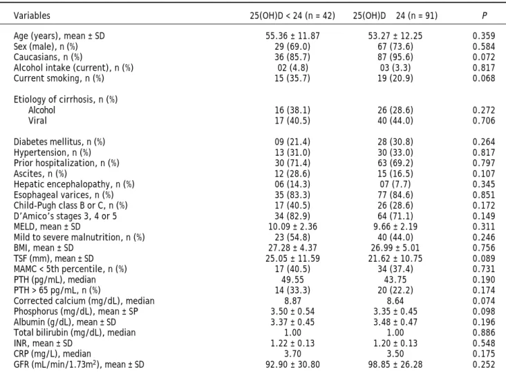
Table 2 shows the bivariate analysis of the fac-tors associated with 25(OH)D levels below the lower tertile. There were no statistically significantly asso-ciations between the studied variables and lower 25(OH)D levels. However, there was a trend towards
Table 2. Bivariate analysis of factors associated with 25(OH)D levels below the lower tertile (< 24 ng/mL).
Variables 25(OH)D < 24 (n = 42) 25(OH)D ≥ 24 (n = 91) P
Age (years), mean ± SD 55.36 ± 11.87 53.27 ± 12.25 0.359
Sex (male), n (%) 29 (69.0) 67 (73.6) 0.584
Caucasians, n (%) 36 (85.7) 87 (95.6) 0.072
Alcohol intake (current), n (%) 02 (4.8) 03 (3.3) 0.817
Current smoking, n (%) 15 (35.7) 19 (20.9) 0.068
Etiology of cirrhosis, n (%)
Alcohol 16 (38.1) 26 (28.6) 0.272
Viral 17 (40.5) 40 (44.0) 0.706
Diabetes mellitus, n (%) 09 (21.4) 28 (30.8) 0.264
Hypertension, n (%) 13 (31.0) 30 (33.0) 0.817
Prior hospitalization, n (%) 30 (71.4) 63 (69.2) 0.797
Ascites, n (%) 12 (28.6) 15 (16.5) 0.107
Hepatic encephalopathy, n (%) 06 (14.3) 07 (7.7) 0.345
Esophageal varices, n (%) 35 (83.3) 77 (84.6) 0.851
Child-Pugh class B or C, n (%) 17 (40.5) 26 (28.6) 0.172
D’Amico’s stages 3, 4 or 5 34 (82.9) 64 (71.1) 0.149
MELD, mean ± SD 10.09 ± 2.36 9.66 ± 2.19 0.311
Mild to severe malnutrition, n (%) 23 (54.8) 40 (44.0) 0.246
BMI, mean ± SD 27.28 ± 4.37 26.99 ± 5.01 0.756
TSF (mm), mean ± SD 25.05 ± 11.59 21.62 ± 10.75 0.089
MAMC < 5th percentile, n (%) 17 (40.5) 34 (37.4) 0.731
PTH (pg/mL), median 49.55 43.75 0.190
PTH > 65 pg/mL, n (%) 14 (33.3) 20 (22.2) 0.174
Corrected calcium (mg/dL), median 8.87 8.64 0.074
Phosphorus (mg/dL), mean ± SP 3.50 ± 0.54 3.35 ± 0.45 0.098
Albumin (g/dL), mean ± SD 3.37 ± 0.45 3.48 ± 0.47 0.196
Total bilirubin (mg/dL), median 1.00 1.00 0.886
INR, mean ± SD 1.22 ± 0.13 1.20 ± 0.13 0.548
CRP (mg/L), median 3.70 3.50 0.175
GFR (mL/min/1.73m2), mean ± SD 92.90 ± 30.80 98.85 ± 26.28 0.252
lower proportion of Caucasians (85.7 vs. 95.6%, P = 0.072), higher proportion of currently smoking (35.7
vs. 20.9%, P = 0.068), greater TSF (25.05 ± 11.59 mm vs. 21.62 ± 10.75 mm, P = 0.089), higher phos-phorus levels (3.50 ± 0.54 mg/dL vs. 3.35 ± 0.45 mg/dL, P = 0.098) and higher median of corrected calcium levels (8.87 mg/dL vs. 8.64 mg/dL, P = 0.074).
Variables with P < 0.100 in the bivariate analysis were included in the multiple logistic regression analysis (Table 3). Levels of 25(OH)D below the lower tertile were independently associated with greater TSF (OR 1.041, IC95% 1.003-1.081, P = 0.034) and non-Caucasian race (4.056, IC95% 1.005-16.369, P = 0.049).
Evaluation of the calcium-PTH-vitamin D axis
PTH levels above the reference value (65 pg/mL) were found in 24.6% of the patients (25.3% among Child-Pugh A and 23.3% among Child-Pugh B/C subjects, P = 0.800). There was no correlation be-tween the PTH and 25(OH)D (P = 0.193). A small positive correlation was observed between PTH lev-els and age (r = 0.184; P = 0.036) and creatinine levels (r = 0.191; P = 0.029). There were no associ-ations with the other studied variables (data not shown).
Among those with 25(OH)D levels lower than 24 ng/dL, 31.7% showed high PTH (> 65 pg/mL). Indi-viduals with a combination of 25(OH)D lower than 24 ng/dL and PTH higher than 65 pg/mL showed no differences when compared to the remaining sub-jects regarding the study variables.
Corrected calcium below 8.5 mg/dL was observed in 41 patients and, among these, PTH levels were el-evated in only 11 subjects. Hypocalcemia with high PTH was not associated with severity of the liver disease, 25(OH)D levels, variables related to the nu-tritional status or other laboratory variables (data
not shown). Only one patient presented hypercal-cemia (corrected calcium higher than 10.2 mg/dL), with 25(OH)D of 19.8 ng/dL and normal PTH.
Levels of 25(OH)D during hospitalization for acute decompensation of cirrhosis
Thirty patients that had been included in the above analysis were previously hospitalized for acute decompensation of cirrhosis and had samples for 25(OH)D measurements collected in the first 48 h of hospitalization. The median interval between the hospitalization and outpatient assessment was of 105 days. During their hospital stay, 3 patients were classified as Child-Pugh A, 19 as B and 8 as C. Upper gastrointestinal bleeding was present on ad-mission in 26 patients, hepatic encephalopathy in 16 and ascites in 11 cases. Bacterial infections were di-agnosed at hospitalization in four patients. As com-pared to outpatient assessment, significantly lower 25(OH)D levels were observed at admission (22.90 ± 9.53 ng/mL vs. 29.46 ± 7.06 ng/mL, P < 0.001) (Figure 1). Likewise, lower 25(OH)D levels at hos-pitalization were noticed in 26 out of 30 patients
Table 3. Multiple logistic regression analysis of factors associated with 25(OH)D levels below the lower tertile (variables
with P < 0.100 in bivariate analysis).
Variables OR CI 95% P
TSF 1.041 1.003-1.081 0.034
Phosphorus 1.535 0.652-3.613 0.326
Corrected calcium 1.706 0.704-4.133 0.237
Non-Caucasian race 4.056 1.005-6.369 0.049
Current smoking 2.177 0.902-5.256 0.084
25(OH)D: 25-hydroxyvitamin D. OR: odds ratio. CI: confidence interval. TSF: triceps skin-fold thickness.
Figure 1. Levels of 25(OH)D for 30 patients with cirrhosis in two different moments. As compared to outpatient assess-ment, significantly lower 25(OH)D levels were observed at ad-mission for acute decompensation of cirrhosis (P < 0.001).
Hospitalization Outpatient assessment 70
60
50
40
30
20
10
0
25(OH)D levels (ng/mL)
p < 0.001
included in this analysis (87%). Admission for acute decompensation of liver disease was also associated with higher MELD (13.65 ± 3.29 vs. 10.84 ± 2.39, P < 0.001), INR (1.45 ± 0.25 vs. 1.27 ± 0.15, P < 0.001), total bilirubin (1.85 ± 1.34 mg/dL vs. 1.33 ± 0.84 mg/dL, P = 0.015), CRP median (6.78 mg/L vs. 3.50 mg/L, P = 0.009) and lower albumin (2.69 ± 0.52 g/dL vs. 3.26 ± 0.43 g/dL, P < 0.001). There were no significant differences in 25(OH)D levels when Child-Pugh C patients were compared the remaining subjects (P = 0.243). Levels of 25(OH)D during acute decompensation were also not associated with other laboratory variables related to hepatic dys-function (MELD, INR, albumin and total bilirubin), with CRP or with the estimated GFR.
DISCUSSION
The main currently known physiological func-tions of vitamin D are the increase of intestinal cal-cium absorption and the stimulation of calcal-cium transport from bones and kidneys to the circulation. Thus, vitamin D deficiency affects bone development, leading to rickets in children (or osteomalacia in adults) and to an increased risk of osteoporosis.20
Recently, the extraskeletal effects of vitamin D have been brought to attention. Vitamin D deficiency has been associated to increased risk of neoplastic, car-diovascular, autoimmune and infectious diseases.21-25
In the case of liver diseases, lower 25(OH)D levels have been associated with greater histologic severity in chronic hepatitis C,26 greater degree of hepatic
dysfunction,11 and higher risk of non-alcoholic fatty
liver disease, hepatic osteodystrophy and hepatocel-lular carcinoma.27-29 However, not all studies have
found these associations, and the impact of other variables, such as body composition parameters, have not been taken into consideration. Therefore, there are still uncertainties about the real meaning of 25(OH)D levels in patients with chronic liver diseases.
In the present study, significantly lower levels of 25(OH)D were observed in patients with cirrhosis as compared to the control group (27.03 ± 6.22 ng/mL
vs. 32.34 ± 11.38 ng/mL, P = 0.018). In addition, 25(OH)D levels lower than 30 ng/mL were found in 69.9% of the subjects, and levels lower than 20 ng/ mL were found in 14.3%. These results are in agree-ment with most of previous studies, in which 25(OH)D levels below 30 ng/mL were observed in the majority of cirrhotic patients.8 However, the
prevalence of 25(OH)D levels below 20 ng/mL ob-served here was smaller than suggested by previous
studies, in which it ranged from 64 to 85%.9,11,30
This difference can probably be explained by the fact that these studies were carried out in European countries in which a higher prevalence of vitamin D deficiency is observed.31 Moreover, in two of these
studies the data collection period was extended for all seasons of the year, which may limit the inter-pretation of results due to the influence of seasonal-ity on 25(OH)D levels.31
Among those individuals with 25(OH)D levels be-low the be-lower tertile (24 ng/mL), there was a trend toward lower proportion of Caucasians, higher pro-portion of currently smoking, greater TSF, higher phosphorus and higher median of corrected calcium. However, in the logistic regression analysis, only greater TSF and non-Caucasian race were associat-ed with 25(OH)D below the lower tertile. The TSF thickness measurement is considered an appropriate method for estimating body fat.32 Although there are
no studies evaluating the relationship between body fat deposits and 25(OH)D levels in cirrhotic pa-tients, data from population-based studies suggest an inverse relation between 25(OH)D and adiposi-ty.33,34 This can be a result from the sequestrating
effect of a high subcutaneous fat on circulating vita-min D, explained by the liposolubility of this com-pound.35,36 Therefore, the inverse association
between TSF and 25(OH)D levels observed here may be explained by the effects of adipose tissue on circu-lating 25(OH)D, regardless of the high prevalence of malnutrition (nearly 50%) and of TSF bellow de 50th percentile (25.6%). Similarly, there is a rela-tionship between lower 25(OH)D levels and black race, probably due to lower rates of 7-dehydrocholes-terol photoconversion in individuals with more pig-mented skin.36,37 These findings were also observed
in a study including 118 individuals, most of which with HCV-related liver disease, that found that black race had the higher odds ratio for 25(OH)D levels lower than 7 ng/mL.38
Levels of PTH above 65 pg/mL were found in 24.6% of the patients and were not associated with the severity of liver disease nor with 25(OH)D lev-els. This is in accordance with the majority of previ-ous studies, in which a relationship between the severity of liver disease and PTH levels was not ob-served.10,30,39,40 In the present study,
vitamin D levels, the absence of a compensatory in-crease of PTH levels in cirrhotic patients with low 25(OH)D levels has already been described.39
Possi-ble explanations for this include polymorphism of the vitamin D receptor gene and suppression of PTH secretion by L-amino acids that activate calcium-sensing receptors.39 However, further studies are
needed to elucidate this phenomenon.
In this study, 25(OH)D levels were not associated with variables related to the severity of liver disease, such as MELD, Child-Pugh classification and D’Amico’s stage of cirrhosis. These findings are in disagreement with the majority of previous studies, in which lower 25(OH)D levels had been related to the severity of the hepatic disease.9-11,28,39,41 A
possi-ble explanation for these differences is the low number of patients with more advanced liver disease (Child-Pugh C) included in the present study. Nev-ertheless, the lack of association with other parame-ters such as MELD, D’Amico’s classification and laboratory tests (INR, albumin, total bilirubin) sug-gests that, in this sample, the degree of hepatocellu-lar dysfunction had little impact on the levels of 25(OH)D. Moreover, most of the studies that inves-tigated 25(OH)D levels in cirrhosis performed only bivariate analysis and included patients hospitalized for complications of the disease. Several factors in-fluence 25(OH)D levels, such as skin pigmentation, adiposity, dietary factors and renal function, and they should be taken into account when investigat-ing factors associated with vitamin D status.42
When the subgroup that had previously been ad-mitted for complications of liver disease was com-pared in two different time points, significantly lower 25(OH)D levels were found during hospitaliza-tion. This reduction occurred in parallel with the worsening of hepatic parameters and rise of CRP levels. However, as in the outpatient evaluation, during hospitalization, 25(OH)D levels were not as-sociated with the laboratory variables related to he-patic failure (Child-Pugh, MELD, INR, albumin and total bilirubin). Although these findings are limited by the possibility that 25(OH)D levels flutuate over time, the fact that lower 25(OH)D levels at hospital-ization were noticed in 87% of cases (26/30 patients) suggests that vitamin D levels might have been affected by specific factors related to hospitalization. These lower levels evidenced at the time of acute decompensation of cirrhosis may indicate a significant role of liver function in the maintenance of serum 25(OH)D concentration. It is still possible that other factors, especially systemic inflammation, may explain these findings. A recent study showed that
25(OH)D behave like negative acute phase reactant, suggesting that hypovitaminosis D may be more a consequence than a cause of inflammatory process-es.43 This theory seems to be especially interesting
in the setting of chronic liver diseases, since cirrho-sis is associated with increased bacterial transloca-tion, endotoxemia and, consequently, chronic inflammation, which seems to be more intense in the patients with more advanced disease.44
It can be concluded that, in this group of stable cirrhotic patients with relatively preserved liver function, the prevalence of hypovitaminosis D was high and it was associated with level of adiposity and non-Caucasian race, but not with the severity of liver disease. Moreover, admission due to acute de-compensation of cirrhosis was associated with lower 25(OH)D concentrations. Although this finding may suggest a role of the liver function in maintaining 25(OH)D levels, the absence of influence of variables related to liver dysfunction suggests that the rela-tionship between 25(OH)D levels and the severity of the disease in cirrhotic patients seem to be more complex than previously supposed. New studies in-vestigating the role of other phenomena such as en-dotoxemia and chronic inflammatory state are necessary for the clarification of this issue.
CONFLICT OF INTEREST
Potential conflict of interest and funding support: none to declare.
Previous presentation and/or publication: noth-ing to report
REFERENCES
1. Blachier M, Leleu H, Peck-Radosavljevic M, Valla DC, Rou-dot-Thoraval F. The burden of liver disease in Europe: a review of available epidemiological data. J Hepatol 2013; 58: 593-608.
2. Schuppan D, Afdhal NH. Liver cirrhosis. Lancet 2008; 371: 838-51.
3. Pinzani M, Rosselli M, Zuckermann M. Liver cirrhosis. Best Pract Res Clin Gastroenterol 2011; 25: 281-90.
4. Lopez-Larramona G, Lucendo AJ, Gonzalez-Castillo S, Te-nias JM. Hepatic osteodystrophy: An important matter for consideration in chronic liver disease. World J Hepatol 2011; 3: 300-7.
5. Holick MF, Binkley NC, Bischoff-Ferrari HA, Gordon CM, Hanley DA, Heaney RP, Murad MH, et al. Evaluation, treat-ment, and prevention of vitamin D deficiency: an Endocrine Society clinical practice guideline. J Clin Endocrinol Me-tab 2011; 96: 1911-30.
fat-soluble vitamins A and D. J Biol Chem 2010; 285: 14486-94.
8. Stokes CS, Volmer DA, Grunhage F, Lammert F. Vitamin D in chronic liver disease. Liver Int 2013; 33: 338-52. 9. Putz-Bankuti C, Pilz S, Stojakovic T, Scharnagl H, Pieber
TR, Trauner M, Obermayer-Pietsch B, et al. Association of 25-hydroxyvitamin D levels with liver dysfunction and mor-tality in chronic liver disease. Liver Int 2012; 32: 845-51. 10. Miroliaee A, Nasiri-Toosi M, Khalilzadeh O, Esteghamati A,
Abdollahi A, Mazloumi M. Disturbances of parathyroid hor-mone-vitamin D axis in non-cholestatic chronic liver disease: a cross-sectional study. Hepatol Int 2010; 4: 634-40. 11. Malham M, Jorgensen SP, Ott P, Agnholt J, Vilstrup H,
Borre M, Dahlerup JF. Vitamin D deficiency in cirrhosis re-lates to liver dysfunction rather than aetiology. World J Gastroenterol 2011; 17: 922-5.
12. Addolorato G, Leggio L, Ferrulli A, Cardone S, Vonghia L, Mirijello A, Abenavoli L, et al. Effectiveness and safety of baclofen for maintenance of alcohol abstinence in alcohol-dependent patients with liver cirrhosis: randomised, dou-ble-blind controlled study. Lancet 2007; 370: 1915-22. 13. The classic: the state of calcium in the fluids of the body.
I. The conditions affecting the ionization of calcium. Fran-klin C. McLean and A. Baird Hastings. Clin Orthop Relat Res 1970; 69: 4-27.
14. Robertshaw M, Lai KN, Swaminathan R. Prediction of cre-atinine clearance from plasma crecre-atinine: comparison of five formulae. Br J Clin Pharmacol 1989; 28: 275-80. 15. Pugh RN, Murray-Lyon IM, Dawson JL, Pietroni MC,
Wil-liams R. Transection of the oesophagus for bleeding oesophageal varices. Br J Surg 1973; 60: 646-9.
16. Kamath PS, Wiesner RH, Malinchoc M, Kremers W, Therneau TM, Kosberg CL, D’Amico G, et al. A model to predict survival in patients with end-stage liver disease. Hepatology 2001; 33: 464-70.
17. Zipprich A, Garcia-Tsao G, Rogowski S, Fleig WE, Seuffer-lein T, Dollinger MM. Prognostic indicators of survival in patients with compensated and decompensated cirrhosis. Liver Int 2012; 32: 1407-14.
18. Morgan MY, Madden AM, Soulsby CT, Morris RW. Derivation and validation of a new global method for assessing nutri-tional status in patients with cirrhosis. Hepatology 2006; 44: 823-35.
19. Frisancho AR. New norms of upper limb fat and muscle are-as for are-assessment of nutritional status. Am J Clin Nutr 1981; 34: 2540-5.
20. Christakos S, Dhawan P, Peng X, Obukhov AG, Nowycky MC, Benn BS, Zhong Y, et al. New insights into the function and regulation of vitamin D target proteins. J Steroid Bio-chem Mol Biol 2007; 103: 405-10.
21. Pilz S, Tomaschitz A, Obermayer-Pietsch B, Dobnig H, Pie-ber TR. Epidemiology of vitamin D insufficiency and cancer mortality. Anticancer Res 2009; 29: 3699-704.
22. Pilz S, Tomaschitz A, Ritz E, Pieber TR. Vitamin D status and arterial hypertension: a systematic review. Nat Rev Cardiol 2009; 6: 621-30.
23. Pilz S, Tomaschitz A, Drechsler C, Dekker JM, Marz W. Vi-tamin D deficiency and myocardial diseases. Mol Nutr Food Res 2010; 54: 1103-13.
24. Shoenfeld N, Amital H, Shoenfeld Y. The effect of melanism and vitamin D synthesis on the incidence of autoimmune disease. Nat Clin Pract Rheumatol 2009; 5: 99-105. 25. Yamshchikov AV, Desai NS, Blumberg HM, Ziegler TR,
Tang-pricha V. Vitamin D for treatment and prevention of infec-tious diseases: a systematic review of randomized controlled trials. Endocr Pract 2009; 15: 438-49.
26. Petta S, Camma C, Scazzone C, Tripodo C, Di Marco V, Bono A, Cabibi D, et al. Low vitamin D serum level is
relat-ed to severe fibrosis and low responsiveness to interfer-on-based therapy in genotype 1 chronic hepatitis C. Hepatology 2010; 51: 1158-67.
27. Targher G, Bertolini L, Scala L, Cigolini M, Zenari L, Falez-za G, Arcaro G. Associations between serum 25-hydroxy-vitamin D3 concentrations and liver histology in patients with non-alcoholic fatty liver disease. Nutr Metab Cardio-vasc Dis 2007; 17: 517-24.
28. Crawford BA, Kam C, Donaghy AJ, McCaughan GW. The heterogeneity of bone disease in cirrhosis: a multivariate analysis. Osteoporos Int 2003; 14: 987-94.
29. Falleti E, Bitetto D, Fabris C, Cussigh A, Fontanini E, For-nasiere E, Fumolo E, et al. Vitamin D receptor gene poly-morphisms and hepatocellular carcinoma in alcoholic cirrhosis. World J Gastroenterol 2010; 16: 3016-24. 30. Monegal A, Navasa M, Guanabens N, Peris P, Pons F, Martinez
de Osaba MJ, Rimola A, et al. Osteoporosis and bone min-eral metabolism disorders in cirrhotic patients referred for orthotopic liver transplantation. Calcif Tissue Int 1997; 60: 148-54.
31. Mithal A, Wahl DA, Bonjour JP, Burckhardt P, Dawson-Hughes B, Eisman JA, El-Hajj Fuleihan G, et al. Global vita-min D status and detervita-minants of hypovitavita-minosis D. Osteoporos Int 2009; 20: 1807-20.
32. Wang J, Thornton JC, Kolesnik S, Pierson RN, Jr. Anthro-pometry in body composition. An overview. Ann N Y Acad Sci 2000; 904: 317-26.
33. Snijder MB, van Dam RM, Visser M, Deeg DJ, Dekker JM, Bouter LM, Seidell JC, et al. Adiposity in relation to vita-min D status and parathyroid hormone levels: a popula-tion-based study in older men and women. J Clin Endocrinol Metab 2005; 90: 4119-23.
34. Hypponen E, Power C. Hypovitaminosis D in British adults at age 45 y: nationwide cohort study of dietary and life-style predictors. Am J Clin Nutr 2007; 85: 860-8.
35. Mawer EB, Backhouse J, Holman CA, Lumb GA, Stanbury SW. The distribution and storage of vitamin D and its me-tabolites in human tissues. Clin Sci 1972; 43: 413-31. 36. Au LE, Rogers GT, Harris SS, Dwyer JT, Jacques PF,
Sa-check JM. Associations of Vitamin D Intake with 25-Hy-droxyvitamin D in Overweight and Racially/Ethnically Diverse US Children. J Acad Nutr Diet 2013.
37. Clemens TL, Adams JS, Henderson SL, Holick MF. Increased skin pigment reduces the capacity of skin to synthesise vitamin D3. Lancet 1982; 1: 74-6.
38. Arteh J, Narra S, Nair S. Prevalence of vitamin D defi-ciency in chronic liver disease. Dig Dis Sci 2010; 55: 2624-8.
39. Fisher L, Fisher A. Vitamin D and parathyroid hormone in outpatients with noncholestatic chronic liver disease. Clin Gastroenterol Hepatol 2007; 5: 513-20.
40. Crosbie OM, Freaney R, McKenna MJ, Hegarty JE. Bone density, vitamin D status, and disordered bone remode-ling in end-stage chronic liver disease. Calcif Tissue Int 1999; 64: 295-300.
41. Chen CC, Wang SS, Jeng FS, Lee SD. Metabolic bone dis-ease of liver cirrhosis: is it parallel to the clinical severity of cirrhosis? J Gastroenterol Hepatol 1996; 11: 417-21. 42. Prentice A. Vitamin D deficiency: a global perspective.
Nutr Rev 2008; 66: S153-S164.
43. Waldron JL, Ashby HL, Cornes MP, Bechervaise J, Razavi C, Thomas OL, Chugh S, et al. Vitamin D: a negative acute phase reactant. J Clin Pathol 2013; 66: 620-2.