Annals of Hepatology 8(1) 2009: S18-S24
S18
www.medigraphic.com
Annals of Hepatology 2009; 8(1): Supplement: S18-S24Annals of
Hepatology
Metabolic syndrome and non-alcoholic
fatty liver disease
Paloma Almeda-Valdés; Daniel Cuevas-Ramos; Carlos Alberto Aguilar-Salinas
Departamento de Endocrinología y Metabolismo del Instituto Nacional de Ciencias Médicas y Nutrición, Mexico City.
Address for correspondence: Carlos A. Aguilar Salinas
Departamento de Endocrinología y Metabolismo del Instituto Nacional de Ciencias Médicas y Nutrición, Mexico City. Phone: 52-5-5133891
Fax: 52-5-5130002
E-mail: [email protected]
Manuscript received and accepted: 26 September 2008 Abstract
The development of nonalcoholic fatty liver disease (NAFLD) is strongly associated with the metabolic syn-drome as reflected by the fact that approximately 90% of the patients with NAFLD have more than one feature of metabolic syndrome and about 33% have three or more criteria. The physiopathology, epidemiology and therapeutic considerations of the disease are reviewed here. Lipotoxicity plays a predominant role in the pathophysiology of both entities. It leads to accumula-tion of triglycerides in the liver as a result of an imbal-ance among the uptake, synthesis, export, and oxida-tion of fatty acids. Both condioxida-tions are very common in Mexico. Using the Adult Treatment Panel diagnostic criteria, the 1994 prevalence of the metabolic syn-drome was 26.6%.Although the prevalence of NAFLD is not known, but it can be estimated from the preva-lence of obesity (30%). Since NAFLD is found in over two thirds of the obese subjects, this condition may exist in 20% of the adult population. The treatment of both conditions should be based in an integral approach, in-cluding the adoption of a healthy lifestyle, weight loss and may be pharmacotherapy. In summary, NAFLD is the hepatic expression of the metabolic syndrome. The study and treatment of these disorders could not be viewed as separate issues.
Key words: Non alcoholic fatty liver disease, metabol-ic syndrome, hepatmetabol-ic esteatosis, insulin resistance, tryglicerides.
The metabolic syndrome is a condition characterized by a cluster of alterations including glucose intolerance/ insulin resistance, abdominal obesity, atherogenic
dys-lipidemia (low concentrations of high-density lipopro-tein-cholesterol and high concentrations of triglycer-ides), elevated blood pressure, a proinflammatory and a prothrombotic state. It increases morbidity and mortality, especially due to cardiovascular disease.1,2 On table I the
diagnostic criteria for the syndrome according to differ-ent organizations are shown.
According to some groups, insulin resistance is the main feature of the metabolic syndrome. Insulin resis-tance refers to a condition in which a greater amount of insulin are required to elicit a normal biologic response at a cellular, organ or whole-body level. As insulin resis-tance increases, a greater degree of compensatory hyper-insulinemia is present.1,3
In Mexico there is a high prevalence of the metabolic syndrome. Using the Adult Treatment Panel (ATP III) di-agnostic criteria, a prevalence of 26.6% was estimated and according to the WHO criteria the prevalence was 13.61%. A significant proportion of the total cases (35.2%) are less than 40 years of age and in people with-out diabetes the age distribution is shifted to a younger group regardless the diagnostic criteria used. The meta-bolic syndrome was more common in women than in men (13.39% vs 13.79%) according to the WHO criteria and more common in men (28.5% vs 25.2%) according to the ATP III. The number of cases was significantly lower with the WHO definition; in fact only 43.4% of the subjects who fulfilled the ATP III criteria were diagnosed using the WHO definition.4
Regardless of its definition, the metabolic syndrome is an important public health concern due to its high preva-lence and because its association with an increase in mor-bidity and mortality mainly due to cardiovascular disease.
Nonalcoholic fatty liver disease
Nonalcoholic fatty liver disease (NAFLD) refers to a histological spectrum of liver damage from simple ste-atosis to advanced fibrosis and cirrhosis in individuals without a relevant alcohol consumption (< 20 g/day).5
It has been recognized as a major cause of liver-related morbidity and mortality.6 Nonalcoholic
steatohepati-tis (NASH) is believed to be an intermediate stage in the progression from steatosis to cirrhosis. Histologi-cal changes of NASH are characterized by steatosis,
Artemisa
www.medigraphic.com
mixed inflammatory cell infiltration, necrosis and pro-gressive fibrosis, ultimately leading to cirrhosis and end-stage liver disease. These are similar to alcohol-induced hepatitis. The probability of developing ad-vanced hepatic fibrosis is significantly greater in indi-viduals with steatohepatitis than in those with simple steatosis.7,8 In most cases, fatty liver does not progress
to end-stage liver disease, but approximately 20% to 30% of cases with NAFLD have histological signs of fibrosis, inflammation and necrosis, indicating the presence of NASH. These patients are at higher risk of developing cirrhosis, terminal liver failure and hepato-cellular carcinoma.9
In Mexico the prevalence of NAFLD is not known, but it can be estimated from the prevalence of obesity and type 2 diabetes mellitus. The 2006 National Survey of Health and Nutrition in Mexico found that nearly 30 percent of the population (34.5% of women and 24.2% of men) is obese. Estimations consider that steatosis is found in over two thirds of the obese population, mean-ing that around 20% of the population may have NAFLD. In addition, according to the Survey the preva-lence of diabetes mellitus in the adult population was 7% and NAFLD is found in about 50% of patients with diabetes.10
It has been estimated that high proportions of the cryptogenic cirrhosis cases have the clinical features and are related to NAFLD.
Pathophysiology of the metabolic syndrome
Insulin resistance is a key event in the pathophysiolo-gy of the metabolic syndrome. Hyperinsulinemia, caused by an increased insulin secretion by the pancreatic beta cells and decreased insulin degradation by the liver, is a compensatory phenomenon to insulin resistance. Hyper-insulinemia leads to an increase in fat mass, lipogenesis, and is associated with increased concentrations of free fatty acids (FFA). These changes are linked with further reduction in insulin signaling and an increase in both he-patic glucose and lipid production.11
A disruption in lipid metabolism has been implicated in the pathogenesis of insulin resistance. FFA inhibits carbohydrate oxidation, insulin-stimulated glucose up-take and its incorporation into glycogen due to a de-crease in the activity of glycogen synthase.12,13 Also, a
disturbance in fatty acid partitioning could contribute to insulin resistance due to the inhibition of cytosolic long-chain fatty (LCFA) acyl CoA oxidation.14,15 The
mecha-nism involves malonyl-CoA, which inhibits carnitine
Table I. Diagnostic criteria for the metabolic syndrome.
National Cholesterol Education American Association Program´s Adult Treatment World Health Organization of Clinical Endocrinologists
Panel III report (ATP III)* (WHO)† (AACE)‡
Abdominal obesity, waist circumference Insulin resistance, identified by 1 of the following: Overweight/obesity Men >102 cm Type 2 diabetes BMI ≥ 25 kg/m2
Women > 88 cm Impaired glucose tolerance
Glucose uptake below the lowest quartile for background population under investigation under hyperinsulinemic, euglycemic conditions
Triglycerides Plus any 2 of the following: Elevated triglycerides
≥ 150 mg/dL Antihipertensive medication and or high blood pressure ≥ 150 mg/dL (≥ 140 mmHg systolic or ≥ 90 mmHg diastolic)
HDL cholesterol Plasma triglycerides ≥ 150 mg/dL Low HDL cholesterol
Men < 40 mg/dL Men < 40 mg/dL
Women < 50 mg/dL Women < 50 mg/dL
Blood pressure ≥ 130/≥ 85 mmHg HDL cholesterol < 35 mg/dL in men or Elevated blood pressure < 39 mg/dL in women ≥ 130/85 mmHg
Fasting glucose BMI > 30 kg/m2 and/or waist:hip ratio > 0.9 2-Hour postglucose challenge
≥ 100 mg/dL in men, > 0.85 in women > 140 mg/dL
Urinary albumin excretion rate ≥ 20 µg/min Fasting glucose between 110 and 126 or albumin:creatinine ratio ≥ 30 mg/g mg/dL
Other risk factors
Family history of type 2 diabetes, hypertension, or CVD
Polycystic ovary syndrome Senderaty lifestyle Advancing age
Ethnic groups having high risk for type 2 diabetes or CVD
* When 3 of 5 of the listed characteristics are present, a diagnosis of metabolic syndrome can be made
www.medigraphic.com
palmitoyltransferase (CPT). CPT controls the transfer of LCFA acyl-CoA from the cytosol into the mitochondria where they are oxidized.3 There is an association between
elevations of malonyl-CoA and insulin resistance. An in-crease in its concentration leads to an inin-crease in cytoso-lic LCFA acyl CoAs, its esterification, formation of dia-cylglycerol, triglycerides, ceramide, and reactive oxygen species, all linked to insulin resistance.14,16,17
The excess of nonesterified fatty acids and the in-creased intracellular lipid content correlates with the presence of insulin resistance.18 The adipocyte is as a
reservoir of fuel stored as triglycerides during caloric abundance for subsequent release during periods of ca-loric need. When the adipocyte no longer functions as a store for lipids, fatty acids are deposited as triglycerides in ectopic sites such as muscle, liver and visceral fat. Abnormal accumulation of fat in the muscle and other tissues plays an important role in the etiology of insulin resistance.15,19
The adipocyte, in addition to its role as a reservoir of fuel, is an active endocrine organ that produces and se-cretes a number of cytokines (adipokines) that contribute to the regulation of metabolic processes. Insulin resis-tance states are characterized by elevated expression and production of adipokines, among the most relevant proinflammatory cytokines secreted by the adipose tis-sue are tumor necrosis factor-alpha (TNF-α), resistin and plasminogen activator inhibitor (PAI), which contribute to the alterations that lead to insulin resistance.20
Adiponectin is produced exclusively by adipose tis-sue and it has been linked to insulin resistance in hu-mans. Its concentrations are decreased in patients with obesity, insulin resistance, type 2 diabetes and NAFLD.6
It has been demonstrated that adiponectin decreases insu-lin resistance by increasing fatty-acid oxidation, reduc-ing triglyceride content in skeletal muscle and liver and suppressing hepatic glucose production.21
Adiponectin activates AMPK (a fuel sensoring enz-ime) and stimulates AMPK-mediated events such glu-cose transport and fatty acid oxidation in muscle.21,22
TNF-α has a role antagonizing the effects of adiponec-tin, contributing to insulin resistance. Furthermore, some data suggest that adiponectin may have a protec-tive role in liver injury in alcoholic and nonalcoholic fatty liver disease in mice due to its antagonistic effects against TNF-α.23
Summarizing, the systemic effects of decreased insu-lin sensitivity reflect the lipotoxic effects of fatty acids as well as an imbalance of the cytokines produced by ad-ipose tissue.
Endothelial dysfunction is another feature that has been observed in humans with the metabolic syndrome. Endothelial dysfunction is thought to be one component of the inflammatory process that initiates atherogenesis. It is manifested as an impaired endothelium-dependent relaxation and increases in circulating adhesion
mole-cules like vascular cell adhesion molecule (VCAM) 1, in-tracellular adhesion molecule (ICAM) 1, and E-selectin. E-selectin is induced by inflammatory cytokines, where-as ICAM-1 and VCAM-1 are expressed by endothelial cells in response to inflammatory cytokines, elevated lev-els of FFA and oxidized low-density lipoprotein choles-terol.24,25
There is a clear association between hepatic lipid deposition and several features of insulin resistance (hyperinsulinemia, hypertriglyceridemia and low HDL concentration) and less hepatic insulin sensitivity is associated with more liver adipose tissue content.26
The molecular mechanisms associated with the devel-opment of hepatic insulin resistance have been studied and include a decrease in insulin-stimulated tyrosine phosphorylation of IRS-1 and IRS-2, which blocks the ability of insulin to activate glycogen synthase, di-minishes the ability to store glycogen and increases gluconeogenesis.27
The increases in intracellular triglyceride content in tissues of humans with insulin resistance and metabolic syndrome can be related to increases in the uptake of FFA from plasma, an enhanced rate of de novo fatty acid syn-thesis, and a dysregulation of intracellular lipid partition-ing in which fatty acid oxidation is impaired and its es-terification enhanced. The metabolic and inflammatory changes observed in the liver of patients with nonalco-holic fatty liver disease resemble those of lipotoxicity in other organs.16
Insulin resistance is usually compensated by islet beta-cells adaptation with both increased beta-cell num-ber and increased beta-cell function. Both abnormally high glucose and lipids have been implicated in the fail-ure of beta-cells. Postprandial hyperglycemia and dyslip-idemia are features that occur before the development of insulin secretory defect. Increases in plasma FFA increase short-term insulin secretion whereas prolonged increases in the concentration of saturated fatty acids and glucose together cause dysfunction and damage to the beta cell resulting in apoptosis.28
The increase in free FFA from adipocyte lypolisis or from excess dietary intake activates protein kinase C (PKC), which phosphorylates serine residues on the in-sulin receptor and inin-sulin receptor substrates (IRS), im-pairing tyrosine phosphorylation required for normal signaling to increase glucose transport, glycogen syn-thesis and other insulin-stimulated events. In the liver the ability of insulin to inhibit gluconeogenesis and glycogenolysis is impaired; all these events increase he-patic insulin resistance which led to elevated glucose production and hyperglycemia.16 Hyperinsulinemia
www.medigraphic.com
The pathophysiology of the metabolic syndrome is complex; the evidence points to insulin resistance as a central factor in the initial disturbance and its final mani-festations.
Pathophysiology of the non-alcoholic fatty liver disease
Traditionally the pathogenesis of NAFLD has been proposed related first to factors that lead to hepatic ste-atosis and next to “a second hit” that leads to hepatic damage progression. The development of NAFLD re-quires the accumulation of triglycerides within the hepa-tocytes. The triglycerides, integral components of lipo-protein particles synthesized by the liver and small intes-tine, are a source of stored energy in adipose tissue and skeletal muscle. Dietary FFA and released from adipose depots are used for hepatic triglyceride synthesis. In the hepatocyte they undergo oxidation or they become ester-ified into triglycerides. A: diacylglycerol acyltransferase (DGAT) is the enzyme that catalyzes de final step in trig-lyceride synthesis; ultimately trigtrig-lycerides exit the liver as VLDL.30
This accumulation of triglycerides in hepatic and oth-er tissues results from an imbalance among the uptake, synthesis, export, and oxidation of fatty acids.11 In states
of energy excess, hepatic DGAT activity and triglyceride synthesis are increased. Triglycerides are exported from the liver in lipoprotein particles and derived to extrahe-patic tissues.30
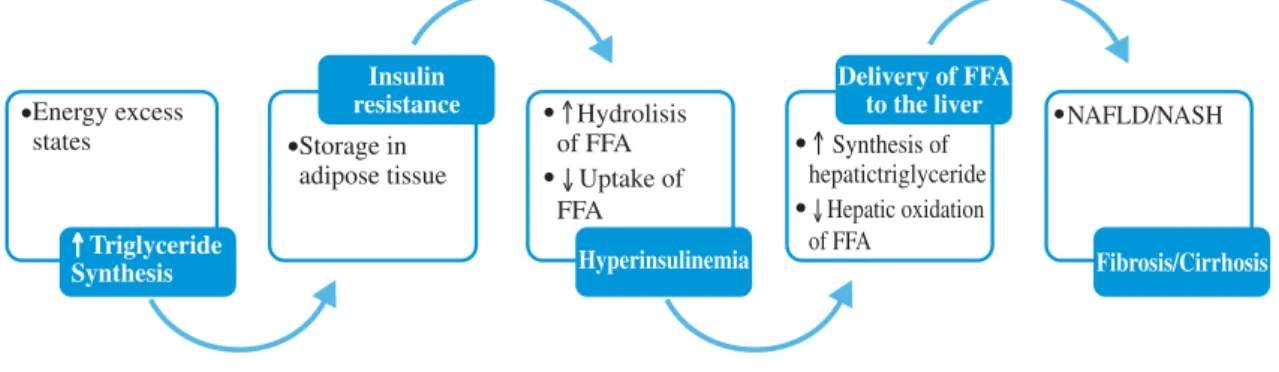
Within adipose tissue, lipoprotein lipase hydrolyzes liver-derived triglyceride and liberates FFA which are transported into adipocytes. The FFA derived from vis-ceral adipocytes, reach the liver through the portal vein, overload hepatocytes with lipid and promote hepatic triglyceride storage. In insulin resistant states there is an increase in lipolysis in adipose depots and inhibition of FFA uptake, increasing FFA delivery to the liver. On fig-ure 1 the mechanisms of hepatic fat deposition are illus-trated.29,30
Studies in animal models demonstrated improvements in hepatic steatosis when triglyceride synthesis was in-hibited. Interestingly, in a recent animal model, synthe-sis of triglycerides was a protective mechanism limiting the progression of NAFLD to NASH and fibrosis. The au-thors hypothesize that triglyceride accumulation is a hallmark of livers that are more exposed to FFA and he-patic VLDL synthesis is a protective mechanism.30
Alteration in the secretion of adipocytokines from adi-pocytes contributes to metabolic and inflammatory ab-normalities such as alteration in the rate of the synthesis of triglycerides in the hepatocytes and increased lipoly-sis in central adipose tissue. As mentioned before, low circulating levels of adiponectin have been linked to sev-eral components of the metabolic syndrome. In the liver, adiponectin increases insulin sensitivity by decreasing
the expression of hepatic gluconeogenic enzymes (phos-phoenolpyruvate carboxykinase and glucose-6-phos-phatase) and the rate of endogenous glucose production. In addition, adiponectin suppresses lipogenesis and acti-vates free fatty acid oxidation.31,32
Tumor necrosis factor α, an adipocyte-derived cytok-ine, is an important mediator of insulin resistance, it im-pairs insulin signaling by decreasing the tyrosine kinase activity of the insulin receptor (IR). Through serine resi-dues phosphorylation in the IRS-1, reduces the capacity of IRS-1 to be phosphorylated by the IR and induces a state of cellular insulin resistance.33 It has been proposed
that overproduction of TNF-α plays a key role in the pathogenesis of fatty liver; by increasing mitochondrial generation of reactive oxygen species, promotes hepato-cyte apoptosis and recruits inflammatory cells to the liv-er.30 In ob/ob mice, a model for NAFLD, fatty liver
dis-ease is significantly improved by inhibition of TNF-α production.8
Leptin is also produced by adipose tissue in propor-tion to adipose tissue mass, when leptin acpropor-tion is lack-ing, whether a consequence of its deficiency or due to leptin resistance, generalized steatosis develops in liv-er, heart, pancreatic islets, kidneys and skeletal muscle. Leptin might function as an antisteatotic hormone aimed at preventing free fatty acid entry and fat accu-mulation in non-adipose tissues.34 Additionally, leptin
appears to be essential for developing fibrosis in re-sponse to chronic liver injury due to induction of trans-forming growth factor β 1.35
Once hepatic steatosis is present other factors promote progression to NASH. Patients at risk for more severe dis-ease are those with several features of the metabolic syn-drome, indicating that insulin resistance might play a role in disease progression.11 Oxidative stress has been
recognized as a mechanism responsible for progression of liver damage. The oxidative stress includes reactive oxy-gen species (ROS) production and lipid peroxidation eventually leading to NASH. The potential sources for the ROS include hepatic cytochrome P450 2E1, mito-chondria and iron overload.7 Mitochondrial damage also
produce ROS which may trigger steatohepatitis and fi-brosis by lipid peroxidation, cytokine induction and in-duction of Fas ligand.5 Excessive fatty acid oxidation is
a major contributor to oxidative stress in the liver. Hepat-ic stellate cells are activated during chronHepat-ic liver injury and produce excessive extracellular matrix leading to fi-brosis; they also produce ROS contributing even more to the oxidative stress.7
Linkage of the metabolic syndrome to non-alco-holic fatty liver disease
NAFLD can be considered as the hepatic representa-tion of the metabolic syndrome.29 The development of
syn-www.medigraphic.com
ESTE DOCUMENTO ES ELABORADO POR MEDI-GRAPHIC
drome as reflected by the fact that approximately 90% of the patients with NAFLD have more than one feature of metabolic syndrome and about 33% have three or more criteria.36 Furthermore, with the addition of each of the
components of the metabolic syndrome the risk of steato-sis increases exponentially.37
Nonalcoholic fatty liver disease has been consistently associated with obesity (60-95%), type 2 diabetes melli-tus (28-55%) and dyslipidemia (27-92%).11
The presence of the metabolic syndrome is associat-ed with a potentially progressive and severe liver dis-ease. In addition, the likelihood of developing NASH increases with the severity of obesity.11,36 There is a
universal association between NASH an insulin resis-tance regardless of body mass index, suggesting that insulin resistance is a central factor in the pathogene-sis of NASH.38
The elevated expression of TNF-α in the liver seen in NAFLD can represent a link between the development of insulin resistance and hepatic steatosis. TNF-α has been proposed as an important mechanism of peripheral insu-lin resistance in obesity and type 2 diabetes. It is liked to increased oxidative stress and cell death in the liver, po-tentiating the development of liver fibrosis and progres-sion to NASH.7
Therapeutic considerations
The diagnosis and treatment of the metabolic syn-drome is a public health problem. The synsyn-drome is asso-ciated with an increased morbidity and mortality mainly due to cardiovascular disease. The treatment of patients with NAFLD should include the identification and treat-ment of the associated metabolic abnormalities to ame-liorate the cardiovascular risk and to improve NALFD.
Many therapeutic options such as diet and exercise are often difficult to maintain successfully. A primary target of intervention for metabolic syndrome should
be weight reduction and the prevention of obesity. Long-term maintenance of weight loss is best achieved when regular physical activity is included in the weight-reduction regimen. Weight reduction has been associated with improvement in liver function tests and in histological findings related to fatty liv-er.39 The rate of weight loss should be gradual,
be-cause in patients with a high degree of fatty infiltra-tion, rapid weight loss may promote inflammatory hepatitis and fibrosis due to an increase in adipose tissue lipolysis and the release of FFA contributing to the exacerbation of preexistent liver steatosis, at least in some patients. A target of weight loss of about 10% of bodyweight over six months seems reason-able.29,40-42 The standard exercise recommendation is a
daily minimum of 30 minutes of moderate intensity physical activity.43
There is no established drug treatment for steatohepa-titis. Multiple pharmacologic interventions have been at-tempted with variable success including pentoxifilline, orlistat, vitamin E, ursodeoxycholic acid and lipid-lower-ing agents. Further randomized, well controlled trials are required to determine the efficacy of these drugs. There are two classes of drugs currently available that reduce insulin resistance. These are metformin and insulin sensi-tizing drugs (thiazolidinediones). Long term treatment with metformin significantly reduced transaminase con-centrations and liver size.44 Thiazolidinediones have
been shown to improve insulin sensitivity in patients with type 2 diabetes, they decrease serum aminotrans-ferase levels, decrease liver fat, and improve the histolog-ical features of steatohepatitis (steatosis, ballooning ne-crosis, and centrilobular inflammation).45
The control of serum glucose and lipids levels should be part of the management. Lipid lowering agents like st-atins and fibrates are useful for the treatment of dyslipi-demia and may improve liver enzymes and hepatic in-flammation.42
Figure 1. Triglyceride synthesis and NAFLD.
Triglyceride synthesis increases in states of energy excess. Insulin resistance and hyperinsulinemia lead to increase lipolysis of triglyceride depots in adipose tissue, amplifying the deriver of FFA to the liver. Insulin further stimula-tes liver triglyceride synthesis while inhibiting fatty acid oxidation inhibiting production of VLDL.
Energy excess
states Storage in
adipose tissue
Hydrolisis of FFA
↓Uptake of FFA
Synthesis of hepatictriglyceride
↓Hepatic oxidation of FFA
NAFLD/NASH
Triglyceride Synthesis
Insulin resistance
Hyperinsulinemia
Delivery of FFA to the liver
www.medigraphic.com
Conclusions
Non alcoholic fatty liver disease is a manifestation of the metabolic syndrome. The physiopathology reviewed in this article focused on the lipotoxicity associated with metabolic syndrome and considers NAFLD as a final ex-pression of this abnormality that can have significant liv-er morbidity and mortality. The treatment of both condi-tions as with all the metabolic disturbances that charac-terize the metabolic syndrome should be in an integral fashion, including the prevention and reduction of the cardiovascular risk associated with the syndrome.
References
1. Reaven G. Metabolic Syndrome: Pathophysiology and implica-tions for management of cardiovascular disease. Circulation 2002; 106:86-288.
2. Grundy S, Brewer B, Cleeman J, Smith S, Lenfant C. Definition of metabolic syndrome: Report of the National Heart, Lung, and Blood Institute/American Heart Association Conference on Sci-entific Issues Related to Definition. Circulation 2004; 109: 433-438.
3. Ruderman N, Cacicedo J, Itani S, Yagihashi N, Saha A, Ye J, Chen K, Zou M, Carling D, Boden G, Cohen R, Keaney J, Kraegen E, Ido Y. Malonyl-CoA and AMP-activated protein kinase (AMPK): possible links between insulin resistance in muscle and early endothelial cell damage in diabetes. Biochem Soc Trans 2003; 31: 202-206.
4. Aguilar-Salinas C, Rojas R, Gómez-Pérez F, Valles V, Ríos-Torres J, Franco A, Olaiz G, Rull J, Sepúlveda J. High prevalence of metabolic syndrome in Mexico. Arch Med Res 2004; 35:76-81. 5. Angulo P. Nonalcoholic fatty liver disease. N Engl J Med 2002;
346: 1221-1231.
6. Pagano C, Soardo G, Esposito W, Fallo F, Basan L, Donnini D, Federspil G, Sechi L, Vettor R. Plasma adiponectin is decreased in nonalcoholic fatty liver disease. Eur J Endocrinol 2005; 153: 113-118.
7. Jiang J, Torok N. Nonalcoholic steatohepatitis and the metabolic syndrome. Metab Syndr Relat Disord 2008; 6: 1-8.
8. Li Z, Yang S, Lin H, Huang J, Watkins P, Moser A, DeSimone C, Song X, Diehl A. Probiotics and antibodies to TNF inhibit in-flammatory activity and improve nonalcoholic fatty liver dis-ease. Hepatology 2003; 37: 343-359.
9. Matteoni C, Younossi Z, Gramlich T, Boparai N, Liu Y, McCullough A. Nonalcoholic fatty liver disease: A spectrum of clinical and pathological severity. Gastroenterology 1999; 116: 1413-1419.
10. Olaiz-Fernández G, Rivera-Dommarco J, Shamah-Levy T, Rojas R, Villalpando-Hernández S, Hernández-Avila M, Sepúlveda-Amor J. Encuesta Nacional de Salud y Nutrición 2006. Cuernavaca, México: Instituto Nacional de Salud Pública. 2006. 11. Bugianesi E, McCullough A, Marchesini G. Insulin resistance: A metabolic pathway to chronic liver disease. Hepatology 2005; 42: 987-1000.
12. Boden G, Jadali F, White J, Liang Y, Mozzoli M, Chen X, Coleman E, Smith C. Effects of fat on insulin-stimulated carbohydrate metabolism in normal men. J Clin Invest 1991; 88: 960-966. 13. Roden M, Price T, Perseghuin G, Petersen K, Rothman D, Cline
G, Shulman G. Mechanism of free fatty acid-induced insulin resistance in humans. J Clin Invest 1996; 97: 2859-2865. 14. Ruderman N, Saha A, Vavvas D, Witters L. Malonyl-CoA, fuel
sensing, and insulin resistance. Am J Physiol Endocrinol Metab 1999; 276: 1-18.
15. McGarry J. Dysregulation of fatty acid metabolism in the etiol-ogy of type 2 diabetes. Diabetes 2002; 51: 7-18.
16. Ruderman N, Prentki M. AMP kinase and malonyl-CoA: Targets for therapy of the metabolic syndrome. Nat Rev Drug Discov 2004; 3: 340-351.
17. Park H, Kaushik V, Constant S, Prentki M, Przybytkowski E, Ruderman N, Saha A. Coordinate regulation of malonyl-CoA decarboxylase, sn-glycerol-3-phosphate acyltransferase, and acetyl-CoA carboxylase by AMP-activated protein kinase in rat tissues in response to exercise. J Biol Chem 2002; 277: 32571-32577.
18. Boden G, Shulman G. Free fatty acids in obesity and type 2 diabetes: defining their role in the development of insulin resistance and β-cell dysfunction. Eur J Clin Invest 2002; 32: 14-23.
19. Unger R. Minireview: Weapons of lean body mass destruction: The role of ectopic lipids in the metabolic syndrome. Endocri-nology 2003; 144: 5159-5165.
20. Grundy S, Hansen B, Smith S, Cleeman J, Kahn R. Clinical man-agement of metabolic syndrome: Report of the American Heart Association/National Heart, Lung, and Blood Institute/American Diabetes Association Conference on Scientific Issues Related to Management. Circulation 2004; 109: 551-556.
21. Yamauchi T, Kamon J, Minokoshi Y, Ito Y, Waki H, Uchida S, Yamashita S, Noda M, Kita S, Ueki K, Eto K, Akanuma Y, Froguel P, Foufelle F, Ferre P, Carling D, Kimura S, Nagai R, Kahn B, Kadowaki T. Adiponectin stimulates glucose utilization and fatty-acid oxidation by activating AMP-activated protein kinase. Nat. Med 2002; 8: 1288-1295.
22. Tomas E, Tsao T, Saha A, Murrey H, Zhang C, Itani S, Lodish H, Ruderman N. Enhanced muscle fat oxidation and glucose trans-port by ACRP30 globular domain: Acetyl-CoA carboxylase in-hibition and AMP-activated protein kinase activation. Proc Natl Acad Sci USA 2002; 99: 16309-16313.
23. Xu A, Wang Y, Keshaw H, Xu L, Lam K, Cooper G. The fat-derived hormone adiponectin alleviates alcoholic and nonalco-holic fatty liver diseases in mice. J Clin Invest 2003; 112: 91-100. 24. Meigs J, Hu F, Rifai N, Manson J. Biomarkers of endothelial dysfunction and risk of type 2 diabetes mellitus. JAMA 2004; 291: 1978-1986.
25. Hennig B, Meerarani P, Ramadass P, Watkins B, Toborek M. Fatty acid-mediated activation of vascular endothelial cells. Me-tabolism 2000; 49: 1006-1013.
26. Seppälä-Lindroos A, Vehkavaara S, Häkkinen A, Goto T, Westerbacka J, Sovijärvi A, Halavaara J, Yki-Järvinen H. Fat accumulation in the liver is associated with defects in insulin suppression of glucose production and serum free fatty acids independent of obesity in normal men. J Clin Endocrinol Metab 2002; 87: 3023-3028.
27. Varman S, Liu Z, Qu X, Elder B, Bilz S, Befroy D, Romanelli A, Shulman G. Mechanism of hepatic insulin resistance in non-alcoholic fatty liver disease. J Biol Chem 2004; 279: 32345-32353.
28. El-Assaad W, Buteau J, Peyot M, Nolan C, Roduit R, Hardy S, Joly E, Dbaibo G, Rosenberg L, Prentki M. Saturated fatty acids synergize with elevated glucose to cause pancreatic β-cell death. Endocrinology 2003; 144: 4154-4163.
29. Rector RS, Thyfault JP, Wei Y, Ibdah JA. Non-alcoholic fatty liver disease and the metabolic syndrome: An update. World J Gastroenterol 2008; 14: 185-192.
30. Choi S, Diehl A. Hepatic triglyceride synthesis and nonalcoholic fatty liver disease. Curr Opin Lipidol 2008; 19: 295-300. 31. Combs T, Berg A, Obici S, Scherer P, Rossetti L. Endogenous
glucose production is inhibited by the adipose-derived protein Acrp30. J Clin Invest 2001; 108: 1875-188.
32. Rajala M, Scherer P. Minireview: The adipocyte-At the cross-roads of energy homeostasis, inflammation, and atherosclerosis. Endocrinology 2003; 144: 3765-3773.
www.medigraphic.com
34. Unger R. Lipotoxic diseases. Annu Rev Med 2002; 53: 319-336.35. Leclercq IA, Farrell GC, Schriemer R, Robertson GR. Leptin is essential for the hepatic fibrogenic response to chronic liver in-jury. J Hepatol 2002; 27: 206-213.
36. Marchesini G, Bugianesi E, Forlani G, Cerrelli F, Lenzi M, Manini R, Natale S, Vanni E, Villanova N, Melchionda N, Rizzetto M. Nonalcoholic fatty liver, steatohepatitis, and the metabolic syn-drome. Hepatology 2003; 37: 917-923.
37. Marceau P, Biron S, Hould F, Marceau S, Simard S, Thung S, Kral J. Liver pathology and the metabolic syndrome X in severe obesity. J Clin Endocrinol Metab 1999; 84: 1513-1517. 38. Chitturi S, Abeygunasekera S, Farrell G, Holmes-Walker J, Hui J,
Fung C, Karim R, Lin R, Samarasinghe D, Liddle C, Weltman M, George J. NASH and insulin resistance: Insulin hypersecretion and specific association with the insulin resistance syndrome. Hepatology 2002; 35: 373-379.
39. Ueno T, Sgawar H, Sujaku K, Hashimoto O, Tamaki S, Torimura T, Inuzuka S, Sata M, Tanikawa K. Therapeutic effects of re-stricted diet and exercise in obese patients with fatty liver. J Hepatol 1997; 247: 103-107.
40. Luyckx F, Desaive C, Thiry A, Dewé W, Scheen A, Gielen JE, Lefèbvre P. Liver abnormalities in severely obese subjects: Ef-fect of drastic weight loss after gastroplasty. Int J Obes 1998; 22: 222-226.
41. Stratopoulos C, Papakonstantinou A, Terzis I, Spiliadi C, Dimitriades G, Komesidou V, Kitsanta P, Agyrakos T,
Hadjiyannakis E. Changes in liver histology accompanying mas-sive weight loss after gastroplasty for morbid obesity. Obes Surg 2005; 15: 1154-1160.
42. Adams L, Angulo P. Treatment of non-alcoholic fatty liver dis-ease. Postgrad Med J 2006; 82: 315-322.
4 3 . Grundy S, Cleeman J, Merz N, Brewer B, Clark L, Hunninghake D, Pasternak R, Smith S, Stone N. Implications of recent clini-cal trials for the national cholesterol education program adult treatment panel III guidelines. Circulation 2004; 110: 227-2 3 9 .
44. Marchesini G, Brizi M, Bianchi G, Tomassetti S, Zoli M, Melchionda N. Metformin in non-alcoholic steatohepatitis. Lan-cet 2001; 358: 893-394.
45. Belfort R, Harrison S, Brown K, Darland C, Finch J, Hardies J, Balas Bogdan, Gastaldelli A, Tio F, PUlcini J, Berria R, Ma J, Dwivedi S, Havranek R, Fincke C, DeFronzo R, Bannayan G, Schenker S, Cusi K. A Placebo-controlled trial of pioglitazone in subjects with nonalcoholic steatohepatitis. N Engl J Med 2006; 355: 2297-2307.
46. Fan C, Pan J, Usuda N, Yeldandi A, Rao S, Reddy J. Steatohepatitis, spontaneous peroxisome proliferation and liver tumors in mice lacking peroxisomal fatty acyl-CoA oxidase. J Biol Chem 1998; 273: 15639-15645.