Otras secciones de este sitio:
☞ ☞ ☞ ☞
☞ Índice de este número
☞ ☞ ☞ ☞
☞ Más revistas
☞ ☞ ☞ ☞
☞ Búsqueda
Others sections in this web site:
☞ ☞ ☞ ☞
☞ Contents of this number ☞
☞ ☞ ☞
☞ More journals ☞
☞ ☞ ☞ ☞ Search Artículo:
Hepatic manifestations of Epstein-Barr viral infection
Copyright © 2005: Mexican Association of Hepatology
ANNALS OF HEPATOLOGY
Number 3 July-September 2 0 0 5
Volume 4
N Méndez-Sánchez et al. Hepatic manifestations of Epstein-Barr viral infection 205
edigraphic.com
Annals of Hepatology 2005; 4(3): July-September: 205-209
Annals of Hepatology
Abstract
Little information exists in the international scientific or medical literature about the hepatic manifestations and complications of Epstein-Barr virus (EBV). The aim of this study was to describe a series of patients with hepatic manifestations of EBV infection. Our sample population was a series of patients whose he-patic dysfunction was correlated with a documented EBV infection. Serum concentrations of IgG and IgM antibodies against the EBV viral capsid antigen (anti-EBV VCA IgG), (anti-EBV early antigen, and (anti-EBV nuclear antigen (EBV-EBNA), and heterophilic antibodies were determined. The expression of latent membrane protein (LMP 1) was also evaluated in each patient. RESULTS. The study included nine patients (six men, three women) with a mean age of 43.5 years. Five pa-tients presented with recent clinical pictures sugges-tive of acute EBV infection. Five patients began with a cholestatic pattern. Two patients required liver biop-sies. Those liver biopsies showed positive immunohis-tochemical staining for LPM 1. No fatalities were at-tributed to EBV infection. In conclusion, the bilirubin levels of patients with acute EBV infection differed from those reported in the medical literature. EBV in-fection should be considered in the differential diagno-sis of patients with liver abnormalities or diverse he-patic manifestations, increased levels of aminotrans-ferases, or a transitory cholestatic pattern with a favorable outcome.
Key words: Epstein-Barr virus infection, hepatic manifestations, cholestasis.
Case Report
Hepatic manifestations of Epstein-Barr
viral infection
Nahum Méndez-Sánchez;1 Cecilia Aguilar-Domínguez;1 Norberto C. Chávez-Tapia;1 Misael Uribe1
1Departments of Biomedical Research and Gastroenterology & Liver
Unit. Medica Sur Clinic & Foundation. Mexico City, Mexico.
Address for correspondence:
Nahum Méndez-Sánchez, M.D., Ph.D. Departments of Biomedical Research, Gastroenterology & Liver Unit, Medica Sur Clinic & Foundation, Puente de Piedra 150, Col. Toriello Guerra, Mexico City, Mexico. Phone: (+525) 55606-6222, ext. 4215 Fax: (+525) 55666-4031 and 55606-1651; e-mail: [email protected]
Manuscript received and accepted: June 20, 2005.
Introduction
Epstein–Barr virus (EBV) was discovered in cells from the tissue of a Burkitt’s lymphoma by Epstein, Achong, and Barr. More than 90% of healthy people
car-ry EBV.1–3 Half these patients present with fever,
lym-phadenopathy, and exudative pharyngitis. Other manifes-tations include splenomegaly (50%), palatal petechiae,
hepatomegaly (more than 20%), and jaundice (5%).4,5
Hepatitis is a common characteristic of infection by EBV, although severe hepatocellular liver injury is rare and its
pathogenesis uncertain.6 In the acute phase, high serum
aminotransferase levels are present in 80% of patients,
whereas jaundice is observed in only 6.6% of patients.7
The main difference between EBV hepatitis and viral hepatitis A, B, or C is that the former does not infect hepatocytes, gallbladder epithelium, or vascular
endothe-lium.8–10 Ninety percent of patients develop mild hepatitis
in the second or third week of symptomatic infection, as well as high serum alkaline phosphatase levels, whereas serum bilirubin levels greater than 8 mg/dL are observed
in only 3% of patients.3–5,11 Little information exists in the
international scientific or medical literature about the he-patic manifestations and complications of EBV. There-fore, the aim of this study was to describe a series of pa-tients with hepatic manifestations associated with EBV infection.
Methods
Annals of Hepatology 4(3) 2005: 205-209 MG
206
edigraphic.com
sustraídode-m.e.d.i.g.r.a.p.h.i.ccihpargidemedodabor
(EBV-EBNA; Mayo Medical Laboratories), and hetero-philic antibodies (Monospot; Meridian Diagnostics, Cin-cinnati, OH). Immunohistochemical studies were per-formed in patients for whom liver biopsies were indicat-ed, including assessment of the expression of latent membrane protein (LMP 1).
Results
We included nine patients in the study, six of whom were male and three female. Their mean age was 43.5 ± 17.15 (range, 19–73 years). The overall fatality rate was 0%.
Underlying illness
Two patients had underlying illnesses. One male pa-tient had ulcerative colitis and a female papa-tient had hyper-thyroidism. The diseases were controlled in both patients.
Clinical features
Fatigue and adynamia were the most common clini-cal features of patients upon admission, and were noted in eight patients (88.8%). Other symptoms, which were either identified on admission or developed during hos-pitalization, are shown in Table I. Gastrointestinal symptoms, including abdominal pain, nausea, and diar-rhea were noted in seven patients (77.7%) at baseline. Fever and respiratory symptoms were present in five (55.5%) and four (44.4%) patients, respectively, on ad-mission. On hematological tests, cytopenia involving one or two cell lines simultaneously was observed in four patients (44.4%), and leukopenia (white cell count
< 4,000 cells/µL) and thrombocytopenia (< 150,000 platelets/µL) were the two most common hematological abnormalities on admission. Hepatic abnormalities, identified by elevated serum liver enzyme and/or biliru-bin levels, were found in seven patients (77.7%). Coag-ulopathy with prolonged prothrombin time was noted in one patient (11.1%) (Table II).
Associated infections
Serology tests for EBV were performed in all patients. Five patients (55.5%) presented a recent clinical picture suggestive of acute EBV infection (patients 1, 6, 7, 8, and 9) (Table III). Five patients (55.5%) began with cholesta-sis (patients 1, 5, 7, 8, and 9), and one of them underwent the molecular adsorbent recycling system (MARS) be-cause of severe cholestasis. Patient 5 was thought to rep-resent a reactivation or atypical prep-resentation of EBV in-fection, because of the presence of only VCA IgG and EBNA antibodies. However, an acute process was con-firmed by hepatic inflammation identified on biopsy. The remaining three patients (33.3%), patients 1, 2, and 3, had recently reported an upper respiratory tract infection, fol-lowed by a clinical picture suggestive of EBV infection. These patients showed few biochemical changes, with the exception of patient 2 who had high levels of serum alka-line phosphatase and gammaglutamyl transpeptidase (GGT), with an EBV serological profile positive for VCA IgG and EBNA. EBV infection was diagnosed in this pa-tient by discounting other etiologies.
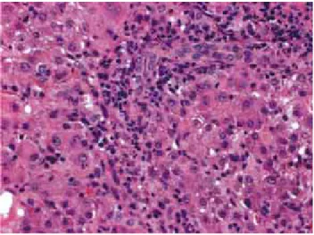
Two patients (22.2%) underwent biopsies due to un-certain diagnoses and severe clinical pictures at presenta-tion (patients 5 and 9). Their liver biopsies showed
in-Table I. Clinical and demographic characteristics of patients.
Variables Patient 1 Patient 2 Patient 3 Patient 4 Patient 5 Patient 6 Patient 7 Patient 8 Patient 9
Sex M M F F F M M M M
Age (yr) 41 33 23 57 44 46 19 56 73
Evolution (days) 3 7 15 7 5 3 30 43 30
Symptoms:
Adynamia + + + + + – + + +
Anorexia – – – – – – – – +
Abdominal pain + – + + + + – + –
Nausea – – – + + + + + –
Diarrhea – – – + – – – + –
Upper respiratory tract infection + + + – – + – – –
Cephalalgia + – – – + – – – –
Fever + + + – + + – – –
Jaundice + – – – + + + + +
Hepatodynia + + + + + + – + –
Hepatomegaly + – – – + – + – –
Splenomegaly – – – – – – + – –
Rash – – – – – + – – –
Paresthesia – – – – – – + – –
Pruritus – – – – – – – + –
207
edigraphic.com
:rop odarobale FDPVC ed AS, cidemihparG
arap
acidémoiB arutaretiL :cihpargideM
sustraídode-m.e.d.i.g.r.a.p.h.i.c
flammatory changes with many apoptotic hepatocytes and cholestasis in a pattern suggestive of viral cellular infec-tion. Positive immunohistochemical staining for LMP 1 in EBV-positive lymphocytes and epithelium demonstrated infection by this virus (Figure 1), as well as the clinical manifestations of infection.
Hepatitis A viral infection was found in patient 8, who presented one month earlier without complications. Dur-ing hospitalization, pleural and pericardial effusions were documented in patient 6.
Treatments and outcomes
All patients were treated with supportive care, and empirical antibiotic treatments were used initially only in some patients. However, once bacterial, parasitical, and/or fungal etiologies were discounted, treatments were continued only as symptomatic support where in-dicated. In some patients, ursodeoxycholic acid, ade-nosylmethionine, and cholestyramine were adminis-tered as therapeutic agents to treat cholestasis (patients 1, 5, 8, and 9). Patient 6 had complications of pneumo-nia and pleural and pericardial effusions, which were treated with cephalosporin and aminoglycoside antibi-otics and surgical intervention, respectively. Patient 9 displayed a persistent progressive increase in bilirubin,
which was treated with two sessions of MARS, which successfully reduced to bilirubin levels. No fatalities were attributed to EBV infection. Eighty-eight percent of patients had favorable outcomes, with symptomatic support only.
Discussion
EBV infection is highly prevalent, and more than 90% of the world’s population carry EBV throughout their lives. In most patients, primary infection occurs
subclini-cally during childhood.2 However, when the disease is
ac-quired during adolescence or adulthood, there are non-specific symptoms, although some reports have integrat-ed a distinctive symptomatic triad of fever, sore throat,
and lymphadenopathy.1 However, the patients described
in the present report did not show these characteristics. Hepatitis caused by EBV is common, mild, and self-limit-ing, although fulminant hepatic failure has been reported in 17 patients worldwide, with an overall mortality of
85%.12 In the present series, five (55%) of the nine
pa-tients with documented EBV infection presented with se-vere cholestasis, and increased levels of total bilirubin of > 26 mg/dL. Those patients were treated with MARS and we observed a decrease in hepatic encephalopathy indi-cated by a clearance of toxins, an increase in mean
arteri-Table II. Biochemical characteristics of patients at baseline.
Variables Patient 1 Patient 2 Patient 3 Patient 4 Patient 5 Patient 6 Patient 7 Patient 8 Patient 9
Hemoglobin (g/dL) 14.3 14.5 14.7 14 7.9 12.8 16.6 14.4 11.8
Platelets (mm-3) 157,000 156,000 318,000 289,000 457,000 133,000 184,000 209,000 238,000
Leukocytes (mm-3) 8,000 3,500 11,100 5,100 5,900 11,600 9,600 8,300 6,000
Lymphocytes (mm-3) 880 1,435 444 1,900 1,121 1,276 1,344 1,411 1,440
Atypical lymphocytes (%) 0 1 0 0 0 0 0 0 0
Monocytes (mm-3) 960 280 1,110 500 250 560 384 498 120
Prothrombin time (s) 13.7 11.2 13.3 10.8 18.7 13.7 10.6 13.5 14
Albumin (g/dL) 2.4 3.46 3.21 3.7 3.02 2.85 3.74 2.49 2
Bilirubin (mg/dL) 14.03 0.6 1.04 0.45 6.69 1.8 4.22 26.33 26
Bilirubin direct (mg/dL) 8.74 0.14 0.21 < 0.1 4.52 0.83 0.12 15.46 16
ALT (UI/L) 109 65 91 33 605 362 18 49 99
AST (UI/L) 232 34 105 33 621 292 16 74 143
Alkaline phosphatase (UI/L) 633 144 74 78 159 111 214 166 151
GGT (UI/L) 1,009 160 58 68 95 180 20 64 60
ALT, alanine aminotransferase; AST, aspartate aminotransferase; GGT, gammaglutamyl transpeptidase.
Table III. Immunological markers of patients.
Variables Patient 1 Patient 2 Patient 3 Patient 4 Patient 5 Patient 6 Patient 7 Patient 8 Patient 9
Heterophilic Abs – – – – – – – – –
Anti-EA + – – – – – – – –
Anti-viral capsid IgG EBV + + + + + + + + –
Early diffuse Ag EBV – – – – – + – – –
EBNA Abs + + + + + + + + +
Anti-viral capsid IgM EBV – – – – – – – – –
Hepatitis virus serology Anti-VHA IgG – Anti-VHA IgG – Anti-VHA IgG Anti-VHA IgG – – –
Annals of Hepatology 4(3) 2005: 205-209 MG
208
edigraphic.com
al pressure, and improvement in liver and renal function,as was observed in patient 9. In these patients, bilirubin levels differed somewhat from those reported in the sci-entific and medical literature. Whereas jaundice is rare as a presenting feature, apparent in only 6.6% of patients in
many series,13–15 most of our patients (66%) showed high
aminotransferase levels that decreased gradually with complete clinical recovery. Although EBV is not a hepa-totrophic virus, mildly elevated serum aminotransferase levels are not uncommon (90% of patients), which is con-sistent with parenchymal liver injury rather than with
de-creased bile flow.13,16 Severe cholestasis is rare, and in
those patients in whom it is present, the mechanism of cholestasis is unknown. We believe that EBV inhibits MRP2, which is the main bilirubin transporter. Further-more, high levels of GGT activity were observed, sug-gesting virus-induced self-limiting cholangiocyte dam-age. On the other hand, serum levels of aminotransferases
are only mildly elevated.17 Other herpes viruses, such as
cytomegalovirus, have been demonstrated to infect bile duct epithelial cells, as well as hepatocytes. However, this is not a consistent finding and was not documented in the present series of patients. These nine patients were immu-nocompetent with no underlying liver disease that might account for the rare development of severe hepatic dys-function. However, one patient was treated with MARS because of an adverse clinical development and progres-sive liver damage. MARS is a therapeutic modality that
appears to be safe in patients with severe cholestasis.18 In
another patient, EBV manifestations were aggravated by the presence of pleural and pericardial effusions, both of which complications are rare according to data from the world scientific and medical literature. Therefore, acute EBV infection should be considered in the differential di-agnosis of patients with transient cholestasis and in-creased alkaline phosphatase levels, whose symptoms peak in the second week after symptom onset.
Two patients described in the present report, represent-ing 22% of cases, showed increased gallbladder wall thickening on abdominal ultrasound. This finding has been reported previously in association with EBV
infec-tion, and may reflect the severity of hepatitis.19,20
All patients were treated with supportive care. In this series, no steroids were used because this therapeutic approach is controversial. However, some reports have shown a moderate clinical benefit in patients with hepa-titis associated with EBV, suggesting the importance of immunological injury in the pathogenesis of
EBV-asso-ciated hepatitis.21 Another important finding was that all
patients were negative for heterophilic antibodies. These are not specific reactants directed against EBV. Accord-ing to other reports, approximately 15% to 20% of pa-tients with EBV-associated IgM are negative for hetero-philic antibodies. The appropriate interpretation of EBV in terms of the markers VCA IgG and IgM, early anti-gen, and EBNA is important because their presence or absence reflects the stage of evolution of the disease, as either acute, recent, chronic, or persistent. We were able to identify an early stage of the disease in two patients characterized by the presence of early antigen. In anoth-er two patients, biopsies positive for LMP 1 of EBV and high titers of antibodies directed against IgG VCA and EBNA suggested possible chronic active EBV infec-tions. Because EBV must reactivate from latency to complete its life cycle, this situation is rare. The liver is the most common target organ, and has a very poor prognosis; patients die from hematological or non-he-matological disorders within a few years. In these pa-tients, a polymerase chain reaction assay can be used to detect the gene for BZLF1, which can be detected from one copy of EBV DNA. Studies of EBV DNA titers have suggested that it may correlate with disease
activi-ty more accurately than do serological data.21
In conclusion, EBV infection must be considered in the differential diagnosis of patients who present with hy-pertransaminasemia or cholestasis suggestive of this dis-ease or with diverse hepatic manifestations together with increased aminotransferase levels or transitory cholestasis and high levels of alkaline phosphatase (with a character-istic peak in the second week after the onset of symp-toms), with a favorable outcome. It is important to high-light the major role that MARS plays in the medical treat-ment of some patients who develop severe cholestasis or acute liver failure.
References
1. Cohen JI. Epstein-Barr virus infection. N Engl J Med 2000; 343: 481-92.
2. Schaller RJ, Counselman FL. Infectious mononucleosis in young children. Am J Emerg Med 1995; 13: 438-40.
3. Hickey SM, Strasburger VC. What every pediatrician should know about infectious mononucleosis in adolescents. Pediatr Clin North Am 1997; 44: 1541-56.
209
edigraphic.com
4. Devereaux CE, Bemiller T, Brann O. Ascites and severe hepatitis com-plicating Epstein-Barr infection. Am J Gastroenterol 1999; 94: 236-40. 5. Mendez-Sanchez N, Uribe M. Infectious mononucleosis hepatitis: a
case-report. Ann Hepatol 2004; 3: 75-76.
6. Jimenez-Saenz M, Perez-Pozo JM, Leal-Luna A, Herrerias-Gutierrez JM. Lethal liver failure in an elderly patient with hepatitis B superinfected with Epstein-Barr virus. Eur J Gastroenterol Hepatol 2002; 14: 1283-4. 7. Kimura H, Nagasaka T, Hoshino Y, et al. Severe hepatitis caused by
Epstein-Barr virus without infection of hepatocytes. Hum Pathol 2001; 32: 757-62.
8. Yuge A, Kinoshita E, Moriuchi M, Ohno Y, Haga H, Moriuchi H. Persistent hepatitis associated with chronic active Epstein-Barr vi-rus infection. Pediatr Infect Dis J 2004; 23: 74-6.
9. Nobili V, Comparcola D, Sartorelli MR, Devito R, Marcellini M. Autoimmune hepatitis type 1 after Epstein-Barr virus infection. Pediatr Infect Dis J 2003; 22: 387.
10. Schuster VH, Muschen M. Epstein-Barr virus and the B cell: a secret romance. Trends Microbiol 2003; 11: 243-5.
11. Rajwal S, Davison S, Wyatt J, McClean P. Primary Epstein-Barr vi-rus hepatitis complicated by ascites with Epstein-Barr vivi-rus reacti-vation during primary cytomegalovirus infection. J Pediatr Gastroenterol Nutr 2003; 37: 87-90.
12. Patel S, Zuckerman M, Smith M. Real-time quantitative PCR of Epstein-Barr virus BZLF1 DNA using the LightCycler. J Virol Meth-ods 2003; 109: 227-33.
13. Hinedi TB, Koff RS. Cholestatic hepatitis induced by Epstein-Barr virus infection in an adult. Dig Dis Sci 2003; 48: 539-41. 14. Valentini P, Angelone DF, Miceli Sopo S, Ngalikpima CJ, Ranno O.
Cholestatic jaundice in infectious mononucleosis. Minerva Pediatr 2000; 52: 303-6.
15. Carbonero CMJ, Torronteras SR, Cintado BC. [Infectious mononucleo-sis: study on hospitalized children]. An Esp Pediatr 1999; 51: 664-6. 16. Massei F, Palla G, Ughi C, Macchia P, Maggiore G. Cholestasis as a
presenting feature of acute Epstein-Barr virus infection. Pediatr In-fect Dis J 2001; 20: 721-2.
17. Bernstein CN, Minuk GY. Infectious mononucleosis presenting with cholestatic liver disease. Ann Intern Med 1998; 128: 509. 18. Mendez-Sanchez N, Chavez-Tapia NC, Espinoza B, Herrera-Gomar
M, Zamora-Valdes D, Uribe M. Acute liver failure and the Molecu-lar Adsorbents Recirculating System: early experience in a tertiary care hospital in Mexico City. Ann Hepatol 2004; 3: 164-6. 19. Barlow G, Kilding R, Green ST. Epstein-Barr virus infection
mimick-ing extrahepatic biliary obstruction. J R Soc Med 2000; 93: 316-8. 20. Murray PG, Young LS. Epstein-Barr virus infection: basis of
ma-lignancy and potential for therapy. Expert Rev Mol Med 2001; 2001: 1-20.