Annals of Hepatology 6(4) 2007: 208-213
208
medigraphic.com
Annals of Hepatology 2007; 6(4): October-December: 208-213
Annals of Hepatology
Concise Review
Mechanisms of angiogenesis in chronic
inflammatory liver disease
María Chaparro;1 Paloma Sanz-Cameno;1 María Trapero-Marugán;1 Luisa García-Buey;1 Ricardo Moreno-Otero1
1 Hepatology Department and Ciberehd. University Hospital La
Princesa.Universidad Autónoma de Madrid. Spain. Abbreviations
E-selectin, endothelial selectin; ECM, extracellular matrix; EC, endothelial cells; NO, nitric oxide; VEGF, vascular endothelial growht factor; aFGF and bFGF, acidic and basic fibroblast growth factors; HGF, hepatocyte growth factor; KDR, kinase insert domain receptor; Flt-1, fms-like tyrosine kinase receptor; CHC, chronic hepatitis C; HCV, hepatitis C virus; Ang, angiopoietin; ALT, alanine aminotransferase; PBC, primary biliary cirhosis; PSC, primary sclerosing cholangitis; AIH, autoimmune hepatitis; HCC, hepatocellular carcinoma. Address for correspondence:
Dra. María Chaparro Sánchez. Unidad de Hepatología.
Hospital Universitario de La Princesa. C/Diego de León 62., 28006. Madrid. Tel: +34 913093911; fax:+34 914022299. E-mail: [email protected]
Manuscript received and accepted: 27 August 2007
Abstract
Intrahepatic hypoxia may occur during the inflam-matory and fibrotic processes that characterize sev-eral chronic liver diseases of viral and autoimmune origin. As a consequence, new vascular structures are formed to provide oxygen and nutrients. Angiogenesis involves a tightly regulated network of cellular and molecular mechanisms that result in the formation of functional vessels. Of particular importance are growth factors and molecules involved in matrix re-modeling and cell migration, as weel as vessel matu-ration-related factors. In recent years a number of studies have investigated the expression and function of many pro- and antiangiogenic molecules in chron-ic liver diseases and liver regeneration. This review examines the potential pathogenic role of angiogene-sis in the context of viral hepatitis, autoinmmune hep-atitis, primary biliary cirrhosis and hepatocellular carcinoma.
Key words: Chronic liver disease, adhesion molecules, angiogenesis, vascular endothelial growth factor, an-giopoietins.
Introduction
Angiogenesis, the formation of new vascular struc-tures from preexisting ones, occurs in several organs dur-ing multiple pathophysiological situations.1 It has be-come one of the most thoroughly investigated patophys-iological phenomena in the last few years because of the key roles it plays in disease pathogenesis and its poten-tial as a therapeutic target. Although mostly studied in relation to cancer, angiogenesis is also known to occur in pathologies characterized by chronic inflammation. Re-cent work from our group and others has demonstrated that chronic diseases of the liver do not represent an ex-ception to this rule.
In 1995, our group2 studied by immunohistochemistry the intrahepatic expression pattern of vascular adhesion molecules (CD31, E-selectin, VCAM-1, cadherin 5 and endoglin) in viral chronic hepatitis. A novel and striking finding in this study was the detection of CD31+ and cadherin 5+ endothelial cells with microvessel morphol-ogy in the inflamed portal tracts from patients with viral chronic hepatitis. Furthermore, the expression pattern of both CD31 and cadherin 5 molecules on endothelial cells was always similar in enlarged portal tracts of these patients, acquiring a characteristic form of capillary tube formation. The assumption for an endothelial cell speci-ficity was further reinforced, because cadherin 5 is a se-lective endothelial cell marker. It was also interesting to note that the presence of these portal microvessels was more important in those cases where portal tracts were more inflamed. By contrast, a weak immunoreactivity for CD31, cadherin 5, and endoglin was only found on sinu-soidal endothelial cells and on portal vascular endotheli-um in normal liver tissue. This work gave important in-formation about the potential pathogenetic role of angio-genesis in the context of liver disease.
Mechanisms of angiogenesis
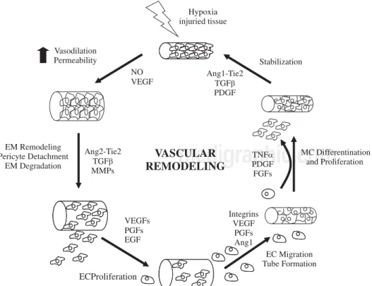
Hypoxia and inflammation are the main stimulous for angiogenesis. Hypoxia promotes the angiogenesis by sig-nalling through hypoxia inducible transcription fac-tors.1,3 During inflammation, vascular permeability is in-creased and monocytes, macrophages, platelets, mast cells, and other leukocytes (able to produce angiogenic
Artemisa
medigraphic.com
lular and molecular mechanisms involved.Endothelial budding is facilitated by vasodilation, loos-ening of interendothelial contacts, and leakiness of preex-isting vessels, which allows extravasation of plasma pro-teins that, together with extracellular matrix components (ECM), lay down a provisional scaffold for migrating endot-helial cells (EC). Nitric oxide (NO), whose angiogenic prop-erties have been characterized,8 is the main factor responsi-ble for vasodilation, whereas vascular endothelial growth factor (VEGF) increases vascular permeability. Next, the basement membrane (mainly collagen IV and laminin) and the ECM (collagen I and elastin) must be degraded to allow subsequent EC migration and proliferation. This is per-formed by specialized proteinases, including plasminogen activator. ECM proteolysis leads to the exposure of cryptic epitopes and release of ECM-embended factors that pro-mote EC migration and proliferation.9,10
EC proliferate in response to growth factors secreted by EC or surrounding cells (including hepatic stellate cells, leukocytes, hepatocytes and Kupffer cells). The most thoroughly characterized is VEGF, a multifunction-al protein that binds to 2 tyrosine kinase receptors: ki-nase insert domain receptor (KDR) and fms-like tyrosine kinase receptor (Flt-1).11 The VEGF promoter contains hypoxia-inducible factors-responsive elements. VEGF
bFGF); hepatocyte growth factor (HGF); and transform-ing growth factor (TGF).7,13
EC proliferate in an ordered manner that leads to the formation of a lumen.14,15 A structured 3-dimensional net-work of vessels of uniform size is then organized by care-fully regulated mechanisms involving signaling path-ways that determine branching, formation of basement membrane and ECM components, and cell migration and differentiation. For nascent vessels to mature, pericytes must be recruited, and a new basement membrane and ECM must be generated to provide structural stabiliza-tion.15 Physical forces and multiple molecules contribute to these processes.
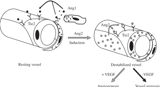
Besides induction of EC proliferation, effective angio-genesis also requires stabilization of the nascent vessels, establishment of interndothelial junctions and formation of a lumen.1 Angiopoietin 1 (ang-1) stabilizes neovessels by binding to the Tie-2 receptor, thereby affecting junc-tional molecules16 and facilitating communication be-tween EC and mural cells.17 However, an excess of Ang-1 makes vessels too tight and inhibits sprouting. Ang-2 may exert opposing effects (Figure 2): in the absence of VEGF, Ang-2 acts as an antagonist of Ang-1, destabilizes vessels and causes EC death, leading to vessel regression, but it fa-cilitates sprouting in the presence of VEGF.17
medigraphic.com
Most molecular mechanisms of angiogenesis arecom-mon to the liver and other organs. Physiological hepatic angiogenesis occurs during liver regeneration, leading to the formation of new functional sinusoids, whereas pathological angiogenesis occurs in fibrosis and is char-acterized by the appearance of capillarized vascular structures. Most chronic liver diseases are characterized by fibrosis and inflammation. During the fibrogenic pro-cess, an excessive amount of ECM is synthesized and ac-cumulated. Fibrotic tissue offers resistance to blood flow and to the delivery of oxygen, thus becoming hypoxic. Stimulaton of hypoxia-inducible factors leads to an an-giogenic switch, to the upregulation of proanan-giogenic factors, and to the formation of neovessels.2
Angiogenesis in chronic viral hepatitis
Angiogenesis occurs in the liver of patients infected with hepatitis B or C virus.1,2 The pathophisiological sig-nificance of chronic viral hepatitis-associated angiogen-esis is presently unclear; it has been proposed to exert a beneficial role by contributing to tissue repair and regen-eration after liver damage.2 It has also been suggested to represent a risk factor for progression to hepatocellular carcinoma in patients with chronic hepatitis C.4
The molecular mechanisms involved in chronic viral hepatitis-associated angiogenesis have not been fully identified. Local production of NO as a result of the over-expression of inducible NO synthase in the livers of pa-tients infected with hepatitis C or B virus may partici-pate in the angiogenic response by inducing vasodilata-tion.18,19 VEGF and HGF, whose expression is increased during chronic viral hepatitis,20-22 may contribute to en-hancing vascular permeability, as suggested by the effect described for these two growth factors. VEGF has been re-ported to induce NO-mediated vasodilation,11 most prob-ably contributing to the progression of neoangiogene-sis.2 HGF effects include a decrease and redistribution of
VE-cadherin (which participates in intercellular con-tacts), and others linking molecules between VE-cad-herin and the actin cytoskeleton. Additionally, viral pro-teins may also play a role in inducing a disruption of in-terendothelial junctions23 through mechanisms involving Src kinases, molecules required for vascular permeability during angiogenesis.24 In this way, Sanz-Cameno et al de-cribed for the first time the ability of hepatitis B virus (HVB), through HBx protein, to promote Ang-2 expres-sion in liver tissue of chronically infected patients,25 and Lara-Pezzi et al found that matrix metalloproteinase 2 is also up-regulated by HBx protein.26 Binding between EC and ECM may be altered in the livers of chronic viral hepatitis patients.27 Integrin avβ3, expressed by activated EC, shows increased tissular expression in chronic hepa-titis C (CHC).22 Interestingly, when EC are induced to migrate by HGF, integrin avβ3 accumumaltes at the lead-ing edge,22 which represents the anchoring zone required for pulling the cellular body28(Table I).
Based on this knowledge, Xalcedo et al29 designed a study in order to test the hypothesis that the evaluation of angiogenesis markers might provide additional, valu-able information on the evolution of liver lesion in CHC after antiviral therapy. They included 36 naïve HCV-in-fected patients and 15 healthy individuals. HCV-infect-ed patients presentHCV-infect-ed a «proangiogenic» profile of angio-genesis soluble markers (VEGF and Ang-2), different from that in healthy controls, while sTie-2 serum levels were similars in both groups. Their results showed a sig-nificant correlation between the grade of inflammation in biopsies and serum levels of VEGF, which suggest that inflammation and angiogenesis processes are strongly as-sociated in those patients.
Interestingly, treatment of CHC patients with pegylat-ed interferon plus ribavirin significantly rpegylat-educpegylat-ed VEGF and Ang-2 levels with respect to pretreatment values. Mean sTie-2 levels were significantly increased after therapy. Taken together, these data indicate that
medigraphic.com
ral combination therapy determines a shift toward an«antiangiogenic» profile of serum markers in CHC pa-tients. The therapy-induced decrease in VEGF levels was significantly correlated with that in ALT and alkaline phospatase serum levels in responder patients, but not in nonresponders. A similar correlation was found between the decreases in the serum levels of Ang-2 and ALT. These significant relationships suggest some degree of association between angiogenesis and the extent of liver damage (Figure 1).
Their novel findings have important implications.29 Firstly, the demonstration of a close relationship between variations in serum levels of angiogenesis markers and re-sponse to therapy in CHC patients conceptually supports the clinical role of angiogenesis in chronic inflammatory liver disease. Secondly, the determination of VEGF, Ang-2, and sTie-2 in parallel to the viral load and fibrosis markers may provide complementary information to ass-es rass-esponse to treatment and disease prograss-ession, which could be helpful to define the optimal follow-up thera-peutic strategy for each patient. Further prospective stud-ies with larger cohorts of patients are required to investi-gate whether these markers may have prognostic value. Additionally, based on these findings, it may be worth investigating in detail the antiangiogenic effects of pe-gylated interferons and, hypothetically, to consider whether antiangiogenic drugs targeting VEGF or Ang-2 could be of additional value for CHC patients with ad-vanced liver disease and those presently considered as «difficult to treat».
Angiogenesis in autoimmune liver disease
Controversy exists regarding intrahepatic vascula-ture in autoimmune liver disease. Some authors have re-ported a tendency to vasopenia and decreased peribil-iary capillary plexus in the livers of primary bilperibil-iary cir-rhosis (PBC), primary sclerosing cholangitis (PSC), and
autoimmune hepatitis (AIH) patients.30,31 This has been attributed to destruction of vascular estructures by au-toimmune mechanisms, similar to the process under-gone by bile ducts in PBC and PSC. However, evidence of angiogenesis has been observed in PBC and AIH. An-giogenesis may occur at a later stage, in response to the hypoxia caused by depletion of existing vessels, al-though the precise kinetics of the process have not been investigated.
PBC is a chronic inflammatory liver disease of multi-factorial etiopathogenesis, characterized by the presence of an intrahepatic mononuclear cell infiltrate, as well as circulating autoantibodies.32,33 It has been suggested that immunological mechanisms involving T-lymphocyte-mediated lysis are important in the characteristic bile-duct and hepatocellular damage occuring in PBC.34-36 Medina et al37 evaluated the phenotype of cellular infil-trates, and the expression of lymphocyte activation, anti-gen recognition and cell-adhesion molecules in the livers of PBC patients; and they investigated and characterized at the molecular level the pattern of reactivity of proan-giogenic factors involved in the formation of neovessels in PBC liver samples. The ocurrence of angiogenesis in the livers of PBC patients was a novel finding of this study. CD31 and VE-cadherin positive EC assemble to form new vascular structures, mainly in portal and peri-portal areas, in association with inflammatory infiltrates and fibrosis. They observated an enhanced expression of VEGF, Ang-1, Ang-2, their receptor Tie-2 and endoglin, which suggests their involvement in EC proliferation and nascent vessels stabilization.
AIH is a chronic progressive liver disease character-ized by serological changes (hypergammaglobulinemia, autoantibodies) and interface hepatitis on histological analysis.1 Tubular structures reflecting formation of new vasculature are observed in inflamed portal tracts of AIH patients.38 As in other inflammatory liver diseases, an up-regulation of inducible NO synthase leading to an
en-Induce leucocyte adhesion
Ang-1 Tie-2 sTie-2 Stabilizes intercellular contacts Stabilization of nascent vessels Inhibit permeability
Ang-2 Tie-2 sTie-2 In the presence of VEGF, it increases Vessel regresion capillary diameter and remodels basal lamina
In the absence of VEGF, it desestabilizes Vessels and causes EC death
aFGF, bFGF, FGF-R1, FGF-R2 Angiostatin Induce EC proliferation Vessel growth TGF-α,HGF EGF-R, c-Met endostatin
medigraphic.com
ESTE DOCUMENTO ES ELABORADO POR MEDI-GRAPHIC
hanced production of NO occurs in AIH, and this may participate in angiogenic processes.39 However, informa-tion about the mechanisms of angiogenesis in AIH is still very limited.
Angiogenesis in hepatocellular carcinoma
Zhang et al40 evaluated the expression of angiogenic factors in hepatocellular carcinoma (HCC) compared with the nontumour liver tissue. They found a high expression of Ang-2 and low expression Ang-1 in HCC samples in comparison with nontumorous tissue, indicating that they play a key role in the carcinogenesis and progresion of HCC via angiogenesis. Pappeti and Herman described that tumorous angiogenesis is very different from the physiological process.41 During this process, vascular quiescence and stabilization are mediated by Ang-1, Ang-2 and Tie-2 system. Therefore, the pathologic state of imbalanced Ang-2/Ang-1 ratio in the presence of VEGF plays a critical role in the transformation of non-cancerous liver to liver cancer by initiating early neovas-cularization. Vajkoczy et al42 reported that tumors in their very early stage are initiated by host vessels via VEGF, VEGF receptor-2 and Ang-2.
In conclusion, expression of Ang-2 against Ang-1 through the Tie2 receptor in the presence of VEGF plays a critical role in initiating early neovascularization and induces transformation of noncancerous liver to HCC.
Conclusions
In recent years, it has become increasingly evident that pathological neoangiogenesis processes occur in chronic inflammatory and fibrotic liver diseases (and probably will be discovered in other nontumoral hepatic alter-ations). The challenge for the upcoming years is the char-acterization of the molecular basis and pathways of angio-genic disorders in an integrated manner. The angiogenesis in chronic inflammatory liver diseases seems to have a prognostic value in the evaluation of disease progression. A better understanding of the process may also lead to the design of efficient and safe antiangiogenic therapies using appropriate combinations of inhibitors of angiogenesis. Antiangiogenic therapy is an effective novel treatment for HCC, which is of great clinical significance.43
References
1. Medina J, Arroyo AG, Sanchez-Madrid F, Moreno-Otero R. Angiogenesis in chronic inflammatory liver disease. Hepatology
2004; 39(5): 1185-95.
2. Monzon C, Sanchez-Madrid F, Buey L, Garcia-Arroyo A, Garcia-Sanchez A, Moreno-Otero R. Vascular adhe-sion molecule expresadhe-sion in viral chronic hepatitis: evidence of neoangiogenesis in portal tracts. Gastroenterology 1995; 108(1): 231-41.
3. Pugh CW, Ratcliffe PJ. Regulation of angiogenesis by hypoxia: role of the HIF system. Nat Med 2003; 9(6): 677-84.
4. Simpson KJ, Henderson NC, Bone-Larson CL, Lukacs NW, Hogaboam CM, Kunkel SL. Chemokines in the pathogenesis of liver disease: so many players with poorly defined roles. Clin Sci (Lond) 2003; 104(1): 47-63.
5. Folkman J. Angiogenesis in cancer, vascular, rheumatoid and other disease. Nat Med 1995; 1(1): 27-31.
6. Carmeliet P. Mechanisms of angiogenesis and arteriogenesis. Nat Med 2000; 6(4): 389-95.
7. Carmeliet P. Angiogenesis in health and disease. Nat Med 2003; 9(6): 653-60.
8. Murohara T, Asahara T, Silver M, Bauters C, Masuda H, Kalka C, et al. Nitric oxide synthase modulates angiogenesis in response to tissue ischemia. J Clin Invest 1998; 101(11): 2567-78. 9. Hiraoka N, Allen E, Apel IJ, Gyetko MR, Weiss SJ. Matrix
metalloproteinases regulate neovascularization by acting as peri-cellular fibrinolysins. Cell 1998; 95(3): 365-77.
10. Jackson C. Matrix metalloproteinases and angiogenesis. Curr Opin Nephrol Hypertens 2002; 11(3): 295-9.
11. Ferrara N, Gerber HP, LeCouter J. The biology of VEGF and its receptors. Nat Med 2003; 9(6): 669-76.
12. Silvestre JS, Tamarat R, Ebrahimian TG, Le-Roux A, Clergue M, Emmanuel F, et al. Vascular endothelial growth factor-B pro-motes in vivo angiogenesis. Circ Res 2003; 93(2): 114-23. 13. Yancopoulos GD, Davis S, Gale NW, Rudge JS, Wiegand SJ,
Holash J. Vascular-specific growth factors and blood vessel for-mation. Nature 2000; 407(6801): 242-8.
14. Kalluri R. Basement membranes: structure, assembly and role in tumour angiogenesis. Nat Rev Cancer 2003; 3(6): 422-33. 15. Jain RK. Molecular regulation of vessel maturation. Nat Med
2003; 9(6): 685-93.
16. Thurston G, Rudge JS, Ioffe E, Zhou H, Ross L, Croll SD, et al. Angiopoietin-1 protects the adult vasculature against plasma leak-age. Nat Med 2000; 6(4): 460-3.
17. Carlson TR, Feng Y, Maisonpierre PC, Mrksich M, Morla AO. Direct cell adhesion to the angiopoietins mediated by integrins. J Biol Chem 2001; 276(28): 26516-25.
18. Majano PL, Garcia-Monzon C, Lopez-Cabrera M, Lara-Pezzi E, Fernandez-Ruiz E, Garcia-Iglesias C, et al. Inducible nitric oxide synthase expression in chronic viral hepatitis. Evidence for a virus-induced gene upregulation. J Clin Invest 1998; 101(7): 1343-52.
19. Majano P, Lara-Pezzi E, Lopez-Cabrera M, Apolinario A, Moreno-Otero R, Garcia-Monzon C. Hepatitis B virus X protein transactivates inducible nitric oxide synthase gene promoter through the proximal nuclear factor kappaB-binding site: evi-dence that cytoplasmic location of X protein is essential for gene transactivation. Hepatology 2001; 34(6): 1218-24.
20. Shimoda K, Mori M, Shibuta K, Banner BF, Barnard GF. Vascu-lar endothelial growth factor/vascuVascu-lar permeability factor mRNA expression in patients with chronic hepatitis C and hepatocellular carcinoma. Int J Oncol 1999; 14(2): 353-9.
21. Okano J, Shiota G, Kawasaki H. Expression of hepatocyte growth factor (HGF) and HGF receptor (c-met) proteins in liver diseases: an immunohistochemical study. Liver 1999; 19(2): 151-9. 22. Medina J, Caveda L, Sanz-Cameno P, Arroyo AG, Martin-Vilchez
S, Majano PL, et al. Hepatocyte growth factor activates endothe-lial proangiogenic mechanisms relevant in chronic hepatitis C-associated neoangiogenesis. J Hepatol 2003; 38(5): 660-7. 23. Lara-Pezzi E, Roche S, Andrisani OM, Sanchez-Madrid F,
Lopez-Cabrera M. The hepatitis B virus HBx protein induces adherens junction disruption in a src-dependent manner. Oncogene 2001; 20(26): 3323-31.
24. Eliceiri BP, Paul R, Schwartzberg PL, Hood JD, Leng J, Cheresh DA. Selective requirement for Src kinases during VEGF-induced angiogenesis and vascular permeability. Mol Cell 1999; 4(6): 915-24.
25. Sanz-Cameno P, Martin-Vilchez S, Lara-Pezzi E, Borque MJ, Salmeron J, Munoz de Rueda P, et al. Hepatitis B virus promotes angiopoietin-2 expression in liver tissue: role of HBV x protein.
medigraphic.com
chronic hepatitis. Eur J Clin Invest 1995; 25(2): 71-8.
28. Kiosses WB, Shattil SJ, Pampori N, Schwartz MA. Rac recruits high-affinity integrin alphavbeta3 to lamellipodia in endothelial cell migration. Nat Cell Biol 2001; 3(3): 316-20.
29. Salcedo X, Medina J, Sanz-Cameno P, Garcia-Buey L, Martin-Vilchez S, Borque MJ, et al. The potential of angiogenesis soluble markers in chronic hepatitis C. Hepatology 2005; 42(3): 696-701.
30. Washington K, Clavien PA, Killenberg P. Peribiliary vascular plexus in primary sclerosing cholangitis and primary biliary cir-rhosis. Hum Pathol 1997; 28(7): 791-5.
31. Matsunaga Y, Terada T. Peribiliary capillary plexus around in-terlobular bile ducts in various chronic liver diseases: An immu-nohistochemical and morphometric study. Pathol Int 1999; 49(10): 869-73.
32. Talwalkar JA, Lindor KD. Primary biliary cirrhosis. Lancet 2003; 362(9377): 53-61.
33. Medina J, Jones EA, Garcia-Monzon C, Moreno-Otero R. Immunopathogenesis of cholestatic autoimmune liver diseases.
Eur J Clin Invest 2001; 31(1): 64-71.
34. Eggink HF, Houthoff HJ, Huitema S, Wolters G, Poppema S, Gips CH. Cellular and humoral immune reactions in chronic active liver disease. II. Lymphocyte subsets and viral antigens in liver biopsies of patients with acute and chronic hepatitis B. Clin Exp Immunol 1984; 56(1): 121-8.
37. Medina J, Sanz-Cameno P, Garcia-Buey L, Martin-Vilchez S, Lopez-Cabrera M, Moreno-Otero R. Evidence of angiogenesis in primary biliary cirrhosis: an immunohistochemical descriptive study. J Hepatol 2005; 42(1): 124-31.
38. Medina J, Garcia-Buey L, Moreno-Otero R. Review article: immunopathogenetic and therapeutic aspects of autoimmune hepatitis. Aliment Pharmacol Ther 2003; 17(1): 1-16.
39. Sanz-Cameno P, Medina J, Garcia-Buey L, Garcia-Sanchez A, Borque MJ, MartVilchez S, et al. Enhanced intrahepatic in-ducible nitric oxide synthase expression and nitrotyrosine accu-mulation in primary biliary cirrhosis and autoimmune hepatitis. J Hepatol 2002; 37(6): 723-9.
40. Zhang ZL, Liu ZS, Sun Q. Expression of angiopoietins, Tie2 and vascular endothelial growth factor in angiogenesis and progres-sion of hepatocellular carcinoma. World J Gastroenterol 2006; 12(26): 4241-5.
41. Papetti M, Herman IM. Mechanisms of normal and tumor-de-rived angiogenesis. Am J Physiol Cell Physiol 2002; 282(5): C947-70.
42. Vajkoczy P, Farhadi M, Gaumann A, Heidenreich R, Erber R, Wunder A, et al. Microtumor growth initiates angiogenic sprout-ing with simultaneous expression of VEGF, VEGF receptor-2, and angiopoietin-2. J Clin Invest 2002; 109(6): 777-85. 43. Pang R, Poon RT. Angiogenesis and antiangiogenic therapy in