CorSalud 2014 Jul-Sep;6(3):229-234
RNPS 2235-145 © 2009-2014 Cardiocentro “Ernesto Che Guevara”, Villa Clara, Cuba. Todos los derechos reservados. 229
Sociedad Cubana de Cardiología
______________________
Artículo Breve
Detección precoz de cardiotoxicidad inducida por antraciclinas
Dr. Geoffrey Chibuzor Nwuruku, Dr. Juan A. Prohías Martínez, Dra. Ángela M. Castro Arca,
Dr. Oyantay Mérida Álvarez, Dr. Joel Brooks Tamayo y Dr. Ricardo A. García Hernández
Servicio de Cardiología. Hospital “Hermanos Ameijeiras”. La Habana, Cuba.
Full English text of this article is also available
INFORMACIÓN DEL ARTÍCULO
Recibido: 21 de noviembre de 2013 Aceptado: 07 de enero de 2014
Conflictos de intereses
Los autores declaran que no existen conflictos de intereses
Abreviaturas
DTI: Doppler tisular
Ɛ:strain
FEVI: fracción de eyección de ventrícu-lo izquierdo
PAI: presión de la aurícula izquierda
SR:strain rate
Versiones On-Line:
Español - Inglés
RA García Hernández Hospital Hermanos Ameijeiras San Lázaro 701, e/ Belascoaín y Marqués González.
Centro Habana 10300.
La Habana, Cuba. Correo electrónico: ramador@infomed.sld.cu
RESUMEN
Introducción:El cáncer es la enfermedad más temible conocida por la humanidad. La cardiotoxicidad, es una complicación del tratamiento antineoplásico, la cual puede ser detectada precozmente mediante ecocardiograma.
Objetivo: Identificar las variables ecocardiográficas relacionadas con la aparición de cardiotoxicidad por antraciclinas.
Método: Se realizó un estudio descriptivo, prospectivo, de corte longitudinal con todos los pacientes que ingresaron en el servicio de Hematología del Hospital Clínico-Quirúrgico “Hermanos Ameijeiras”, durante el período comprendido entre enero de 2010 hasta enero de 2012. Fueron estudiados 28 pacientes, los cuales recibieron quimioterapia con antraciclinas. La información general de cada paciente, así como la inherente al ecocardiograma transtorácico, fue obtenida durante el ingreso hospitala-rio, al mes, a los 6 y a los 12 meses.
Resultados:El 69,3 % de los pacientes que desarrollaron cardiotoxicidad eran mayo-res de 45 años y existió un predominio del sexo masculino (76,9 %). El 56,8 % pmayo-resen- presen-tó cardiotoxicidad a dosis menor de 550 mg/m2 (p=0.032). Los valores del strain rate/ Ɛ* en los pacientes que presentaron cardiotoxicidad, se redujeron significativamente al mes [0.8638/0.2 (p= 0.043) y 13.77/4.1 (p=0.031)]; mientras que la FEVI, permane-ció normal [54,6±4 (p=0.036)]. En relapermane-ción al volumen/presión de la aurícula izquier-da, existió un incremento en los valores de referencia (21,13 ± 5,08 ml y 10,91 ± 0,57 mmHg), aunque sin significación estadística (p=0.217 y p=0.728).
Conclusiones: Para el diagnóstico precoz de cardiotoxicidad la técnica de strain rate/ Ɛ ha sido útil.
Palabras clave: Cardiotoxicidad, Antraciclinas, Ecocardiograma, Strain rate
Early detection of anthracycline-induced cardiotoxicity
ABSTRACT
Introduction:Cancer is the most dreaded disease known to mankind. Cardiotoxicity is a complication of antineoplastic treatment, which can be detected early by echocar-diogram.
Objective:Toidentifyechocardiographicvariablesrelatedtotheoccurrenceof cardio-toxicity by anthracycline.
Detección precoz de cardiotoxicidad inducida por antraciclinas
tientsadmittedtotheHematologyDepartmentofHermanosAmeijeirasSurgicalCli- nical Hospital, from January 2010 to January 2012. 28 patients who received chemo-therapy with anthracyclines were studied. The general information of each patient, as well as the information concerning the transthoracic echocardiogram, was obtained during hospitalization, at one, 6 and 12 months.
Results:69.3% of patients who developed cardiotoxicity were older than 45 years and there was a predominance of males (76.9%). 56.8% had cardiotoxicity at a dose lower than 550 mg/m2 (p = 0.032). Strain rate/Ɛ values in patients who developed cardio-toxicity were significantly reduced at one month [0.8638/0.2 (p = 0.043) and 13.77/ 4.1 (p=0.031)]; while LVEF remained normal [54.6±4 (p=0.036)]. Regarding volume/ pressure of the left atrium, there was an increase in the reference values (21.13 ± 5.08 mland10.91 ± 0.57 mmHg),although withoutstatisticalsignificance(p = 0.217 and p = 0.728).
Conclusions: Strain rate/Ɛ technique has been helpful for early diagnosis of cardio-toxicity.
Key words: Cardiotoxicity, Anthracyclines, Echocardiography, Strain rate
INTRODUCCIÓN
El cáncer es la enfermedad más temible conocida por la humanidad. Algunas complicaciones surgen más por la terapia que por la enfermedad per se. Sin embargo, no debe haber dudas en cuanto a la relación ries-go/beneficio en el tratamiento antineoplásico1. Entre
las terapias antineoplásicas, las antraciclinas son las mejores estudiadas y constituyen las más empleadas en el tratamiento de muchas neoplasias de tipo hema-tológico2. El factor más importante que limita el uso de
estas drogas es la cardiotoxicidad, que está definida como la reducción de la fracción de eyección de ven-trículo izquierdo (FEVI) mayor de 10 % de su valor límite normal de 55 %. Esta definición, se utiliza como criterio estricto de suspensión del tratamiento3.
La cardiotoxicidad puede ser aguda (durante la ad-ministración del fármaco o inmediatamente después), precoz (desde días hasta 12 meses posterior a su ad-ministración) o tardía (más de 12 meses)4. La forma
aguda ocurre en menos del 1 % de los pacientes y es, generalmente identificada por la presencia de hipo-tensión, taquicardia, arritmias, pericarditis y disminu-ción de la contractilidad miocárdica. No se requiere seguimiento cardíaca durante esta etapa, pues es transitoria y usualmente reversible5. Otros autores han
coincidido en que la toxicidad precoz, es claramente dosis dependiente6,7; sin embargo, existen otros
fac-tores de riesgo como la administración intravenosa, dosis única elevada, radioterapia previa sobre el me-diastino, uso concomitante de otro fármaco cardio-tóxico, sexo femenino, edades extremas de la vida y el daño miocárdico subclínico preexistente8-10.
A partir de la gran utilidad que nos aporta la eco-cardiografía, se diseña el presente estudio con el obje-tivo de evaluar, mediante ecocardiografía, las altera-ciones cardiovasculares que aparecen con el empleo de antraciclinas; así como determinar la relación entre la dosis acumulativa de quimioterapia y la aparición de cardiotoxicidad por estos fármacos.
MÉTODO
Se realizó un estudio descriptivo, prospectivo, de corte longitudinal, con los 28 pacientes que ingresaron en el servicio de Hematología del Hospital Clínico-Quirúrgico “Hermanos Ameijeiras”, durante el período compren-dido entre enero de 2010 hasta enero de 2012. A todos se les solicitó el consentimiento informado escri-to. Los criterios de inclusión fueron: edad mayor de 18 años, diagnóstico de linfoma (Hodgkin y no Hodgkin) o leucemia mieloide aguda, y tratamiento con fármacos antineoplásicos de tipo adriamicina o rubidomicina, exclusivamente.
La información general de cada paciente, así como la inherente al ecocardiograma transtorácico, fue ob-tenida durante el ingreso hospitalario, al mes, a los 6 y a los 12 meses.
Chibuzor Nwuruku G, et al.
CorSalud 2014 Jul-Sep;6(3):229-234 231
ventricular, justo antes de la apertura de la válvula mitral desde dos vistas ortogonales apicales (2 y 4 cámaras, respectivamente). Para el registro del flujo-grama mitral y la obtención de la velocidad pico de la onda E, se utilizó el Doppler pulsado en vista apical (4 cámaras). El registro espectral mitral se obtuvo a una velocidad de barrido de 100 mm/s. Desde la misma proyección, se activó el Doppler tisular (DTI) y a nivel del anillo mitral medial se obtuvo la E’. Posterior-mente, se procedió a estimar la presión de la aurícula izquierda (PAI) mediante la fórmula PAI= [1,24(E/E’) + 1,91]. Para el strain (Ɛ) y strain rate (SR)*, se mantuvo activado el DTI color con el volumen
muestra colocado en el septum medio-apical, luego de adquirir al menos 3 ciclos (con óptima señal electrocar-diográfica), se colocó una línea M vir-tual en el espesor de la pared, y se re-ajustó su anchura para evitar el registro del volumen sanguíneo y de ese modo, optimizar la relación señal-ruido.
Para cumplir los objetivos propues-tos, se resumió la información, y se in-trodujo en una base de datos creada en el programa SPSS versión 16.0, para
ello se utilizó el por ciento como medida de resumen para datos cualitativos, y el promedio y desviación estándar para variables cuantitativas. Se empleó la prueba exacta de Fisher para evaluar la asociación entre variables cualitativas en relación a la presencia de cardiotoxicidad; al considerar el tamaño de la muestra, se empleó la prueba de Mann Whitney para la comparación de los promedios. Se tuvo en cuenta el nivel de significación estadística, y se estableció como significativo el 95 % de probabilidad asociada, es decir, p <0.05.
Los resultados, que se compararon con autores nacionales y extranjeros, se muestran en tablas y gráficos.
RESULTADOS
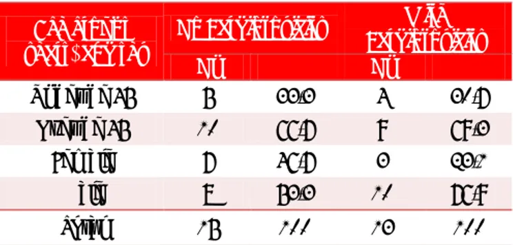
La tabla 1 muestra que el 69,3 % de los pacientes que
desarrollaron cardiotoxicidad (9/13), eran mayores de 45 años, y 10 (76,9 %), eran del sexo masculino.
El 53,8 % de estos presentó cardiotoxicidad a dosis menor de 550 mg/m2 (Tabla 2).
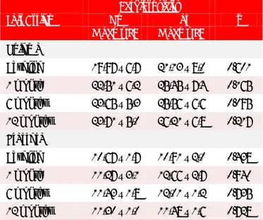
En la tabla 3 se observa la relación del SR, Ɛ y la FEVI, en relación a la cardiotoxicidad. Nótese que tras laadministracióndeunciclodeantraciclinas,los valo-
Tabla 1. Relación entre la edad el sexo y la cardiotoxicidad
por antraciclinas.
Grupos de edad (años) y sexo
Sin
Cardiotoxicidad Cardiotoxicidad Con
Nº % Nº %
Menores de 45 5 33,3 4 30,7
Mayores de 45 10 66,7 9 69,3
Femenino 7 46,7 3 23,1
Masculino 8 53,3 10 76,9
Total 15 100 13 100
Fuente: Hoja de Recolección de datos.
Tabla 3. Relación entre variables ecocardiográficas y
cardiotoxicidad.
Variables No Cardiotoxicidad Sí p
Media ± DE Media ± DE
Strain rate
Basal (ingreso) 1,2567 ± 0,7 1,1933 ± 0,6 0.140 1 mes 1,4193 ± 0,9 0,8638 ± 0,2 0.043 6 meses 1,1933 ± 0,6 0,8792 ± 0,3 0.132 12 meses 1,3087 ± 0,71 0,7400 ± 0,24 0.260
Ɛ
Basal (ingreso) 24,73 ± 13,1 22,92 ± 3,0 0.500 1 mes 21,13 ± 20,2 13,77 ± 4,1 0.031 6 meses 20,33 ± 17,7 14,88 ± 3,2 0.119 12 meses 22,48 ± 12,6 14,24 ± 3,2 0.979 FEVI
Basal (ingreso) 55 ± 4 50 ± 8 0.150
1 mes 52,5 ± 5 54,6 ± 4 0.036
6 meses 53,4 ± 5 53,8 ± 6 0.413
12 meses 56,8 ± 5 52,7 ± 6 0.715
Fuente: Hoja de Recolección de datos p < 0.05
Tabla 2. Relación entre la dosis de antraciclinas y la cardiotoxicidad.
Dosis Acumulativas Cardiotoxicidad Sin Cardiotoxicidad Con p
Nº % Nº %
Menor de 550 mg/m2 2 13,3 7 53,8 0.032
Mayor de 550 mg/m2 13 86,7 6 46,2 0.716
Total 15 100 13 100
Detección precoz de cardiotoxicidad inducida por antraciclinas
res medios del SR y Ɛ se redujeron significativamente
al mes [0,8638 ± 0,2 (p=0.043) y 13,77 ± 4,1 (p=0.031), respectivamente], mientras que la FEVI permaneció dentro de los límites normales [54,6 ± 4 (p=0.036)].
En los pacientes que desarrollaron cardiotoxicidad
(Tabla 4), existió un discreto aumento en los valores
de volumen (21,13 ± 8,0 ml) y presión (10,91 ± 2,0 mmHg) de la AI. Aunque no se evidenciaron diferen-cias significativas entre ambos grupos.
Tabla 4. Relación entre volumen/presión de la aurícula
izquierda.
Aurícula izquierda
Cardiotoxicidad
p
No Sí
Media ± DE Media ± DE
Volumen
Basal (ingreso) 19,97 ± 6,7 21,13 ± 8,0 0.601 1 mes 22,51 ± 6,2 25,45 ± 7,4 0.165 6 meses 23,65 ± 5,3 25,56 ± 6,6 0.095 12 meses 23,71 ± 5,0 26,21 ± 6,8 0.217 Presión
Basal (ingreso) 10,67 ± 1,7 10,91 ± 2,0 0.438 1 mes 11,27 ± 3,1 12,66 ± 2,7 0.940 6 meses 11,43 ± 1,8 12,01 ± 1,2 0.735 12 meses 11,30 ± 1,0 11,48 ± 1,6 0.728
Fuente: Hoja de Recolección de datos p > 0.05
DISCUSIÓN
En esta investigación se encontró que los pacientes más susceptibles para desarrollar cardiotoxicidad fue-ron los mayores de 45 años y del sexo masculino. Tal resultado contrasta con el encontrado por Grenier et al.11, donde los pacientes menores de 18 años fueron
más propensos a desarrollar este tipo de complica-ción. Sin embargo, otro autor12 plantea que en edades
extremas (menores de 18 y mayores de 65 años), apa-rece mayor vulnerabilidad para desarrollar cardiotoxi-cidad, pues consideran que los miocitos de los pa-cientes jóvenes son más susceptibles los fármacos antineoplásicos, así como en el caso de los pacientes adultos, el daño miocárdico subclínico preexistente.
El riesgo de cardiotoxicidad clínica se incrementa con dosis acumulativas de antraciclinas. Existen estu-dios13 que han registrado su aparición con dosis
acu-mulativa menor de 400 mg/m2, y otro informe14,
seña-la, que la incidencia de cardiotoxicidad se aproxima al 30 % con dosis acumulativa de 500 mg/m2. Nuestros
resultados concuerdan con la literatura revisada, aun-que llama la atención, el elevado porcentaje de casos (53,8 %) que desarrolló esta complicación, lo que pro-bablemente pudo estar influenciado por el tamaño muestral, el mayor número de pacientes que recibie-ron dosis acumulativa menor a 500 mg/m2, además de
la variabilidad individual.
Existen autores que plantean que la mecánica lon-gitudinal del ventrículo izquierdo depende predomi-nantemente del subendocardio, que es más vulnerable y sensible a la presencia de enfermedad miocárdica6,15, 16. Por otro lado, la disminución de la distensibilidad
conlleva a alteraciones en la relajación longitudinal, lo que produce un retardo progresivo de la torsión ventricular, que altera la función diastólica y elevalas presiones de llenado ventricular, en una fase en que la FEVI se mantiene normal6. Esta situación promueve el
empleo de otras técnicas ecocardiográficas para iden-tificar precozmente la aparición de cardiotoxicidad6,15, 16. Basado en ello, otros autores, han demostrado que
existe una reducción significativa del SR y Ɛ, con dosis acumulativas muy bajas de fármacos antineoplásicos, en tanto otras variables ecocardiográficas, como la FEVI y el Doppler mitral, permanecen invariables17.
Nuestros resultados coinciden con la literatura revisa-da, donde existió una reducción del SR/Ɛ, siendo muy significativo, después de finalizado un ciclo de trata-miento de antraciclinas. Por lo anteriormente comen-tado se debe señalar, que las técnicas de SR/Ɛ, podrían alertarnos sobre la presencia de una disfunción ven-tricular subyacente, asociada a la quimioterapia, a pesar de una FEVI conservada18.
El discreto aumento (no significativo) del volumen y la presión de la AI se debe a que cuando la aurícula se vacía hacia un ventrículo rígido (por el incremento de su presión telediastólica), ambos parámetros se incre-mentan para mantener un adecuado volumen eyecti-vo, debido a que durante la diástole ventricular, la AI queda expuesta directamente a las presiones del ven-trículo izquierdo: indicador de la duración y gravedad de la disfunción diastólica19.
pre-Chibuzor Nwuruku G, et al.
CorSalud 2014 Jul-Sep;6(3):229-234 233
sión en la AI no fueron significativos. Probablemente en esto influyeron las manipulaciones terapéuticas que se realizaron a algunos pacientes, una vez diag-nosticada la cardiotoxicidad, lo cual pudo ocasionar la reducción notable de la precarga y las presiones de llenado en sentido general20.
CONCLUSIONES
Las técnicas de SR/Ɛ fueron útiles para el diagnóstico precoz de cardiotoxicidad.
RECOMENDACIONES
Es necesario realizar otros estudios con mayor número de pacientes. Se considera que el criterio para la de-tección precoz de cardiotoxicidad, no debe circunscri-birse solo a la reducción de la FEVI (mayor de 10 %), sino que debe tomarse en cuenta el empleo de otras herramientas ecocardiográficas (Ɛ/SR), en aras de pro -mover una valoración integral del paciente.
Nota del Editor
* Strain, y strain rate son palabras del idioma inglés, cuyo significado, en el contexto de la ecocardiografía, es defor-mación miocárdica y deformación miocárdica en el tiempo, respectivamente (tomado de:
http://www.echobasics.de/strain-cas.html).
CorSalud ha decidido utilizarlas en su idioma original debi-do a la alta frecuencia de uso en cardiología y porque consi-dera que no es razonable sustituirlas por otros términos que quizás no expresen con claridad las características de este tipo de variable ecocardiográfica.
REFERENCIAS BIBLIOGRÁFICAS
1. Cardinale D, Colombo A, Lamantia G, Colombo N, Civelli M, De Giacomi G, et al. Anthracycline-induced cardiomyopathy: clinical relevance and response to pharmacologic therapy. J Am Coll Cardiol. 2010;55(3):213-20.
2. Takemura G, Fujiwara H. Doxorubicin-induced car-diomyopathy: from the cardiotoxic mechanisms to management. Prog Cardiovasc Dis. 2007;49(5):330-52.
3. Hunt SA, Abraham WT, Chin MH, Feldman AM, Francis GS, Ganiats TG, et al. Focused update incorporated into the ACC/AHA guidelines for the Diagnosis and Management of Heart Failure in Adults: A Report of the American College of Car-diology Foundation/American Heart Association
Task Force on Practice Guidelines Developed in Collaboration with the International Society for Heart and Lung Transplantation. J Am Coll Cardiol. 2009;53(15):e1-e90.
4. Minotti G, Salvatorrelli E, Menna P. Pharmacolo-gical foundations of cardio-oncology. J Pharmacol Exp Ther. 2010;334(1):2-8.
5. Yeh ET, Bickford CL, Pharm D BC. Cardiovascular complications of cancer therapy: incidence, patho-genesis, diagnosis and management. J Am Coll Cardiol. 2009;53(24):2231-47.
6. Eidem BS. Identification of anthracycline cardiotoxi-city: left ventricular ejection fraction is not enough. J Am Soc Echocardiogr. 2008;21(12):1290-2.
7. Von DD, Layard MW, Basa P, Davis HL Jr, Von Hoff AL, Rozencweig M, et al. Risk factors for doxo-rubicin-induced congestive heart failure. Ann Intern Med. 1979;91(5):710-7.
8. Gharib MI, Burnett AK. Chemotherapy-induced car-diotoxicity: current practice and prospects of pro-phylaxis. Eur J Heart Fail. 2002;4(3):235-42.
9. Monsuez JJ, Charniot JC, Vighat N, Artigou JY. Car-diac side-effects of cancer chemotherapy. Int J Cardiology. 2010;144(1):3-15.
10.Lyu YL, Kerrigan JE, Lin CP, Azarova AM, Tsai YC, Ban Y, et al. Topoisomerase II beta mediated DNA double-strand breaks: implications in doxorubicin cardiotoxicity and prevention by dexrazoxane. Can-cer Res. 2007;67(18):8839-46.
11.Grenier MA, Lipshultz SE. Epidemiology of antracy-cline cardiotoxicity in children and adults. Semin Oncol. 1998;25(4 Suppl 10):72-85.
12.Lipshultz SE, Alvarez JA, Scully RE. Anthracycline associated cardiotoxicity in survivors of childhood cancer. Heart. 2008;94(4):525-33.
13.Swain SM, Whaley FS, Ewer MS. Congestive heart failure in patients treated with doxorubicin: a re-trospective analysis of three trials. Cancer. 2003;97: 2869-79.
14.Schimmel KJ, Richel DJ, Van den Brink RB,Guchelaar HJ. Cardiotoxicity of cytotoxic drugs. Cancer Treat Rev. 2004;30(2):181-91.
15.Mercuro G, Caddeddu C, Piras E, Dessì M, Madeddu C, Deidda M, et al. Early epirubicin myocardial dysfunction revealed by serial Doppler echocardio-graphy. Correlation with inflammatory and oxi-dative stress markers. Oncologist. 2007;1(9):1124-33.
Detección precoz de cardiotoxicidad inducida por antraciclinas
Gietema JA. Cardiovascular toxicity caused by can-cer treatment: strategies for early detection. Lancet Oncol. 2009;10(4):391-9.
17.Marwick TH, Leano RL, Brown J, Sun JP, Hoffmann R, Lysyansky P, et al. Myocardial strain measure-ment with 2-dimensional speckel-tracking echocar-diography: Definition of normal range. JACC Cardio-vasc Imaging. 2009;2(1):80-4.
18.Hare JL, Brown JK, Leano R, Jenkins C, Woodward N, Marwick TH. Use of myocardial deformation imagingtodetectpreclinicalmyocardialdysfunction
before conventional measures in patients under-going breast cancer treatment with trastuzumab. Am Heart J. 2009;158(2):294-301.
19.Pritchett AM, Jacobsen SJ, Mahoney DW, Rode-heffer RJ, Bailey KR, Redfield MM. Left atrial volume as an index of left atrial size: a population based study. J Am Coll Cardiol. 2003;41(6):1036-43. 20.Sawaya H, Sebag IA, Plana JC, Januzzi JL, Ky B,
CorSalud 2014 Jul-Sep;6(3):229-234
RNPS 2235-145 © 2009-2014 Cardiocentro Ernesto Che Guevara, Villa Clara, Cuba. All rights reserved. 229
Cuban Society of Cardiology
______________________
Brief Article
Early detection of anthracycline-induced cardiotoxicity
Geoffrey Chibuzor Nwuruku, MD; Juan A. Prohías Martínez, MD; Ángela M. Castro Arca,
MD; Oyantay Mérida Álvarez, MD; Joel Brooks Tamayo, MD; and Ricardo A. García
Hernández
, MD
Cardiology Department. Hermanos Ameijeiras Hospital. Havana, Cuba.
Este artículo también está disponible en español
ARTICLE INFORMATION
Received: November 21, 2013 Accepted: January 7, 2014
Competing interests
The authors declare no competing interests
Acronyms
DTI: Doppler tissue imaging
Ɛ:strain
LAP: left atrial pressure
LVEF: left ventricle ejection fraction
SR:strain rate
On-Line Versions:
Spanish - English
RA García Hernández Hospital Hermanos Ameijeiras San Lázaro 701, e/ Belascoaín y Marqués González.
Centro Habana 10300.
La Habana, Cuba. E-mail address: ramador@infomed.sld.cu
ABSTRACT
Introduction:Cancer is the most dreaded disease known to mankind. Cardiotoxicity is a complication of antineoplastic treatment, which can be detected early by echocar-diogram.
Objective:Toidentifyechocardiographicvariablesrelatedtotheoccurrenceof cardio-toxicity by anthracycline.
Method: A descriptive, prospective and longitudinal study was conducted with all pa-tientsadmittedtotheHematologyDepartmentofHermanosAmeijeirasSurgical Cli-nical Hospital, from January 2010 to January 2012. 28 patients who received chemo-therapy with anthracyclines were studied. The general information of each patient, as well as the information concerning the transthoracic echocardiogram, was obtained during hospitalization, at one, 6 and 12 months.
Results:69.3% of patients who developed cardiotoxicity were older than 45 years and there was a predominance of males (76.9%). 56.8% had cardiotoxicity at a dose lower than 550 mg/m2 (p = 0.032). Strain rate/Ɛ values in patients who developed cardio-toxicity were significantly reduced at one month [0.8638/0.2 (p = 0.043) and 13.77/ 4.1 (p=0.031)]; while LVEF remained normal [54.6±4 (p=0.036)]. Regarding volume/ pressure of the left atrium, there was an increase in the reference values (21.13 ± 5.08 mland10.91 ± 0.57 mmHg),although withoutstatisticalsignificance(p = 0.217 and p = 0.728).
Conclusions: Strain rate/Ɛ technique has been helpful for early diagnosis of cardio-toxicity.
Key words: Cardiotoxicity, Anthracyclines, Echocardiography, Strain rate
Detección precoz de cardiotoxicidad inducida por antraciclinas
RESUMEN
Introducción:El cáncer es la enfermedad más temible conocida por la humanidad. La cardiotoxicidad, es una complicación del tratamiento antineoplásico, la cual puede ser detectada precozmente mediante ecocardiograma.
Objetivo: Identificar las variables ecocardiográficas relacionadas con la aparición de cardiotoxicidad por antraciclinas.
Early detection of anthracycline-induced cardiotoxicity
todos los pacientes que ingresaron en el servicio de Hematología del Hospital Clínico-Quirúrgico “Hermanos Ameijeiras”, durante el período comprendido entre enero de 2010 hasta enero de 2012. Fueron estudiados 28 pacientes, los cuales recibieron quimioterapia con antraciclinas. La información general de cada paciente, así como la inherente al ecocardiograma transtorácico, fue obtenida durante el ingreso hospitala-rio, al mes, a los 6 y a los 12 meses.
Resultados:El 69,3 % de los pacientes que desarrollaron cardiotoxicidad eran mayo-res de 45 años y existió un predominio del sexo masculino (76,9 %). El 56,8 % pmayo-resen- presen-tó cardiotoxicidad a dosis menor de 550 mg/m2 (p=0.032). Los valores del strain rate/ Ɛ* en los pacientes que presentaron cardiotoxicidad, se redujeron significativamente al mes [0.8638/0.2 (p= 0.043) y 13.77/4.1 (p=0.031)]; mientras que la FEVI, permane-ció normal [54,6±4 (p=0.036)]. En relapermane-ción al volumen/presión de la aurícula izquier-da, existió un incremento en los valores de referencia (21,13 ± 5,08 ml y 10,91 ± 0,57 mmHg), aunque sin significación estadística (p=0.217 y p=0.728).
Conclusiones: Para el diagnóstico precoz de cardiotoxicidad la técnica de strain rate/ Ɛ ha sido útil.
Palabras clave: Cardiotoxicidad, Antraciclinas, Ecocardiograma, Strain rate
INTRODUCTION
Cancer is the most dreaded disease known to man-kind. Some complications arise more due to therapy than due to the disease per se. However, there should be no doubt as to the risk/benefit ratio in the anti-neoplastic treatment of cancer1. Among the
antineo-plastic therapies, anthracyclines are the best studied and the most used in the treatment of many he-matologic malignancies2. The major factor limiting the
use of these drugs is cardiotoxicity, which is defined as the reduction of left ventricle ejection fraction (LVEF) greater than 10% of its normal limit of 55%. This definition is used as a strict criterion for stopping treatment3.
Cardiotoxicity can be acute (during drug admi-nistration or immediately after), early (from days to 12 months after administration) or late (more than 12 months)4. The acute form occurs in less than 1% of
patients and is generally identified by the presence of hypotension, tachycardia, arrhythmia, pericarditis and decreased myocardial contractility. No cardiac mo-nitoring is required during this stage, as it is usually transient and reversible5. Other authors have agreed
that early toxicity is clearly dose dependent6,7;
how-ever, there are other risk factors such as intravenous administration, single high dose, prior radiotherapy on the mediastinum, concomitant use of other cardio-toxic drugs, female sex, extreme ages of life and preexisting subclinical myocardial damage8-10.
This study was designed based on the great
use-fulness provided by echocardiography, with the aim to assess, by echocardiography, the cardiovascular changes that occur with the use of anthracyclinesin our hospital; and determine the relationship between the cumulative dose of chemotherapy and the appear-ance of cardiotoxicity caused by these drugs.
METHOD
A descriptive, prospective andlongitudinal study was conducted with all patients admitted to the Hema-tology Department of Hermanos Ameijeiras Surgical Clinical Hospital, from January 2010 to January 2012. All were requested to sign written informed consent. Inclusion criteria were age over 18 years, diagnosis of lymphoma (Hodgkin and non-Hodgkin) or acute mye-loid leukemia, and treatment with antineoplastic drugs of the Adriamycin or Rubidomycin type, exclusively.
The general information of each patient, as well as the information concerning the transthoracic echocar-diogram, was obtained during hospitalization, at one, 6 and 12 months.
Chibuzor Nwuruku G, et al.
CorSalud 2014 Jul-Sep;6(3):229-234 231
of the mitral valve from two orthogonal apical views (2 and 4 chambers, respectively). Pulsed Doppler in apical view (4-chambers) was used to record mitral flow chart and obtain peak velocity of E wave. The mitral spectral recording was obtained at a scanning speed of 100mm/s. From the same projection, Dop-pler tissue imaging (DTI) was activated, and at the medial mitral annulus the E’ was obtained. Subse-quently, we proceeded to estimate the left atrial pressure (LAP) using the LAP formula = [1.24 (E/E ') + 1.91]. For the strain (Ɛ) and strain rate (SR), DTI color
remained activated with the sample volume placed in the mid-apical septum after acquiring
at least 3 cycles (with optimal ECG signal), a virtual M line was placed in the thickness of the wall, and its width was adjusted to prevent the regis-tration of blood pool and thereby op-timize the noise-signal ratio.
To meet the objectives, the infor-mation was summarized and placed in a database created in SPSS version 16.0, for this purpose the percent was used as summary measure for qua-litative data, and the average and
stan-dard deviation forquantitative variables. Fisher's exact test was used to assess the association among qua-litative variables in relation to the presence of car-diotoxicity. Considering the sample size, the Mann Whitney test was used for comparison of averages. The level of statistical significance was taken into account and the 95% of associated probability was established as significant, i.e., p <0.05.
The results, which were compared with national and foreign authors, are shown in tables and graphs.
RESULTS
Table 1 shows that 69.3% of patients who developed
cardiotoxicity (9/13) were older than 45, and 10 (76.9%) were male.
53.8% of these showed cardiotoxicity at a dose lower than 550 mg/m2 (Table 2).
In Table 3 the relationship of SR, Ɛ and LVEF, in
relation to cardiotoxicity is observed. It can be noted that after administration of an anthracycline cycle, the mean values of SR and Ɛ decreased significantly at one
month[0.8638 ± 0.2(p =0.043) and 13.77± 4.1(p =
Table 1. Relation between age, sex and cardiotoxicity by
anthracycline.
Age groups
(years) and sex No Cardiotoxicity
With Cardiotoxicity
Nº % Nº %
Under de 45 5 33,3 4 30,7
Over de 45 10 66,7 9 69,3
Female 7 46,7 3 23,1
Male 8 53,3 10 76,9
Total 15 100 13 100
Source: Data Collection Sheet.
Table 3. Relation between echocardiographic variables and
cardiotoxicity.
Variables No Cardiotoxicity Yes p
Mean ± SD Mean ± SD
Strain rate
Baseline 1,2567 ± 0,7 1,1933 ± 0,6 0.140 1 month 1,4193 ± 0,9 0,8638 ± 0,2 0.043 6 months 1,1933 ± 0,6 0,8792 ± 0,3 0.132 12 months 1,3087 ± 0,71 0,7400 ± 0,24 0.260 Ɛ
Baseline 24,73 ± 13,1 22,92 ± 3,0 0.500 1 month 21,13 ± 20,2 13,77 ± 4,1 0.031 6 months 20,33 ± 17,7 14,88 ± 3,2 0.119 12 months 22,48 ± 12,6 14,24 ± 3,2 0.979 LVEF
Baseline 55 ± 4 50 ± 8 0.150
1 month 52,5 ± 5 54,6 ± 4 0.036
6 months 53,4 ± 5 53,8 ± 6 0.413 12 months 56,8 ± 5 52,7 ± 6 0.715
Source: Data Collection Sheet p < 0.05
Table 2. Relationship between dose of anthracyclines and cardiotoxicity.
Cumulative dose No Cardiotoxicity Cardiotoxicity With p
Nº % Nº %
Less than 550 mg/m2 2 13,3 7 53,8 0.032
Greater than 550 mg/m2 13 86,7 6 46,2 0.716
Total 15 100 13 100
Early detection of anthracycline-induced cardiotoxicity
0.031), respectively] whereas LVEF remained within normal limits [54.6 ± 4 (p = 0.036)].
In patients who developed cardiotoxicity (Table 4), there was a slight increase in volume (21.13 ± 8.0 ml) and pressure values (10.91 ± 2.0 mmHg) of LA. Al-though no significant differences between both groups were evident.
Table 4. Volume/pressure ratio of the left atrium.
Left Atrium No Cardiotoxicity Yes p
Mean ± SD Mean ± SD
Volume
Baseline 19,97 ± 6,7 21,13 ± 8,0 0.601 1 month 22,51 ± 6,2 25,45 ± 7,4 0.165 6 months 23,65 ± 5,3 25,56 ± 6,6 0.095 12 months 23,71 ± 5,0 26,21 ± 6,8 0.217 Pressure
Baseline 10,67 ± 1,7 10,91 ± 2,0 0.438 1 month 11,27 ± 3,1 12,66 ± 2,7 0.940 6 months 11,43 ± 1,8 12,01 ± 1,2 0.735 12 months 11,30 ± 1,0 11,48 ± 1,6 0.728
Source: Data Collection Sheet p > 0.05
DISCUSSION
This research found that the most susceptible patients to develop cardiotoxicity were males over 45 years. This result contrasts with that found by Grenier et al.11, where patients under 18 were more likely to
develop this complication. However, another author12
states that in extreme ages (under 18 and over 65), there is more vulnerability to develop cardiotoxicity, since they consider that myocytes from young patients are more susceptible to antineoplastic drugs, as well as in the case of adult patients, to preexisting sub-clinical myocardial damage.
The risk of clinical cardiotoxicity increases with cumulative doses of anthracyclines. Studies13 have
re-corded its appearance with cumulative dose lower than 400 mg/m2, and another report14 states that the
incidence of cardiotoxicity approaches 30% with cu-mulative dose of 500 mg/m2. Our results agree with
the literature reviewed, although the high percentage of cases (53.8%) that developed this complication is noteworthy, which probably could have been in-fluenced by the sample size, the higher number of patients who received cumulative doses lower than 500 mg/m2, in addition to individual variability.
Some authors suggest that left ventricular longitu-dinal mechanics depends predominantly on the sub-endocardium, which is more vulnerable and sensitive tothepresenceofmyocardialdisease6,15,16.Inaddition,
the decreased compliance leads to alterations in the longitudinal relaxation, which causes a progressive delay of ventricular torsion, altering diastolic function and raising the ventricular filling pressures, in a phase in which LVEF remains normal6. This situation
pro-motes the use of other echocardiographic techniques to early identify the onset of cardiotoxicity6,15,16. Based
on this, other authors have shown that there is a
sig-nificant reduction in the SR and Ɛ, with very low cumulative doses of antineoplastic drugs, while other echocardiographic variables such as LVEF and mitral Doppler remain unchanged17. Our results are
con-sistent with the literature reviewed, where there was
a reduction in the SR/Ɛ, being very significant after the end of a cycle ofanthracyclines. Based on the above
mentioned it should be noted, that SR/Ɛ techniques
could warn us about the presence of an underlying ventricular dysfunction associated with chemotherapy, despite a preserved LVEF18.
The modest increase (not significant) in the volume and pressure of the LA occurs because when the atrium empties into a rigid ventricle (by increasing its diastolic pressure), both parameters are increased to maintain an adequate ejection volume, and consider-ing that durconsider-ing ventricular diastole the LA is directly exposed to the pressures of the left ventricle, these parameters are indicators of the duration and severity of diastolic dysfunction19.
Chibuzor Nwuruku G, et al.
CorSalud 2014 Jul-Sep;6(3):229-234 233
CONCLUSIONS
SR/Ɛ techniques were useful for early diagnosis of
cardiotoxicity.
RECOMMENDATION
Further studies with more patients are needed. It is considered that the criterion for early detection of cardiotoxicity should not be limited only to reduced LVEF (greater than 10%), but the use of other echocardiographic tools (Ɛ/SR) should be taken into account, in order to promote a comprehensive assessment of the patient.
REFERENCES
1. Cardinale D, Colombo A, Lamantia G, Colombo N, Civelli M, De Giacomi G, et al. Anthracycline-induced cardiomyopathy: clinical relevance and response to pharmacologic therapy. J Am Coll Cardiol. 2010;55(3):213-20.
2. Takemura G, Fujiwara H. Doxorubicin-induced car-diomyopathy: from the cardiotoxic mechanisms to management. Prog Cardiovasc Dis. 2007;49(5):330-52.
3. Hunt SA, Abraham WT, Chin MH, Feldman AM, Francis GS, Ganiats TG, et al. Focused update incorporated into the ACC/AHA guidelines for the Diagnosis and Management of Heart Failure in Adults: A Report of the American College of Car-diology Foundation/American Heart Association Task Force on Practice Guidelines Developed in Collaboration with the International Society for Heart and Lung Transplantation. J Am Coll Cardiol. 2009;53(15):e1-e90.
4. Minotti G, Salvatorrelli E, Menna P. Pharmacolo-gical foundations of cardio-oncology. J Pharmacol Exp Ther. 2010;334(1):2-8.
5. Yeh ET, Bickford CL, Pharm D BC. Cardiovascular complications of cancer therapy: incidence, patho-genesis, diagnosis and management. J Am Coll Cardiol. 2009;53(24):2231-47.
6. Eidem BS. Identification of anthracycline cardiotoxi-city: left ventricular ejection fraction is not enough. J Am Soc Echocardiogr. 2008;21(12):1290-2.
7. Von DD, Layard MW, Basa P, Davis HL Jr, Von Hoff
AL, Rozencweig M, et al. Risk factors for doxo-rubicin-induced congestive heart failure. Ann Intern Med. 1979;91(5):710-7.
8. Gharib MI, Burnett AK. Chemotherapy-induced car-diotoxicity: current practice and prospects of pro-phylaxis. Eur J Heart Fail. 2002;4(3):235-42.
9. Monsuez JJ, Charniot JC, Vighat N, Artigou JY. Car-diac side-effects of cancer chemotherapy. Int J Cardiology. 2010;144(1):3-15.
10.Lyu YL, Kerrigan JE, Lin CP, Azarova AM, Tsai YC, Ban Y, et al. Topoisomerase II beta mediated DNA double-strand breaks: implications in doxorubicin cardiotoxicity and prevention by dexrazoxane. Can-cer Res. 2007;67(18):8839-46.
11.Grenier MA, Lipshultz SE. Epidemiology of antracy-cline cardiotoxicity in children and adults. Semin Oncol. 1998;25(4 Suppl 10):72-85.
12.Lipshultz SE, Alvarez JA, Scully RE. Anthracycline associated cardiotoxicity in survivors of childhood cancer. Heart. 2008;94(4):525-33.
13.Swain SM, Whaley FS, Ewer MS. Congestive heart failure in patients treated with doxorubicin: a re-trospective analysis of three trials. Cancer. 2003;97: 2869-79.
14.Schimmel KJ, Richel DJ, Van den Brink RB,Guchelaar HJ. Cardiotoxicity of cytotoxic drugs. Cancer Treat Rev. 2004;30(2):181-91.
15.Mercuro G, Caddeddu C, Piras E, Dessì M, Madeddu C, Deidda M, et al. Early epirubicin myocardial dysfunction revealed by serial Doppler echocardio-graphy. Correlation with inflammatory and oxi-dative stress markers. Oncologist. 2007;1(9):1124-33.
16.Altena R, Perik PJ, van Veldhuisen DJ, de Vries EG, Gietema JA. Cardiovascular toxicity caused by can-cer treatment: strategies for early detection. Lancet Oncol. 2009;10(4):391-9.
17.Marwick TH, Leano RL, Brown J, Sun JP, Hoffmann R, Lysyansky P, et al. Myocardial strain measure-ment with 2-dimensional speckel-tracking echocar-diography: Definition of normal range. JACC Cardio-vasc Imaging. 2009;2(1):80-4.
18.Hare JL, Brown JK, Leano R, Jenkins C, Woodward N, Marwick TH. Use of myocardial deformation imagingtodetectpreclinicalmyocardialdysfunction before conventional measures in patients under-going breast cancer treatment with trastuzumab. Am Heart J. 2009;158(2):294-301.
Rode-Early detection of anthracycline-induced cardiotoxicity
heffer RJ, Bailey KR, Redfield MM. Left atrial volume as an index of left atrial size: a population based study. J Am Coll Cardiol. 2003;41(6):1036-43. 20.Sawaya H, Sebag IA, Plana JC, Januzzi JL, Ky B,