Guideline
Guidelines for medical treatment of acute Kawasaki disease: Report of
the Research Committee of the Japanese Society of Pediatric
Cardiology and Cardiac Surgery (2012 revised version)
Research Committee of the Japanese Society of Pediatric Cardiology and
Cardiac Surgery Committee for Development of Guidelines for Medical Treatment of Acute Kawasaki Disease
The primary purpose of practical guidelines is to contribute to timely and appropriate diagnosis and treatment of a given disease or condition, in addition to providing current medical informa-tion on pathogenesis and treatment, as determined by specialists in the field. Guidelines, however, should not be considered pro-cedure manuals that limit the treatment options of practitioners, because treatment modalities other than those recommended in such guidelines are often required. Such treatment choices are the result of comprehensive analysis of all medical circumstances, including patient condition, treatment option, and disease sever-ity. Furthermore, certain drugs shown to be useful in studies conducted in other countries may not yet have been approved for
use here in Japan. The results of clinical research (including randomized controlled trials) must be verified in subsequent research, and the safety and effectiveness of a particular treat-ment may take several months to confirm.
Evidence classification
Recent clinical guidelines typically provide evidence levels based on study design and reported effectiveness.
Level (class) based on study design
These are defined as follows: class Ia, systematic reviews, meta-analyses; class Ib, randomized controlled trials; class IIa, non-randomized controlled trials; class IIb, other quasi-experimental studies; class III, non-experimental reports (comparative studies, correlation studies, case studies); and class IV, opinions of com-mittees of experts and authorities.
Classification (grade) based on efficacy
These are given as follows: grade A, highly recommended; grade B, recommended; grade C, recommended, but evidence is uncer-tain; and grade D, contraindicated.
The present guidelines will use these classification systems in reviewing the available evidence for the various treatments.
Background of the present revision of the treatment guidelines
In July 2003, the Scientific Committee of the Japanese Society of
Pediatric Cardiology and Cardiac Surgery published its
Treat-ment Guidelines for Acute Kawasaki Disease(KD). These guide-lines were designed to present, in a clinically relevant manner, the findings of Ministry of Health research done from 1998 through 2000 by the Onishi group at Kagawa Medical University (working under the official title, “The Pediatric Pharmaceutical Investigation Research Group”). This research had been pub-lished as “Research designed to identify and solve problems in the suitable use of pharmaceuticals for pediatric medical treat-ment: pharmaceuticals in cardiology” and had originally been conducted to provide clinical data for the approval of single-use i.v. immunoglobulin (IVIG).
During the 9 years that have passed since the publication of the previous guideline, new data have been collected, and reports on new drug treatments have been published. Members of the International Kawasaki disease Symposium have been waiting
Correspondence: Tsutomu Saji, MD, First Department of Pediatrics, Toho University, 6-11-1 Omori-Nishi, Ota-Ku, Tokyo 143-8541, Japan. Email: saji34ben@med.toho-u.ac.jp
Principal Author
Tsutomu Saji, Department of Pediatrics, Toho University Omori Medical Center, Tokyo, Japan
Section Authors
Mamoru Ayusawa, Department of Pediatrics and Child Health, Nihon University School of Medicine, Tokyo, Japan
Masaru Miura, Division of Cardiology, Tokyo Metropolitan Children’s Medical Center, Tokyo, Japan
Tohru Kobayashi, Department of Pediatrics, Gunma University Gradu-ate School of Medicine, Maebashi, Gunma, Japan
Hiroyuki Suzuki, Department of Pediatrics, Wakayama Medical Uni-versity, Wakayama, Japan
Masaaki Mori, Department of Pediatrics, Yokohama City University Medical Center, Yokohama, Kanagawa, Japan
Masaru Terai, Department of Pediatrics, Yachiyo Medical Center, Tokyo Women’s Medical University, Tokyo, Japan
Shunichi Ogawa, Department of Pediatrics, Nippon Medical School, Tokyo, Japan
Associate Member
Hiroyuki Matsuura, Department of Pediatrics, Toho University Omori Medical Center, Tokyo, Japan
External Evaluation Committee
Tomoyoshi Sonobe, Department of Pediatrics, Japan Red Cross Medical Center, Tokyo, Japan
Shigeru Uemura, Cardiovascular Center, Showa University Northern Yokohama Hospital, Yokohama, Kanagawa, Japan
Kenji Hamaoka, Pediatric Cardiology and Nephrology, Kyoto Prefec-tural University of Medicine Graduate School of Medical Science, Kyoto, Japan
Hirotaro Ogino, Department of Pediatrics, Kansai Medical University, Osaka, Japan
Masahiro Ishii, Department of Pediatrics, Kitasato University School of Medicine, Tokyo, Japan
Received 10 October 2013; accepted 4 February 2014.
for a revision of the previous Japanese guideline. Thus, the Sci-entific Committee was restructured and assigned the task of revis-ing the guideline.
Purpose and methods
Data on IVIG that have accumulated since it was approved and first marketed have confirmed the efficacy and safety of single-use IVIG therapy. In addition, the incidence of coronary artery lesions (CAL) has gradually decreased every year since IVIG
treatment was introduced in Japan.1The incidence of giant
coro-nary artery aneurysms (CAA), however, has remained almost unchanged, which highlights the importance of timely use of second- and third-line treatments for IVIG-resistant patients.
In developing the present guideline, we carefully reviewed the most recent available literature, classified evidence and effi-cacy, and revised suggested treatment methods, including pro-cedures for selecting first-, second- and third-line medications, with a special focus on off-label uses. For example, the previous guideline did not mention new therapeutic agents such as infliximab (IFX), cyclosporin A (CsA), or methotrexate (MTX). In the present edition, risk/benefit considerations are also clearly presented, based on data collected in and outside Japan. Despite the publication of almost 200 reports every year on KD, there is still no universally accepted treatment for IVIG resistance. This is also the case, however, for many other disorders, such as autoimmune disease and rheumatoid conditions, given that no single medication will benefit all patients in the same way. Thus, to ensure optimal outcome, physicians must treat each patient individually.
Diagnosis and treatment of incomplete KD
In the published results of the 21st Nationwide Survey of KD by Jichi Medical School a total of 23 730 cases of KD were reported
in Japan during the 2 year period 2009–2010.1Diagnosis of KD
follows the criteria outlined in the fifth edition of the diagnosis
guidelines for KD,2which requires that at least five of the
fol-lowing six principal symptoms are present: (i) fever persisting≥5
days (including fever that subsides before the fifth day in response to therapy); (ii) bilateral conjunctival congestion; (iii) changes in lips and oral cavity: reddening of lips, strawberry tongue, diffuse injection of oral and pharyngeal mucosa; (iv) polymorphous exanthema; (v) changes in peripheral extremities: reddening of palms and soles, indurative edema (initial stage); membranous desquamation from fingertips (convalescent stage); and (vi) acute non-purulent cervical lymphadenopathy.
Kawasaki disease, however, may also be diagnosed when only four of the aforementioned symptoms are present, if during the period of illness either 2-D echocardiography or coronary angi-ography shows CAA, including dilation of coronary artery, and other causes of CAA can be excluded. A diagnosis of KD is possible even if five or more of the principal symptoms are not present, if other conditions can be excluded and KD is suspected – a condition known as incomplete KD. Indeed, approximately 15–20% of KD patients have incomplete KD. But, even if a patient has four or fewer of the principal symptoms, the illness should not be regarded as less severe, because cardiovascular
abnormalities are not rare in patients with incomplete KD. For this reason, even patients with fewer than five of the aforemen-tioned symptoms should be evaluated for KD. Early treatment is essential, particularly when fever is present, because CAL devel-opment in such cases is not uncommon. Diagnosis of incomplete KD is not a simple matter of adding up the number of overt KD symptoms: the importance and individual characteristics of each symptom of the illness must be correctly assessed. For example, redness and crusting at a bacille Calmette–Guérin (BCG) inocu-lation site in infants younger than 1 year and multilocular
cervi-cal lymphadenopathy in children aged≥4 years are characteristic
features of KD.
Basic pathology
The 2012 Revised International Chapel Hill Consensus Confer-ence Nomenclature of Vasculitides defines KD as an arteritis associated with mucocutaneous lymph node syndrome,
pre-dominantly affecting medium and small arteries.3There is very
little damage to veins. The location of pathological changes clearly differentiates KD from other vasculitis syndromes, given that the principal danger of KD is inflammatory vasculitis of the coronary arteries. Edematous lesions develop in the intima media, and vascular fragility increases due to partial rupture of the internal and external elastic lamina. As a result, the arterial wall can no longer withstand its internal pressure, particularly diastolic pressure, and becomes distended and deformed, leading in severe cases to aneurysm formation. Only a few other diseases cause distension of coronary arteries. These include vasculitis resulting from Epstein–Barr virus infection, lupus, classical periarteritis nodosa, and atherosclerotic lesions. Calcification can also occur in rare cases and affect coronary arteries, for example in cases of renal dialysis in adults and herpes infection in newborns.
Patients with KD may develop multiple lesions in the proxi-mal region and vessels branching out from it. As aneurysms begin to calcify, further pathological distension or development of aneurysms and intimal thickening may develop 2–3 years later in areas with previously disrupted internal and external elastic
lamina.4
Coronary artery lesions
The principal characteristics of KD are dilation of coronary arteries and CAA. Most CAA occur in the proximal region and its
branches, and arteries with a CAA measuring≥8 mm in diameter
are very unlikely to regain their normal morphology. Right CAA may lead to occlusion or recanalization, and left CAA may pro-gress to stenotic lesions.
Suitable pharmaceuticals for treating KD
Treatment of acute KD
The principal objective in treating acute KD is minimizing the risk of developing CAL. In practice, this means quickly suppress-ing the acute-phase inflammatory reaction caused by KD. Except in cases of very mild KD, IVIG should be started before illness day 7. Histological studies have shown that arteritis typically develops by 8 or 9 days after KD onset. Therefore, treatment should begin before this point, to suppress arteritis and hasten resolution of fever and normalization of inflammation markers. In patients with incomplete KD, IVIG should also be begun as soon as possible after a diagnosis of KD, especially if fever is present. In approximately 80% of cases, fever should be lowered
to ≤37.5°C within 48 h of starting IVIG. In 40% of
IVIG-resistant patients, fever can be reduced to ≤37.5°C with
addi-tional IVIG of 1 g/kg. Persistent fever after 48 h of starting IVIG should be regarded as evidence of IVIG-resistant KD. Prevention of CAA in such patients may largely depend on the selection of subsequent treatment.
In addition to CAL, other cardiovascular complications may develop in patients with acute KD, including myocarditis, pericardial effusion, valvular regurgitation, and, rarely, arrhyth-mia. Specific treatment may be required for these sequelae, as well as for cardiac dysfunction or heart failure. Furthermore, other symptom-specific treatment may be required for systemic complications such as edema, hypoalbuminemia, electrolyte imbalances (i.e. hyponatremia), paralytic ileus, hepatic dysfunc-tion, cholecystitis, impaired consciousness, convulsions, anemia, diarrhea, vomiting, and dehydration. Particularly during high-dose IVIG infusion, care must be taken to prevent volume over-load so as to protect the patient from complications such as heart failure.
There is currently no universally accepted classification system to evaluate KD severity and need for IVIG use, although many such scoring systems have been proposed. Initial attempts were
made by Asai and Kusakawa,5which were followed by the Iwasa
score6 and Harada score.7 More recently, predictive models
designed to evaluate the possibility of IVIG resistance were
pro-posed, including the Kobayashi score,8Egami score,9and Sano
score.10In general, such predictive models consider factors such as
age, gender, days of illness, white blood cell count, %neutrophils, hematocrit, platelet count, C-reactive protein (CRP), aspartate aminotransferase (AST), alanine aminotransferase (ALT), total bilirubin, sodium, and albumin. Recently, a randomized controlled trial found that IVIG plus steroid as initial therapy for patients predicted to be at high risk for IVIG resistance improved clinical
and coronary arterial outcomes.11–13 The effectiveness of such
predictive models, however, has not been confirmed in large-scale prospective cohort studies or meta-analyses, and controversy remains as to whether initial therapy with IVIG plus steroids is the optimal treatment.
Choice of treatment for IVIG-resistant patients
Several second-line treatment options are available if fever per-sists or has reappeared at 24 h after first-line treatment. The
efficacy of these second-line treatments for resistance to first-line treatment is currently being investigated by researchers in many countries, but evidence remains limited due to the lack of randomized controlled trials.
Options for second-line treatment include additional IVIG, i.v. methylprednisolone pulse (IVMP), prednisolone (PSL), IFX, ulinastatin (UTI), CsA, MTX, and plasma exchange (PE). The decision to use any of these treatments requires careful consid-eration of patient characteristics. At present, the most commonly
used second-line treatment is additional IVIG,1which is
some-times given in combination with other medications. As for ster-oids, a retrospective study noted a high incidence of giant
aneurysms.14 That small uncontrolled case study reported that
several patients had received steroids before rupture of coronary arteries, which suggests that physicians should carefully consider the decision to use steroids for patients with KD if CAA are already present. When steroids, biologics, or immunosuppres-sants are given to infants, there is also a risk of long-term side-effects, and questions remain regarding the general safety of such medications. Thus, a careful risk/benefit evaluation should be done to consider the likelihood of such adverse effects versus the possibility of CAA formation.
Algorithm for selecting optimal treatment
To decrease the risks of first-line IVIG resistance and CAA, it seems reasonable to consider risk stratification using predictive models and to select more-aggressive initial treatment for patients at high risk of IVIG resistance. Such patients should be treated with 2 g/kg of IVIG in combination with either 2 mg/kg per day PSL or 30 mg/kg per day IVMP. If the patients fail to respond to these treatments, a third-line treatment will be upgraded to a second-line treatment.
Because few studies have assessed the efficacy of medications other than IVIG retreatment, it is impossible at this time to assign an objective order of these treatment options. The present guide-lines, however, offer evidence levels and grades to assist physi-cians in selecting appropriate alternatives. Various methods of calculating KD patient risk scores and, thereby, estimating KD severity have been developed at a number of institutions by different physicians, based on their particular experience with
KD.8–10 The Japanese Society of Pediatric Cardiology and
Cardiac Surgery does not intend to limit the treatment options available to clinicians, especially when such options have already received ethics committee approval at their institution. Instead, the judgment of physicians in selecting treatments should be respected, for practical reasons as well. Such treatments may be given after a physician has established a sufficient basis for selecting a given treatment and received informed consent/assent from the family/patient (Fig. 1).
Immunoglobulin
Purpose
Currently, the most effective anti-inflammatory treatment for KD
is early IVIG.15–17The latest systematic review by the Cochrane
Collaboration states that CAL development can be reduced by a
Fig
.
1
Algorithm
for
the
treatment
of
acute
Ka
w
asaki
disease
(KD).
ASA,
acetyl
salic
ylic
acid;
CRP
,
C-reacti
v
e
protein;
IFX,
infliximab;
IVIG,
i.v
.
immunog
lob
ulin;
IVMP
,
i.v
.
methylprednisolone
pulse;
PSL,
prednisolone;
UTI,
Mechanism of action
Because the causes of KD are unknown, the mechanisms under-lying the therapeutic benefits of IVIG remain speculative. Table 1
lists the hypothesized mechanisms of action.19–22
Indications
I.v. immunoglobulin is suitable for almost all cases of typical acute KD, that is, when KD is diagnosed based on the presence of the principal symptoms specified in the criteria of the diagnostic
guideline for KD2and the patient is at risk for CAL. For patients
with symptoms that only partially fulfill the diagnostic criteria, incomplete KD may be diagnosed – if other diseases or condi-tions can be excluded – after which IVIG should be started as
quickly as possible due to the risk of CAL.17
In cases of less severe KD or spontaneous defervescence, clinicians may refrain from IVIG, in accordance with the consid-erations detailed in the Ministry of Health Group Committee
guidelines for IVIG (Harada score)7and disease severity
stand-ards established at the physician’s institution.
Data from the 21st Nationwide Survey of KD show that IVIG
was given to 89.5% of patients.1
Treatment method and dosage
Period of treatment
I.v. immunoglobulin should be started on or before the seventh day after KD onset. It is essential to quickly reduce inflammation and duration of fever, definitely before illness day 8 or 9, when CAL begin to appear. Markers of systemic inflammation, for example CRP and neutrophil count, should be lowered as well.
One study compared patients receiving IVIG on the fifth of illness day or earlier with those who received IVIG on the sixth through ninth days of illness. Although duration from treatment onset to defervescence was slightly longer overall among those receiving IVIG earlier, total duration of fever was shorter. More-over, the groups did not differ in incidence of fever recurrence or additional IVIG treatment, or in number of days of hospitaliza-tion. Furthermore, 1 year after appearance of symptoms, those
who had received IVIG earlier had a lower incidence of CAL.23
Dosage
The suggested IVIG dosage for acute KD is 2 g/kg per day (single use), 1 g/kg per day for 1 or 2 days continuously (modi-fied single use), or 200–400 mg/kg per day, over 3–5 days (divided dosing).
Studies in a number of countries have shown that, as com-pared with divided-dose regimens, a single dose of 2 g/kg per day significantly reduced CAL incidence, more quickly normalized inflammation markers, and was more effective in reducing
fever.4,5As for 1 g/kg/day use, if clinical efficacy is seen on the
first day, it might not be necessary to continue treatment into the second day.
The 21st Nationwide Survey of KD found that a single dose of 2 g/kg per day IVIG was used in 85% of reported cases and that 1 g/kg per day was given for 1 or 2 days in 6.2% and in 7.7% of
cases, respectively.1
There is no consensus in Japan as to whether older/larger children should be treated with 2 g/kg IVIG or a lower dose.
As for 2 g/kg regimen, the treatment rate varies slightly for different products, although IVIG is typically given over a period of approximately 12 h in North America. In Japan, one product permits use within a similar 12 h period, but the total volume of 2 g/kg IVIG is usually given over a period of 24 h. Because volume overload might occur when the treatment rate is too fast, which can lead to cardiac dysfunction, it is important to adhere to the recommended treatment rate and carefully observe patient hemodynamics.
Product types and directions for use
At present, four brands of IVIG are approved for KD in Japan (Table 2): two are processed with polyethylene glycol (PEG), one is sulfonated, and one is processed to ensure a pH of 4 (acidic). No major differences in efficacy have been reported. Table 2 lists the characteristics of these products, as described in their respec-tive product inserts.
The principal differences are as follows.
Table 1 Immunoregulatory effects of IVIG19–22
I. Fc receptor-mediated effects
Blockade of Fc receptors on macrophages and effector cells Antibody-dependent cellular cytotoxicity
Induction of inhibitory FcγRIIB receptors Promotes clearance of antibodies that block FcRn II. Anti-inflammatory effects
Attenuation of complement-mediated damage Decrease in immune complex-mediated inflammation Induction of anti-inflammatory cytokines
Inhibition of activation of endothelial cells Neutralization of microbial toxins Reduction in steroid requirements Modulation of matrix metalloproteinases III. Effect on B cells and antibodies
Control of emergent bone marrow B-cell repertoires Negative signaling through Fcγreceptor
Selective downregulation/upregulation of antibody production Neutralization of circulating autoantibodies by anti-idiotypes IV. Effect on T cells
Regulation of T-helper cell cytokine production Neutralization of T-cell superantigens
Regulation of apoptosis V. Effect on dendritic cells
Inhibition of differentiation and maturation Regulation of inflammatory cytokine production VI. Other
Mutually interacts with immunological molecules Suppression of autoantibody production against vascular
endothelial cells
Acceleration of phagocytosis arising from binding of neutrophils and macrophages (opsonin effect) Suppression of inflammation-related gene S100 mRNA Suppression of MCP-1 receptor CCR2 gene expression
produced by macrophages
T able 2 IVIG medications Product name Kenk etsu V enilon-I (for i.v . use) Kenk etu Glo v enin-I (for i.v . use) Kenk etu V enoglob ulin IH (for i.v . use) Nisseki Polyglobin N (for i.v . use) Generic name Freeze-dried sulfonated human normal immunoglob ulin Freeze-dried polyethylene glycol-treated human normal immunoglob ulin Polyethylene glycol-treated human normal immunoglob ulin pH 4-treated acidic human normal immunoglob ulin Compan y (manuf acturer/distrib utor) Kak etsuk en-T eijin Pharma Limited Nihon Pharmaceutical–T ak eda Pharmaceutical Japan Blood Products Or ganization-Mitsubishi T anabe Pharma Japan Blood Products Or ganization-Japan Red Cross Society Form of medication Freeze-dried preparation Freeze-dried preparation Liquid medication Liquid medication Constituents (in 2.5 g o f product) Sulfonated human immunoglob ulin G Glycin Human plasma alb umin D-mannitol Sodium chloride 2500 mg 1 125 mg 125 mg 500 mg 450 mg Polyethylene glycol treated human immunoglob ulin G
D-mannitol Glycin Sodium
(1) The sulfonated product (Kenketsu Venilon-I; Teijin, Tokyo, Japan) contains serum albumin, and its sodium concentration is identical to that of saline (154 mEq/L).
(2) The two products processed with PEG come in freeze-dried
(Kenketsu Glovenin-I; Nihon Shinyaku, Kyoto, Japan) and liquid (Venoglobulin IH; Japan Blood Products Organiza-tion, Tokyo, Japan) form. The suggested infusion rate for PEG-processed IG is slightly slower than that of the sulfonated product. Kenketsu Glovenin-I has a sodium con-centration of 154 mEq/L. Because liquid preparations are usually refrigerated until use, they must be warmed to at least room temperature beforehand.
(3) The pH 4-processed IG (Nisseki Polyglobin-N; Japan Red
Cross Society, Tokyo, Japan) comes in liquid form and should also be warmed to at least room temperature before use. During injection, it is essential that the liquid does not leak out of the vein, because this may cause necrosis of the skin. Furthermore, because the preparation contains maltose, the plasma glucose dehydrogenase method should not be used to measure blood sugar after injection, given that this method can be affected by the presence of maltose.
Close monitoring and a slower infusion rate are required during the first 30–60 min, given that all the aforementioned products might result in anaphylaxis during treatment. If no adverse reac-tions occur during the first hour of treatment (rate, 0.01 mg/kg
per min), the maximum rate (<0.03 mg/kg per min) of 2 g/kg
may then be used over a course of 12–20 h.
IVIG retreatment for IVIG-resistant patients
Although IVIG is the established first-line treatment for KD, approximately 15–20% of all KD patients (16.6% of patients in
the 21st Nationwide Survey of KD1) have persistent or
recrudes-cent fever after 2 g/kg of IVIG, and there has been considerable debate regarding the optimal second-line treatment for such patients. The 21st Nationwide Survey of KD reported that addi-tional IVIG was given to a large majority (91.5%) of the 3231 IVIG-resistant patients reported during the survey period. Steroid was given together with IVIG in 29.0% of patients, IFX in 4.3%, immunosuppressants in 3.7%, and PE in 2.2% of patients. IVIG retreatment alone was effective in approximately half of the
patients.24
In recent years, various scoring systems have been developed to evaluate the likelihood of IVIG resistance at the time of
diag-nosis. Representative scoring systems are listed in Table 3.8–10If
such scores suggest that patients are at high risk of IVIG resist-ance, more aggressive primary therapy in combination with the usual first-line treatment of 2 g/kg IVIG plus aspirin can be
considered. In the RAISE study, Kobayashi et al. found that
IVIG plus PSL, started at 2 mg/kg per day and halved every 5 days, was effective in preventing CAL formation and initial
treat-ment failure.8,13In addition, Egamiet al. and Ogataet al. as well
as Sano et al. and Okada et al. reported the effectiveness of
methylprednisolone (MP; 1–3 doses of 30 mg/kg of IVMP) in
combination with IVIG.9–12As compared with patients receiving
only IVIG plus aspirin, defervescence was significantly more
likely, and the incidence of CAL was significantly lower, among patients receiving IVIG plus steroids. Although further research is necessary, it seems advisable to adapt this risk-stratified strat-egy for severe cases so as to reduce the number of IVIG-resistant patients and further lower the incidence of CAL.
Effectiveness
I.v. immunoglobulin was found to be quite safe and, at present, has the greatest effectiveness. For these reasons, its effectiveness has been widely recognized both in Japan and in other countries, and it is also included in the recommendations of many relevant textbooks.
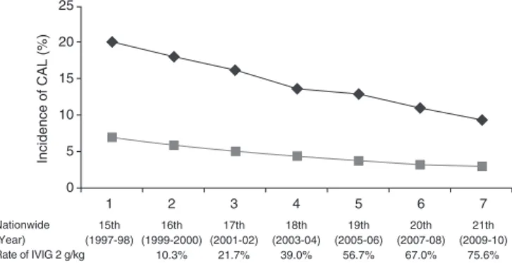
The incidence of cardiac complications reported in the latest Nationwide KD survey decreased to approximately half that in 1997–1998, when patients only rarely received 2 g/kg IVIG. During the acute phase of the illness, that is, until approximately 1 month after disease onset, the incidence of cardiac complica-tions was 9.3%, including dilation, 7.26%; valvular insufficiency, 1.19%; coronary aneurysm, 1.04%; giant coronary aneurysm, 0.24%; coronary artery stenosis, 0.03%; and myocardial
infarc-tion, 0.01%. Even during the convalescent phase, that is, >28
days after disease onset, complications persisted in 3.0% of patients, including dilation, 1.90%; aneurysm, 0.78%; valvular insufficiency, 0.29%; giant aneurysm, 0.22%; stenosis, 0.03%; and myocardial infarction, 0.02%. Furthermore, the number of deaths in Japan within 2 years of KD onset was 51 during the 10 year period 1991–2000, which decreased by more than 60% to 19 cases with the introduction of 2 g/kg IVIG during the subsequent
10 year period, 2001–2010 (Fig. 2).1
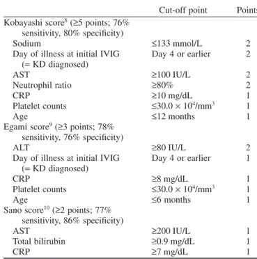
Table 3 Representative scoring systems for evaluating potential IVIG resistance
Cut-off point Points Kobayashi score8(≥5 points; 76%
sensitivity, 80% specificity)
Sodium ≤133 mmol/L 2
Day of illness at initial IVIG (=KD diagnosed)
Day 4 or earlier 2
AST ≥100 IU/L 2
Neutrophil ratio ≥80% 2
CRP ≥10 mg/dL 1
Platelet counts ≤30.0×10⁴/mm3 1
Age ≤12 months 1
Egami score9(≥3 points; 78% sensitivity, 76% specificity)
ALT ≥80 IU/L 2
Day of illness at initial IVIG (=KD diagnosed)
Day 4 or earlier 1
CRP ≥8 mg/dL 1
Platelet counts ≤30.0×10⁴/mm3 1
Age ≤6 months 1
Sano score10(≥2 points; 77% sensitivity, 86% specificity)
AST ≥200 IU/L 1
Total bilirubin ≥0.9 mg/dL 1
CRP ≥7 mg/dL 1
Side-effects
I.v. immunoglobulin is derived from human plasma and is con-sidered to have very few adverse effects and a high level of safety (Table 4). It is necessary, however, to carefully explain the pos-sibilities of rare side-effects to patients and/or their families and to obtain their informed consent before treatment.
In Japan, there have been no reports of viral contamination of any IVIG product. Donated blood is carefully screened to confirm the absence of HBs antigens, HCV antibodies, HIV-1 antibodies, HIV-2 antibodies, and HTLV-1 anti-bodies and to verify normal ALT. Furthermore, when plasma is pooled, the nucleic acid amplification testing (NAT) is used to test for HIV, hepatitis B virus (HBV), hepatitis C virus (HCV), hepatitis A virus, and human parvovirus B19, and only plasma that tests negative for all these infections is used. Using present pharmaceutical production processes, the absence of viruses that are undetectable even by NAT (e.g. abnormal prion proteins and human parvovirus B19), cannot be determined with 100% cer-tainty, but there have been no reports of viral infection due to IVIG. Side-effects are infrequent but include post-treatment chills and shivering, shock (such as cyanosis and hypotension),
anaphylactic reactions, aseptic meningitis,25hemolytic anemia,26
hepatic dysfunction, jaundice, acute renal failure, thrombocyto-penia, and pulmonary edema. Thus, patients should be careful
monitored for these side-effects. Particularly immediately after the start of i.v. treatment and when the infusion rate is increased, the physician should monitor for coldness and shivering, altered consciousness, discomfort, trembling, cyanosis, hypotension, and shock. Finally, cardiac dysfunction or even acute heart failure may develop during acute KD, so close attention should be paid to patient vital signs, and to preventing sudden increases in circulating blood volume, throughout the duration of i.v.
treatment.27,28
Other considerations when using IVIG are as follows.
(1) Patients with IgA deficiency: allergic reactions may occur in
response to IVIG in patients with anti-IgA antibodies.
(2) Patients with renal damage: risk of further impairment of
renal function.
(3) Patients with cerebral or cardiovascular damage or a history
of these conditions: blood viscosity may increase when high-dose IVIG is given rapidly, thus leading to thromboembolic events such as cerebral or myocardial infarction.
(4) Patients at risk for thromboembolism: rapid use of
high-dose IVIG could increase blood viscosity and lead to thromboembolic events.
(5) Patients with hemolytic anemia, blood loss anemia, immune
deficiencies, or immunosuppressive disorders: the possibility of human parvovirus B19 infection cannot be completely excluded. If such infection occurs, severe systemic effects such as fever and sudden or persistent anemia may result.
(6) Patients with reduced cardiac function: high-dose IVIG may
lead to cardiac dysfunction or could worsen existing heart failure.
A post-marketing survey of IVIG for KD noted that among 7259 patients who received IVIG treatment, 484 had a total of 697 adverse events (9.6%) and only 68 patients experienced 78 severe
adverse events (1.1%; Table 5).29
Evidence levels
First-line IVIG treatment: class Ia, grade A.
Additional IVIG treatment in IVIG-resistant patients: class III, grade B.
Combined therapy with IVIG and steroid as first-line treat-ment for suspected IVIG-resistant patients: class Ib, grade B.
15th 16th 17th 18th 19th 20th 21th (1997‐98) (1999‐2000) (2001‐02) (2003‐04) (2005‐06) (2007‐08) (2009‐10)
10.3% 21.7% 39.0% 56.7% 67.0% 75.6% Rate of IVIG 2 g/kg
Incidence of CAL (%)
Nationwide (Year)
0
1 2 3 4 5 6 7
5 10 15 20 25
Fig. 2 Incidence of coronary artery lesions (CAL) vs rate of 2 g/kg i.v. immunoglobulin (IVIG) treatment. ,<30 days; ,≥30 days.
Table 4 General side-effects of immunoglobulin
High incidence Rare
General Fatigue, fever, facial erythema, coldness Anaphylaxis
Systemic side-effects Loss of appetite, myalgia, arthralgia, swollen joints Common cold symptoms, anaphylaxis, blepharedema Neurological Headache, migraine, dizziness Aseptic meningitis, weakness, abnormal sensations Respiratory Shortness of breath, cough, bronchial spasms Pleural effusion, blood transfusion-related lung disorders,
pulmonary edema
Cardiovascular Hypotension, hypertension, chest pain Irregular pulse, myocardial infarction Gastrointestinal Loss of appetite, nausea, vomiting, abdominal pain,
diarrhea
Taste disorder
Renal Renal tubular disorders, renal failure
Dermatological Urticaria, erythema, pimples, pruritus Multiform exudative erythema
Methylprednisolone pulse
Purpose
I.v. methylprednisolone pulse is usually given because of its powerful and rapid immunosuppressive effect (Table 6). Among available steroids, MP treatment is often selected for high-dose i.v. infusion because it is less likely to disrupt electrolyte balance. IVMP is widely used in treating severe pediatric illnesses such as rheumatic disease and kidney disease and is also used in treating confirmed and suspected IVIG-resistant KD.
Mechanism of action
Steroids bind with glucocorticoid receptors in cytoplasm and
regulate nuclear expression of proteins such as NF-κB, which
produces an anti-inflammatory effect referred to as genomic
action.30When high-dose MP is given i.v., however, the saturation
point of these glucocorticoid receptors is greatly exceeded; thus, mechanisms other than genomic action are thought to contribute to its efficacy. Such mechanisms may include acting through
proteins that dissociate from complexes with cytosolic
glucocorticoid receptors, membrane-bound glucocorticoid recep-tors, and functional modification of membrane-bound protein after interlocation of the cell membrane. These mechanisms
precede genomic action.30,31
When used for KD patients, the effects of IVMP are very rapid, which suggests that non-genomic mechanisms stimu-late immunocytological activity and suppress inflammatory cytokines. In confirmed and suspected IVIG-resistant patients, IVMP was reported to limit production of cytokines involved in
inflammation and CAL,32 and to reduce transcription at the
genetic level.33
Indications
Patients suspected of being IVIG resistant on the basis of clinical symptoms and laboratory findings.
Patients found to be IVIG resistant after first-line IVIG treatment.
It should be noted that IVMP treatment for KD is an off-label use.
Treatment method and dosage
In patients with kidney disease or connective-tissue disease, the standard dose of IVMP is 20–30 mg/kg IVMP, given once a day
over a period of 2–3 h, for 1–3 consecutive days.31 For KD
patients, studies of IVMP in combination with first-line IVIG
investigated a single dose of 30 mg/kg IVMP.11,12,34Studies of
second-line IVIG treatment in IVIG-resistant patients
investi-gated the same IVMP dose given once a day, for 1–3 days.32,33,35–39
Because the half-life of IVMP is only 3 h,31some studies used
additional therapy with PSL started at 1–2 mg/kg per day and
gradually tapered over a period of 1–3 weeks.38,39
Effectiveness
First-line therapy with IVIG plus IVMP for all KD patients has
not been proven to prevent CAL.40There is, however, no evidence
that IVMP increases CAL incidence. In a double-blind randomized controlled trial comparing IVIG plus IVMP with IVIG plus placebo, no significant differences were found in factors such as duration of fever, incidence of additional treat-ment, incidence of CAL, and coronary artery diameter, as
indi-cated by Z score.34 A post-hoc analysis of patients requiring
additional treatment, however, found that the incidence of CAL was significantly lower among those who had received IVIG plus IVMP, which suggests that the combined regimen had been effec-tive among IVIG-resistant patients. Studies have also reported that suspected IVIG-resistant patients (as determined by Egami score or Sano score) who received first-line IVIG plus IVMP had earlier defervescence and a significantly lower rate of CAL than
did those who had received IVIG alone.11,12
For patients resistant to initial IVIG, some studies compared IVMP as a second-line treatment to additional treatment with IVIG and found that duration of fever was shorter after IVMP but
that CAL incidence was similar.32,36–40The researchers, however,
highlighted the fact that IVMP therapy was less expensive than
retreatment of IVIG.36,37Nevertheless, the finding of equal
effi-cacy for IVIG and IVMP has not been shown in non-inferiority trials and requires confirmation. One study reported that IVIG-resistant patients who did not respond to additional IVIG had a lower rate of CAL after subsequent IVMP followed by PSL
treatment.39
Side-effects
The reported side-effects of IVMP treatment for KD patients include sinus bradycardia (6–82%), hypertension (10–91%),
hyperglycemia (6–55%), and hypothermia (6–9%).39,41
There-fore, patient vital signs must be very carefully monitored during IVMP, including monitoring of electrocardiogram and blood pressure.
To avoid development of gastrointestinal ulcer, patients can be given H2 blockers and/or other antacid agents. Additional
heparin can also be given as thrombosis prophylaxis.38,39
Never-theless, the necessity of these medications has not been proven.
Evidence levels
Initial IVIG plus IVMP for all KD patients: class Ib, grade C.
Table 5 Post-marketing survey of adverse effects of Ig for KD (no. treatments, 7259)
Side-effect No. events
Hepatic dysfunction 69
Abnormal findings of liver enzyme tests 40
Pruritus, rash 78
Hypothermia 50
Hypotension 19
Aseptic meningitis 19
Pallor 15
Cyanosis 14
Heart failure 13
Shock 13
Peripheral coldness 13
Hemolytic anemia 4
Initial IVIG plus IVMP for suspected IVIG-resistant patients: class Ib, grade B.
Second-line IVMP use for IVIG-resistant patients: class IIb, grade B.
Prednisolone
Purpose
The primary purpose of PSL therapy is to take advantage of its powerful anti-inflammatory effects (Table 6). PSL may quickly resolve KD vasculitis and suppress the potential risk for remodeling of coronary arteries.
Mechanism of action
Prednisolone is the most widely used synthetic corticosteroid hormone, and its glucocorticoid action is stronger than that of cortisol. Through cytoplasmic steroid receptors, PSL inhibits gene transcription of inflammatory cytokines and promotes gene
transcription of anti-inflammatory cytokines.30 PSL also
sup-presses inflammation by inhibiting production of inflammatory
cytokines (e.g. tumor necrosis factor-α [TNF-α], interleukin
[IL]-6, IL-8), chemokines, and cell adhesion molecules. In addi-tion, PSL stimulates production of anti-inflammatory proteins
such as lipocortin, IL-1 receptor antagonists, β-2 adrenergic
receptors, and IκB kinase.
Indications
Patients suspected of being IVIG resistant, based on evaluation of clinical symptoms and laboratory findings.
Patients found to be IVIG resistant after first-line IVIG treatment.
PSL treatment for KD is an off-label use.
Treatment method and dosage
When used in combination with initial IVIG, 2 mg/kg per day
of PSL is given i.v. in three divided doses.13After defervescence
and improvement in the patient’s general condition, PSL can be given orally. After CRP normalizes, the patient is continued for 5 days on the same dosage in three divided doses of 2 mg/kg per day. Thereafter, if fever does not recur, the dosage of PSL is decreased to 1 mg/kg per day in two divided doses on the subsequent 5 days and then a single dose of 0.5 mg/kg per day on the final 5 days. If fever recurs after dose reduction, addi-tional treatment should be considered, including an increase in PSL dose, IVIG retreatment, or other treatments. The most
common periods for relapse are 4–5 days after the
start of PSL and after the dose reduction from 2 mg/kg to 1 mg/kg.
For patients resistant to initial IVIG, the regimen for second-line PSL should, in principle, involve the same dosages and timings as specified for first-line PSL therapy.
Effectiveness
Although corticosteroids are the treatment of choice for other forms of vasculitis, their use has been limited in KD. In 1975, a case–control study showed that fatal cases were more frequently
treated with PSL as compared with matched non-fatal cases.42In
addition, a retrospective study found that PSL had a detrimental
effect when used as initial therapy.14 Finally, a prospective
randomized controlled trial of three groups (receiving either aspirin, flurbiprofen, or PSL plus dipyridamole) did not confirm the efficacy of PSL. These results led to PSL being
contraindicated for KD in the 1980s.43 A retrospective study,
however, in the 1990s of a PSL plus aspirin regimen found this combination to be useful in preventing CAL and shortening
dura-tion of fever,44which led to a reconsideration of PSL therapy. In
2006, a prospective randomized controlled trial comparing initial IVIG plus PSL to initial IVIG alone reported a significantly lower
incidence of CAL in the IVIG plus PSL group.45A subsequent
retrospective study suggested that risk stratification of initial
treatment might be possible using the Kobayashi score;8,46
there-fore, a randomized controlled trial to assess immunoglobulin plus steroid efficacy for KD (RAISE study) was carried out. The RAISE Study showed that among patients with a Kobayashi
score ≥5, initial treatment with IVIG plus PSL significantly
decreased the incidence of CAL and rate of resistance to initial
treatment.13 Although its external validity remains unproven,
initial therapy with IVIG plus PSL for patients at high risk of IVIG resistance could become the standard therapy for severe KD.
Reports have also shown the effectiveness of PSL as a
second-line therapy for IVIG-resistant patients.47 One study however,
reported that PSL therapy might induce CAL formation in IVIG-resistant patients, given that more days have elapsed since the
onset of illness.48No randomized controlled trials have assessed
PSL therapy for IVIG-resistant patients; thus, the efficacy of PSL for this subgroup is unknown.
Side-effects
According to the product labeling, PSL may lead to side-effects such as shock (0.08%), infection (2.54%), Legg-Calvé-Perthes disease (0.36%), gastrointestinal perforation (0.02%), gastroin-testinal hemorrhage (0.80%), gastroingastroin-testinal ulcer (0.02%), diabetes (3.95%), posterior subcapsular cataract (0.09%), pan-creatitis (0.03%), congestive heart failure (0.02%), and impaired hepatic function (1.21%), as well as circulatory collapse, arrhyth-mia, secondary adrenocortical insufficiency, osteoporosis, myo-pathy, thrombosis, increased intracranial pressure, seizure,
abnormal mental function, glaucoma, central serous
chorioretinopathy, esophagitis, and jaundice (incidences
unknown).
Prednisolone is contraindicated for patients with (i) infections for which there is no effective antimicrobial agent, such as sys-temic mycoses; (ii) severe infections accompanied by reduced renal function or chronic renal failure; or (iii) a history of acute myocardial infarction.
Evidence levels
Initial IVIG plus PSL for suspected IVIG-resistant patients: class Ib, grade B.
Biologics (infliximab)
Purpose
The serum concentration of TNF-αis elevated in KD patients,
and several reports have shown a significant association between KD severity and incidence of CAA. IFX suppresses inflammation
by blocking the action of TNF-α(Table 6).
Mechanism of action
Infliximab was originally developed in mice as a mouse antibody
with human TNF-α. IFX is a chimeric monoclonal antibody and
is produced by bonding 25% V-region – a specific antibody derived from mice – with 75% C-region of the human
immuno-globulin G1κ-chain. Because each IFX molecule contains 25%
mouse protein, anti-chimeric antibodies (neutralizing antibodies) develop in approximately 40% of patients; thus, among patients undergoing repeated use, its efficacy decreases and allergic reac-tions might occur. Production of neutralizing antibodies is inhib-ited in patients with rheumatoid arthritis (RA) who receive IFX in
combination with MTX. IFX binds specifically to TNF-α, not to
TNF-β. The mechanisms of action are believed to be as follows:
(i) neutralize soluble TNF-αand block binding of TNF-αto TNF
receptors (p55 and p75); (ii) bind membrane-associated TNF-α
expressed on the surface of TNF-α-producing cells, inducing
apoptosis through complement-dependent cytotoxicity and antibody-dependent cellular cytotoxicity and inhibiting
produc-tion of TNF-α; and (iii) dissociate TNF-αbound to receptors. As
a result of these mechanisms, IFX suppresses activation of inflammatory cells and production of inflammatory cytokines such as IL-1 and IL-6.
Indications
IVIG-resistant patients.
The use of IFX for treating KD is off-label.
Treatment method and dosage
In Japan, IFX is presently approved for use in adults with (i) RA; (ii) inflammatory bowel disease (IBD; Crohn’s disease, ulcera-tive colitis); (iii) intractable uveitis accompanying Behçet disease; (iv) pruritus; and (v) ankylosing spondylitis (AS). In Europe and the USA, it has also been approved for use in treating
Crohn’s disease in children aged≥6 years.49
Children treated with IFX usually receive one dose of 5 mg/ kg. In patients with Crohn’s disease, however, there are reports of other dosages such as 3 mg/kg or 6 mg/kg. For adults with RA, 3–10 mg/kg IFX is given i.v. once every 8 weeks. IFX has a half-life of approximately 9.5 days and is usually given by i.v. drip infusion mixed in 200–500 mL of saline, over a period of at least 2 h. Unlike RA, KD is an acute disease, and MTX and steroids are not usually given as they would be for RA. A single-dose IFX regimen is recommended because KD is an acute disease, unlike RA, and MTX or steroids are not usually con-comitantly used. Studies in the USA have not established a lower age limit for IFX use, but there is no assurance of complete safety when IFX is given to newborns and infants.
Effectiveness
The first experience of the effectiveness of IFX for treating KD
was reported in 2004 by Weisset al., who used it with positive
results to treat a 3-year-old patient who had not responded to
treatment with IVIG and IVMP at the 45th day of illness.50Later,
several reports confirmed the effectiveness of IFX in suppressing inflammation among patients resistant to both IVIG and IVMP. These reports suggested that IFX is safe and effective within a
relatively short time.51–61IFX lowered serum levels of
inflamma-tory markers such as IL-6, CRP, and soluble TNF-α receptor
1.52,62By 2009, a total of 39 cases (patient age range, 1 month–13
years; CAA development, 22 of 39) of IFX use in treating KD
that did not respond to IVIG and/or IVMP had been reported.58In
the USA, IFX was used in approximately 1% of the 4811 IVIG-resistant cases, and its use had increased from 0% in 2001 to
2.3% by 2006.63 In a recent review of additional treatment for
IVIG-resistant patients, either additional IVIG, 3 days of IVMP,
or IFX was recommended.64 The effectiveness of anti-TNF-α
antibody in reducing vasculitis severity was demonstrated in an
animal model of KD vasculitis.65
In Japan, 6 years have passed since IFX was first used as an
off-label treatment for a patient who failed to respond to IVIG.52
The Japanese Society of Kawasaki Disease surveyed the use of IFX during 2006–2011 and found a total of 192 patients treated with IFX during that period. It was effective in around 80% of cases but was unsuccessful in reducing fever in 10–15% of cases. Experimental studies have not reported any severe side-effects; thus, IFX appears to be relatively safe for use in most patients. In general, the incidence of CAA is lower when IFX is used before the 10th day after onset.
Side-effects
After IFX had been approved for RA, it was given to>5000 adult
patients with RA in Japan. Adverse events were reported in 28% of these patients within 6 months of first use; 6.2% of these were severe adverse events, including bacterial pneumonia (2.2%, 108
patients), Pneumocystis pneumonia (0.4%, 22 patients), sepsis
(0.2%, 10 patients), tuberculosis (0.3%, 14 patients), and severe
infusion reaction (0.5%, 24 patients; Table 7).66–76As for patients
with juvenile idiopathic arthritis (JIA), there is a report that adverse events were more frequent at lower doses (3 mg/kg) than
at higher doses (6 mg/kg).69There are limited data, however, on
the safety of IFX in children. Therefore, the indication of IFX for KD should be determined only after carefully assessing the risk– benefit balance on a case-by-case basis.
Infusion-associated reaction
Because IFX is a chimeric monoclonal antibody, it might induce anaphylactic reactions. For this reason, patients receiving IFX should be carefully observed for symptoms such as fever, rash, pruritus, and headache, along with regular monitoring of vital signs. The patient should also be carefully monitored for other side-effects, such as respiratory distress, bronchial spasms,
angioedema, cyanosis, hypoxia, and urticaria.70
Premedication with acetaminophen and/or antihistamines is
As for long-term IFX treatment, in a study of 163 patients with JIA (68 receiving IFX and 95 receiving etanercept; mean age, 17 years; mean treatment period, 22.9 months), there were 71 adverse events, and 62.9% of the events occurred in patients treated with IFX. In contrast, another report found IFX to be safe
and well-tolerated, with few side-effects.73Among patients with
JIA who had been receiving IFX for 1 year, the incidence of infusion reaction was 3.3% among those who had been receiving
a dose of 3 mg/kg and 7% among those receiving 6 mg/kg.74,75In
addition, neutralizing human antichimeric antibodies (HACA) were found in many patients who developed an infusion-associated reaction. HACA was also found in 7.1–12.1% of
pediatric patients with Crohn’s disease.76
Delayed hypersensitivity symptoms were seen≥3 days after
repeated use of IFX (24 h–3 weeks after treatment), including myalgia, rash, fever, fatigue, arthralgia, pruritus, edema of the hands and face, dysphagia, urticaria, pharyngeal pain, and head-ache. Table 7 lists the points of concern when giving IFX to pediatric patients. For these reasons, additional use of IFX in patients with acute KD is not recommended.
Exacerbation of heart failure
Infliximab worsened symptoms of heart failure in adults with New York Heart Association (NYHA) class III or IV disease and
left ventricular ejection fraction<50%. Even among NYHA class
II patients, IFX should be used with caution because serum brain natriuretic peptide is elevated in acute KD, which suggests asymptomatic cardiac impairment, including subclinical myocar-ditis, cardiac hypofunction, pericardial effusion, and
atrioven-tricular valvular regurgitation.70
Exacerbation of infectious diseases
The possibility of worsening of infectious disease is especially important for infants who have not yet been vaccinated against BCG. QuantiFERON (QFT-TB Gold; Japan BCG Laboratory, Tokyo, Japan) testing is not affected by BCG vaccination or mycobacterial infection, but a false-positive result may occur if a patient has a history of past infection. Although pediatric patients sometimes show false-negative results, QuantiFERON testing may nevertheless be useful. It is essential to conduct a careful diagnostic interview, including questions on infections in family members and the patient’s BCG vaccination status. Findings from chest radiography or computed tomography, if required, are also important.
As for live vaccines other than BCG, such as the rotavirus vaccine, use of IFX should be postponed if the patient has had
such a vaccination<2 months previously or has had vaccines for
measles–rubella, mumps, or chickenpox <1 month previously.
IFX is contraindicated if any active infection is present. Unfortunately, evidence is limited regarding the interval nec-essary between inoculation with a live vaccine and IFX treat-ment. Some specialists suggest an interval of 2–3 months to ensure patient safety.
Development of malignant tumors
When etanercept was used to treat 1200 patients with JIA, five patients developed malignancies, including Hodgkin lymphoma, non-Hodgkin lymphoma, thyroid carcinoma, yolk-sac cancer, and cervical dysplasia of the uterus. All these patients, however, had also been treated with other immunosuppressants, and two had received adalimumab and IFX as well. Before IFX is given, the possible side-effects should be carefully explained to the patient
and/or family, and written informed consent should be obtained.71
The US Food and Drug Administration reported that 48 patients developed malignant carcinomas (of which half were lymphomas)
after receiving anti-TNF-αagents, and 11 patients died. Among
the patients, IFX was given to 31, etanercept to 15, and adalimumab to two patients; 88% of the patients developing malignant carcinomas had also received other
immunosuppres-sants (e.g. azathioprine and MTX).72The present data do not show
a conclusive association between IFX and malignant disease.
Carriers of hepatitis B and C
Among adult patients with rheumatic diseases, asymptomatic carriers of HBV or chronic hepatitis may experience reactivation
of HBV or de novo hepatitis.77,78Thus, testing for HBs antigens
and HBs and HBc antibodies is necessary before IFX treatment. Because HBV carrier status and presence of chronic viral hepa-titis are associated with higher risk of activation of these viruses and exacerbation of existing hepatitis, IFX use in such patients should be avoided, as recommended by the Japan College of
Rheumatology.78
Screening for HCV infection should be done before IFX treat-ment. IFX is also contraindicated for patients with active hepa-titis C. Patients who are positive for HCV but do not have active hepatitis should be carefully monitored if IFX is used. Although the safety of IFX for hepatitis C patients has not been confirmed,
Table 7 Severe adverse effects and contraindications of anti-TNF-αtreatment for children66–76
Severe adverse effects
Overresponse at treatment site Infusion reaction
Varicella infection
Latent infections (tuberculosis etc.)
Neurological demyelination diseases (multiple sclerosis etc.) Neuropsychiatric side-effects
Fatigue, headache, vertigo, depression, anxiety Pain amplification syndrome
Malignant tumors Immunogenicity Contraindications
Complete contraindications Active infections
Recurrent infections and history of chronic infections Existing untreated tuberculosis
Multiple sclerosis, optic neuritis
Combined use with anakinra (anti-IL-1 receptor antagonist) Active or recent (previous 10 years) malignant tumor (except
skin tumors) Relative contraindications
Pregnancy, breastfeeding HIV, HBV, or HCV infection
there are no reports in Japan or other countries of IFX worsening hepatitis C. Nevertheless, consultation with a pediatric liver spe-cialist is recommended before beginning IFX treatment.
Other
Infliximab is contraindicated in patients with demyelination dis-orders or allergy to IFX. For patients with KD, severe complica-tions due to IFX are likely to be uncommon because IFX is mostly given as one dose and because KD patients usually have no other chronic active infectious disease. Many children, however, become susceptible to acute infectious disease at early infancy thus, IFX should be used only after careful examination for active infections such as pneumonia, otitis media, and urinary tract infections. In addition, long-term follow up of possible side-effects is required.
Evidence levels
When used for IVIG-resistant patients: class IIb, grade C.
Ulinastatin
Purpose
The principal action of UTI is to reduce inflammatory vascular lesions caused by proteolysis, edema, necrosis, and hemorrhage
(Table 6).79
Mechanism of action
Ulinastatin is a human urinary trypsin inhibitor, purified from human urine. UTI is a polyvalent enzyme inhibitor – a serine protease inhibitor – with a molecular weight of 67 000 kDa and blocks various protein-degrading pancreatic enzymes, including trypsin. UTI is produced by many organs, including liver, kidney, pancreas, lungs, heart, adrenals, stomach, large intestine, brain, and testes.
Suppression of TNF-α
Ulinastatin suppresses production and secretion of inflammatory
cytokines, for example TNF-α, IL-6, and IL-8 from neutrophils
or TNF-α from monocytes.80 It also inhibits expression of
intercellular adhesion molecule-1 on the surface of vascular
endothelial cells activated by TNF-α, thereby playing a
protec-tive role with regard to endothelial cells.
Blocking of neutrophil elastase
Ulinastatin has a dual action, first blocking elastase release, espe-cially from neutrophils and platelets, and then deactivating elastase as it is released. UTI removes oxygen radicals and reduces the activity of cytokines and cell adhesion factors. By stabilizing lysosome membranes, UTI suppresses the release of various protein-degrading enzymes. Finally, it also blocks the release of inflammatory cytokines of myocardial inhibitory factor
containing TNF-αand hypercoagulopathy.81
Indications
IVIG-resistant patients.
Initial treatment in combination with IVIG. Its use in KD treatment is off-label.
Treatment method and dosage
Although optimal dosage has not been determined for pediatric patients, several reports show that a dose of 5000 U/kg given 3–6 times/day, not exceeding 50 000 units/dose, is suitable for KD patients. UTI has a half-life of only 40 min when given i.v. at 300 000 U/10 mL. UTI is officially approved to treat two condi-tions: (i) acute pancreatitis in the earlier phase (adult dosage, 25 000–50 000 units i.v. 1–3 times/day with dose tapering there-after); and (ii) acute circulatory collapse (adult dosage, 100 000 units i.v. 1–3 times/day).
Effectiveness
Ulinastatin has been reported to inhibit mRNA transcription of prostaglandin H2 and thromboxane A2 in polynuclear
leukocytes.82 It also prevented neutrophil-induced damage to
endothelial cells.83The first use of UTI was reported in 1993,
after which several case studies were reported. These reports appeared to support the effectiveness and safety of UTI treatment under certain conditions, such as (i) when given as a single dose to patients with clinically mild disease; (ii) when it allowed a reduction in IVIG dose in the context of combination therapy; and (iii) when IVIG was ineffective due to non-response or
resist-ance to IVIG.84,85Although these studies enrolled only a small
number of patients, and there have been no well-designed clinical studies of UTI, it has been recognized and used as an additional
option for treating IVIG-resistant patients.86Recent retrospective
cohort studies showed that as a first-line treatment UTI in com-bination with IVIG plus aspirin was less likely to require second-line treatment and had a lower risk of CAA among patients at
high risk for IVIG resistance, as defined by Kobayashi score.87
Side-effects
The most important side-effect of UTI is anaphylactic shock. UTI should be used carefully if the patient has a history of drug allergies or allergic reactions to products containing gelatin or a past history of UTI use. Other side-effects include liver dysfunc-tion (0.5%), leukopenia (0.2%), rash, pruritus (0.1%), diarrhea (0.1%), angialgia (0.1%), increased AST and/or ALT, eosino-philia, and vascular pain at the injection site. Also, if UTI is given along the same route as IVIG and the medications are thus mixed, the drug will become white and turbid. To avoid this, a different i.v. route can be selected. Alternatively, IVIG may be paused and the i.v. route can be flushed with saline before and after UTI infusion, after which IVIG infusion can continue.
Evidence level
First-line treatment with IVIG plus UTI: class IIa, grade B. IVIG-resistant patients: class IIb, grade C.
Immunosuppressants
Cyclosporin A
Purpose
In 2008, Onouchi et al. reported a susceptibility gene of KD:
inositol triphosphate (Table 6).88ITPKC suppresses T-cell
activ-ity through the calcineurin/nuclear factor of activated T-cells (calcineurin/NFAT) cascade. Patients with suppressed ITPKC function may produce more cytokines, such as IL-2. For this reason, ITPKC was thought to be a critical gene contributing to IVIG resistance and development of CAA. CsA is used to block calcineurin function and suppress cytokine production.
Several studies evaluated the efficacy of CsA in IVIG-resistant
patients.89–91Accumulating evidence of its effectiveness spurred
multicenter observational studies in Japan and other countries, and the results of these studies indicate that CsA is safe and
well-tolerated.90,91
Mechanism of action
Cyclosporin A binds and inhibits calcineurin, which has a major role in signal transduction that results in increased T-cell activity. By dephosphorylating NFAT, the transcription factor for IL-2 genes, the nuclear import of NFAT is blocked, and production of
cytokines such as IL-2 is inhibited.92
Indications
IVIG-resistant patients.
Its use in treating KD is off-label.
Treatment method and dosage
Usually, 4 mg/kg per day of Neoral® (Novartis Pharmaceuticals UK, Surrey, UK) is given orally in two divided doses before
meals.90The required dose is drawn into a 1 mL syringe and can
be given to infants. Outside Japan, some researchers believe that the absorption of CsA is reduced during acute KD. Thus, they start patients on i.v. 3–5 mg/kg per day. After resolution of fever, 10 mg/kg per day of Neoral is given orally in two divided doses
of 5 mg/kg.91In principle, before the fifth dose on the third day,
the trough level of CsA should be monitored to confirm that it is within the therapeutic range (60–200 ng/mL). If it is not within the therapeutic range and fever remains, the dose may be
increased by 5–8 mg/kg per day.90There is no established
dura-tion of treatment, but CsA is usually given until CRP again normalizes, or for a period of 10–14 days. This period may be
extended if the dose is tapered.91Therapeutic doses of aspirin
30–50 mg/kg per day should be given in combination with CsA until defervescence is confirmed.
Effectiveness
Cyclosporin A has not been evaluated in prospective randomized trials, but observational studies of its use as a third-line treatment in IVIG-resistant patients showed that fever was reduced within 72 h in most patients receiving CsA, and CRP returned to
normal.90,91Additional IVIG, however, was occasionally required
for cases in which CsA was ineffective.90It should be noted that
there are no reports of its use in infants younger than 4
months.90,91
Side-effects
There have been no reports of severe side-effects in treating KD. In approximately 40% of patients, asymptomatic hyperkalemia
was observed in serum samples 3–7 days after treatment. Because plasma samples did not show evidence of hyperkalemia,
these may have been cases of pseudohyperkalemia.90There have
also been reports of hypomagnesemia,91but no reports have noted
arrhythmias due to electrolyte imbalances. Other side-effects reported in patients receiving long-term CsA include hirsutism and hypertension in a few patients.
Evidence level
Class III, grade C.
Methotrexate
Purpose
In 2008, Lee et al. reported that MTX reduced fever and
sup-pressed inflammation in IVIG-resistant patients.93
Mechanism of action
Methotrexate (4-amino-N10-methylpteroyl glutamic acid) is a folic acid antagonist. Pharmacologically, MTX (i) inhibits syn-thesis of purine bodies; (ii) increases adenosine release; (iii) inhibits production of inflammatory cytokines; (iv) suppresses lymphoproliferation; and (v) suppresses migration and adhering of neutrophils; and (vi) suppresses serum immunoglobulin. The mechanism by which low-dose MTX suppresses inflammation, however, has not been confirmed.
Indications
IVIG-resistant patients.
Use of MTX in treating KD is off-label.
Treatment method and dosage
Dosage: 10 mg/m2, given orally once a week. Do not provide
folic acid supplements. MTX is given until defervescence. In the
report by Leeet al. describing the use of MTX, the median total
dosage was 20 mg/m2(range, 10–50) given in two divided
doses.93
Effectiveness
Although there have been no prospective randomized trials of MTX, in a case series describing 17 IVIG-resistant patients who received MTX, fever recurred 7 days after the start of MTX in three patients and 14 days after the start of MTX in one patient. Fever resolved, however, in all four of these patients after they received their second or third dose of MTX. Finally, there was no fever recurrence after MTX was discontinued.
Side-effects
The side-effects of MTX at standard doses include gastrointesti-nal disturbances, hair loss, and myelosuppression, but these
side-effects were not seen at low doses.93In general, side-effects could
include shock or anaphylaxis, myelosuppression, infection, hepatic dysfunction, and acute renal failure.
Evidence levels