Departamento de Bioquímica y Biología Molecular e
Instituto de Biología y Genética Molecular (UVa-CSIC)
TESIS DOCTORAL:
Función de los canales de K
+
y Ca
2+
en
un modelo de hipertensión esencial
Presentada por Sendoa Tajada Esteban para optar al grado de
doctor por la Universidad de Valladolid
Abbreviations
5‐HT Serotonin
ABC ATP‐Binding Cassette
Ach Acetylcholine
Ad Adventitia
AID Alpha Interaction Domain
ANP Atrial Natriuretic Peptide
Ao Aorta
AOTF Accusto‐Optic Tunable Filter
Ar Artery
ATP Adenosine triphosphate
BAPTA 1,2‐bis(o‐aminophenoxy)ethane‐N,N,N',N'‐tetraacetic acid
BID Beta Interaction Domain
BKCa Large conductance Ca2+‐dependent potassium channels
BP Blood Pressure
BPH Blood Pressure High
BPN Blood Pressure Normal
BSA Bovine Serum Albumin
cAMP Cyclic Adenosine Monophosphate
CGRP Calcitonin‐Gene‐Related‐Peptide
CICR Calcium‐Induced Calcium Release
ClC Chloride channel
CNN1 Calponin 1
CO Cardiac Output
CS Cardiovascular System
Ct Threshold cycle
DAG Diacylglycerol
DHP Dihydropyridine
DHPs Dihydropyridines
DMEM Dulbecco’s Modified Eagle’s Medium
DP Diastolic Pressure
DTE 1,4‐Dithioerythritol
DTT Dithiothreitol
EC50 Half maximal effective concentration
EDHF Endothelium‐Derived Hyperpolarizing Factor
EGTA Ethylene Glycol Tetraacetic Acid
EK Potassium equilibrium potential
EMCCD Electron Multiplying Charge Coupled Device
eNOS Endothelial Nitric Oxide Synthase
ER Endoplasmic Reticulum
F Fluorescence intensity of each pixel
Fo Average resting fluorescence intensity
G/V Conductance‐Voltage relationship
GAPDH Glyceraldehyde‐3P‐dehydrogenase
GBP Gabapentin
GDP Guanosine diphosphate
GFP Green Fluorescent Protein
Glb Glibenclamide
Gmax Maximum conductance
GTP Guanosine triphosphate
HEPES 4‐(2‐hydroxyethyl)‐1‐piperazineethanesulfonic acid
HTN Hypertension
HVA High‐Voltage Activated
I Intima
I/V Intensity‐Voltage relationship
IKCa Intermediate conductance Ca2+‐dependent potassium channels
IP3 Inositol triphosphate
IP3R Inositol triphosphate recepptor
KATP ATP‐dependent potassium channels
KCa Ca2+‐dependent potassium channels
KIR Inward rectifier potassium channels
Kv Voltage‐dependent potassium channels
L Lumen
LA Left atrium
LTCCs L‐type calcium channels
LV Left ventricle
LVA Low‐Voltage‐Activated
M Media
MHC Myosin Heavy Chain
MII Myosin II‐type
MLC Myosin Light Chain
MLCK Myosin Light Chain Kinase
MLCP Myosin Light Chain Phosphatase
MP Mean Pressure
MT Myogenic Tone
NAd Noradrenaline
NDPs Nucleotide diphosphates
Nif Nifedipine
NMDG N‐Methyl‐D‐Glucamine
NO Nitric Oxide
NOS3 Nitric Oxide Synthase 3
nPs Activity of Ca2+ sparklets
PA Pulmonary Artery
PAF Platelet Activating Factor
PGE E type prostoglandins
PGF F type prostoglandins
PGI2 Prostacyclin
Phe Phenylephrine
Pi Phosphate group
PI3K Phosphatidyl Inositol‐3 Kinase
Pin Pinacidil
PIP2 Phosphatidylinositol 4,5‐bisphosphate
PKA Protein Kinase A
PKG Protein Kinase G
PLC Phospholipase C
PM Plasmatic Membrane
PMCA Plasma Membrane Ca2+‐ATPase pump
Po Open probability
PSS Physiological Saline Solution
qPCR Quantitative real time Polymerase Chain Reaction
RA Right Atrium
RAAS Rennin‐Angiotensin‐Aldosterone System
ROCs Receptor Operated Channels
RP18S Ribosomal Protein 18S
RT Room Temperature
RV Right Ventricle
RyR Ryanodine Receptor
SACS Stretch‐Activated Channels
SCG Superior Cervical Ganglion
SDS‐PAGE Sodium Dodecyl Sulfate PolyAcrylamide Gel Electrophoresis
SEM Standard Error of the Mean
SERCA Sarcoplasmic Reticulum Ca2+‐ATPase pump
SHR Spontaneously Hypertensive Rats
SKCa Small conductance Ca2+‐dependent potassium channels
SMDS Smooth Muscle Dissociation Solution
SOCs Store Operated Channels
SP Systolic Pressure
SR Sarcoplasmic Reticulum
STOCs Spontaneous Transient Outward Currents
SUR Sulfonylurea Receptors
TBS
Tris Buffered Saline
TEA Tetraethylammonium
TGM Tris Glycine Methanol
TIRF Total Internal Reflection Fluorescence
TLDA TaqMan Low Density Array
TM Transmembrane helices
TPR Total Peripehrical Resistance
TRPs Nonspecific cationic channels
V Vein
VDCCs Voltage‐Dependent Calcium Channels
VM Membrane potential
VSM Vascular Smooth Muscle
VSMCs Vascular Smooth Muscle Cells
VWA Von Willebrand factor type A
INDEX
1 INTRODUCTION ... 1
1.1 HYPERTENSION ... 2
1.2 OVERVIEW OF THE CARIOVASCULAR SYSTEM ... 3
1.3 BLOOD VESSELS ... 4
1.4 THE VASCULAR WALL ... 6
1.4.2 The Tunica Intima ... 6
1.4.2 The Tunica Media ... 7
1.4.3 Tunica Adventitia... 7
1.5 VASCULAR SMOOTH MUSCLE ... 7
1.6 VASCULAR SMOOTH MUSCLE CELLS STRUCTURE ... 8
1.6.1 Caveolae ... 9
1.6.2 Sarcoplasmic reticulum ... 10
1.6.3 Contractile machinery ... 12
1.6.4 Gap‐junctions ... 14
1.7 VASCULAR SMOOTH MUSCLE FUNCTION AND REGULATION ... 15
1.7.1 Crossbridge cycle ... 15
1.7.2 Intracellular calcium homeostasis ... 17
1.7.3 Excitation contraction coupling in vascular smooth muscle ... 18
1.8 VASCULAR TONE ... 20
1.8.1 Regulation of vascular tone ... 22
1.9 VSMCs ION CHANNELS AND VASCULAR TONE ... 26
1.9.1 Potassium channels ... 28
1.9.2 Calcium channels ... 33
1.9.3 Other channels ... 37
1.10 ION CHANNELS AND HYPERTENSION ... 39
1.10.1 Vascular ion channels remodelling during hypertension ... 39
1.10.2 Effect of hypertension on VM ... 40
1.10.3 Effect of hypertension on potassium channels ... 41
1.10.4 Effect of hypertension on calcium channels ... 44
2 OBJETIVES ... 48
3 MATERIALS & METHODS ... 52
3.1.3 Myography set‐up ... 57
3.1.4 TIRF imaging‐set‐up ... 58
3.2 SOLUTIONS ... 60
3.2.1 Electrophysiological solutions ... 60
3.2.2 VSMCs isolation solutions ... 62
3.2.3 HEKs culture media ... 63
3.2.4 Western‐blot solutions ... 64
3.2.5 Myography solutions ... 65
3.2.6 Calcium imaging solutions ... 65
3.3 PLASMIDS ... 66
3.4 DRUGS ... 66
3.5 METHODS ... 67
3.5.1 Hypertension mouse model ... 67
3.5.2 Blood pressure measurements ... 67
3.5.3 VSMCs isolation ... 68
3.5.4 RNA isolation and real‐time PCR ... 69
3.5.5 Western blot... 76
3.5.6 Electrophysiological methods ... 78
3.5.7 Myography methods ... 83
3.5.8 Calcium imaging methods ... 87
3.5.9 Maintenance and transfection of cultured HEK cells ... 90
4 RESULTS ... 92
4.1 ESSENTIAL HYPERTENSION MOUSE MODEL ... 94
4.1.1 Characterization of VSMC excitability in BPN and BPH mesenteric arteries ... 95
4.2 CHARACTERIZATION OF INWARD‐RECTIFIER CHANNELS IN VSMCs FROM BPN AND BPH MICE…. ... 99
4.2.1 mRNA expression profile of inward‐rectifier channel subunits ... 99
4.2.2 Expression of KIR and KATP channels‐encoding proteins in BPN and BPH VSMCs ... 102
4.2.3 Functional characterization of KIR and KATP channels in isolated VSMCs... 103
4.2.4 Characterization of KIR and KATP channel contribution to vascular tone in pressurized mesenteric arteries from BPN and BPH mice ... 106
4.3.1 Functional characterization of LTCCs channels in isolated VSMCs ... 109
4.3.2 mRNA expression profile of LTCCs ancillary subunits ... 114
4.3.3 Functional characterization of α2δ LTCCs accessory subunit in isolated VSMCs ... 116
4.3.4 Specific interaction of gabapentin with the α2δ accessory subunit of VSMCs LTCCs ... 116
4.3.5 Dose response effects of GBP in native VSMCs and in LTCC HEK transfected cells . 118 4.3.6 Expression of LTCCs proteins in BPN and BPH VSMCs ... 120
4.4 DIFFERENCES IN LTCC ACTIVITY MEASURED AT PHYSIOLOGICAL MEMBRANE POTENTIALS ... 121
4.4.1 Ca2+ sparklets site density ... 121
4.4.2 Ca2+ sparklets open probability ... 122
4.4.3 Ca2+ sparklets dwell time ... 123
4.4.4 Ca2+ sparklet amplitude distribution ... 124
4.5 FUNCTIONAL CHARACTERIZATION OF Ca2+ SPARKS AND BKCa ACTIVITY IN BPN AND BPH CELLS ... 126
4.5.1 Sparks frequency and amplitude ... 127
4.5.2 STOCs frequency and amplitude ... 128
4.5.3 BK channel Ca2+ sensitivity ... 129
5 DISCUSSION ... 132
5.1 Vascular bed distribution of inward rectifier K+ channels ... 136
5.2 Functional expression of KIR and KATP channels in mesenteric arteries ... 137
5.3 Changes in KIR and KATP channels in essential hypertension ... 138
5.4 Contribution of the BPN/BPH model for understanding the molecular basis of hypertension ... 139
5.5 Calcium channels remodelling in hypertension ... 140
5.6 LTCCs functional expression ... 142
5.7 Changes in the composition of LTCCs in hypertension ... 144
5.8 Functional activity of LTCCs in basal conditions ... 147
5.9 Uncoupling in Ca2+ SPARKs and BK STOCs ... 150
5.10 General conclusion ... 151
6 CONCLUSIONS ... 154
7 REFERENCES ... 158
8 APPENDIX ... 174
8.1 Poiseuille’s Law ... 176
8.4.1 Patch‐Clamp configurations ... 178
8.4.2 Special configurations ... 179
9 RESUMEN ... 181
1.1INTRODUCCIÓN ... 183
1.2 OBJETIVOS ... 183
1.3 MATERIALES ... 184
1.4 RESULTADOS ... 184
1.4.1 Estudio de los canales de K+ ... 184
1.4.2 Estudio de los canales de Ca2+ ... 185
1.4.3 Acoplamiento spark‐STOCs ... 186
1.5 DISCUSION ... 186
1.1 HYPERTENSION
Hypertension (HTN) or arterial hypertension, is defined as a chronic elevation of systolic blood
pressure (BP) above 140 mmHg or diastolic BP over 90 mmHg or both in adults (Carretero &
Oparil, 2000). Its high prevalence (close to 30% of adult population in developed countries
(Chobanian et al., 2003)), makes hypertension a relevant risk factor contributing to
cardiovascular morbidity and mortality. HTN is associated with functional and structural
cardiovascular pathologies such as ischemic heart disease, cerebrovascular disease, heart
failure and renal failure, and is estimated to cause more than 7 million premature deaths per
year worldwide.
About 95 per cent of all cases of hypertension are diagnosed as essential hypertension (or
primary hypertension), in which it is not possible to identify a single, specific cause. The
remaining cases are forms of hypertension secondary to other pathologies, such as renal
artery stenosis, or pheochromocytoma. Essential hypertension tends to be familiar and is likely
to be the consequence of a complex interaction between enviromental and genetic factors.
More than 50 genes have been examined in association studies with hypertension, and the
number is constantly growing. The genetic influence upon hypertension is not fully understood
at the moment, although the majority of the studies support the concept that the inheritance
is probably multifactorial. The prevalence of essential hypertension increases with age, and in
these cases is also more likely to be multifactorial, involving mechanisms such as an increase in
the stiffening of the arteries, a decrease in glomerular filtration rate or the developing of renal
microvascular disease resulting in decreasing efficiency of sodium excretion. In addition to
age, other factors contributing to the development of essential hypertension include obesity,
renin elevation and metabolic syndrome or sodium rich diets. The complexity of this disease
makes also difficult to create animal models of essential hypertension, and the best models to
reproduce human hypertension are those strains obtained with a combination of genetic
factors, by phenotypic selection.
Hypertension is a risk factor for all clinical manifestations of atherosclerosis. In addition, it is an
independent predisposing factor for heart failure, coronary artery disease, stroke, renal
disease and peripheral arterial disease. It is the most important risk factor for cardiovascular
morbility and mortality in industrialized countries. Appropriate treatment, and even
prevention, of hypertension depends upon better understanding of the underlying causes and
1.2
OVERVIEW
OF
THE
CARIOVASCULAR
SYSTEM
The function of the cardiovascular system (CS) is to transport and distribute essential
substances to the body tissues and to remove metabolic products. The circulatory system also
maintains the homeostasis in all the tissue of the body for optimal survival and function of the
cells. Also, the CS contributes to regulation of body temperature and pH, participates in
humoral communication and defense against infections and allows the adjustments of O2 and
nutrients as a function of tissue needs. The heart and circulation in turn are controlled to
provide the necessary cardiac output and arterial pressure to maintain the adequate tissue
blood flow (Levy & Pappano, 2007).
The CS has two main components: the heart and blood vessels. The heart consists of two
pumps in series: the right ventricle to propel blood through the lungs for exchange of O2 and
CO2 (The pulmonary circulation) and the left ventricle to propel blood to all other tissues of
the body (The systemic circulation) (Figure 1.1). The blood vessels consist of a series of
distributing tubes, and an extensive system of thin vessels that permit rapid exchange between
the tissues and the vascular channels.
Figure 1.1. Overview of the cardiovascular system. The right side of the heart, pulmonary circulation, left side of the heart, and systemic circulation are arranged in series. RA, right atrium; RV, right ventricle; PA, pulmonary artery; Ao,aorta; LA, left atrium; LV, left ventricle (Klabunde, 2005).
The pulmonary circulation represents a “loop” through the lungs that is involved in the
exchange of gases between the blood and alveoli. The systemic circulation is the circulation of
the blood to all parts of the body except the lungs. The blood leaving the lungs enters the left
atrium by way of the pulmonary veins. Blood then flows from the left atrium into the left
RV RA
LA
LV CARDIOVASCULAR SYSTEM
PA Ao
Pulmonar circulation
ventricle. The left ventricle ejects the blood into the aorta, which then distributes the blood to
all the organs via the arterial system. The aorta gives raise to named conduit arteries. Theses
branch repeatedly to form tiny arteries, which branch into even narrower vessels of high
resistance, the arterioles. Arterioles branch into a vast number of fine, thin walled capillaries,
which are the primary site of exchange. Blood flow from the capillaries enters venules, which
form veins, returning blood flow to the right atrium via large systemic veins (Klabunde, 2005).
1.3
BLOOD
VESSELS
Blood vessels of the cardiovascular system can be classified by size, function or cell
composition.
Figure 1.2. Cross sectional area of the different vascular bed (Aaronson & Ward, 2004).
Figure 1.2 represents different type of blood vessel, seen in cross section. Each blood vessel
has different roles as detailed below:
a. Arteries (elastics and musculars): the function of the arteries is to transport blood
under high pressure to the tissues. For this reason, the arteries have strong vascular
walls, and the velocity of blood flow within them is elevated.
b. Resistance vessels (little arteries and arterioles): are the last small branches of the
arterial system; they act as control conduits through which blood is released into the
capillaries. The arteriole has a strong muscular wall that can induce large changes in
the inner diameter of the vessel by contracting or relaxing, thus having the capability
of vastly altering blood flow in each vascular bed in response to metabolic needs.
c. Capillaries: the function of the capillaries is to exchange fluid, nutrients, electrolytes,
hormones, and other substances between the blood and the interstitial fluid. To serve
this role, the capillary walls are very thin and have numerous minute capillary pores
permeable to water and other small molecular substances.
1.5mm 0.5mm
2μm 1μm
15μm 1mm
Wall thickness 2mm
30mm 5mm
20mm 5μm
20 μm 4mm
Lumen Diameter 25mm
Vena cava Vein
Venule Capillary
Arteriole Muscular
artery Ascending
d. Venules: the venules collect blood from the capillaries, and they gradually coalesce
into progressively larger veins.
e. Veins: the veins function as conduits for transport of blood from the venules back to
the heart; equally important, they serve as a major reservoir of extra blood. Because
the pressure in the venous system is very low, the venous walls are thin. Even so, they
are muscular enough to contract or expand and thereby act as a controllable reservoir
for the extra blood, either a small or a large amount, depending on the needs of the
circulation.
The main factor that drives blood along blood section is the pressure gradient (Figure 1.3).
Ventricular ejection raises aortic pressure to ≈ 100 mm Hg. Also, because heart pumping is
pulsatile, the arterial pressure alternates between a systolic pressure level of 120 mm Hg and
a diastolic pressure level of 80 mm Hg. As the blood flows through the systemic circulation, its
mean pressure falls progressively so that the pressure gradient dissipates completely at the
entrance of the venae cavae into the right atrium of the heart (Levick J.R., 2003).
Figure 1.3. Mean pressure in systemic curculation of human. The drop in mean pressure across the main arteries is only ≈ 2 mmHg. The large pressure drop across the terminal arteries and arterioles (diameter20‐400 μm) shows that they are the main resistance vessels.Adapeted from (Levick J.R., 2003)
Changes in the diameter of blood vessels regulate arterial blood pressure, alter blood flow
within organs, regulate capillary blood pressure, and distribute blood volume within the body.
Changes in vascular diameters are brought about by activation of vascular smooth muscle cells
outside of the blood vessel, vasoactive substances released by cells that line the blood vessels
and intrinsic stretch‐activated responses (Klabunde, 2005).
1.4
THE
VASCULAR
WALL
The wall of all blood vessels, both arteries and veins, consists of three layers: the tunica intima
(innermost coat), tunica media (middle coat) and tunica adventitia (outer coat) (Figure 1.4).
Figure 1.4. A. Structure of the wall of a small artery, vein and capilar. B. mmunofluorescence micrograph of mouse aorta. On the left is the lumen (L) of the artery. The intima (I) is evident as a single layer of red‐ staining endothelial cells. The media (M) contains dense layers of elastin, whereas the elastin in the adventitia (Ad) consists of fine fibers. The vein (V) on the top right shows the presence of endothelial cells but no elastin, whereas the small artery (Ar) directly below shows both. Scale bar = 100 μm (Wagenseil & Mecham, 2009).
1.4.2
The
Tunica
Intima
The intima is a sheet of flattened endothelial cells resting on a thin layer of connective tissue.
Endothelial cells lining the vascular lumen of the vessel are sealed to each other by “tight
junctions”, restricting the diffusion of large molecules across the endothelium. The endothelial
cells play a crucial role in controlling vascular permeability, vasoconstriction, angiogenesis
(growth of new blood vessels) and hemostatic balance (Aird, 2007).
1.4.2
The
Tunica
Media
The media is separated from the intima by a fenestrated (perforated) sheath, the internal
elastic lamina, mostly composed of elastin. The media contains smooth muscle cells (VSMCs)
embedded in an extracellular matrix composed mainly of collagen, elastin and proteoglycans.
This layer consists of spindle‐shaped, smooth muscle cells, arranged helically, so that the
vascular lumen narrows when they contract. Individual cells are long enough to wrap around
small arterioles several times (McGrath et al., 2005).
Adjacent VSMCs form gap junction. These are areas of close cellular contact in which arrays of
large channels called connexons span both cell membranes, allowing ions to flow from one cell
to another. The VSMCs therefore form a syncytium, in which depolarization spreads from each
cell to its neighbors.
1.4.3
Tunica
Adventitia
The adventitia is a connective tissue sheath with no distinct outer border. Its role is to tether
the vessel loosely to the surrounding tissue. An external elastic lamina separates the tunica
media from the adventitia. The adventitia of most vessels contains sympathetic nerve endings.
Each terminal has numerous bead‐like swellings (‘varicosities’) that release a vasoconstrictor
agent, noradrenaline, which regulates local resistance and blood flow (Levick J.R., 2003). In
large arteries and veins the adventitia also contains small blood vessels, called vasa vasorum
(literally ‘vessels of vessels’), which nourish the thick media (Aaronson & Ward, 2004).
1.5
VASCULAR
SMOOTH
MUSCLE
Vascular smooth muscle (VSM) is composed of cells (fibers) that are smaller than skeletal
muscle fibers. VSM muscle can generally be divided into two major types, multi‐unit smooth
muscle and unitary (or single‐unit) smooth muscle (Figure 1.5).
Multi‐Unit Smooth Muscle. This type of smooth muscle is composed of discrete, separate
smooth muscle fibers. Each fiber operates independently of the others and often is innervated
by a single nerve ending, (as for skeletal muscle fibers). Some examples of multi‐unit smooth
muscle are the ciliary muscle of the eye, the iris muscle of the eye, and the piloerector muscles
that cause erection of the hairs when stimulated by the sympathetic nervous system.
Unitary Smooth Muscle is a mass of hundreds to thousands of smooth muscle fibers that
contract together as a single unit. The fibers usually are arranged in sheets or bundles, and
one muscle fiber can be transmitted to the next. In addition, the cell membranes are joined by
many gap junctions through which ions can flow freely from one muscle cell to the next so that
action potentials or ion fluxes without action potentials can travelfrom one fiber to the next
and cause the muscle fibers to contract together. This type of smooth muscle is also known as
syncytial smooth muscle because of its interconnections among fibers. It is also called visceral
smooth muscle because it is found in the walls of most viscera of the body, including the gut,
bile ducts, ureters, uterus, and many blood vessels.
Figure 1 5. Multi‐unit (A) and unitary (B) smooth muscle (Guyton, 2005).
1.6
VASCULAR
SMOOTH
MUSCLE
CELLS
STRUCTURE
Vascular smooth muscle cells (VSMCs), are located in the tunica media of arteries, arterioles,
venules and veins, embedded in a matrix of collagen, elastin and various glycoproteins. The
VSMCs are mononucleated and typically have a spindle or irregular elongate cylinder and its
diameter decreases gradually in the end. These cells have a diameter between 5‐10 μm and a
length ranging between 50 and 200 μm (Figure 1.6)
Figure 1.6. Smooth muscle cell ultrastructure. Modified from (Aaronson & Ward, 2004) Guyton pagina 99
Cuerpo denso Banda densa Retículo sarcoplásmico
Filamentos de miosina Filamentos de actina
Núcleo
Sarcoplasmic reticulum
Dense body Dense band
Actin filaments Myosin filaments
There are substantial differences in the way contraction is regulated in different blood vessels.
At the level of the arteriole, where the media consists of a single layer of cells, smooth muscle
cell organization is relatively simple. The small diameter of the arteriole and the length of a
smooth muscle cell (≈100 μm) enable the cell to completely wrap the lumen. However, to
ensure that the cell tips do not overlap, they are oriented slightly tilted with respect to the
main axes of the vessel, therefore creating a partial helix. The next cell completes the helix and
so on, giving a general helical or spiral appearance to the organization (McGrath et al., 2005).
In resistance arteries, where there are multiple cell layers and a wider lumen, individual
smooth muscle cells are set close to perpendicular (±10 deg) with the axis of flow. However,
the smooth muscle cells are positioned in a diagonal offset pattern which creates the helical
arrangement using groups of cells (Arribas et al., 2007) (Figure 1.7).
Figure 1.7. Nuclear stained mouse mesenteric artery. A live segment of artery was mounted on a perfusion myograph and maintained at 70 mmHg. All three cell types (nuclei) are visible. Smooth muscle cell nuclei run vertically. Adventitial nuclei are bright and roughly circular. Endothelial cell nuclei are fainter and are mostly associated with the position of the smooth muscle cells. White line represents a ‘possible’ path describing the helical nature of the cellular arrangement (McGrath et al., 2005).
In both cases the elongated shape of the VSMCs allows them to wrap helically around the
vessel, so that changes in their contractility (tone) contract or dilate the vessel. This
contraction is possible by a series of intracellular structures described below.
1.6.1
Caveolae
In the VSMCs membrane surface there are numerous small invaginations, called caveolae,
which significantly increase the surface area of the cell by up to 75%. The cytoplasmic surface
of each caveola has a striated coat of protein, caveolin‐1, and the caveolar membrane is
enriched in cholesterol and sphingomyelin. Caveolae are often in close proximity to the
sarcoplasmic reticulum. There is evidence that caveolae are involved in the calcium transport
and protein trafficking between the inner membrane and the cell surface (Levick J.R., 2003).
basic functions: contraction (Drab et al., 2001), proliferation (Schwencke et al., 2005) and
celular metabolism (Raikar et al., 2006). The exact function of caveolae is controversial, and
more roles are continuously suggested. As several membrane proteins and signaling molecules
(such as β‐adrenergic receptors, G proteins, L‐type Ca2+ channels (LTCCs), ATP‐dependent
potassium channels (KATP channels), adenyl cyclase and protein kinase C) have been found to
locate preferentially in caveolae, it has been suggested that they could represent
“signalosomes” where biochemical pathways are integrated. A recent study revealed that
caveolae‐associated LTCCs contribute to pro‐hypertrophic signaling in cardiomyocytes
(Makarewich et al., 2012).
1.6.2
Sarcoplasmic
reticulum
The smooth endoplasmic reticulum of VSM, or sarcoplasmic reticulum (SR), is the main
releasable store of Ca2+ ions of the cell and can accumulate Ca2+ concentrations in the range of
10‐100 μM (Gorlach et al., 2006). Since the SR is relatively poorly developed, only 4% of the
cell volume, the Ca2+ store is not very big, especially in small resistance vessels. Indeed, tonic
vasoconstriction in resistance vessels requires not only store release but also extracellular Ca2+
influx.
The proximity between caveolae and SR suggest a rudimentary analog of the transverse tubule
system of skeletal muscle. Therefore it has been proposed that membrane depolarization is
transmitted into the caveolae and it can induce calcium release from the abutting sarcoplasmic
tubules in the same way that action potentials in skeletal muscle transverse tubules cause
release of calcium ions from the skeletal muscle longitudinal sarcoplasmic tubules.
Figure 1.8. Sarcoplasmic tubules in a large smooth muscle fiber showing their relation to invaginations in the cell membrane called caveolae Guyton (2005).
Calcium can be released from the internal stores through two different pathways that have a
different contribution to the physiological function of the VSMCs: Sarcoplasmic reticulum
a. IP3 Receptor
At some places within the cell, the cell membrane is in close proximity (around 15 nm) to the
SR, which facilitates release of calcium from store by agonists, such as noradrenaline. The
activation of the sarcolemmal agonist receptor triggers the formation of soluble factor, inositol
triphosphate (IP3), which quickly diffuses to the nearby SR, where IP3 receptors are linked to
Ca2+ release channels (IP3‐Ca2+ release channels). IP3 thus releases the SR Ca2+ store. The
subsequent global increase of cytosolic Ca2+ leads to increased vascular tone.
b. Ryanodine Receptor
The SR membrane also has a second type of Ca2+ release channel, the ryanodine receptor
(RyR). At rest, ryanodine receptors spontaneously release brief bursts of Ca2+, the so called Ca2+
sparks. These Ca2+ sparks will only raise the Ca2+ concentrations locally, in the subsarcolemmal
region. This local signal activates nearby sarcolemmal Ca2+‐dependent K+ channels (BKs),
leading to hyperpolarization; so the RyR Ca2+ spark does not cause a direct contraction (Nelson
et al., 1995).
Figure 1.9. Phenylephrine binding to α1 receptor activatin the G‐protein Gq and phospholipase C (PLC) to cleave phosphatidylinositol 4,5‐bisphosphate (PIP2) into inositol triphosphate (IP3) and diacylglycerol (DAG). IP3 causes intracellular Ca2+ release from the endoplasmic reticulum.
Phenylephrine (agonist)
α1 Receptor
βγ PIP
2 DAG
PIP3
PLC
α
Ca2+
Cl
-VD
CC
s
Ca2+
Depolarization
+ + + + + +
1.6.3
Contractile
machinery
Contractile machinery of VSMCs is formed by two contractile proteins: actin and myosin.
Monomers of these proteins polymerize into thin (actin) and thick filaments (myosin).
a. Actin thin filaments
The thin filaments that form part of the contractile machinery are predominantly composed of
α‐ and γ‐actin (Draeger et al., 1990). In contrast, β‐actin constitutes an important structural
protein of the cytoskeleton just below the plasma membrane, playing an integral role in
development of the mechanical tension generated during contracion (Gunst & Zhang, 2008).
The actin filaments contain tropomyosin but lack of the Ca2+ sensitivity regulation protein,
troponin, which is present in skeletal and striated muscle. Instead of troponin, smooth muscle
cells have large amounts of another regulatory protein called calmodulin.
The vascular myocyte can shorten by a half or more, whereas striated muscle fibres shorten by
only about one‐third. The enhanced shortening of VSMCs is due to differences in the length of
actin filaments and in the structure of myosin filaments. The ratio between myosin and actin
filaments (number of filaments counted on transverse sections) in the smooth muscle is 10:1
(Gabella, 1984), and also differs with the skeletal muscle where the ratio is 2:1.
b. Myosin filaments
The myosin filaments bind to actin and possess ATPase enzyme activity. These filaments are
mainly formed by myosin II‐type (MII) (Eddinger & Meer, 2007). MII is a hexamer molecule
composed of two heavy chains (MHC) and two pairs of myosin light chains (MLC). The MII
hexamer consists of three differentiated regions. The tail domain is made up of the C‐terminal
ends of the MHCs, which are intertwined in an α‐helical rod and form the major constituents
of the thick myosin filaments. The head domain is composed of the globular N‐terminal end of
the MHCs that protrudes laterally from the filament. The head constitutes the “motor domain”
that contains the actin‐binding region as well as the ATP hydrolysis site that provides the
energy required for force production. The intermediate neck domain is the region creating the
angle between the head and tail. This hinge‐like lever arm is the site of non‐covalent binding of
the MLCs—one from each pair binds to each MHC. The head and neck domains, along with the
MLCs, that lean outward from the thick filaments are called cross‐bridges to reflect their
function as the parts of the myosin macromolecule that interact with the actin filaments
during contractile activity. Myosin head organization in these filaments also differs with the
bridges arranged so that the bridges on one side hinge in one direction and those on the other
side hinge in the opposite direction. This allows the myosin to pull an actin filament in one
direction on one side while simultaneously pulling another actin filament in the opposite
direction on the other side. This organization allows smooth muscle cells a better efficiency in
the contraction (Craig & Woodhead, 2006).
c. Intermediate filaments
The VSM possesses other filaments that are not directly involved in the contraction,
intermediate filaments. These filaments are composed of a large number of cytoskeletal
proteins, although desmin and vimentin are the predominant constituents (Tang, 2008).
Intermediate filaments form the structural network of the cytoskeleton and are largely
responsible for the shape and spatio‐temporal organization within the cell. Although there is
no much information about the function of these filaments, they are believed to play
important roles in signal transduction, contractile activity and other important processes
(Taggart & Morgan, 2007;Tang, 2008).
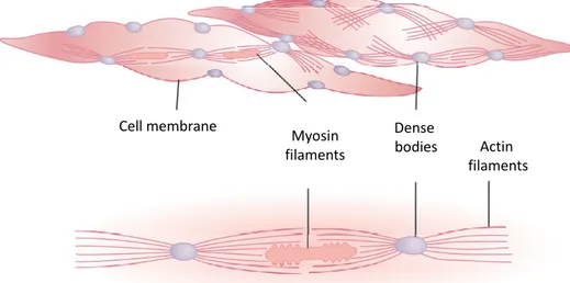
d. Filaments organization
Smooth muscle does not have the same striated arrangement of actin and myosin filaments as
is found in cardiac or skeletal muscle. The filaments are arranged in a more disorganized
structure, but remaining a myosin filament surrounded by several actin filaments. The actin
filaments are rooted in dense bands on the inner cell membrane and dense bodies in the
cytoplasm, which function like Z‐lines in cardiac myocytes. These structures are composed of
α‐actinin, the same protein as Z lines. The dense bodies are not aligned across the cell, but act
to bridge thin filaments together along the contractile plane of the cell. Dense bodies serve as
anchors from which the thin filaments can exert force to bring the polar cell membranes
towards each other resulting in cell shortening. Interestingly, dense bodies also are associated
with β‐actin, which is the type found in the cytoskeleton, suggesting that dense bodies may
integrate the functions of the contractile machinery and the cytoskeleton during contraction
(Aguilar & Mitchell, 2010). In comparison, dense bands are associated with the plasmatic
membrane (PM) and they are composed of a large number of proteins including α‐actinin,
vinculin and cytoskeletal actin. The actin filaments of the contractile machinery become
tethered to the cytoskeleton by virtue of these dense bands, which thus play an important role
The structure of an individual contractile unit within a smooth muscle cell is composed of ≈10
actin filaments radiating from two dense bodies, the ends of these filaments overlap a myosin
filament located midway between the dense bodies. This contractile unit is similar to the
contractile unit of skeletal muscle.
Figure 1.10. Physical structure of smooth muscle. The upper left‐hand fiber shows actin filaments radiating from dense bodies. The lower lefthand fiber and the right‐hand diagram demonstrate the relation of myosin filaments to actin filaments(Guyton 2005).
1.6.4
Gap
‐
junctions
Similar to cardiac myocytes, vascular smooth muscle cells are electrically connected by gap
junctions, also called nexus. The gap junctions is composed of six connexin molecules forming
a hemi‐tube (connexon), and the hemi‐tubes of adjacent cells join end‐to‐end, connecting the
cytoplasm of the two cells. These low‐resistance intercellular connections allow propagating
responses along the length of the blood vessels, however the spread is decremental and
extends only about 3 mm along the vessel longitudinally (Christ et al., 1996). That is why an
electrical depolarization and contraction of a local site on an arteriole can result in
depolarization at a distant site along the same vessel, indicating cell‐to‐cell propagation of the
depolarizing currents (Christ et al., 1996;Levick J.R., 2003).
The innermost myocytes of the tunica media also form gap junctions with the endothelial cells.
Although its role has been a source of great controversy in the literature, these heterocellular
or myoendothelial gap junctions transmit hyperpolarizing signals from the lining endothelium
to the vascular myocytes (de Wit et al., 2008)
Cell membrane
Myosin
filaments Actin filaments Dense
1.7
VASCULAR
SMOOTH
MUSCLE
FUNCTION
AND
REGULATION
1.7.1
Crossbridge
cycle
VSMCs contraction depends on crossbridge formation between thick myosin filaments and
thin actin filaments. This process promotes sliding of the actin filaments along the myosin
filaments, shortening the cell and causing muscle contraction.
Figure 1.11. The contractile elements are composed of myosin thick filaments and actin thin filaments anchored to dense bodies. The movement of thin filaments caused by phosphorylation of myosin light chains and subsequent ATP hydrolysis by the myosin II ATPase decreases the distance between anchor points. Myosin II is a hexamer composed of two heavy chains, two essential light chains and two regulatory light chains. Phosphorylation of the two regulatory light chains causes formation of a cross bridge between actin and myosin filaments and also creates a change in the angle of the neck region of myosin II, which causes motion of the actin thin filaments resulting in shortening of the cell (Aguilar & Mitchell, 2010).
Vascular contraction depends primarily on global cytosolic Ca2+ ion concentration, which
triggers myosin filaments activation. An increase in free intracellular calcium can result from
either increased entry of calcium into the cell through L‐type calcium channels (LTCCs) or
release of calcium from internal stores (e.g., sarcoplasmic reticulum). The rise in cytosolic Ca2+
concentration causes the formation of the Ca2+‐calmodulin complex. Calmodulin is a
cytoplasmic protein related to troponin C, that binds 4 Ca2+ ions (Johnson et al., 1996). Ca2+‐
calmodulin complex activates the enzyme myosin light chain kinase (MLCK). The light chain is a
component of the myosin head involved in crossbridge formation with actin, and vascular
myosin. When MLCK is activated, an immediate and marked increase in phosphorylation in the
machinery (Somlyo & Somlyo, 2003). MLCK transfers a phosphate group from ATP to the
myosin light chain, enabling the myosin head to form a crossbridge with actin, thenthe myosin
head rotates to generate tension, as in striated muscle (Onishi et al., 1983;Craig et al., 1983)
Vascular relaxation occurs through myosin light chain phosphatase (MLCP), which turns off the
myosin motor. When cytosolic Ca2+ concentration falls, MLCK activity declines. Meanwhile the
MLCP increases its activity, removing the phosphate group of myosin light chain. This detaches
preexisting crossbridges, and as new ones cannot form, the myocyte relaxes, leading to
vasodilatation (Hartshorne et al., 2004). Thus, the degree of contraction of smooth muscle
degree is regulated by the balance between MLCK and MLCP.
Figure 1.12. Regulation of vascular smooth muscle contraction by myosin light chain kinase (MLCK). Increased intracellular calcium, by either increased entry into the cell (through L‐type Ca2+ channels) or release from the sarcoplasmic reticulum (SR), forms a complex with calmodulin, activating MLCK, which phosphorylates myosin light chains (MLC), causing contraction. Cyclic adenosine monophosphate (cAMP) inhibits MLCK, thereby causing relaxation. ATP, adenosine triphosphate; Pi, phosphate group(Klabunde, 2005).
One important characteristic of smooth muscle is that muscle tension can be maintained for a
long period with little energy consumption by regulating the cross‐bridge cycle velocity. This
regulation is determined by the balance between myosin phosphorylation and
dephosphorylation, which relies on Ca2+‐dependent activation process. Furthermore, the cross‐
bridge cycle in the VSM maintains contraction for longer periods of time than skeletal muscle,
because the fraction of time that the cross‐bridges remain attached to the actin filaments (the
so called latch‐state) is longer in smooth muscle. In this way, VSMC sustains the tension with a
low ATP consumption rate. In adittion, there are Ca2+‐independent mechanisms that can
modify contractile machinery‐Ca2+ sensitivity by modulating the activity of the MLCP
(Martinez‐Lemus et al., 2009;Murphy & Rembold, 2005).
Ca2++ Calmodulin
Ca2+‐Calmodulin
L‐Type Calcium channel
Ca2+
SR
MLC
Pi
ATP
MLCK
cAMP
‐
+
1.7.2
Intracellular
calcium
homeostasis
The degree of contraction of VSMCs, that determines arterial tone, is tightly controlled by the
concentration of free cytosolic calcium. In the VSM [Ca2+]i is typically in the range 100‐350 nM
(Gorlach et al., 2006) and these basal levels are mainly determined by 3 mechanism:
1. Extracelular Ca2+ entry. Extracelular Ca2+ enters the myocyte through Ca2+‐conducting
channels in the sarcolema. These channels can be classified in two different groups:
i. Voltage‐dependent calcium channels (VDCCs) such as L‐ and T‐type calcium
channels (Nelson et al., 1990;Sonkusare et al., 2006b).
ii. Voltage independent calcium channels. These latter include (1) receptor
operated channels (ROCs), (2) capacitative or store‐operated channels (SOCs)
and (3) mechanosensitive or stretch‐activated channels (SACs).
Voltage‐dependent Ca2+ channels (VDCCs) have a low but finite open‐state probability
under basal conditions, allowing a small extracellular Ca2+ influx. This design allows a
tightly regulation of Ca2+ influx and therefore an excellent control of vascular basal
tone, and blood pressure (Sonkusare et al., 2006b). Receptor operated channels
(ROCs) are regulated by agonist‐receptor interaction independently of a previous
change in membrane potential and in most cases, there is a transduction protein that
mediates between receptor activation and channel opening (Bolton, 1979;Zhou et al.,
2006). Store‐operated channels (SOCs) are activated by the emptying of intracellular
Ca2+ stores (primarily the endoplasmic reticulum). Ca2+ store depletion activates Ca2+
entry from the extracellular space through plasma membrane channels to refill the
store, as in a capacitor (Putney, Jr., 1986;Parekh & Penner, 1997;Sanders, 2001).
Finally, stretch‐activated channels are activated when tension or stretch is generated
in the SMC plasma‐membrane (Guharay & Sachs, 1984;Park et al., 2003).
2. Stored Ca2+ release. Calcium is released from the sarcoplasmic reticulum (SR) through
the IP3‐dependent Ca2+ release channels, when the myocyte is stimulated by agonist
such as noradrenaline (Berridge, 2008). Noradrenaline and adrenaline (via α1‐
adrenoceptors), angiotensin II (via AT1 receptors), endothelin‐I (via ETA receptors), and
acetylcholine (via M3 receptors) activate phospholipase C through the Gq‐protein,
causing the formation of soluble factor, inositol trisphosphate (IP3) from PIP2. IP3 then
quickly diffuses to the nearby SR, where IP3 receptors are linked to Ca2+ release
channels (IP3‐Ca2+ release channels). IP3 thus directly stimulates the SR to release the
time, the SOCs are activated, increasing extracellular calcium influx (the capacitative
calcium entry (Putney, Jr., 1986;Berridge, 1995)).
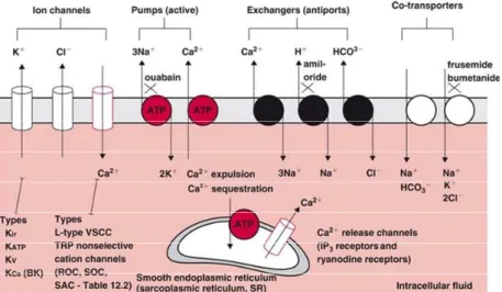
Figure 1.13. Proposed mechanisms involved in the regulation of Ca2+ homeostasis in VSMC.a: DAG activates ROC‐mediated Ca2+ influx and IP3 activates IP3R‐mediated Ca2+ release. b: Depletion of Ca2+ from the SR activates SOC‐mediated Ca2+ influx. c: Membrane depolarization leads to VDCC‐mediated Ca2+ influx. d: A high level of local [Ca2+]i is capable of triggering RyR‐ mediated Ca2+ release, a positive feedback mechanism known as Ca2+‐ induced Ca2+ release (CICR). The low [Ca2+]i under resting conditions is achieved and maintained mainly by active Ca2+ sequestration into the SR by the Ca2+‐ATPase (SERCA) and Ca2+ extrusion by the Ca2+‐ ATPase in the plasma membrane (Landsberg & Yuan, 2004).
3. Ca2+‐ATPase pumps. Cytoplasmic Ca2+ concentrations are tightly regulated by pumps
(such as the sarcoplasmic–endoplasmic reticulum Ca2+ ATPase (SERCA) and plasma
membrane Ca2+ ATPase (PMCA)) (Milner et al., 1992). SR uptake from de cytoplasm by
SERCA is called Ca2+ sequestration and the extracellular transfer by the PMCA is called
Ca2+ expulsion. Na+‐Ca2+ exchanger also contributes to Ca2+ expulsion, although in
VSMCs Ca2+‐ATPases predominate. At rest, there is a small but continuous influx of
calcium through both VDCCs (because of their low open probability at resting
membrane potential) and nonspecific cationic channels (TRPs), so that sarcolemmal
Ca2+ pumps have to expel Ca2+ continuously, otherwise Ca2+ would accumulate in the
cell.
1.7.3
Excitation
contraction
coupling
in
vascular
smooth
muscle
Vascular smooth muscle contraction can be initiated by electrical, chemical and mechanical
stimuli (Davis & Hill, 1999). The processes involved in this contraction are called
electromechanical coupling, pharmacomechanical coupling and mechanical coupling
respectively.
Agonist
Receptor
IP3
IP3R a
Ca2+ ROC
Ca2+ DAG
SOC VDCC
Ca2+ Ca 2+
RyR
b
Ca2+ d
SR c
K+
Membrane depolarization
KV
Ca2+
Mg2+
Mg2+
Ca2+
Serca PMCA
a. Electromechanical coupling
Smooth muscle membrane potential (VM) regulates muscle contractility by modulating Ca2+
influx through VDCCs. At rest, VSMCs potential is around ‐40 to ‐60 mV (Nelson & Quayle,
1995). In this range, calcium channels open probability increases exponentially, so that within
a narrow range of membrane potential, influx of calcium through L‐type calcium channels
changes dramatically. VSMC depolarization increases the opening VDCCs leading to
contraction, while hyperpolarization has the opposite effect.
The VM is the net result of the activity of a number of active and passive ion transport
mechanisms in the cell membrane. Among them, several types of potassium channels such as
calcium‐dependent potassium channels (BKCa), voltage‐dependent potassium channels (Kv),
inward rectifier potassium channels (KIR), ATP‐dependent potassium channels (KATP) and some
ion‐pumps (Na+‐K+ ATPase) have been shown to modify membrane potential in VSMCs (see
below).
Figure 1.14. Dependence of contractile tone on membrane potential in isolated artery.Membrane potential was altered by estracellular H+ (U), extracellular K+ ( ), extracellular Ca2+ (z), noradrenaline ({) and oxigen tension (¯). The highlighted box indicates membrane potential and tone in the “resting” basal state. (Siegel et al., 1991).
b. Pharmacomechanical coupling
Binding of different agonist (circulating hormones, neurotransmitters, metabolites…) to its
specific receptors on the cell membrane initiates a cascade of biochemical events leading to
coupled receptor activation and subsequent second messenger generation. Second messenger
systems playing a major role in the contractile function of VSMCs are the
phosphatidylinositide (described in section 1.7.2 part 2) and the cAMP pathways (Brown et
al., 1989). The latter involves adrenaline‐dependent vasodilatation in those tissues where β2‐
adreceptors are strongly expressed. The β2‐adrenoceptors is coupled to G protein, which
activates membrane‐bound adenylyl cyclase. The activation of adenylyl cyclase catalyzes the
conversion of ATP into cyclic adenosine monophosphate (cAMP). cAMP activates the
phosphorylating enzyme protein kinase A (PKA). PKA induces vascular relaxation by multiple
actions: (1) phosphorylation of phospholamban, (2) phosphorylation of KIR, KATP and KCa
channels, which increases their open state probability or (3) inhibition of MLCK.
Figure 1.15. Pharmacomechanical coupling. Phosphatidylinositede and
cAMP‐PKA pathways modulating
potassium channels and hence
vascular response ina vascular
smooth muscle cell. PLC,
phospholipase C; DAG,
diacylglycerol; PKC, protein kinase C; PKA, protein kinase A; PKG, protein kinase G(Ko et al., 2008).
c. Mechanichal coupling
A physical distortion of the cell or the cell membrane can activate second messenger systems
and ion channels. Some TRP channels have been suggested to be activated by the tension of
the vessel wall (Sharif‐Naeini et al., 2008), contributing to the contractile response of the
vessel to stretch, the myogenic response.
1.8 VASCULAR
TONE
The contractile activity of VSMCs in the walls of small arteries and arterioles is the major
determinant of total peripherical resistance (TPR) in the vascular circulation. Vascular tone
regulates the caliber of the vessel, and hence blood flow. According to Poiseuille’s law (see
inversely proportional to tube radius raised to the fourth power, r4. Thus, small changes in the
diameter of the vessels will lead to dramatic changes in resistance.
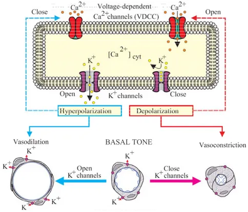
Blood vessels are normally in a state of partial contraction called basal tone, from which they
can constrict further or dilate depending on the tissue demand for blood (Nelson & Quayle,
1995).
Figure 1.16. Membrane potential and vascular tone. Inhibition of K+ channels causes membrane depolarization and leads to artery contraction. Modified from Jackson (Jackson, 2000).
The average intracellular Ca2+ concentration in VSMCs in the wall of those vessels under basal
tone is around 100‐300 nM, several orders of magnitude lower than the extracellular fluid.
Membrane potential, through activation of VDCCs, is a primary determinant of cytoplasmic
Ca2+and vascular tone (Knot & Nelson, 1998). Vascular tone plays an important role in the
regulation of blood pressure and the distribution of blood flow between and within the tissues
and organs of the body. Regulation of the contractile activity of vascular smooth muscle cells in
the systemic circulation is dependent on a complex interplay of vasodilator and
vasoconstrictor stimuli from circulating hormones, neurotransmitters, endothelium‐derived
factors, and blood pressure (Jackson, 2000). All of these signals are integrated by vascular
muscle cells to determine the activity of the contractile apparatus of the muscle cells and
hence the diameter and hydraulic resistance of a blood vessel.
BASAL TONE
1.8.1
Regulation
of
vascular
tone
The processes that regulate vascular tone fall into two classes: intrinsic and extrinsic. Intrinsic
regulation is regulation by factor located entirely within an organ or tissue. Extrinsic regulation
is regulation by factors from outside the organ. Vascular regulation involves a hierarchy of
control process, each of which can override and modify the lower one.
Figure 1.17. Overview of vascular control. Vascular tone is determined by many different competing vasoconstrictor and vasodilator influences acting on the blood vessel. These influences can be separated into extrinsic factors that originate from outside of the organ or tissue in which the blood vessel is located, and intrinsic factors that originate from the vessel itself or the surrounding tissue (Levick J.R., 2003).
a. Intrinsic mechanism
i The myogenic response
Myogenic response is a fundamental process for the development of resting vascular tone,
converting mechanical force into an adaptive electrical and chemical biological response. The
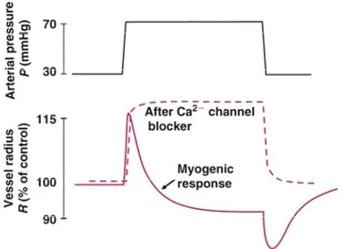
myogenic response was first described by Sir William Bayliss in 1906. When blood pressure is
raised acutely in an artery or arteriole, the pressure at first distends the vessel. Within
seconds, however, most systemic arterioles and arteries react and undergo a well‐sustained
contraction. This vital pressure‐sensitive mechanism, called the myogenic tone (MT) or
myogenic response, allows a constant blood flow despite changes in arterial pressure (Davis &
Hill, 1999). Conversely, a fall in pressure triggers a fall in vascular tone and vasodilation. This
independent of the endothelium or the nervous system. The myogenic response is important
because a significant portion of the arterial tone is caused by transmural pressure and because
it stabilizes tissue blood flow and capillary filtration pressure if arterial pressure changes
(autoregulation).
Figure 1.18. Change in diameter of isolated cerebral artery upon raising luminal pressure P. Initial passive stretch is followed by active contraction‐the bayliss myogenic response. This is abolished by L‐ type channel blockers (Levick J.R., 2003).
The initial myogenic response is mediated by depolarization and Ca2+ concentration. When an
arterial myocyte is stretched, it depolarizes to around ‐40mV. This activates L‐type Ca2+
channels, leading to a rise in cytosolic free [Ca2+] and contraction. The depolarization is
attributed to stretch‐activated channels, namely TRP cation channels, volume‐regulated
chloride channels and ENaC‐like channels (epithlelial Na+ channels) because the myogenic
response is impaired by these channels specific blockers.
The local (myocyte and endothelial) factors that maintain tonic arterial constriction, or ‘tone’, can be studied in isolated, cannulated small arteries. These arteries develop spontaneous MT
when the lumen is pressurized. Indeed, the level of tone in isolated arteries is often
comparable to that observed in the same vessels in vivo (Hill et al., 2001), and may even be
used to predict BP changes.
ii Endothelial secretions
Endothelium produces vasoconstrictor endothelin and the vasodilators nitric oxide (NO),
endothelium‐derived hyperpolarizing factor (EDHF) and prostacyclin (PGI2). These are
Nitric oxide (NO) is a freely diffusible gas with vasodilator properties. NO is generated from L‐
arginine by endothelial nitric oxide synthase (eNOS). eNOS activity is stimulated tonically by
the shear stress exerted by flowing blood, and can be enhanced by agonist such as
acetylcholine and inflammatory mediators.
Figure 1.19. Regulation of NO production and its effect on neigbouring VSMC. eNOS endothelial nitric oxide synthase; ER endoplasmic reticulum; PDE‐5 phosphodiesterase 5; PI3 kinase, phosphatidyl inositol‐ 3 kinase (Levick J.R., 2003).
NO casuses vasodilation by two mechanisms: (1) NO diffuses rapidly into neighboring VSMCs,
where it binds to the enzyme guanylyl cyclase. The activated guanylyl cyclase catalyzes the
production of cyclic guanosine monophosphate (cGMP). The cGMP activates kinases such as
protein kinase G (PKG) that promote vascular relaxation. (2) High concentrations of NO directly
activate BKCa channels in the VSMCs membrane. This hyperpolarizes the smooth muscle,
leading to vascular relaxation.
iii Metabolic vasoactive factors
The metabolic vasodilators act on the resistance vessels within the active tissue. Among the
factors that increase in active tissues, and have a vasodilating effect, during metabolic
hyperemia are: K+ ions, H+ ions (acidosis), hypoxia, adenosine, ATP, phosphate ions,