A Trojan
horse
strategy
to reverse
drug-resistance
in brain
tumors
Martha Leonor Pinzón Daza
Doctorado en Ciencias
UNIVERSIDAD DEL ROSARIO DOCTORADO EN CIENCIAS BIOMEDICAS
UNIVERSITÁ DEGLI STUDI DI TORINO DOTTORATO IN MEDICINA MOLECOLARE
A "Trojan horse" strategy to reverse drug-resistance in brain tumors
Martha Leonor Pinzón Daza
DIRECTOR: CO-DIRECTOR:
Chiara Riganti, PhD Ruth Garzón, PhD
Assintant Professor of Biochemistry Principal Professor of Biochemistry Departament
of Oncology Faculty of Natural Science and Mathematics
Medicine School, University of Turin, Italy Universidad del Rosario, Bogotá, Colombia
UNIVERSITÁ DEGLI STUDI DI TORINO DOTTORATO IN MEDICINA MOLECOLARE
A "Trojan horse" strategy to reverse drug-resistance in brain tumors
1
CONTENTABSTRACT (English) 2
ABSTRACT (Italian) 4
ABSTRACT (RESUMEN) (Spanish) 6
LIST OF PUBLICATIONS 8
ABREVIATIONS 9
ACKNOWLEDGEMENTS 11
1. Justification 12
2. Theorical framework 14
2.1. Brain tumors 14
2.2. Blood Brain Barrier 15
2.3. Multidrug resistance genes and ABC transporters 17
2.4. ABC transporters and hypoxia 20
2.5. Therapeutic approach to brain cancer, the use of nanoparticles
and liposomes (Article II) 21
2.5.1. Statins and mechanism of action 25
2.6. ABC transporters and signaling pathway 27
3. Hypothesis 29
4. General objective 30
4.1. Specific objectives 30
5. General discussion 30
6. Conclusions and perspectives 35
2
ABSTRACT
Malignant gliomas represent one of the most aggressive forms of Central Nervous System (CNS) tumors. According to the WHO classification of brain tumors, astrocytomas have been categorized into four grades, determined by the underlying pathology. Malignant (or high-grade) gliomas include anaplastic glioma (WHO grade III) as well as glioblastoma multiforme (GBM; WHO grade IV). These are the most aggressive brain tumors with the worst prognosis (1). The therapeutic management of CNS tumors is based on surgery, radiotherapy and chemotherapy, depending on the characteristics of the tumor, the clinical stage and age (2), (3), however none of the standard treatments is completely safe and compatible with an acceptable quality of life (3), (4). Chemotherapy is the first choice in disseminated tumors, like invasive glioblastoma, high-risk medulloblastoma or multiple metastasis, but the prognosis in these patients is very poor (2),(3). New targeted therapies (2), anti-angiogenic therapies (3), (4) or gene therapies show a real benefit only in limited groups of patients with known specific molecular defects (4). Thereby, the development of new pharmacological therapies for brain tumors is mandatory. Malignant gliomas are frequently chemoresistant and this resistance seems to depend on at least two mechanisms: first, the poor penetration of many anticancer drugs across the blood-brain barrier (BBB), the blood-cerebrospinal fluid barrier (BCSFB) and the blood-tumor barrier (BTB), due to their interaction with several ATP-binding cassette (ABC) drug efflux transporters that are overexpressed by the endothelial or epithelial cells of these barriers. Second, ABC drug efflux transporters in tumor cells confer multidrug resistance (MDR) on several other solid tumors; they are present on CNS tumors too and their role in gliomas is under investigation (5). Drug delivery across the blood-brain barrier (BBB) is one of the vital problems in targeted therapy treatments. Recent studies have shown that some small molecules used in these therapies are substrates of P-glycoprotein (Pgp), as well as other efflux pumps like multidrug resistance-related proteins (MRPs) and breast-cancer resistance related protein (BCRP), which extrude several anticancer drugs and will not allow drugs to reach the tumor (1).
DOXOrubicin (DOXO), a drug widely used in anti-cancer therapy, is a substrate of Pgp and BCRP, and it is very effective to attack the vitro brain tumor cells, but has a limited clinical use for its low delivery across BBB and the resistance of tumors. On the other hand BBB cells and brain tumor cells also have surface proteins, such as Low Density Lipoprotein Receptor (LDLR), which could be used as a therapeutic target. The importance of this study is based on the generation of therapeutical strategies to promote the passage of drugs through the BBB and the intratumoral accumulation, and at the same time, on the analysis of cellular mechanisms that induce increased expression of ABC transporters, to be used as therapeutic targets.
3
through the BBB, overcoming the resistance of tumor and reducing the side effects of DOXOrubicin dose. In addition this strategy can be considered as a new strategy to increase the effectiveness of different drugs in several brain tumors and ensures high efficiency even in a hypoxic environment, characteristic of cancer cells, where the expression of Pgp transporter was increased.
Taking advantage of another signaling pathway recognized as a modulator of Pgp activity this study presents not only the strategy of the Trojan horse, but also a second therapeutic proposal related to the use of Temozolomide plus DOXOrubicin. This strategy showed that temozolomide (TMZ) penetrated the BBB in a way involved the Wnt/GSK / -catenin signaling pathway, which modulates the expression of Pgp transporter. It was demonstrated that the TMZ reduces Wnt3 protein and mRNA allowing raising the hypothesis that this drug decreases Wnt3 gene transcription in BBB cells, decreasing -catenin pathway activation by its phosphorylation, reducing -catenin nuclear translocation and binding to the promoter of the mdr1 gene. Taking together the results of this study allowed the recognition of three basic mechanisms related to the down-regulation of Pgp and associated strategies: the first was the use of statins, which led to the transporter nitration decreasing its activity by NFκB pathway; the second one was based on the use of temozolomide, which by methylating Wnt3 gene reduces the activity of the -catenin signaling pathway, decreasing the expression of Pgp transporter; the third one consisted on the cross-talk between the Wnt/GSK / -catenin axis and the Wnt/RhoA/RhoA kinase as a modulator of mdr1 transcription: we demonstrated that RhoA protein kinase promoted the activation of the protein PTB1, which by phosphorylating
GSK induced phosphorylation of -catenin, priming it for destruction by the proteasome, avoiding the binding to the promoter of the mdr1 gene and therefore reducing Pgp expression. In conclusion, the therapeutic startegies proposed in this work increased the cytotoxicity of tumour cells by increasing permeability not only in the BBB, but also in tumor barrier. Also, the "Trojan horse" strategy could be useful for the therapy of other diseases associated with the central nervous system. On the other hand, these studies indicate that recognition of mechanisms associated with the expression of ABC transporters could be a key tool in the development of new anti-cancer therapies.
4
ABSTRACTIl glioma maligno rappresenta una delle forme più aggressive di tumore del sistema nervoso centrale (SNC). In base alla classificazione WHO dei tumori cerebrali, gli astrocitomi sono stati suddivisi in quattro gradi, in base ai loro caratteri patologici. I gliomi maligni o di altro grado comprendono il glioma anaplastico (grado III WHO) ed il glioblastoma multiforme (GBM; gradi IV WHO). Questi sono i tumori più aggressivi e dalla prognosi peggiore (1). Il trattamento terapeutico dei tumori del SNC è basato su chirurgia, radioterapia e
chemioterapia, in base alle caratteristiche del tumore, allo stadio clinico ed all età , ,
tuttavia nessuno di questi trattamenti standard è sicuro e compatibile con una qualità di vita accettabile (3), (4). La chemioterapia è la prima scelta terapeutica nei tumori disseminati, come il glioblastoma invasivo, il medulloblastoma ad alto rischio o le metastasi multiple, ma
la prognosi di questi pazienti è molto scadente , . Le nuove targeted therapies , le terapie anti-angiogeniche (3), (4) o le terapie geniche mostrano un reale beneficio solo in un gruppo limitato di pazienti con specifici difetti molecolari (4). Pertanto, è necessario sviluppare nuove strategie farmacologiche per i tumori cerebrali. I gliomi maligni sono spesso chemioresistenti e tale resistenza è dovuta a due fattori: primo, la bassa penetrazione di molti farmaci antitumorali attraverso la barriera ematoencefalica (BEE), la barriera emato-cerebrospinale (BECS) e la barriera emato-tumorale (BET), a causa della loro interazione con numerosi trasportatori di efflusso ATP Binding Cassette (ABC), che sono iperespressi nelle cellule endoteliali o epiteliali di queste barriere. In secondo luogo, i trasportatori di efflusso ABC conferiscono un fenotipo multidrug resistance (MDR) a molti tumori solidi; essi sono presenti anche nei tumori del SNC e la loro funzione nei gliomi è oggetto di studio (5). Il
delivery di farmaci attraverso la BBB è uno dei problemi critici nelle targeted therapies .
Studi recenti hanno mostrato che molte molecole usate in tali terapie sono substrati della glicoproteina P (Pgp) e di altre pompe di efflusso, come le MDR-related proteins (MRP) e la breast-cancer resistance related protein (BCRP), che estrudono numerosi farmaci antitumorali impedendo loro di raggiungere il tumore (1).
La DOXOrubicina (DOXO), un farmaco ampiamente usato nella terapia antitumorale, è substrato di Pgp e BCRP; è molto efficiente in vitro contro le cellule derivate da tumori cerebrali, ma ha un uso clinico limitato a causa del suo basso passaggio attraverso la BBB e della resistenza tumorale. D altra parte, le cellule di BBB e dei tumori cerebrali hanno anche proteine di superficie, come il recettore per le lipoproteine a bassa densità (RLDL) che può essere usato come bersaglio terapeutico. Questo studio è stato incentrato sulla creazione di strategie terapeutiche che incrementino il passaggio dei farmaci attraverso la BEE ed il loro
accumulo intratumorale e sull analisi dei meccanismi molecolari che inducono l aumentata
espressione dei trasportatori ABC, al fine di sfruttarli come bersagli terapeutici.
)n questo lavoro ho dimostrato che l uso di una nuova strategia cavallo di Troia , basata sulla
DOXO liposomiale, può evitare che il farmaco sia estruso dai trasportatori ABC presenti sia sulla BEE sia sulle cellule tumorali. La formulazione liposomiale ha permesso di sfruttare il
RLDL come punto di attracco, per garantire l entrata nella BEE e nelle cellule tumorali
mediante endocitosi. Tale approccio è stato combinato con l uso di statine, farmaci anti
5
per garantire una buona efficienza anche in ambienti ipossici, caratteristici dei tumori, dove
l espressione della Pgp è aumentata.
Sfruttando un altra via di segnale riconosciuta come modulatrice dell attività di Pgp, il mio
lavoro presenta anche un secondo approccio terapeutico basato sulla combinazione di temozolomide (TMZ) e DOXO. Tale studio ha mostrato che la TMZ può modulare sulla BEE la via di segnale Wnt/GSK3/ -catenina, che regola l espressione della Pgp. (o dimostrato che la TMZ riduce sia il mRNA che la proteina Wnt3, lasciando ipotizzare che tale farmaco riduca la
trascrizione di Wnt nelle cellule di BEE, diminuendo di conseguenza l attivazione della -catenina tramite fosforilazione, la sua traslocazione nucleare ed il suo legame sul promotore del gene mdr1.
Nell insieme lo studio ha permesso di identificare tre meccanismi che possono ridurre la
quantità di Pgp e possono essere usati come strategie terapeutiche: primo, l uso di statine, che inducono nitrazione di Pgp e diminuzione della sua attività, tramite la via di NFkB;
secondo, l uso della TMZ, che metilando il gene Wnt , riduce l attività della -catenina e
l espressione della Pgp. )l terzo si basa sul cross-talk tra la via Wnt/GSK3/ -catenina e la via Wnt/RhoA/RhoA cinasi, come modulatore della trascrizione del gene mdr1: ho dimostrato
che RhoA cinasi attiva la proteina PTPB ; quest ultima – tramite defosforilazione di GSK3 – promuove la fosforilazione di -catenina, indirizzandola alla degradazione proteasomiale,
impedendone il legame sul promotore del gene mdr e riducendo così l espressione di Pgp.
In conclusione, le strategie terapeutiche proposte in questo lavoro hanno aumentato la citotossicità da chemioterapici nelle cellule tumorali aumentando la permeabilità non solo
della BEE, ma anche del tumore. La strategia cavallo di Troia proposta può essere utile per
la terapia anche di altre patologie del SNC. Inoltre, questo studio suggerisce che la conoscenza
dei meccanismi che regolano l espressione dei trasportatori ABC può essere un utile
6
RESUMEN
7
Este estudio demostró que el uso de una nueva estrategia basada en el Caballo de Troya , donde se combina la droga DOXOrubicina, la cual es introducida dentro de un liposoma, salvaguarda la droga de manera que se evita su reconocimiento por parte de los transportadores ABC tanto de la BBB como de las células del tumor. La construcción del liposoma permitió utilizar el receptor LDLR de las células asegurando la entrada a través de la BBB y hacia las células tumorales a través de un proceso de endocitosis. Este mecanismo fue asociado al uso de estatinas o drogas anticolesterol las cuales favorecieron la expresión de LDLR y disminuyeron la actividad de los transportadores ABC por nitración de los mismos, incrementando la eficiencia de nuestro Caballo de Troya. Por consiguiente demostramos que el uso de una nueva estrategia o formulación denominada ApolipoDOXO más el uso de estatinas favorece la administración de fármacos a través de la BBB, venciendo la resistencia del tumor y reduciendo los efectos colaterales dosis dependiente de la DOXOrubicina. Además esta estrategia del "Caballo de Troya", es un nuevo enfoque terapéutico que puede ser considerado como una nueva estrategia para aumentar la eficacia de diferentes fármacos en varios tumores cerebrales y garantiza una alta eficiencia incluso en un medio hipóxico, característico de las células cancerosas, donde la expresión del transportador Pgp se vió aumentada. Teniendo en cuenta la relación entre algunas vías de señalización reconocidas como moduladores de la actividad de Pgp, este estudio presenta no solo la estrategia del Caballo de Troya, sino también otra propuesta terapéutica relacionada con el uso de Temozolomide más DOXOrubicina. Esta estrategia demostró que el temozolomide logra penetrar la BBB por que interviene en la via de señalización de la Wnt/GSK3/ -catenina, la cual modula la expresión del transportador Pgp. Se demostró que el TMZ disminuye la proteína y el mRNA de Wnt3 permitiendo plantear la hipótesis de que la droga al disminuir la transcripción del gen Wnt3 en células de BBB, incrementa la activación de la vía fosforilando la -catenina y conduciendo a disminuir la -catenina nuclear y por tanto su unión al promotor del gen mdr1. Con base en los resultados este estudio permitió el reconocimiento de tres mecanismos básicos relacionados con la expresión de los transportadores ABC y asociados a las estrategias empleadas: el primero fue el uso de las estatinas, el cual condujo a la nitración de los transportadores disminuyendo su actividad por la via del factor de transcripción NFκB; el segundo a partir del uso del temozolomide, el cual metila el gen de
Wnt3 reduciendo la actividad de la via de señalización de la la -catenina, disminuyendo la expresión del transportador Pgp. El tercero consistió en la determinación de la relación entre el eje RhoA/RhoA quinasa como un modulador de la via (no canónica) GSK3/ -catenina. Se demostró que la proteína quinasa RhoA promovió la activación de la proteína PTB1, la cual al fosforilar a GSK3 indujo la fosforilación de la -catenina, lo cual dio lugar a su destrucción por el proteosoma, evitando su unión al promotor del gen mdr1 y por tanto reduciendo su expresión. En conclusión las estrategias propuestas en este trabajo incrementaron la citotoxicidad de las células tumorales al aumentar la permeabilidad no solo de la barrera hematoencefálica, sino también de la propia barrera tumoral. Igualmente, la estrategia del
Caballo de Troya podría ser ’til para la terapia de otras enfermedades asociadas al sistema
8
LIST OF PUBLICATIONS
This thesis is based on the following publications, which are indicated in the text by the corresponding Roman numerals:
I. Pinzón-Daza M, Garzón R, Couraud P, Romero Ia, Weksler B, Ghigo D, Bosia A, Riganti C. The association of statins plus LDL receptor-targeted liposome-encapsulated DOXOrubicin increases in vitro drug delivery across blood-brain barrier cells. Br J Pharmacol. 2012 Dec; 167(7):1431-47.
II. Pinzon-Daza ML, Campia I, Kopecka J, Garzon R, Ghigo D, Riganti C. Nanoparticle- and liposome-carried drugs: new strategies for active targeting and drug delivery across blood-brain barrier. Current drug metabolism. 2013;14(6):625-40.
III. Pinzòn-Daza M.L., Kopecka J., Campia I., Garzon R., Riganti C. The LDL-masked DOXOrubicin : an effective strategy for drug delivery across hypoxic blood brain barrier. ScienceJet 2014, 3: 49.
IV. Riganti, C., I. C. Salaroglio, M. L. Pinzon-Daza, V. Caldera, I. Campia, J. Kopecka, M. Mellai, L. Annovazzi, P. O. Couraud, A. Bosia, D. Ghigo and D. Schiffer (2014). "Temozolomide down-regulates P-glycoprotein in human blood-brain barrier cells by disrupting Wnt3 signaling." Cell Mol Life Sci 71(3): 499-516.
9
ABREVIATIONS
ATP ADENOSIN TRIPHOSPHATE AJ ADHERENT JUNCTIONS ABC ATP BINDING CASSETTE BBB BLOOD BRAIN BARRIER
BCRP BREAST CANCER RELATED PROTEIN BCSF BLOOD CEREBRO-SPINAL FLUID
CNS CENTRAL NERVOUS SYSTEM DAU DAUNORUBICIN
DOXO DOXORUBICIN
DNA DEOXIRIBONUCLEIC ACID FPP FARNESYL PIROPHOSPHATE GSH GLUTHATION
GGPP GERANYL GERANYL PIROPHOSPHATE GBM GLIOBLASTOMA
HMG-CoAR 3-HYDROXY-3-METHYLGLUTARYL-COENZYME A REDUCTASE HRE HYPOXIA RESPONSE ELEMENT
HIF- HYPOXIA INDUCIBLE FACTOR-1 ALPHA
HA HUMAN ALBUMIN
LDL LOW DENSITY LIPOPROTEIN
LDLR LOW DENSITY LIPOPROTEIN RECEPTOR LEF LINFOIDS ENHACED FACTOR
LDH LACTATE DESHIDROGENASE MDR MULTIDRUG RESISTANCE
MRP MULTIDRUG RESISTANCE RELATED PROTEIN NOS NITRIC OXIDE SYNTHASE
NP NANOPARTICLES Pgp P-GLICOPROTEIN
PCP PLANAR CELLULAR POLARITY
SREBP STEROL REGULATORY ELEMENT-BINDING PROTEIN SLN SOLID LIPIDIC NANOPARTICLES
TJ TIGHT JUNCTIONS TMZ TEMOZOLOMIDE
10
To the memory of my father, my guide, my example, my light,
my support, my friend and certainly the most wonderful human
11
ACKNOWLEDGEMENTS
My sincere and deep gratitude, who with their voice, their hand, their support, their teachings allowed me conclude this work. First of all I thank the University of Rosario for the opportunity to develop my dissertation work with the ERASMUS MUNDUS COLOMBIA ERACOL programme for which I was granted a scholarship for three years to work at the University of Turin, Italy. The University of Rosario not only gives me the time but also its financial support during my stay abroad.
I also thank the Department of Oncology of the Medical School and the Doctoral program in Molecular Medicine of the University of Turin, Italy: by opening their doors and allow me the full development of the thesis work. I am especially grateful to Dr. Dario Ghigo and the Dr. Amalia Bosia for allowing me to be part of their working group.
I give a very special thanks to my tutor and guide in this process, Dr. Chiara Riganti who gives me her full support, her energy, her time and all the will to make this work not only possible but give important fruits. Her unconditional attitude, her optimism, her permanent support and human warmth were always what drove me, so also the desire to give the best permanently. I thank also my coworkers of the laboratory Ivana Campia, for her permanent help, Johana Kopecka for her warmth, Carolina Belisario for her support inside and outside the laboratory, and especially moments shared in Italy. To my great friend Sophie Doublier who gave me the opportunity to get to know another face of Italy and by offering me a wonderful friendship. Thanks to Iris, Loredana and the other collaborators of the laboratory who I will always remember deeply.
A special thank you is directed to research BIO-BIO group colleagues who gave me their support and accompaniment in this doctorate: Lilia, Fabio, Alejandro, Albita, Alejandrina. I especially thank my boss, colleague and friend Ruth Garzón who has co-directed this thesis and who has always given me their support. Thank you especially for all her suggestions, her desire to promote new ideas and her generous attitude with the entire working group, for her friendship and being a person that so comprehensive: thank you.
12
1. JustificationIt is estimated that each year more than 11 million new cases of cancer, of which about 80%
occur in developing countries are present in the world. In Colombia cancer is the third
leading cause of death, after cardiovascular diseases and violence, so as in 2010, there were
33.450 deaths by cancer, which accounted for 16.9% of all deaths: 16.381 were men and
17.069 women, with a crude rate of 72.9 per 100,000 and 74.1 per 100,000 cancer mortality
respectively, according to documents reported in the ten-year plan for the control of cancer
of Colombia describe in the period of 2012-2021
(http://www.cancer.gov.co/documentos/Plandecenalparaelcontroldelcancer). Worldwide,
deaths caused by tumors account for almost 15% of the total (rate of 65/100,000 people (1).
This incidence depends on different factors: the zone, the age, among other epidemiological
characteristics. It is the case of leukemia and tumors of the central nervous system which are
the main causes of death from cancer among the population under the age of 30 (2, 3). This
incidence in young people is characteristic of brain tumors, i.e. 1 per 1300 in habitants
develops brain cancer before the age of 20. In young people, 25% of cancer mortality is
secondary to a tumor of the central nervous system (6, 7). On the other hand the 23% of
pediatric cancers are brain where medulloblastoma is the most common cause of death
related to cancer in children (8). Taking into account the adult population, 7 cases of
malignant brain cancer per 100 thousand people are diagnosed every year and this rate is
among the 10 leading causes of death due to cancer. (9).
Whitin the 5 fundamental kinds of tumors of the CNS (neuronal tumors, poorly differentiated
neoplasms, meningiomas, gliomas and metastasis), it has been shown that aggressive
glioblastoma, and low and high grade gliomas account for 78% of primary brain tumors
among adults (5), while brain metastasis of solid tumors are also frequent and occur in
approximately 25% of patients with cancer (10).
In addition, the analysis of the demographic characteristics of these lesions has been studied
in Colombia and it is very similar to the world. Each tumor set occurs in a specific
demographic group. Meningiomas and schwannomas appear most commonly in women as
astrocytomas are more frequent in men. Germ cell tumors and astrocytomas are diagnosed
13
more in adults (2). Regarding patient s sex, it has been shown that gliomas occur in men 40% more than women (11) and the average age of presentation is 45 years old.
Taking the above data, it is estimated that the high levels of incidence/mortality for cancer of
the central nervous system and especially gliomas, indicate a poor prognosis, which could be
attributed to very advanced stages at the time of diagnosis or to the lack of efficacy in the
treatment. From these observations, it is important to understand that the high prevalence of
this disease is not only due to the aggressiveness of the tumor but also to deficiency in
therapies that can control it. This limitation can be explained from the presence of a natural
mechanism- the blood brain barrier (BBB) - that the brain uses to regulate the entry of
substances, including anti cancer therapies. In this way the BBB acts as a neuroprotective
shield in order to protect the brain from most substances, maintaining a balanced supply of
nutrients to the brain tissues and filtering the harmful compounds from the brain to the
blood stream (11). Anti cancer drugs such as anthracyclines, used for a long time, do not
show a high effectiveness and the resistance to these agents has become a failure of
chemotherapy, promoting a low prognosis. One of the mechanisms that are postulated as
possible cause of this failure relies on the multidrug resistance (MDR) phenotype, which
leads to a low citiotoxicity by anticancer agents; because they can not cross the BBB and can
not accumulate in tumor. Studies based on cells in vitro and clinical studies have shown that
MDR occurs in central nervous system (CNS) (12) cancer cells. In addition it has been
recognized that the key mechanism of the MDR is based on the overexpression of human
tumors membrane transporters, such as: the P-glycoprotein (Pgp) and the related MDR
(MRPs), known as ABC transporters proteins (ABC: ATP binding cassette), which function as
ATP-dependent pumps that bind to anti cancer drugs and pump them outside the BBB or
tumor. The CNS tumors have a MDR phenotype, due to overexpression of Pgp and MRPs,
which may be constitutive or induced by own chemotherapy (4). Therefore, this phenotype is
a strong barrier to the diffusion of chemotherapeutic drugs. This study contributes to
increase the efficiency of anti cancer drugs using the association between ApolipoDOXO and
Statins to improve administration of drugs through BBB, in order to overcome the resistance
of the tumor and reduce the dose-dependent side effects of DOXO in vitro. Additionally this
work serves as basis to contribute to the decrease in deaths from brain cancer affecting a
14
2. Theorical framework2.1. Brain tumors
Brain tumors or Gliomas are named in different ways depending on histological features
observed and compared with mature glial cells. Brain tumors with cells resembling
astrocytes were termed astrocytomas; the tumor cells of ependymoma shared an appearance
with ependymal cells; pinealoma was composed of tumor cells resembling those of the pineal
gland. The names of several tumor entities related to cell types are no longer recognized
today. Examples are spongioblastoma to the spongioblast (or by other authors glioblastoma
referring to the glioblast) and medulloblastoma to the medulloblast. It is noteworthy that this
first comprehensive classification system already outlined the concept of tumors arising from
immature precursor cells or from neuroectodermal stem cells (6). Low grade gliomas,
mostly astrocytomas (World Health Organization (WHO) grade II) are progressively
transforming into malignant gliomas, that is, anaplastic tumors (WHO grade III) and
glioblastoma multiforme (GBM; WHO grade IV). However, most GBM are diagnosed without
any prior record of a tumor of lower grade (7). GBM is a complex mixture of cell types that
includes astrocyte-like and stem-like cells, characterized by rapid growth and diffuse
invasiveness into adjacent brain parenchyma. Resection possibility depends on tumor
location and only the nodular component can be surgically controlled. The infiltrative
component of the tumor, however, is left to chemotherapy and radiotherapy that can impede
tumor progression for a limited time only. GBM patient survival is less than 1 year (8). GBM
has a severe mutated phenotype that consists of large chromosomal alterations; at the
genetic level, the most frequent mutations affect genes involved in the control of cell cycle,
growth, apoptosis, invasion and neovascularization (9). Glioblastoma multiforme refers to a
malignant neoplasm with abundant glial pleomorphism, numerous mitotic figures and giant
cells, vascular hyperplasia, and focal areas of necrosis. It grows as an irregular mass in the
white matter and infiltrates the surrounding parenchyma by coursing along white matter
tracts, frequently involving the corpus callosum and crossing the midline to produce the
characteristic butterfly appearance (10). Of the estimated 17,000 primary brain tumors diagnosed in the United States each year, approximately 60% are gliomas (11). GBM
accounts for approximately half of all newly diagnosed primary brain tumors with an
15
tumor, accounting for approximately 12–15% of all intracranial neoplasms and 50–60% of all astrocytic tumors (12).
Malignant Gliomas are the most common and deadly tumors, however, the survival of
patients with GBM, although it varies individually, has improved from an average of 10
months to 14 months after the diagnosis in the past 5 years since it has improved the level of
care (13). It has been recognized that the origin of brain cancers can be directly in the brain
or outside the CNS in other parts of the body and spread to the brain such as metastasis (14).
Approximately 25% of cancer patients develop brain metastases, and although usually
appear late in the course of the disease, a brain metastasis may cause early symptoms, until
the primary cancer has been identified. In this way the diagnostic and therapeutic
management approach depends on the quantity and location of brain injury and the stage of
the cancer. Patients with brain cancer metastases are rarely cured (15) and this may be due
to the role played by the tumor barrier and the BBB to obstruct the chemotherapeutic
treatment with agents such as paclitaxel and DOXOrubicin (DOXO) . Given the failure of many
chemotherapy drugs that do not permeate the BBB, a new class of agents that have a good
chemotherapeutic activity against metastases in the brain is required. These agents must be
not only BBB permeable and active against metastatic cancer cells, but also non-toxic to the
cells of the CNS (14).
In general, the brain tumors derived from the cells glial respond differentially to treatment
with surgery and radiotherapy or radiotherapy alone, combined with or without
chemotherapy (7). The treatment of malignant brain tumors with conventional approaches is
usually unsuccessful since curative doses generally lead to excessive toxicity in normal cells
of the brain (10).
2.2. Blood Brain Barrier
BBB is constituted by the brain endothelial cells which form the anatomical substrate called
cerebral microvascular endothelium (16). BBB regulates the transport of solutes and other
substances including drugs in and out of the brain, controls leukocyte migration, and
maintains the homeostasis of the brain microenvironment, which is crucial for neuronal
16
with astrocytes, pericytes, neurons, and extracellular matrix, constitute a neurovascular unit that is essential for the health and function of the CNS (17). The transport of solutes and other substances across BBB is strictly constrained through both physical tight junctions
(TJs) and adherent s junctions (AJs) and metabolic barriers (enzymes, diverse transport systems) and excludes also very small, electrically neutral and lipid soluble molecules. Thus,
conventional pharmacological drugs or chemotherapeutic agents are unable to pass through
the barrier (17).
The main structural differences between the endothelium of brain capillaries and other
capillary endothelium are the TJs between adjacent endothelial cells (18), the lack of hollow
fenestrations and pinocitotic vesicles (19). The TJs are multiproteic complex located on the
lateral surface of the brain endothelial cells, they are composed of transmembrane proteins,
as claudin-3, occludin, adhesion molecules and cytosolic proteins, such as zonula
occludens-1/2/3, AF6, 7 H 6, cingulin, claudin-5. In the BBB the presence of TJs and the absence of
fenestrations prevent paracellular molecules delivery (20). The AJs, other complexes of
basolateral membranes composed of vascular endothelial cadherin, p and complex -/ -catenin, contribute to strong cellular adhesion and are required for the assembly of the TJs
(20). They further decrease the paracellular permeability of many substances. Finally, the
absence of pinocitotic vesicles increases the strict control over the processes of transcytosis
through BBB. In general, both the luminal (into the blood) surfaces and the basolateral (to
the brain), contribute to the function of "barrier" of the brain microvascular endothelium
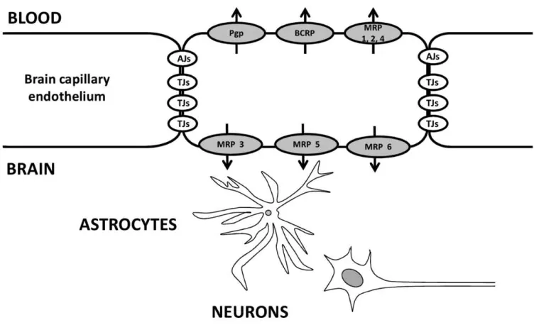
(Figure 1).
Several mentioned features are induced in the endothelial cells by the close proximity and
multiple contacts with pericytes, astrocytes and neurons; also, the simultaneous presence of
endothelial cells, extracellular matrix and cells of the CNS form a physiologically competent
BBB (17).
In addition to the BBB, other crucial barriers to maintain the homeostasis of the brain
parenchyma are the barrier blood - cerebrospinal fluid (BSCF), which is formed by the
epithelial cells of the choroid plexus and regulates the transport of molecules between the
CNS and cerebrospinal fluid (21) and the blood-retina barrier (22). In all these barriers, the
17
strictly regulated. Here ABC transporters are crucial in the process of active transport
through the cells of the BBB.
Figure No. 1. Schematic representation of the blood-brain barrier (BBB). BBB is a structural-functional unit composed by peculiar brain microvascular endothelial cells, devoid of fenestrations, rich of tight junctions (TJs) and adherent junctions (AJs) on lateral surface, and ABC transporters, such as Pgp, MRP1-6, BCRP, on luminal and basolateral side. Most of these features, which determine a tight control on the bi-directional transport of metabolites and xenobiotics, are maintained by the close proximity and multiple contacts between endothelial cells and CNS cells, such as astrocytes and neurons (23).
2.3. Multidrug resistance genes and ABC transporters
The multidrug resistance (MDR), which is the cross resistance toward chemotherapeutic
drugs unrelated for chemical structure and mechanism of action, is the main cause of failure
of the pharmacological therapy in human cancers. Tumors of CNS have often a MDR
phenotype, due to the overexpression of Pgp and MRPs, which can be constitutive or induced
by the chemotherapy itself. Moreover CNS is surrounded by brain-blood barrier (BBB), which
is reach of both Pgp and MRPs. This abundance is a further obstacle to the diffusion of
chemotherapeutic drugs toward brain tumors (24). The MDR gene product Pgp is a 170-kDa
transmembrane protein associated with tumor resistance to chemotherapeutic drugs such as
[image:20.612.115.491.137.366.2]sub-18
lethal levels by actively transporting them out of the cells. Currently, the ABC transporters
family includes more than 200 prokaryotes and eukaryotes proteins, and many of them are
still unknown. The function of ABC transporters is not limited to drug transport, and many
important human ABC transporters are involved in transport and/or regulation of ions or
other substances (25, 26) (Table No.1). Initially discovered in the 1970s as a prototypic
transporter involved in multidrug resistance (MDR) of cancer cells (27), Pgp was also the first
ABC transporter that was detected in endothelial cells of the human BBB in 1989 (24), (28).
Subsequently Pgp was localized in brain capillary endothelial cells of several species,
including monkeys, rats, mice, cattle, and pigs, suggesting that Pgp may serve as general
defense mechanism in the mammalian BBB, protecting the brain from intoxication by
potentially harmful lipophilic compounds from natural sources and other lipophilic
xenobiotics which otherwise could penetrate the BBB by simple diffusion without any
limitation (29). The exact localization of Pgp in the BBB has been the object of numerous
investigations using various experimental approaches such as in situ hybridization and immunohistochemistry. In the absence of Pgp in the BBB in mdr1a knockout mice, the brain penetration of Pgp substrate drugs can increase up to 10- to 100-fold (30).
Furthermore, blockade of BBB Pgp by cerebral application of Pgp inhibitors significantly
increases the brain concentration of various drugs, again being in line with Pgp functioning as
an efflux transporter in the BBB. Expression of Pgp confers drug resistance to numerous
antitumor agents, including DOXOrubicin, daunorubicin, actinomycin D, etoposide,
teniposide, colchicine, taxol, vincristine, and vinblastine (31). In contrast to Pgp, details about
other BBB ABC transporters are much more limited (32). The ABCC family (with the first
member, MRP1, discovered in cancer cells in 1992) currently has 12 members (including
MRP1-9), which act as organic anion transporters, but can also transport neutral organic
drugs (33). As a consequence, Pgp and MRPs have overlapping substrate specificity, so that
several drugs are substrates for both transporter families (34).
Like Pgp, MRPs are located in several normal tissues, including the BBB and BCSFB.
Resistance to numerous anticancer agents is associated with overexpression of the 170-kDa
Pgp or the 190-kDa MRPs. The role of MRPs in BBB permeability has been demonstrated by
experiments in which inhibitors of MRPs, such as probenecid or MK-571, were shown to
19
endothelial cells (35). It is of great importance also the expression of BCRP (Breast Cancer
Related Protein) transporter in BBB and tumor cells. BCRP was first discovered in a
chemotherapy-resistant breast cancer cell line, but there is no indication that its expression is
specific for breast cancer cells or that BCRP should play a significant role only in
[image:22.612.94.515.240.484.2]chemotherapy resistance in breast cancer.
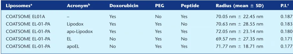
Table 1. Substrates of ATP-binding cassette (ABC) transporters
present in blood-brain barrier (BBB)
The tissue distribution of BCRP shows extensive overlap with that of Pgp, suggesting that
both transporters similarly confer protection from potentially harmful xenobiotics in various
tissues (26). In the brain, BCRP has been detected in capillary endothelial cells of humans
mainly at the luminal surface (36). Based on mRNA analysis, BCRP was more strongly
expressed on BBB than Pgp or MRP1 (33). The role of BCRP in brain uptake was recently
demonstrated by using mdr1a knockout mice, in which BCRP was inhibited by GF120918, resulting in reduced brain uptake of prazosin and mitoxantrone. Interestingly, mdr1a
knockout mice had about three times more BCRP in the brain microvessels than normal mice,
20
transporters also share a low specificity for substrates, i.e. each transporter can recognize
different molecules and the same substrate can be extracted by different transporters (38)
(Table 1). The vast majority of ABC transporters involved in multidrug resistance or their
mRNAs have been identified in cells of the BBB and BSCF; the relative abundance in these
barriers and the prevalent location luminal or basolateral membrane, is unknown for all
transporters (23). Cells overexpressing ABC transporters have a different intracellular drug
distribution and the reasons of this are not so clear. There are several possibilities involving
both direct and indirect effects. First, the transporter may be expressed not only on the
plasma membrane, but also on the vesicular membranes, where it actively contributes to
drug transport from the cytoplasm into the vesicles. Another explanation is suggested by the
observation that vesicular sequestration is a saturable process strongly dependent on the
cytoplasmic drug concentration (39).
There are a limited number of studies on high-grade brain tumors identifying subtypes of
MDR, the level of expression of these transporters, whit a lacking functional detailed and
pharmacological analysis. This lack of knowledge has contributed in large part to the poor
effectiveness of therapies targeting cancer in the CNS. Recently some factors associated with
the expression of ABC transporterst have been identified, among which are: 1) hypoxia; 2)
oxidative stress, 3) the use of chemotherapy; (4) the use of radiotherapy. In some cases the
combination of these effects leads to an increase in the MDR (40).
2.4. ABC transporters and hipoxia
Many reports have demonstrated an increased chemoresistance and radioresistance in
hypoxic cancer cells by activation of the Hypoxia-Inducible Factor- ()F- mediated pathway (40) HIF- is composed of two subunits: , which is constitutively expressed, and
, which is rapidly degraded under normal conditions but becomes stable when the oxygen or iron supply decreases, leading to a net increase in Hypoxia inducible factor alfa. This factor is
constitutively high in the hypoxic areas of tumours. When active, HIF- up regulates several genes involved in processes such as cellular growth, glucose and iron metabolism, pH control,
angiogenesis, matrix remodelling and drug resistance. Since HIF- promotes cellular proliferation, inhibition of apoptosis, invasion and MDR, its expression in tumours is related
21
Thus, different therapeutic approaches have been attempted in order to reduce
HIF-expression. In the lung, most cell types, including bronchial and alveolar epithelium, smooth
muscle and vascular endothelium, overexpress HIF- under hypoxic conditions. (igh levels of HIF- have been described in mesothelioma biopsies of patients, one of the most highly chemoresistant and aggressive tumor, whereas mesothelial cells contain low amounts of
HIF-(41). HIF-1 is associated with multidrug resistance because could regulate transcription
of ABC transporter genes in cancer cells and promote overexpression of Pgp and MRPs in
cancer tissues. These facts have been demonstrated to efflux the intracellular anticancer
drugs outside the cancer cells, resulting in promotion of chemoresistance to taxanes
(paclitaxel or docetaxel) and anthracyclines (DOXO or daunorubicin) (42).
2.5. Therapeutic approach to brain cancer, the use of nanoparticles and liposomes
(Article II)
The antineoplastic drugs have been one of the major successes of cancer medicine. Their use
has helped to increase life, in childhood cancer has increased from more than 30% in the
1960s to 70 and 80% today (43), (44). The first two anthracyclines, daunorubicin (also
known as daunomycin and rubidomycin) and DOXOrubicin (DOXO, also known as
Adriamycin), were isolated in the 1960s from Streptomyces peucetius, a species of actinobacteria. While DAU has been shown to be highly effective against acute lymphoblastic
and myeloblastic leukemias, DOXO has been found to have a much broader anticancer
spectrum, which includes numerous solid tumors in addition to hematological malignancies.
Although more than 40 years old, anthracyclines are still frequently used in clinical practice,
and DOXO in particular, remains as an important component of many current chemotherapy
protocols for treating breast cancer, sarcomas, childhood solid tumors e.g., Wilms tumor ,
leukemias, (odgkin s disease, non-(odgkin s lymphomas, and many other cancers.
)nterestingly, rather than being replaced with novel progressive targeted agents, current
clinical practice tends to combine them with the novel therapeutics to maximize the
therapeutic response (45). Despite their extensive use, the mechanism of anthracyclines
antineoplastic action is still a subject of debate and apparently it is a combination of several
different mechanisms, which accounts for the high efficiency of this class of anticancer drugs
22
Mechanism of action of anthracyclines is attributed to their intercalation between base pairs
of the DNA strands, which prevents replication of rapidly growing cancer cells. However,
more recent studies have shown that at clinically relevant concentrations, intercalation is
unlikely to play a major role. Anthracyclines are called topoisomerase poisons because they act by stabilizing a reaction intermediate in which DNA strands are cut and covalently linked
to topoisomerase II- tyrosine residues, which blocks subsequent DNA resealing. Failure to
relax the supercoiled DNA blocks DNA replication and transcription. Furthermore, breaking
DNA strands may trigger apoptosis of cancer cells, apparently via the p53-dependent pathway. Some studies have also suggested that reactive oxygen species (ROS) formation
and lipid peroxidation participate in the anticancer effects of anthracyclines (45).
According with their structure DOXO differs from daunorubicin only by a single hydroxyl
group. This fact has spurred researchers worldwide to find analogs of DOXO that have less
acute toxicity, e.g. causing less cardiomyopathy, and can be administered orally, and/or have
different, or greater antitumor efficacy. None of these analogs have stronger antitumor
efficacy than the original two anthracyclines, but there are some differences in toxicity.
Methods have been fashioned to keep the peak plasma level of DOXO muted to minimize
cardiotoxicity, but the only apparently effective method available so far (prolonged drug
infusion) is cumbersome. The bisoxopiperazine class of drugs (especially dexrazoxane)
provides protection against anthracycline-induced cardiomyopathy and has much promise
for helping mitigate this major obstacle to prolonged use of the anthracyclines. The DOXO
analogs being evaluated in the 1990s have been selected for their ability to overcome
multidrug resistance in cancer cells. Thirty years after discovery of the anticancer activity of
the first anthracycline, some means of reducing anthracycline toxicity have been devised.
Current studies are evaluating increased doses of epirubicin to improve anthracycline
cytotoxicity, while limiting cardiotoxicity, but at present DOXO still remains in this drug class
as the one having the most proven anticancer effect (49).
Different methods have been developed in order to maintain an effective amount of DOXO
which minimizes the cardiotoxicity; it has been developed derivatives of the drug, to prevent
this and other effects in plasma. Some systems can protect DOXO by macrophages
23
transporters (47), (48). Some DOXO analogues have been evaluated in recent years and have
proven to be effective to prevent resistance to drugs in cancer cells. This is how many
strategies offer a promising alternative therapy for brain cancers (13) (50). Within these
include nanoparticles or liposomes as key elements that used to allow access to the drug to
the brain.
In recent years the nanoparticles (NPs), emerged as a very efficient way of transporting the
drug, due to small size, high solubility and multifunctionality (51). Size ranges between 10
and 1000 nm; they are made with a variety of materials, including carbohydrates like
malto-dextrins or chitosan derivatives (52); fatty acids as beenic and palmitic acid are assembled in
the solid lipid NPs (SLNs)(53); proteins such as human albumin (HA)(54), among others, are
also conjugated. Some of them are amphiphilic molecules micelles with their oriented
towards the external surface hydrophilic and hydrophobic towards the inner center groups.
Tumor specific drug delivery has become increasingly interesting in cancer therapy, as the
use of chemotherapeutics is often limited due to severe side effects. Conventional drug
delivery systems have shown low efficiency and a continuous search for more advanced drug
delivery principles is therefore of great importance (50).
Liposomes were suggested as drug transporters in cancer chemotherapy by Gregoriadis et al.
in 1974. Since then, the interest in liposomes has increased and liposome systems are now
being extensively studied as drug transporters. Three basic requirements need to be fulfilled
if liposomes are to be successful in delivering drugs specifically to cancerous tissue: (i)
prolonged blood circulation, (ii) sufficient tumor accumulation, (iii) controlled drug release
and uptake by tumor cells with a release profile matching the pharmacodynamics of the drug.
This system is part of the called nanoparticles. In nano aqueous polymeric particles made of
natural or artificial polymers ranging in size between about 10 and 1000 nm, drugs may be
adsorbed to the surface or chemically attached. Drug delivery systems such as microspheres,
liposomes or polymeric NPs may improve the effectiveness and decrease the side effects of
cancer chemotherapy. Some of these transporters possess moderate MDR reversal activity
on their own. To further enhance chemotherapy of drug-resistant malignancies, some
investigators have integrated the approach of encapsulation with the combination of
cytotoxic drugs and chemosensitizer (13). The exact strategy implemented, however, has
24
whereas in other studies, encapsulated cytotoxic drug was administered with free
chemosensitizer (55). Co-encapsulation of anticancer drugs and chemosensitizers by
polymeric particles has been reported to have good efficacy (56). Moreover, lower normal
tissue drug toxicity and fewer drug-drug interactions have generally been observed in the
combination treatments. However, so far, there has been no consensus regarding which
strategy provides the optimal treatment outcome (57).
There have been used many ways to present drug to BBB, especially focusing on DOXO for its
low penetration across BBB. One strategy is the pegylated liposomal DOXO developed to
maintain or enhance the demonstrated antineoplastic effects of DOXO, while improving the
toxicity profile associated with this important cytotoxic agent. Accumulating clinical data
have confirmed the activity of pegylated liposomal DOXO in cancers of the breast and ovary
(49).
Figure No. 2. Nanoparticles- and liposomes-loaded drugs are more easily delivered across blood-brain barrier (BBB) than free drugs with at least three mechanisms: 1) they are favored to enter cells by simple diffusion; 2) they are uptaken by a receptor-triggered endocytosis, if conjugated with specific ligands; 3) they are less effluxed by ABC transporters: indeed nanoparticles and liposomes can directly inhibit the catalytic cycle of ABC transporters and alter the optimal physico-chemical properties of the plasma membrane in which the transporters work. Overall, the drugs loaded on these delivery systems can pass through the BBB and reach the brain parenchyma at therapeutically relevant concentrations (23).
[image:27.612.92.505.340.547.2]25
The abundance of ABC transporters in endothelial cells of brain capillaries of bovine, murine
and human origin may help to clarify by which mechanisms NPs and liposomes protect drugs
from the ABC transporters-mediated efflux (Figure 2). This information could be then
translated into the design of NPs/liposomes with specific physico-chemical properties, useful
not only to increase the delivery across BBB, but to increase the pharmacological efficacy in
every condition for which the ABC transporters represent an obstacle.
The major advantage of using NPs/liposomes-based strategies is their great versatility: once
synthesized a specific carrier formulation, the cargo can be easily varied and can include
drugs, siRNA, gene-delivering systems, and radiotracers. Such versatility enlarges the number
of applications, from the therapeutic use of drug-delivery systems in diseases such as cancer,
epilepsy, infective or neurodegenerative disorders, to the diagnostic use and to theranostic
applications, which couples diagnosis and therapy. If these are the benefits of NPs- and
liposome-based strategies, there are also many unsolved issues, such as the lack of consistent
information about their long term toxicity or the lack of effective strategies assuring a strong
tropism for BBB or CNS cells. These gaps have to be considered as challenges, which will give
the opportunity to continue developing new drug delivery systems, progressively more
effective and selective (58, 59).
2.5.1. Statins and mechanism of action
Statins are competitive inhibitors of 3-hydroxy-3-methylglutaryl coenzyme A (HMG-CoA)
reductase, which catalyzes the rate limiting step in cholesterol synthesis. Therefore, statins
dramatically decrease both cholesterol and isoprenoid intermediates, impairing the
isoprenylation and activity of different enzymes, such as the small G-protein families Ras and
Rho (60). Farnesylpyrophosphate (FPP) and geranylgeranylpyrophosphate (GGPP) serve as
important lipid attachments for the posttranslational modification of heterotrimeric
G-proteins and small GTP binding G-proteins (61). Isoprenylation converts small GTPases from a
cytosolic (inactive) state to a membrane-bound (active) state. Statins reduce the risk of
myocardial infarction and stroke. Increasing evidence suggests that protection conferred by
statins relates not only to cholesterol-lowering but rather to direct effects on endothelium
26
endothelium function is one the earliest clinical effects after initiation of statin treatment
(62).
Most statins are given in the active -hydroxy acid form except for lovastatin and simvastatin, which are administered as inactive lactone prodrugs. Both lactone and acid forms were
observed in the systemic circulation after oral administration of atorvastatin, lovastatin,
simvastatin, and cerivastatin in humans and/or animals, indicating that interconversion
occurs between the lactone and acid forms of these statins. The 2 forms of these statins are
substrates for Pgp transporter (63). To date, there are several lines of preclinical and
epidemiologic evidences to support the anticancer potential of statins. (64). Some
epidemiologic analyses have demonstrated up to a 50% reduction in cancer risk among statin
users and partial or complete responses have been observed in some, but not all, patients
undergoing early phase 1/2 trials. These mixed responses underscore the importance of
reliably identifying the appropriate subset of patients who stand to benefit most from
statin-based anticancer therapy. To ultimately advance statins as anticancer agents, it is therefore
crucial to understand the molecular mechanisms involved in their anticancer activity and to
delineate markers that distinguish the subset of tumors that are sensitive to statin-induced
apoptosis. Statin-induced apoptosis results directly from inhibiting HMGCoA reductase (64),
Once statins have blocked it and depleted the intracellular end-products of the mevalonate
pathway, cytoplasmic transcription factors known as sterol regulatory element-binding
proteins are activated. These transcription factors translocate to the nucleus, bind DNA at
promoter regions containing sterol response elements (SREs), and induce the transcription of
several key target genes, including HMGCR and the low-density lipoprotein receptor (LDLR). Up-regulated LDLR on the cell surface then binds and internalizes extracellular LDL-loaded
cholesterol, thus reducing plasma cholesterol. It is this extraordinary feedback mechanism
that has been successfully exploited to control hypercholesterolemia with statins (63).
A well-characterized pleiotropic effect of statins is the upregulation and activation of
endothelial nitric oxide sinthase (eNOS), one of the three NOS isoforms that catalyze the
conversion of L-arginine to L-citrulline and nitric oxide (NO) with a 1:1 stoichiometry (62).
Statins regulate eNOS via inhibition of geranylgeranylation of the small G-protein Rho (65).
Translocation of inactive Rho from the cytosol to the membrane depends on
27
leads to phosphorylation of myosin light chains required for the formation of actin stress
fibers and focal adhesion complexes. Anchoring of mRNAs to the actin cytoskeleton is
necessary for their stability and translational expression (66). Consequently, Rho-mediated
reorganization of the actin cytoskeleton may regulate the trafficking and subcellular
localization of specific mRNAs.
Rho GTPases play a key role in the regulation of tumor growth, migration, and sensitivity to
anticancer drugs. Some of these functions are dependent on the activity of the transcription
factor nuclear factor-κB NFκB (67), (68) or Wnt3a / -catenin (non canonical) signaling pathway (69). NF-κB is composed of protein dimers, such as the heterodimer p /p , and regulates the expression of genes involved in inflammation, cellular proliferation, and
apoptosis (70). )n resting cells, members of the inhibitory )κB family proteins, like )κB , bind directly to NF-κB dimer in the cytoplasm, preventing its nuclear localization. NF-κB is free to
translocate and bind to DNA on the target genes when )κB is phosphorylated by the )κB
kinase (IKK) complex, ubiquitinated, and degraded by S26 proteasome (71). Statins, by
inhibiting RhoA and its effector Rho kinase, can activate the IKK/NF-κB pathway (72). In addition, DOXO can induce NF-κB translocation in cancer cells via different mechanisms (73). By activating NF-κB, statins and DOXO may enhance the transcription of the inducible nitric-oxide synthase (iNOS) (70). NO is a signaling molecule involved in the control of cellular
growth, differentiation, and apoptosis (70). In previous studies Riganti et al. suggested that
NO is implicated in the DOXO cytotoxicity in HT29 cells and reverts DOXO resistance via the
tyrosine nitration of Pgp and MRP3, two ATP-binding cassette transporters that recognize
DOXO as a substrate. Such a nitration reduces the drug efflux in DOXO-resistant cells (74).
2.6. ABC transporters and signaling pathway
Statins have been recognized as regulators of iNOS expression, nitration and modulation of
the activity of ABC transporters, via RhoA protein and NF-κB transcription factor (74). However it is necessary to study other pathways that may be involved in the expression
and/or activity of ABC transporters modulation, in order to find therapeutic targets
associated with the use of new therapies (systems of nanoparticles or liposomes) to increase
their effectiveness. In this way it has been considered the activity of the protein Rho as a
28
Wnt family secreted glycoproteins is central to a wide array of developmental events across
all animal taxa, including cell proliferation, migration, establishment of cell polarity, and
specification of cell fate (75). Dysregulation of Wnt signaling is known to result in human
disease, and aberrant Wnt activation is a critical step in oncogenesis in many human tumors
(76). Furthermore it has been shown a correlation between Wnt/ -catenin signaling
activation and ABC transporters expression (77) which induce multidrug resistance. Wnt
ligands bind to cellular receptors to activate diverse signaling events, including
-catenin-dependent transcriptional induction (the canonical pathway), stimulated release of
intracellular calcium (the Wnt–Ca2+ pathway), and initiation of the planar cell polarity/convergent extension pathway (the PCP pathway) (76) (72). There are ~20
members of the Wnt family in mammals that can be divided into two major categories:
transforming and non-transforming factors. Transforming Wnts possess the ability to
transform mammary epithelial cells and can induce secondary axis formation in Xenopus.
Transforming Wnts generally activate the canonical, -catenin-dependent signaling wherein
Wnt stimulation regulates cellular -catenin to ultimately induce transcription of target genes
(78). Cytosolic -catenin is continuously degraded in unstimulated cells via phosphorylation– ubiquitination-coupled proteasomal degradation. Receptor binding of canonical Wnts inhibits
this degradation resulting in the accumulation of stabilized -catenin, which can then shuttle
into the nucleus. Nuclear -catenin interacts with members of the T-cell factor
(TCF)/lymphoid enhancing factor (LEF) family of HMG-box transcription factors to induce
expression of Wnt target genes (79). Non-transforming Wnts neither transform cells nor
induce secondary axis formation and are associated with the non-canonical Wnt–Ca2+ and PCP pathways. Wnt signaling in these pathways is -catenin-independent. Non-transforming
Wnts control cytoskeletal changes to affect movement and polarity and do so in part by
promoting the activation of the Rho family of GTPases, including Rho and Rac, which are
important regulators of cytoarchitecture, cell adhesion, transcriptional events, and cell cycle
progression (78). Wnt-induced activation of Rho GTPases is not restricted to non
transforming Wnts, however. The canonical Wnt1 in Xenopus was shown to trigger the
activation of both Rho and Rac, and, in several studies, Wnt3A was reported to promote both
canonical -catenin dependent signaling as well as the activation of RhoA (69). While the
importance of Wnt-triggered Rho activation of non-canonical signaling is well appreciated, its
contribution to canonical signaling has not been examined. For that reason it is important to
29
ABC transporters. In addition a possible modulator of canonical signaling pathway could be
the statins which are important Rho activity regulators.
3. Hypothesis
BBB is the first obstacle for access of drugs targeting cancer cells in the brain. This
impediment is represented by the expression of ABC transporters, which recognize and
extrude the drugs out of the cells, decreasing the effectiveness of the chemotherapy. Taking
as a basis the liposome structure, which accommodates the anti-cancer drug DOXO, it is
possible to create a "Trojan horse". The liposome marked with a human peptide from
APOB100, allows the recognition of the LDL (LDL-R) receptor, which is expressed in the cells
of the BBB. This leads the liposome to cross the BBB, due to its low polarity by a process of
simple diffusion or by endocytosis via LDL-R. In addition, the simultaneous use of statins,
anti-cholesterol drugs, allows a greater expression of LDL-R and also induces a decrease in
the activity of ABC transporters. Statins reduce the Rho GTPase activity and increase the
activation of NOS, induce the synthesis of not favoring the nitration of ABC transporters.
This combination increases the permeability of the "Trojan horse" allowing greater
accumulation and toxicity of drugs in cancer cells. In hypoxia conditions there is an
increasing Pgp expression however our strategy could be useful to reduce ABC transporters
activity and could induce citotoxicity in cancer cells.
In addition to recognize that Temozolomide crosses the BBB, the combined use of this drug
associated with DOXO arised as an alternative therapy. Temozolomide is not substrate of
ABC transporters or evades its action, it is a modulator of GSκ / -catenin signalling, which is involved in the expression of Pgp transporter. This study contributes to clarify how
Temozolomide reduces the activity of the GSκ / -catenin pathway, lowers the expression of Pgp transporter and therefore facilitates the entrance of DOXO. By associating
Temozolomide with the DOXO the permebeality of the latter can be increased through the
30
4. General Objective
The objective of this work was to reverse the MDR phenotype in CNS tumors starting from
the creation and use of a therapeutic strategy that improves the permeability of DOXO
through the BBB, its accumulation and cytotoxic effect in tumors.
4.1. Specific Objectives
A. Generate a tool based on liposomes therapy that improves the permeability of DOXO
through the BBB and reversed the MDR phenotype.
B. Verify that the LDL-mimicking liposome, ApolipoDOXO, binds the LDL receptor,
reversed the MDR phenotype, i.e.:
1. crosses in vitro the BBB
2. accumulates in tumor cells
3. exerts a strong cytotoxic effect.
C. Evaluate the use of combination therapies with DOXO or ApolipoDOXO, in the reversal
of the MDR phenotype, associated with the use of:
1. Statins
2. Temozolomide
D. Evaluate the behavior of the ApolipoDOXO system in the tumor microenvironment
under hypoxic conditions.
E. Determine molecular mechanisms associated with the expression of the MDR
phenotype caused by overexpression of ABC transporters.
5. General discussion
Malignant gliomas, the most common brain tumors known as high-grade tumors, and
systemic metastatic tumors, have high levels of ABC transporters or molecules responsible
for the efflux of drugs used in chemotherapy treatments. These tumors response to
chemotherapy is poor, since many anticancer drugs (DOXO, daunorubicin, etoposide,
teniposide, paclitaxel, docetaxel, vincristine, vinblastine, cisplatin, methotrexate) have a