C Kiafar. Hepatic hydrothorax 313
www.medigraphic.com
Annals of Hepatology 2008; 7(4): October-December: 313-320
Annals of Hepatology
Concise Review
Hepatic hydrothorax: Current concepts of
pathophysiology and treatment options
Camron Kiafar;1 Nooman Gilani1
1Department of Gastroenterology and Hepatology, Carl. T.
Hayden VA Medical Center, Phoenix, Arizona, USA 85012. Abbreviations used:
ESLD- End stage liver disease, TIPS- Transjugular intrahepatic porto-systemic shunt, VATS- Video-assisted thoracoscopy, SBEM- Spontaneous bacterial empyema, SBP- Spontaneous bacterial peritonitis, CPAP- Continuous positive airway pressure, LTx- Liver transplantation.
Address for correspondence: Nooman Gilani, M.D, FACG, FASGE Chief of Endoscopy
Department of Gastroenterology and Hepatology (111G) Carl. T. Hayden VA Medical Center 650 E Indian School Road Phoenix, AZ 85012
Tel: (602) 277-5551 ext 7618 Fax: (602) 222-6562 E-mail: [email protected] [email protected]
Manuscript received and accepted: 25 September and 10 October 2008
Abstract
Pleural effusions develop in 6-10% of patients with end-stage liver disease. Although, commonly seen in con-junction with ascites, isolated hepatic hydrothorax can occur in a small number of patients with cirrhosis. Re-fractory hepatic hydrothorax particularly poses a challenging therapeutic dilemma as treatment options are limited at best in these patients. Current patho-physiologic understanding of this disorder, as a cause, points towards the presence of diaphragmatic defects responsible for the shift of fluid from the peritoneal to the pleural cavity. When sodium restriction and di-uretic treatment fail, liver transplantation remains the most definitive therapy in these refractory cases. How-ever, transjugular intrahepatic porto-systemic shunt (TIPS), or video-assisted thoracoscopic (VATS) repair of the diaphragmatic defects (with or without pleurode-sis) are effective strategies in those who are not trans-plant candidates or those awaiting organ availability. Hepatic hydrothorax, especially when refractory to medical treatment, poses a challenging management dilemma. An early recognition and familiarity with available treatment modalities is crucial to effectively manage this exigent complication of cirrhosis.
Key words: Hepatic hydrothorax, complications of cir-rhosis, pleural effusions in cirrhosis.
Introduction
Hepatic hydrothorax is defined as the presence of pleural fluid (usually greater than 500 cc) in a patient with cirrhosis in the absence of primary cardiac or pulmo-nary disease.1,2 This complication occurs in approximate-ly 6-10% of patients with advanced cirrhosis.3 Although, more commonly associated with alcohol induced liver disease, cirrhosis from all causes can lead to the develop-ment of hepatic hydrothorax. The incidence of a pleural effusion is much higher with the concomitant presence of ascitic fluid. In one series of 330 patients with cirrhosis and ascites, 18 (6%) also had a pleural effusion.4 In an-other study, 6% of 200 patients with cirrhosis had a pleu-ral effusion, but none of the 54 without ascites had de-monstrable pleural fluid.5 Nevertheless, isolated right-sided hydrothorax can occur in a small number of patients.6,7
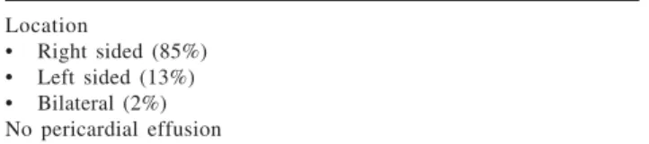
Unlike ascites where large volumes are generally tol-erated due to the capacitance of peritoneal cavity, small volumes (1-2 L) of fluid within the pleural space can lead to significant symptoms. The effusions may affect either single or both pleural spaces, but there is predilection for the right hemithorax. Of interest, regarding the reported cases of hepatic hydrothorax, 85% have been right sided, 13% left-sided and 2 % bilateral.8
Hepatic hydrothorax, hepatopulmonary syndrome and pulmonary hypertension have been recognized as major pulmonary manifestations of cirrhosis. In this review, we present current understanding of patho-physiology, clini-cal manifestations, diagnosis and related complications of hepatic hydrothorax, and discuss therapeutic options available for this disorder.
Pathophysiology
Pleural effusion develops when rate of fluid accumu-lation exceeds its removal. Since its first description by Laennec in early 19th century, several mechanisms have been postulated (Table 1) for the development of hy-drothorax in patients with cirrhosis.1,5,9-12 Regarding the mechanisms postulated; the direct passage of peritoneal fluid via diaphragmatic defects appears more plausible in explaining most cases of hepatic hydrothorax.3,13 This passage of fluid from peritoneal to pleural space has
Artemisa
www.medigraphic.com
been demonstrated by various methods. In fact, Emerson in 1955 was the first to describe such a defect (post-mor-tem) in a patient with hepatic hydrothorax.9 These de-fects can be demonstrated not only grossly, but also mi-croscopically in these patients. Lieberman and cowork-ers introduced CO2 into the peritoneal cavity of patients with hepatic hydrothorax.4 A pneumothorax indicative of a diaphragmatic defect was apparent in these patients on chest radiographs, taken within 48 hours. These defects may also be visualized using thoracoscopy.4,13,14 Intrap-eritoneal injection of methylene blue can be used intra-operatively to demonstrate and localize the defect(s). Furthermore, scintigraphic studies using intraperitoneal instillation of 99mTc-human serum albumin or 99m Tc-sul-phor-colloid have demonstrated transfer into the pleural cavity minutes to hours after administration.15,16 The movement of radioisotope is unidirectional towards the pleural cavity due to negative intrathoracic pressure compared to increased intra-abdominal pressure. These anatomic abnormalities permitting the passage of fluids have also been demonstrated with other imaging tech-niques.17
Microscopic examinations of these defects have re-vealed discontinuities or gaps in the collagen bundles that make up the tendonous portion of the diaphragm.4 When ascitic fluid collects within the peritoneal cavity, it raises the intra-abdominal pressure and tends to stretch the diaphragm; thereby, creating or enlarging these mi-croscopic defects. Increase in abdominal pressure (as a re-sult of ascites) can rere-sult in herniation of peritoneum through these gaps in the pleural cavity. This leads to the formation of pleuro-peritoneal blebs. These blebs are typically less than 1 cm in diameter and tend to rupture, thus, providing free communication between the perito-neal and pleural cavities. These blebs tend to occur more commonly in the right hemidiaphragm. For reasons that are poorly understood but may be related to embryonic development, the left hemidiaphragm is more muscular and relatively resistant to blebs formation.8
Clinical presentation
The clinical presentation is usually dominated by signs and symptoms of cirrhosis and its complications, mainly ascites. Moreover, a pleural effusion may simply be an incidental finding on a chest radiograph performed for unrelated reasons. However, a small subset of cirrhot-ic patients do present primarily with pulmonary com-plaints related to hydrothorax.18 These symptoms may in-clude dyspnea, non-productive cough, pleuritic chest pain or fatigue related to hypoxemia. Severe dyspnea and potential respiratory compromise can occur with large pleural effusions. Furthermore, these large effusions have the potential of causing cardiac tamponade with pro-found systemic hypotension that may require immediate intervention.19
Diagnosis
Clinical suspicion
Diagnosis of hepatic hydrothorax can typically be made on clinical grounds. For example, a patient with es-tablished cirrhosis and ascites who is found to have a right-sided pleural effusion most likely has hepatic hy-drothorax. However, patients can also present with a pleural effusion in the absence of a known diagnosis of cirrhosis and/or history of ascites. Others still, may have fever, respiratory symptoms or left-sided pleural effusion, suggesting an etiology other than the hepatic hydrotho-rax or the presence of spontaneous bacterial empyema (SBEM) (a poorly described complication which will be described later). Therefore, a diagnostic thoracentesis is mandatory in all patients with pleural effusions for two main reasons; first, to exclude the presence of an infec-tion, and second to rule out an alternative diagnosis.
Pleural fluid analysis
Causes of pleural effusions are numerous, and some-times, in a cirrhotic patient cannot be readily distin-guished from other etiologies besides portal hyperten-sion. A nicely done study from Barcelona20 confirmed that many end-stage liver disease patients with pleural ef-fusions do not have hepatic hydrothorax; as 18/60 (30%) patients upon thoracentesis yielded a diagnosis other than hepatic hydrothorax including, SBEM, tuberculo-sis, adenocarcinoma, parapneumonic empyema and undi-agnosed exudates. Other investigators also suggest that both thoracentesis and paracentesis should be performed to ascertain that both fluids are similar in character.3 Al-though, there are no absolute contraindications to thora-centesis, increased caution is warranted in the presence of anticoagulation or a bleeding diathesis with a PT or PTT greater than twice the midpoint of the normal range, a platelet count less than 25,000/mm3 or a serum creati-nine concentration greater than 6 mg/dL. Despite this caution, there is good safety profile for both paracentesis and thoracentesis in those with mild coagulation abnor-malities.20-22 However, in a subset of patients with severe renal failure (creatinine levels 6-14 mg/dL); the risk of average hemoglobin loss was seven-fold higher than their counterparts.21
C Kiafar. Hepatic hydrothorax
www.medigraphic.com
higher in the pleural fluid compared to ascitic fluid.24 The serum-to-pleural fluid albumin gradient is usually greater than 1.1 g/dL, similar to the one resulting from portal hypertension, although, this has not been studied extensively.
Spontaneous bacterial empyema (SBEM) is defined as an infection of a pre-existing pleural effusion (hy-drothorax) in a patient with cirrhosis.25,26 Its estimated in-cidence is around 13% (similar to the inin-cidence reported for spontaneous bacterial peritonitis; SBP) in cirrhotic patients with ascites.25,27 The pathogenesis of SBEM is also similar to that of SBP. The diagnosis of SBEM is es-tablished if the pleural fluid (PF) cultures are positive and a polymorphonuclear (PMN) count is > 250 cells/uL. In patients with negative cultures (and compatible clini-cal course) the diagnosis is made with a pleural fluid PMN count > 500 cells/uL and by excluding a parapneu-monic infection.26 The microorganisms responsible for SBEM (E. coli, Klebsiella, Streptococcus and Enterococ-cus species) appear similar to that of SBP.25 Sese et al demonstrated that patients with hepatic hydrothorax had lower opsonic activity and complement (C3) levels than their counterparts with non-hepatic pleural effusions.28 In addition, all patients who developed SBEM had lower levels of pleural fluid C3 and total protein levels, and had a higher Child-Pugh score than those who did not develop SBEM.
SBEM can present in various ways including as that seen with SBP (abdominal pain), symptoms may be
lo-calized to the chest cavity (dyspnea or pleuritic chest pain), or may be systemic in nature (fever, shock or en-cephalopathy). Similar to hepatic hydrothorax, SBEM can occur in the absence of ascites. Interestingly, up to 40% of SBEM cases may not be associated with SBP.25 The treatment of SBEM is similar to that of SBP (intrave-nous 3rd generation cephalosporins) for 7 to 10 days.25 We prefer cefotaxime at a dose of 2gm Q8h due to its very good published results in cirrhotic patients.29,30 However, despite treatment, mortality remains high at ap-proximately 20%.25
Radiological studies
Nuclear scans can be performed to establish the diag-nosis of hepatic hydrothorax with fairly high accuracy. Intra-peritoneal administration of 99mTc-human serum al-bumin or 99mTc-sulphur colloid could be used to demon-strate the communication between the peritoneal and pleural space as shown in Figure 1.31 The migration of ra-dioisotopes from the peritoneal cavity into the pleural space confirms the presence of a communication and the effusion.15,31 This diagnostic modality can also be used in the absence of ascites via ultrasound guided administra-tion of the isotope (in 500 cc of saline) into the perito-neal cavity.6 The lack of radioisotope to localize in the pleural space should raise concern for a primary cardiop-ulmonary process responsible for patient’s pleural effu-sion. Although, this test has been considered the «gold standard» for identification of hepatic hydrothorax due to very high specificity (up to 100%), its sensitivity re-mains modest ≈ 71%. The sensitivity of the test can be greatly improved (up to 100%) by performing a thora-centesis prior to administration of radioisotopes in order to reduce pleural pressure.32 Various other investigators have used modalities similar to radioisotopes; such as in-traperitoneal injections of dyes and air to diagnose he-patic hydrothorax.4,13
Recent advances in radiological imaging (ultrasound, CT scans and MRI) have enabled investigators to exam-ine in detail the diaphragmatic defects responsible for the development of hepatic hydrothorax.17,33
Invasive techniques
Thoracoscopy can be an alternative diagnostic option to directly visualize the underlying diaphragmatic de-fects. The concomitant administration of dyes in the peri-toneal cavity may increase the probability of locating such defects, in case they are not readily apparent on thoracoscopy.13
Management
Since hepatic hydrothorax and ascites share the same pathophysiological mechanisms; understandably,
thera-Table I. Proposed mechanisms for the development of hepatic hydrothorax.
• Azygous vein hypertension causing formation of collateral anastomoses between portal and azygous systems.1
• Transfer of peritoneal fluid into the pleural space via diaphragmatic defects.9
• Passage of fluid from peritoneal to the pleural space via trans-diaphragamtic lymphatics.5,10
• Hypoalbuminemia resulting in decreased colloid osmotic pressure.11
• Lymphatic leakage from the thoracic duct.12
Table II. Clinical and lab features of hepatic hydrothorax. Location
• Right sided (85%) • Left sided (13%) • Bilateral (2%) No pericardial effusion Laboratory features
• Cell count < 500 cells/mm3
• Total protein concentration < 2.5 g/dL • Total protein pleural fluid to serum ratio < 0.5
• Lactate dehydrogenase pleural fluid to serum ratio < 0.5 • Serum to pleural fluid albumin gradient > 1.1 g/dL
www.medigraphic.com
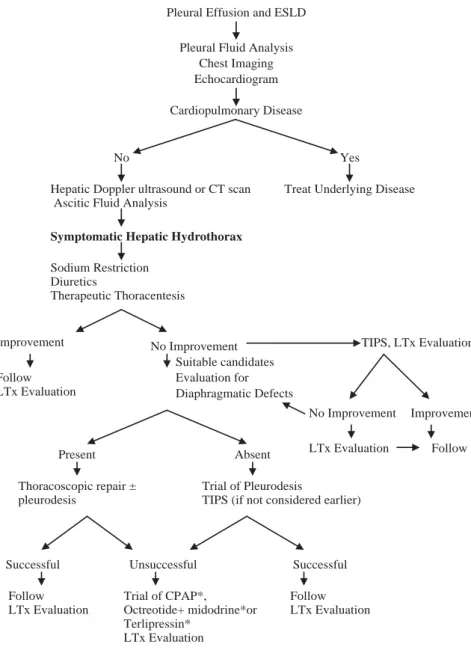
py is directed towards the underlying mechanisms of flu-id accumulation, namely, sodium retention and sinusoi-dal hypertension. Management can involve dietary, phar-macologic and radiological interventions. In addition, surgical approaches aimed at repairing the diaphragmatic defects responsible for pleural fluid accumulation can be considered in selective patients with refractory hy-drothorax. However, a majority of these patients with he-patic hydrothorax have advanced liver disease and may be potential candidates for orthotopic liver transplanta-tion; the only effective cure to date. Thus, the aim of therapy in such patients should be relief of symptoms and prevention of further complications until transplan-tation can be performed. An algorithm for the manage-ment of these patients is illustrated in figure 2, and the various potential therapeutic options available to date are discussed in detail below.
Medical management
Similar to the therapy of ascites, obtaining a negative sodium balance is the primary goal of dietary and phar-macologic management.34 Dietary restriction of sodium intake to 2 g/d (88mEq/d) is the simplest manner by which to achieve a negative sodium balance. In fact, a low sodium diet alone has shown to be effective in elimi-nating ascites in 10-15% of patients.35 However, most pa-tients with ascites, and almost all papa-tients with hepatic hydrothorax require the addition of diuretics (spironolac-tone and/or furosemide). These diuretics are typically maintained at a ratio of 10:4 (spironolactone 100 mg: fu-rosemide 40 mg), and dosages are increased as needed to attain a goal of producing renal excretion of at least 120mEq of sodium per day.36 However, in many patients these goals are not achieved due to diuretic induced elec-trolyte imbalances, renal impairment or precipitation of encephalopathy. When diuretic therapy is deemed not helpful, patients should be considered for orthotopic liv-er transplantation.
Other investigational agents such as terlipressin, oct-reotide and midodrine have been used, mainly in case re-ports, to treat hepatic hydrothorax with moderate bene-fit.37-39 It is postulated that these agents will reduce splanchnic blood flow and hence decrease peritoneal and pleural fluid accumulation. Unfortunately, at present there is not enough evidence to recommend routine use of these agents.
Thoracentesis
Thoracentesis is a simple and relatively safe procedure that can be used not only for diagnostic but therapeutic purposes as well. Thoracentesis can be performed in pa-tients with dyspnea due to hepatic hydrothorax for imme-diate relief of symptoms. In patients with both hepatic hy-drothorax and massive ascites, it is recommended to drain the ascites prior to performing a thoracentesis.2 Further-more, generally, no more than 2 liters of fluid should be removed during the first therapeutic thoracentesis, in order to minimize the risk of unilateral pulmonary edema and/or hypotension.8,40 The effect of routine use of albumin infu-sion in conjunction with thoracentesis has not been estab-lished. Although, in one study investigators routinely in-fused albumin (5 g per each liter of fluid removed); no fur-ther conclusions were drawn about its effect on the patients outcome.20 The major risk of thoracentesis appears to be the development of pneumothorax. In the above study, investigators also showed that diagnostic thoracen-tesis carried a low risk (1%) of pneumothorax, however; the incidence rose to 9% after a therapeutic procedure. De-spite its relative safety, when thoracentesis is required too frequently (< every 2-3 weeks) in patients on maximal so-dium restriction and optimal diuretics, alternative treat-ment options must be considered.8
Figure 1. Anterior view of the chest and upper abdomen after in-jection of 99mTc-sulphur colloid into the peritoneal cavity and
C Kiafar. Hepatic hydrothorax
www.medigraphic.com
Radiologic interventions: Transjugular intrahepa-tic portosystemic shunts (TIPS)
Over the past decade, TIPS has been used more often in patients who have failed medical management or those who require frequent thoracentesis.41-47 By creat-ing a nonselective side-to-side porto-systemic shunt, TIPS decreases portal pressures, and addresses the sinu-soidal hypertension that leads to ascites formation- an essential step for pleural fluid accumulation.48 The use of TIPS for hepatic hydrothorax was first described in a case series by Strauss et al41 in 1994. In this study, 5 pa-tients with Child class C cirrhosis underwent TIPS placement. Two of these patients did not require further thoracentesis, while in the remaining 3, further fluid re-moval was required due to stent occlusion. However, once stent patency was restored, the need for repeat tho-racentesis was eliminated. A larger study by Gordon et
al, evaluated 24 Child class B and C cirrhosis patients following TIPS placement.43 Fourteen of twenty four (58.3%) had complete resolution of symptoms follow-ing TIPS and did not require further thoracentesis; while another 5 (20.8%) required fewer number of tho-racentesis. Despite this superior efficacy, 6 patients (25%) died of either post-procedure complications (1/6) or liver failure (5/6), and 9 (37.5%) developed transient hepatic encephalopathy. Other groups have reported even higher initial success rates (≈ 82%) with TIPS, though; less stringent criteria (such as decrease in effu-sion size or reduced need for thoracentesis) were used in these studies.46 But again, despite symptomatic im-provement in many patients, TIPS unfortunately was as-sociated with complications and did not improve the overall prognosis of these patients. A recent study of 28 patients shows that severity of liver dysfunction is di-rectly related to non responsiveness and higher one year
Figure 2. Proposed algorithm for the diagnosis and management of hepatic hydrothorax. Asterisk: investigational approach. ESLD- End Stage Liver Di-sease, TIPS – Transjugular intrahepatic porto-systemic shunt, LTx – Liver trans-plantation, CPAP – Continuous Positive Airway Pressure.
Pleural Effusion and ESLD Pleural Fluid Analysis
Chest Imaging Echocardiogram Cardiopulmonary Disease
No Yes
Hepatic Doppler ultrasound or CT scan Ascitic Fluid Analysis
Treat Underlying Disease
Symptomatic Hepatic Hydrothorax
Sodium Restriction Diuretics
Therapeutic Thoracentesis
Improvement No Improvement TIPS, LTx Evaluation Suitable candidates
Follow LTx Evaluation
Evaluation for Diaphragmatic Defects
Improvement LTx Evaluation Follow Present Absent
Thoracoscopic repair ± pleurodesis
Trial of Pleurodesis
TIPS (if not considered earlier)
Successful Unsuccessful Successful Follow
LTx Evaluation
Trial of CPAP*,
Octreotide+ midodrine*or Terlipressin*
LTx Evaluation
Follow LTx Evaluation
www.medigraphic.com
ESTE DOCUMENTO ES ELABORADO POR MEDI-GRAPHIC
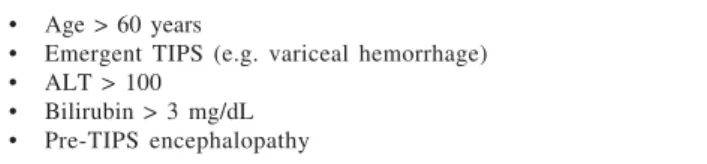
mortality after TIPS placement for refractory HH.49 Thus, because TIPS is associated with many potential risks, it should be considered in selected patients who re-accumulate their effusions rapidly (despite medical treatment) with a Child-Pugh score of less than 10, are younger than 60, and do not have hepatic encephalopa-thy or severe pulmonary hypertension. The factors asso-ciated with poor prognosis after TIPS placement are il-lustrated in Table III.50 Since a high mortality is antici-pated in this patient population in the absence of liver transplantation, it is not surprising that only a third of patients are expected to live and be relapse free at 1 year after the TIPS placement.46,50
Surgical interventions
The surgical approaches for the management of hepat-ic hydrothorax have included; tube thoracostomy with chemical pleurodesis, thoracoscopy to repair diaphrag-matic defects with/without pleurodesis and placement of peritoneovenous shunts.
Pleurodesis: Falchuk et al first described the use of tetracycline induced pleurodesis for 2 patients with re-current hepatic hydrothorax .51 One patient remained free of effusion at 6-month follow up while the other died of variceal hemorrhage 3 weeks after the pleurode-sis. Chemical pleurodesis is not always successful and does carry a modest risk of complications including fe-ver, chest pain, empyema, incomplete re-expansion, pneumonia, and wound infection among others.52 Ikard and colleagues53 reported two unsuccessful attempts with tetracycline pleurodesis, attributed to large flow of ascites across the diaphragm, resulting in a thoracic cavity that could not be kept empty enough to allow pleural coaptation and subsequent chemical pleurode-sis. Because of its low success rate and moderate degree of complications, pleurodesis by itself is rarely per-formed and typically reserved for patients in whom no other options exist. However, the use of continuous positive airway pressure (CPAP) appears effective in keeping the pleural cavity dry after chemical pleurode-sis.54 It is postulated that CPAP will decrease the nega-tive pleural pressure and thus prevent the shift of fluid form the peritoneal to the pleural space. Furthermore, Takahashi55 has shown dramatic improvement in their patient with refractory hepatic hydrothorax using only nasal CPAP, but further studies are needed before this can be routinely recommended.
Chest tube placement: Runyon et al reported their ex-perience with 2 patients with ascites and persistent right-sided pleural effusions, in whom chest tube placement led to massive fluid shifts, protein and electrolyte deple-tion and eventually death of both patients.56 At present, chest tube insertion is considered a relative contraindica-tion for the treatment of hepatic hydrothorax.
Repair of diaphragmatic defects: Thoracoscopy to repair diaphragmatic defects with/without sclerosing the pleural membranes may be an alternative in pa-tients with refractory hepatic hydrothorax who are deemed not candidates for TIPS. Thoracoscopy ap-pears to be more likely to succeed if diaphragmatic de-fects can be identified.57 Mouroux et al employed vid-eo-assisted thoracoscopy (VATS) to close large defects using sutures and biologic glue in combination with talc pleurodesis in 8 patients.13 None of the patients (6/8) with repaired defects developed recurrent hy-drothorax despite the recurrence of ascites. In another study, de Campos et al performed 21 VATS, with talc pleurodesis in 18 patients- all with hepatic hydrotho-rax.52 The overall success rate was 48% despite thora-coscopy revealing defects in only 5 of the 18 patients. However, a higher mortality was noted in this study with 7 of 18 patients dying between 5 and 40 days of follow-up. In contrast, two other studies (15 and 41 patients each) showed almost 75% success rate with VATS assisted talc pleurodesis without resorting to di-aphragmatic repairs,58,59 though, repeat procedure was required in many patients.59 Thoracoscopic repair of diaphragmatic defects using pleural flap or mesh onlay reinforcement also shows good early results in patients with refractory hydrothorax.60 In brief, this procedure (and its variations) may be considered a palliative al-ternative not only to patients requiring frequent thora-centesis, but also an alternative to TIPS. Furthermore, it may also be used as a bridge to liver transplantation, if larger randomized trials confirm the previous results and show a lower mortality rate.
Peritoneo-venous shunts: A peritoneo-venous shunt (Le Veen shunt) to divert ascitic fluid has been used in refractory cases. However, after the initial efficacy, in most cases, the shunt is rendered ineffective over time as the intrathoracic pressure is lower than the central venous pressure resulting in fluid flow towards the pleural space.53 Due to these inconsistent results and frequent complications associated with LeVeen shunt (infection, coagulopathy and bleeding in compromised host), this procedure has become almost obsolete.6
Liver transplantation: When all other therapies fail, liver transplantation is the only option available; which fortunately is curative for most patients with this compli-cation. The short-term complications and long-term prog-nosis in patients undergoing liver transplantation for re-fractory hepatic hydrothorax appears similar to other groups.61
Table III. Factors associated with poor prognosis after TIPS. • Age > 60 years
• Emergent TIPS (e.g. variceal hemorrhage) • ALT > 100
C Kiafar. Hepatic hydrothorax
www.medigraphic.com
ConclusionHepatic hydrothorax is a devastating complication of end-stage liver disease, and in fact, is not that uncom-mon as previously thought. Its management is challeng-ing and frequently associated with poor outcomes in most cases. Non-the-less an early diagnosis is of vital im-portance to establish an appropriate management plan. When other measures fail, liver transplantation remains the treatment of choice. Both TIPS and possibly VATS assisted diaphragm repair, though, temporizing measures, are perhaps the best available “bridging” to liver trans-plantation in selected patients with refractory hepatic hy-drothorax.
References:
1. Morrow CS, Kantor M, Armen RN. Hepatic hydrothorax. Ann Internal Med 1958; 49: 193-203.
2. Straus RM, Boyer TD. Hepatic hydrothorax. Semin Liver Dis
1997; 17: 227-32.
3. Alberts WM, Salem AJ, Solomon DA, Boyce G. Hepatic hy-drothorax, cause and management. Arch Intern Med 1991; 151: 2383-88.
4. Lieberman FL, Hidemura R, Peters RL, Reynolds TB. Pathogen-esis and treatment of hydrothorax complicating cirrhosis with ascites. Ann Intern Med 1966; 64: 341-51.
5. Johnston RF, Loo RV. Hepatic hydrothorax. Studies to deter-mine the source of the fluid and report of thirteen cases. Ann Intern Med 1964; 61: 385-401.
6. Rubinstein D, McInnes IE, Dudley FJ. Hepatic hydrothorax in the absence of clinical ascites: Diagnosis and management. Gas-troenterology 1985; 88: 188-91.
7. Pop CM, Gherasim RM, Dumitrascu DL. Hydrothorax without ascites in liver cirrhosis. Rom J of Gastroenterol 2003; 12: 315-17. 8. Lazaridis KN, Frank JW, Krowka MJ, Kamath PS. Hepatic Hy-drothorax: Pathogenesis, Diagnosis and Management. Am J Med
1999; 107: 262-67.
9. Emerson PA, Davies JH. Hydrothorax complicating ascites. Lan-cet 1955; 1: 487-88.
10. Meigs JV, Armstrong SH, Hamilton HH. A further contribution to the syndrome of fibroma of the ovary with fluid in the abdo-men and chest: Meigs Syndrome. Am J Obstet Gynecol 1943; 46: 19-37.
11. Higgins G, Kelsall AR, O’Brien JRP, Stewart AM, Witts LJ. As-cites in chronic disease of the liver. Q J Med 1947; 16: 263-74. 12. Dumont AE, Mulholland JH. Flow rate and composition of tho-racic duct lymph in patients with cirrhosis. N Eng J Med 1960; 263: 471-74.
13. Mouroux J, Perrin C, Venissac N, Blaive B, Richelme H. Man-agement of pleural effusions of cirrhotic origin. Chest 1996; 109: 1093-96.
14. Huang PM, Chang YL, Yang CY, Lee YC. The morphology of diaphragmatic defects in hepatic hydrothorax: thoracoscopic find-ing. J Thorac Cardiovasc Surg 2005; 130: 141-45.
15. Mittal BR, Maini, A, Das BK. Peritoneopleural communication associated with cirrhotic ascites: scintigraphic demonstration.
Abdom Imaging 1996; 21: 69-70.
16. Benet A, Vidal F, Toda R, Siurana R, De Virgala CM, Richart C. Diagnosis of hepatic hydrothorax in the absence of ascites by intraperitoneal injection of 99m-Tc-Fluor colloid. Postgrad Med J 1992; 68: 153.
17. Zenda T, Miyamoto S, Murata S, Mabuchi H. Detection of dia-phragmatic defect as the cause of severe hepatic hydrothorax
with magnetic resonance imaging. Am J Gastroenterol 1998; 93: 2288-89.
18. Nolop KB. Massive hydrothorax complicating occult cirrhosis.
South Med J 1985; 78: 214-15.
19. Kaplan LM, Epstein SK, Schwartz SL, Cao QL, Pandian NG. Clinical, echocardiographic and hemodynamic evidence of car-diac tamponade caused by large pleural effusions. AM J Respir Crit Care Med 1995; 151: 904-8.
20. Xiol X, Castellote J, Cortes-Beut R, Delgado M, Guardiola J, Sese S. Usefulness and complications of thoracentesis in cirrhotic pa-tients. Am J Med 2001; 111: 67-69.
21. McVay PA, Toy PTCY. Lack of increased bleeding after para-centesis and thorapara-centesis in patients with mild coagulation ab-normalities. Transfusion 1991; 31: 164-71.
22. Runyon BA. Paracentesis of ascitic fluid: a safe procedure. Arch Intern Med 1986; 146: 2259-61
23. Light RW, MacGregor MI, Luchsinger PC, Ball WC. Pleural ef-fusions: The diagnostic separation of transudates and exudates.
Ann Intern Med 1972; 77: 507-13.
24. Sahn SA. State of the art: the pleura. Am Rev Respir Dis 1988; 138: 184-234.
25. Xiol X, Castellvi JM, Guardiola J, Sese E, Castellote J, Perello A, Cervantes X, et al. Spontaneous bacterial empyema in cirrhotic patients: a prospective study. Hepatology 1996; 23: 719-23. 26. Xiol X, Castellote J, Baliellas C, Ariza J, Gimenez RA, Guardiola
J, Casais L. Spontaneous bacterial empyema in cirrhotic patients: Analysis of eleven cases. Hepatology 1990; 11: 365-70. 27. Albillos A, Cuervas-Mons V, Millan I, Canton T, Montes J,
Bar-rios C, Garrido A, et al. Ascitic fluid polymorphonuclear cell count and serum to ascites albumin gradient in the diagnosis of bacterial peritonitis. Gastroenterology 1990; 98: 134-40. 28. Sese E, Xiol X, Castellote J, Rodriguez-Farinas E, Tremosa G.
Low complement levels and opsonic activity in hepatic hydrotho-rax: its relationship with spontaneous bacterial empyema. J Clin Gastroenterol 2003; 36: 75-77.
29. Felisart J, Rimola A, Arroyo V, Perez-Ayuso RM, Quintero E, Gines P, Rodes J. Cefotaxime is more effective than is ampicillin-tobramycin in cirrhotics with severe infections. Hepatology 1985; 5: 457-62 30. Runyon BA, Akriviadis EA, Sattler FR, Cohen J. Ascitic fluid and
serum cefotaxime and desacetyl cefotaxime levels in patients treated for bacterial peritonitis. Dig Dis Sci 1991; 36: 1782-86. 31. Bhattacharay A, Mittal BR, Biswas T, Dhiman RK, Singh B, Jindal
SK, Chawla Y. Radioisotope scintigraphy in the diagnosis of hepatic hydrothorax. J Gastroenterol Hepatol 2001; 16: 317-21. 32. Stewart CA, Hung GL, Ackerman Z, Applebaum DM. Radionu-clide determination of peritoneo-pleural communication in hy-drothorax. J Nucl Med 1991; 32: 924.
33. Sawabe J, Ishida H, Niizawa M. Sonographic demonstration of diaphragmatic fistula in a patient with hepatic hydrothorax. Akita J Med 1992; 19: 615-19 (in Japanese with English translation). 34. Garcia-Tsao G. Current management of the complications of
cirrhosis and portal hypertension: variceal hemorrhage, ascites, and spontaneous bacterial peritonitis. Gastroenterology 2001; 120: 726-48.
35. Garcia N Jr, Sanyal AJ. Minimizing ascites. Complications of cirrhosis signals clinical deterioration. Postgrad Med 2001; 109: 91-93.
36. Roberts LR, Kamath PS. Ascites and hepatorenal syndrome: patho-physiology and management. Mayo Clinic Proc 1996; 71: 874-81. 37. Ibrisim D, Cakaloglu Y, Akyuz F, Karadag A, Ozdil S, Besisik F, Mungan Z, et al. Treatment of hepatic hydrothorax with terlipressin in a cirrhotic patient. Scand J Gastroenterol 2006; 41: 862-65.
38. Pfammatter R, Quattropani C, Reichen J, Goke B, Wagner AC. Treatment of hepatic hydrothorax and reduction of chest tube output with octreotide. Eur J Gastroenterol Hepatol 2001; 13: 977-80.
www.medigraphic.com
with large hepatic hydrothorax and mild ascites. Nephrol Dial Transplant 2005; 20: 2583.
40. Heller BJ, Grathwohl K. Contralateral re-expansion pulmonary edema. South Med J 2000; 93: 828-31.
41. Strauss RM, Martin LG, Kaufman SL, Boyer TD. Transjugular intrahepatic portal systemic shunt for the management of symp-tomatic cirrhotic hydrothorax. Am J Gastroenterol 1994; 92: 1520-22.
42. Chalasani N, Gitlin N. TIPS for patients with refractory hepatic hydrothorax: what is the take home message? Am J Gastroenterol
1997; 92: 2129-30.
43. Gordon FD, Anastopoulos HT, Crenshaw W, Gilchrist B, McEnnif N, Falchuk KR, LoCicero J 3rd, et al. The successful treatment of symptomatic, refractory hepatic hydrothorax with TIPS.
Hepatology 1997; 25: 1366-69.
44. Degawa M, Hamasaki K, Yano K, Nakao K, Kato Y, Sakamoto I, Nakata K, et al. Refractory hepatic hydrothorax treated with transjugular intrahepatic portosystemic shunt. J Gastroenerol
1999; 34: 128-31.
45. Conklin LD, Estrera AL, Weiner MA, Reardon PR, Reardon MJ. Transjugular intrahepatic portosystemic shunt for recurrent he-patic hydrothorax. Ann Thoracic Surg 2000; 69: 609-11. 46. Siegerstetter V, Deibert P, Ochs A, Olschewski M, Blum HE,
Rossle M. Treatment of refractory hepatic hydrothorax with TIPS: long term results in 40 patients. Eur J Gastroenterol Hepatol
2001; 13: 529-34.
47. Spencer EB, Cohen DT, Darey MD. Safety and efficacy of transjugular intrahepatic portosystemic shunt creation for the treatment of he-patic hydrothorax. J Vasc Interv Radiology 2002; 13: 385-90. 48. Conn HO. Transjugular intrahepatic portal-systemic shunts: the
state of the art. Hepatology 1993; 17: 148-58.
49. Wilputte JY, Goffette P, Zech F, Godoy-Gepert A, Geubel A. The outcome after transjugular intrahepatic portosystemic shunt (TIPS) for hepatic hydrothorax is closely related to liver dysfunction: a long-term study in 28 patients. Acta Gastroenterol Belg 2007; 70: 6-10.
50. Chalasani N, Clark WS, Martin LG, Kamean J, Khan MA, Patel NH, Boyer TD. Determinants of mortality in patients with ad-vanced cirrhosis and transjugular intrahepatic portosystemic shunting. Gastroenterology 2000; 118: 138-44.
51. Falchuk KR, Jacoby I, Colucci WS, Rybak ME. Tetracycline-induced pleural symphysis for recurrent hydrothorax complicat-ing cirrhosis. A new approach to treatment. Gastroenterology
1977; 72: 319-21.
52. Milanez de Campos Jr, Filho LO, de Campos WE, Sette H Jr, Fernandez A, Filomeno LT, Jatene FB. Thoracoscopy and talc poudrage in the management of hepatic hydrothorax. Chest 2000; 118: 13-17.
53. Ikard RW, Sawyers JL. Persistent hepatic hydrothorax after peri-toneal jugular shunt. Arch Surg 1980; 115: 1125-27.
54. Drouhin F, Fischer D, Law Koune JD, Boiteau R, Tenaillon A, Labayle D. Treatment of hydrothorax in liver cirrhosis with chemi-cal pleurodesis associated with continuous positive airway pres-sure ventilation. Preliminary results] Gastroenterol Clin Biol 1991; 15: 271-72
55. Takahashi K, Chin K, Sumi K, Sumi K, Nakamura T, Matsumoto H, Niimi A, et al. Resistant hepatic hydrothorax: a successful case with treatment by nCPAP. Respir Med 2005; 99: 262-64. 56. Runyon BA, Greenblatt M, Ming RHC. Hepatic hydrothorax is a
relative contraindication to chest tube insertion. Am J Gastroenterol
1986; 81: 566-67.
57. Ibi T, Koizumi K, Hirata T, Mikami I, Hisayoshi T, Shimizu K. Diaphragmatic repair of two cases of hepatic hydrothorax using video-assisted thoracoscopic surgery. Gen Thorac Cardiovasc Surg 2008; 56: 229-32.
58. Ferrante D, Arguedas MR, VanLeeuwen DJ, Collins BG, van Leeuwen DJ. Video-assisted thoracoscopic surgery with talc pleurodesis in the management of symptomatic hepatic hydrotho-rax. Am J Gastroenterol 2002; 97: 3172-75.
59. Cerfolio RJ, Bryant AS. Efficacy of video-assisted thoracoscopic surgery with talc pleurodesis for porous diaphragm syndrome in patients with refractory hepatic hydrothorax. Ann Thorac Surg
2006; 82: 457-59.
60. Huang PM, Kuo SW, Lee JM. Thoracoscopic diaphragmatic re-pair for refractory hepatic hydrothorax: application of pleural flap and mesh onlay reinforcement. Thorac Cardiovasc Surg
2006; 54: 47-50.