The limited prognostic value of
liver histology in children with biliary atresia
Piotr Czubkowski,* Joanna Cielecka-Kuszyk,** Malgorzata Rurarz,* Diana Kaminska,*Malgorzata Markiewicz-Kijewska,*** Joanna Pawlowska*
* Department of Gastroenterology, Hepatology, Feeding Disorders and Pediatrics, ** Department of Pathology, *** Department of Pediatric Surgery and Transplantation. The Children’s Memorial Health Institute. Warsaw, Poland.
ABSTRACT
Background and rationale for the study. The aim of the study was to determine the prognostic value of histopathological findings with special care to the severity of liver fibrosis at the moment of hepatopor-toenterostomy (HPE) in children with biliary atresia (BA). We performed analysis of 142 wedge liver biopsies taken at the time of HPE. All patients were operated by the same surgical team between 1995 and 2007. According to the outcome 6 months after HPE patients were divided into prognostic groups: group 1-bilirubin level < 2 mg% (n = 65), group 2-bilirubin level > 2 mg% (n = 77). Liver biopsies were re-evaluated according to the extended histopathological protocol and then were compared between the prognostic groups. Survival with native liver (SNL) estimates were performed in regard to severity of liver fibrosis. Results. Survival with native liver estimates after 2, 5 and 10 years in patients after successful operation were 96%, 91%, 75% vs. 30%, 11%, and 5% if operation failed (p < 0.001). There was no difference between groups in the following variables: fibrosis (p = 0.69), portal inflammation (p = 0.99), lobular inflammation (p = 0.95), cholangiolitis (p = 0.23), accumulation of bile pigments (zone 1:p = 0.49; zone 2:p = 0.51; zone 3:p = 0.48), bile plugs in canaliculi (p = 0.12), bile plugs in ducts (p = 0.32), bilirubinostasis in hepatocytes (p = 0.45), bile ductular proliferation (p = 0.59), ductal plate malformation (p = 0.12), focal necrosis (p = 0.44), giant cell transformation (p = 0.45), haematopoesis (p = 0.52), ductopenia (p = 0.46), microabscesses (p = 0.49), ballooning of hepatocytes (p = 0.08). The actuarial 5/10-year SNL was not dependent on severity of liver fibrosis (log-rank test p = 0.84). The severity of fibrosis corresponded neither with the age at HPE nor with the laboratory findings before operation but increased the risk of portal hypertension. Conclusion. Liver histology at the time of HPE is of limited value in prognosis making in BA.
Key words. Biliary atresia. Liver biopsy. Prognosis. Portal hypertension. Liver transplantation. Ductular proliferation. Ductal plate malformation.
Correspondence and reprint request: Malgorzata Rurarz, M.D.
Department of Gastroenterology, Hepatology and Eating Disorders. The Children’s Memorial Health Institute.
Al. Dzieci Polskich 20, 04-730 Warsaw, Poland. Tel.: +48228157397, +48602282469. Fax: +48228157382 E-mail: [email protected]
Manuscript received: February 05, 2014. Manuscript accepted: March 13, 2015.
INTRODUCTION
Biliary atresia (BA) is a progressive disease of ex-tra and inex-trahepatic bile ducts developing in the first weeks of life, with varying occurrence between 1:5,000 and 1:20,000.1-3 It is the main cause of
neo-natal cholestasis and most frequent indication to liv-er transplantation (LTx) in children, representing
over 50% of pediatric cases.4 The treatment of choice
is Kasai hepatoportoenterostomy (HPE) which is a resection of fibrous remnants of biliary ducts fol-lowed by anastomosing a conventional Roux-en-Y loop of jejunum to the fibrous edges of the transect-ed fibrous tissue in the porta hepatis.5 The
restora-tion of intestinal bile flow within the first months after surgery may delay or even stop the progression of disease. Nevertheless, progressive devastating process inevitably leads to LTx in most of the affect-ed children. The pathways of ongoing injury, so as the etiology of disease remain unclear. Pathological inflammatory reaction to some extrinsic factor and genetic predisposition are considered most likely.6
Liver histology usually presents with specific fea-tures discriminating BA from other causes of neona-tal cholestasis in the majority of cases. Typically, varying degree of cholestasis, inflammation, fibrosis
and ductular proliferation is present.2,6 How far
se-verity of these changes predicts outcome of treat-ment was analyzed previously but with inconsistent conclusions.
The aim of this study was to perform thorough analysis of histopathological presentation in chil-dren with BA and to correlate it with an early and long-term outcome, with special emphasis on liver fi-brosis.
MATERIAL AND METHODS
We performed the retrospective chart review of 142 children (86 female/56 male) with BA type III (complete obliteration) who underwent HPE in our institution between 1995 and 2006. The operative wedge liver biopsy was performed in each case. Early outcome was based on the restoration on bile flow and total bilirubin (TB) concentration within 6 months after HPE. Liver biopsies were re-evaluated according to the detailed histopathological protocol and than were compared between the outcome groups. The severity of liver fibrosis was analyzed in regard to survival with native liver, age at HPE, laboratory findings and development of portal hypertension.
All patients received standard postoperative treatment including antibiotic prophylaxis for 6-12 months, ursodeoxycholic acid, nutritional and vita-min supplementation. Steroids were used in some proportion of patients but doses and periods of treat-ment changed over time what precluded estimation of its effects on overall outcome. Patients were un-der regular checks in outpatient clinics. Any suspi-cion of portal hypertension development was followed by endoscopy. Varices were eradicated by EVL (oesophagal variceal ligaton) in all patients with no regard to previous history of bleeding.
Statistical assessment was based on Fisher exact test, Kruskal Walis test and log-rank test with Kap-lan Meier survival estimates as appropriate. The end-point for survival analysis was death, listing for LTx or LTx. The research was approved by Institu-tional Ethical Board.
Histological examination
The liver wedge biopsies from the right lobe were collected during Kasai procedure. Each biopsy was assessed by two experienced pathologist blinded to clinical outcomes. All liver biopsy specimens were fixed in 10% neutral buffered formalin pH 7.4 for 12-24 hours and embedded in paraffin and cut into 4
μm thick sections. Sections displaying at least 10 portal tracts were routinely stained by hematoxylin-eosin, periodic Acid-Schiff method, with and without digestion, Gomori silver stain and Azan method. The ductal and ductular phenotype was examined on paraffin embedded tissue using monoclonal anti-bodies directed against Cytokeratin 7 (OV-TL 1:50 Dako) and 19 (RCK 108 1:100 Dako) and SMA by means of EnVision system DAKO. This method al-lowed to determine changes specific for ductal plate malformation by the positive staining on bile ducts epithelia and abnormal ductal structures. The cells positive for CK7 were recognized as bile ductular ep-ithelial cells even if they did not show any tubular structure. In some cases, a few liver cells in the lob-ule showed a positive reaction for CK7.
Histological criteria
In the course of evaluation histological activity of the disease, each sample was described for fibrosis and inflammation. The fibrosis scores were defined after Ishak as follows:7
• 0: no fibrosis.
• 1: fibrous expansion of some portal areas, with or without short fibrous septa.
• 2: fibrous expansion of most portal areas, with or without short fibrous septa.
• 3: fibrous expansion of most portal areas with oc-casional portal to portal bridging.
• 4: fibrous expansion of most portal areas with marked bridging (portal to portal as well as por-tal to central).
• 5: marked bridging with occasional nodules (in-complete cirrhosis).
• 6: cirrhosis, probable or definite.
Inflammatory changes in portal tracts were clas-sified as grade:
• 0: no inflammation • 1: minimal inflammation.
• 2: mild inflammation (cells in less than one third of portal tracts).
• 3: moderate inflammation (cells in one third to two thirds of portal tracts).
• 4: severe inflammation (dense packing of cells in more than two thirds of portal tracts).
mod-erate (+3) and severe (+4). The following categories of lesions were assessed as present or absent: bile duct proliferation, bile stasis in zones 1-3, cholangi-olitis, microabscesses, focal necrosis, giant cell transformation, bile plugs in ducts, hematopoiesis, ductopenia and balooning of hepatocytes. Presence or absence of malformed ductal plate structures was recorded according to the Desmet description.8
RESULTS
Overall outcome
One-hundred-forty-two neonates with BA type III underwent HPE at the median age of 69 days (27-131). The overall actuarial 5 and 10-year survival with native liver were 38 and 19% respectively, and did not show significant differences according to the age at surgery, p = 0.61 (Figure 1). An early out-come was based on the restoration of bile flow and re-duction of total bilirubin (TB) concentration after HPE as a most reliable predictor of outcome (Figure 2). Survival with native liver estimates after 2, 5 and 10 years in patients after successful operation were 96%, 91%, 75% vs. 30%, 11%, and 5% if operation failed (p < 0.001). Due to this observation patients were grouped according to bilirubin 6 month after HPE:
• Good outcome group: total bilirubin level < 2 mg% (n = 65).
• Poor outcome group: total bilirubin level > 2 mg% (n = 77).
Histopathologic assessment was performed and compared between groups accordingly.
Histological presentation
In general, histological assessment did not show sig-nificant differences between prognostic groups. All vari-ables are summarized in table 1. Nevertheless there were differences in severity of particular parameters.
The degree of fibrosis varied in the examined ma-terial, but predominated moderate fibrosis. Mild fi-brosis (degree 1 and 2) was present in both groups (31% of patients with good vs. 36% with poor prog-nosis) after 6 months. Moderate fibrosis (grade 3 and 4) was the same in both groups (53% of patients with good vs. 53% with poor prognosis) after 6 months and surprisingly severe fibrosis (grade 5 and 6) occurred more frequently in the group of good prognosis after 6 months (17% vs. 10%).
Ductular proliferation was the same in both groups after 6 months (83% vs. 83%). In 13% of all patients we observed signs of ductopenia. Ductal plate malforma-tion was observed more frequently in the group of patients with good prognosis (53% vs. 42%).
Figure 2. Survival estimates in regard to the early outcome of Kasai hepatoportoenterostomy defined by bilirubin concen-tration 6 months after operation.
Figure 1. Survival estimates according to the age at Kasai operation.
0 2 4 6 8 10
1.0
0.9
0.8
0.7
0.6
0.5
0.4
0.3
0.2
0.1
0.0
-0.1
Bilirubin < 2 mg% Bilirubin > 2 mg% Outcome after 6 months
1.0
0.9
0.8
0.7
0.6
0.5
0.4
0.3
0.2
0.1
0.0
0 2 4 6 8 10
< 50 50-70 70-90 > 90
Non-specific inflammatory infiltrates in the por-tal tracts were always present and similar with both groups, in contrast to the inflammatory reaction around bile ducts, which occurred in about 61% vs.
68%. We also observed small foci of inflammation in the lobules of all patients but focal necrosis oc-curred in 71% vs. 71% and 73% vs. 72% of patients with good vs poor prognosis respectively. Microab-scesses, as sign of surgical hepatitis, were seen in 75% of patients with good vs. 86% with poor prog-nosis. The incidence of ballooning of hepatocytes was higher in group 2 and almost reached
signifi-cance (p = 0.08). This is however non-specific parameter and interpretation should be cautious.
Severity of liver fibrosis and survival with native liver
For the further analyses, the liver fibrosis was arranged into groups according to Ishak scale:
• Grades 1-2: mild (n = 48). • Grades 3-4: moderate (n = 75). • Grades 5-6: severe (n = 19).
Table 1. Correlation of histopatology between prognostic groups within 6 months after Kasai hepatoportoenterostomy.
Prognosis Good (n = 65) Bad (n = 77) Prognosis Good (n = 65) Bad (n = 77)
Fibrosis P = 0.69
Grade 1 3 7
Grade 2 17 21
Grade 3 20 25
Grade 4 14 16
Grade 5 6 6
Grade 6 5 2
Inflammation portal tract P = 0.99
Grade 1 5 6
Grade 2 38 45
Grade 3 14 16
Grade 4 8 10
Inflammation lobules P = 0.95
Grade 1 15 15
Grade 2 27 34
Grade 3 11 14
Grade 4 12 14
Cholangiolitis P = 0.23
Yes 40 53
No 25 24
Bile stasis zone 1 P = 0.49
Yes 55 64
No 10 13
Bile stasis zone 2 P = 0.51
Yes 37 43
No 28 34
Bile stasis zone 3 P = 0.48
Yes 42 51
No 23 26
Bile plugs in canaliculi P = 0.12
Grade 1 5 6
Grade 2 26 39
Grade 3 26 17
Grade 4 8 15
Bile plugs in ducts P = 0.32
Yes 27 28
No 38 49
Bile stasis hepatocytes P = 0.45
Grade 1 20 20
Grade 2 18 29
Grade 3 27 28
Bile ductular proliferation P = 0.59
Yes 54 64
No 11 13
Ductal plate malformation P = 0.12
Yes 35 33
No 30 44
Focal necrosis P = 0.44
Yes 48 55
No 17 22
Giant cell transformation P = 0.45
Yes 37 42
No 28 35
Haematopoesis P = 0.52
Yes 36 42
No 29 35
Ductopenia P = 0.46
Yes 8 11
No 57 66
Microabscesses P = 0.49
Yes 37 45
No 28 32
Balooning of hepatocytes P = 0.08
Yes 49 66
aaaaaaaa aaaaaaaa aaaaaaaa aaaaaaaa aaaaaaaa aaaaaaaa aaaaaaaa aaaaaaaa aaaaaaaa aaaaaaaa aaaaaaaa aaaaaaaa aaaaaaaa aaaaaaaa aaaaaaaa aaaaaaaa aaaaaaaa aaaaaaaa aaaaaaaa aaaaaaaa aaaaaaaa aaaaaaaa aaaaaaaa aaaaaaaa aaaaaaaa aaaaaaaa aaaaaaaa aaaaaaaa aaaaaaaa
Figure 3. Corre-lation of age at HPE and laboratory fin-dings before opera-tion with severity of liver fibrosis (grade 1-mild, 2-modera-te, 3-severe).
The severity of fibrosis corresponded neither with age at HPE (median age of 69 days range 27-131) nor with the laboratory findings before operation (mean total bilirubin 10.1 mg% (SD ± 3.4), ALT 134 (SD ± 70) and GGTP 847 (SD ± 514) (Figure 3). Estimates of SNL were the worst in children with severe fibrosis or cirrhosis in the initial biopsy, however differences were not significant statistical-ly. Probability of 5/10-year SNL in mild, moderate and severe fibrosis group was: 36/23, 39/33 and 35/ 13% respectively, p = 0.84 (Figure 4).
Liver fibrosis and portal hypertension
Significant portal hypertension (SPH) was de-fined by at least one of the following: variceal bleed-ing, esophageal varices at least II grade or with red mark spots, or gastric varices. SPH developed in 57
(40%) and variceal bleeding occurred in 37 (26%). Severity of fibrosis correlated with SPH and devel-oped in 6 (11%) with mild, in 35 (47%) with moder-ate and in 16 (76%) patients with severe fibrosis or cirrhosis.
DISCUSSION
In spite of the fact that in most patients HPE serves as a bridge to liver transplantation, still 20-year survival with native liver may be expected in over 20% of patients.9 The principal condition
of favorite long-term outcome is restoration of bile flow after HPE reflected by significant drop in serum bilirubin concentration.10 The outcome
usually correlated with the age at operation, experi-ence of surgical team and anatomical type of BA.2,11 In our analysis we eliminated this variables.
A B C D 40 35 30 25 20 15 10 5 0 2,800 2,600 2,400 2,200 2,000 1,800 1,600 1,400 1,200 1,000 800 600 400 200 0
1 2 3
450 400 350 300 250 200 150 100 50 0 160 140 120 100 80 60 40 20
1 2 3
1 2 3 1 2 3
Age at HPE p = 0.43 Total Bilirubin p = 0.41
Age at operation was not significant as prognostic factor, all patients had type III - complete oblitera-tion and on average, fifteen Kasai operaoblitera-tions are performed annually in our institution.
Liver biopsy, routinely taken during HPE, nat-urally brought the concept of its importance in the assessment of prognosis. Characteristic fea-tures comprise ductular proliferation (83 %), mod-erate fibrosis (53%) with absence of sinusoidal fibrosis and bile stasis and ductal plate malforma-tion in 42 up to 53% of patients.12 Proportions of
these changes may evolve over time however, from
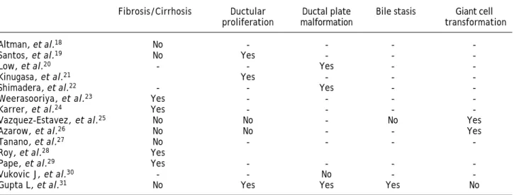
Table 2. Summary of studies in biliary atresia involving liver biopsy parameters as prognostic factors.
Fibrosis/Cirrhosis Ductular Ductal plate Bile stasis Giant cell
proliferation malformation transformation
Altman, et al.18 No - - -
-Santos, et al.19 No Yes - -
-Low, et al.20 - - Yes -
-Kinugasa, et al.21 Yes - -
-Shimadera, et al.22 - - Yes -
-Weerasooriya, et al.23 Yes - - -
-Karrer, et al.24 Yes - - -
-Vazquez-Estavez, et al.25 No No - No Yes
Azarow, et al.26 No No - - Yes
Tanano, et al.27 No - - -
-Roy, et al.28 Yes
Pape, et al.29 Yes - - -
-Vukovic J, et al.30 - - No -
-Gupta L, et al.31 No Yes Yes Yes No
predominant portal inflammation and features of ductular obstruction at the beginning stages to more fibrotic state with destruction of bile ducts later on.13-17 Individual dynamics of histological
presentation carries significant clinical implica-tions. First is that samples taken before 6 weeks of age may be inconclusive and repeated liver biop-sy may be required for diagnosis.17 Second, there
is a difficulty in the assessment of prognostic val-ue of histological features what may account for in-consistent results in previous studies summarized in table 2.18-31
The severity of liver fibrosis, as a major indicator of disease progression, was most commonly analyzed parameter. We showed it did not correspond with the outcome of HPE, however there was only 13% probability of long-term survival in case of advanced liver fibrosis or cirrhosis at the moment of opera-tion. There was no correlation between age at HPE and severity of fibrosis, what is contradictory to previous reports.23
Pape, et al.29 calculated the mean volume of
fi-brosis per number of periportal fields (Vfib) and the Ishak score. Detection of Vfib at the time of HPE was proved to be a valid marker in predicting transplant-free survival in children with BA how-ever the Ishak score showed no correlation with transplant-free survival or Vfib. After extensive analysis, Lampela, et al. did not find correlation be-tween severity of neither fibrosis nor inflammation and survival with native liver, however propor-tions of inflammatory components showed more lymphocytes and less macrophages in those who were transplanted within 2 years. Moreover none of the histologic variables at HPE predicted devel-Figure 4. Survival estimates according to severity of liver
fibrosis at Kasai operation.
p = 0.84
0 1 2 3 4 5 6 7 8 9 10 11 12 Years after HPE
1.0
0.9
0.8
0.7
0.6
0.5
0.4
0.3
0.2
0.1
0.0
Survival with native liver
opment of esophageal varices.15 Our data showed
that risk of portal hypertention increases with se-verity of liver fibrosis.
Ductal plate malformation structures (DPM), are the remnants of impaired remodeling of primitive fe-tal bile ducts during organogenesis.32 These
struc-tures are found in other pathologies as well but in BA they may be associated with poor prognosis.20,22,31
Ductular proliferation usually seen in BA, results from uncontrolled response to chronic cholestasis and supposedly originates from a proliferation of preexisting intrerlobular bile ducts and ductules or from ductular transformation of the periportal hepa-tocytes.33,34 Kinugasa, et al.21 estimated intensity of
ductular proliferation by cytokeratin-7 (CK-7) stain-ing. The number of CK7-positive cells in the bile ductules was microscopically calculated within the 40-μm-thick interstitium along the limiting plate (LP), and the CK7-positive cell number per unit length of the LP was estimated. The higher number of CK7-positive cells correlted with poor bile drain-age after HPE. In other paper the presence of syn-cytial giant cells, lobular inflammation, focal necrosis, bridging necrosis and cholangitis were each associated with failure of HPE and bile in zone 1 with success of operation.26
Discrepancies in current evidence make prognos-tic value of histological assessment unclear. Moreo-ver, subjectivity of examination may bring additional bias. That issue was raised by biliary atresia research consortium (BARC), which identified bile plugs in ducts, giant-cell transforma-tion, extramedullary hematopoiesis, and bile duct proliferation as most reliable features in terms of interobserver agreement.15
The major limitation of our study is retrospective nature of collected data, but liver specimens were re-examined in accordance to the newly prepared protocol. We also lacked histopathology from long-term native liver survivals since we do not perform liver biopsies in these patients routinely.
We concluded that liver histology at the time of HPE is of limited value in prognosis-making and should be interpreted cautiously in regard to the other clinical factors. Patients with severe fibrosis or cirrhosis in initial biopsy are likely to develop portal hypertension.
ACKNOWLEDGEMENTS
We would like to thank Mrs. Krystyna Lech-Kun-kel for major contribution in coordination of the study and specimen management.
REFERENCES
1. Chardot C, Carton M, Spire-Bendelac N, Le Pommelet C, Golmard JL, Auvert B. Epidemiology of biliary atresia in France a national study 1986-96. J Hepatol 1999; 31: 1006-13.
2. Balistreri WF, Grand R, Hoofnagle JH, Suchy FJ, Ryckman FC, Perlmutter DH, Sokol RJ. Biliary Atresia: Current Con-cepts and Research Directions. Summary of a symposium.
Hepatology 1996; 23: 1682-92.
3. Sokol R, Shepherd R, Superina R, Bezerra JA, Robuck P, Ho-ofnagle JH. Screening and outcomes in biliary atresia: summary of a National Institutes of Health workshop.
He-patology 2007; 46: 566-81.
4. Chardot C, Buet C, Serinet MO, Golmard JL, Lachaux A, Roquelaure B, Gottrand F, et al. Improving outcom es of biliary atresia: French national series 1986-2009. J
Hepa-tol 2013; 58: 1209-17.
5. Kasai M, Kimura S, Asakura Y. Surgical treatment of biliary atresia. J Pediatr Surg 1968; 3: 665-75.
6. Hartley JL, Davenport M, Kelly DA. Biliary atresia. Lancet 2009; 374: 1704-13.
7. Ishak K, Baptista A, Bianchi L, Callea F, De Groote J, Gu-dat F, Denk H, et al. Histological grading and staging of chronic hepatitis. J Hepatol 1995; 24: 289-93.
8. Desmet VJ. Congenital diseases of intrahepatic bile ducts: variations on the theme «ductal plate malformation”.
He-patology 1992; 16: 1069-83.
9. Lykavieris P, Chardot C, Sokhn M, Gauthier F, Valayer J, Bernard O. Outcome in adulthood of biliary atresia: a stu-dy of 63 patients who survived for over 20 years with their native liver. Hepatology 2005; 41: 366-71.
10. Goda T, Kawahara H, Kubota A, Hirano K, Umeda S, Tani G, Ishii T, et al. The most reliable early predictors of outcome in patients with biliary atresia after Kasai’s operation. J
Pediatr Surg 2013; 48: 2373-7.
11. McKiernan PJ, Baker AJ, Kelly DA. The Frequency and out-come of biliary atresia in the UK and Ireland. Lancet 2000; 355: 25-9.
12. Bessho K, Bezerra JA. Biliary atresia: will blocking inflam-mation tame the disease? Annu Rev Med 2011; 62: 171-85. 13. Moyer K, Kaimal V, Pacheco C, Mourya R, Xu H,
Shivaku-mar P, Chakraborty R, et al. Staging of biliary atresia at diagnosis by molecular profiling of the liver. Genome
Medi-cine 2010; 2: 33.
14. Lampela H, Kosola S, Heikkila P, Lohi J, Jalanko H, Pakari-nen MP. Native liver histology after successful portoente-rostomy in biliary atresia. J Clin Gastroenterol 2014; 48: 721-8.
15. Yeh MM. Pathologic diagnosis of biliary atresia on liver biopsy: is tissue the issue? J Gastroenterol Hepatol 2009; 24: 933-40.
16. Zani A, Quaglia A, Hadzi N, Zuckerman M, Davenport M. Cytomegalovirus-associated biliary atresia: An aetiologi-cal and prognostic subgroup. J Pediatr Surg 2015; (7). Doi: 10.1016/j.jpedsurg.2015.03.001 [Epub ahead of print].
17. Azar G, Beneck D, Lane B, Markowiz J, Daum F, Kahn E. Atypical morphologic presentation of biliary atresia and value of serial biopsies. J Pediatr Gastroenterol Nutr 2002; 34: 212-15.
18. Altman RP, Lilly JR, Greenfeld J, Weinberg A, van Leeuwen K, Flanigan L. A multivariable risk factor analysis of the portoenterostomy (Kasai) procedure for biliary atresia: twenty-five years of experience from two centers. Ann
19. Santos JL, Kieling CO, Meurer L, Vieira S, Ferreira CT, Lo-rentz A, Silveira TR. The extent of biliary proliferation in liver biopsies from patients with biliary atresia at por-toenterostomy is associated with the postoperative prog-nosis. J Pediatr Surg 2009; 44: 695-701.
20. Low Y, Vijayan V, Tan CE. The prognostic value of ductal plate malformation and other histologic parameters in bi-liary atresia: an immunohistochemical study. J Pediatr 2001; 139: 320-2.
21. Kinugasa Y, Nakashima Y, Matsuo S, Shono K, Suita S, Sueishi K. Bile ductular proliferation as a prognostic fac-tor in biliary atresia: an immunohistochemical assessment.
J Pediatr Surg 1999; 34: 1715-20.
22. Shimadera S, Iwai N, Deguchi E, Kimura O, Ono S, Fumino S, Higuchi K. Significance of ductal plate malformation in the postoperative clinical course of biliary atresia. J Pediatr
Surg 2008; 43: 304-7.
23. Weerasoorya V, White F, Shepherd RW. Hepatic fibrosis and survival in biliary atresia. J Pediatr 2004; 144: 123-5. 24. Karrer FM, Lilly JR, Stewart BA, Hall RJ. Biliary atresia
re-gistry, 1976-89. J Pediatr Surg 1990; 25: 1076-81. 25. Vazquez-Estevez J, Stewart B, Shikes RH, Hall RJ, Lilly JR.
Biliary atresia: early determination of prognosis. J Pediatr
Surg 1989; 24: 48-50.
26. Azarow KS, Phillips MJ, Sandler AD, Hagerstrand I, Superi-na RA. Biliary atresia: should all patients undergo a por-toenterostomy? J Pediatr Surg 1997; 32: 168-72.
27. H. Tanano Æ T. Hasegawa Æ T. Kimura Æ T. Sasaki, H. Kawahara Æ A. Kubota Æ A. Okada. Proposal of fibrosis
index using image analyzer as a quantitative histological evaluation of liver fibrosis in biliary atresia. Pediatr Surg
Int 2003; 19: 52-6.
28. Roy P, Chatterjee U, Ganguli M, Banerjee S, Chatterjee SK, Basu AK. A histopathological study of liver and biliary remnants with clinical outcome in cases of extrahepatic biliary atresia. Indian J Pathol Microbiol 2010; 53: 101-5. 29. Pape L, Olsson K, Petersen C, von Wasilewski R, Melter M.
Prognostic Value of Computerized Quantification of Liver Fibrosis in Children with Biliary Atresia. Liv Transplant 2009; 15: 876-82.
30. Vukovic J, Grizelj R, Bojanic K, Coric M, Luetic T, Batinica S, Kujund•ic-Tiljak M; et al. Ductal plate malformation in patients with biliary atresia. Eur J Pediatr 2012; 171: 1799-804.
31. Gupta L, Gupta SD, Bhatnagar V. Extrahepatic biliary atresia: Correlation of histopathology and liver function tests with surgical outcomes. J Indian Assoc Pediatr Surg 2012; 17: 147-52.
32. Tan CEL, Marie D, Howard ER, Moscoso GJ. Extrahepatic biliary atresia: A first-trimester event? Clues from light microscopy and immunohistochemistry. J Pediatr Surg 1994; 29: 808-14.
33. Nakanuma Y, Ohta G. Immunohistochemical study on bile ductular proliferation in various hepatobiliary diseases.
Liver 1986; 6: 208-11.
34. Thung SN. The development of proliferating ductular structures in liver disease. An immunohistochemical study.
Arch Pathol Lab Med 1990; 114: 407-11.