Annals of Hepatology 7(2) 2008: 136-143
136
www.medigraphic.com
Annals of Hepatology 2008; 7(2): April-June: 136-143
Annals of Hepatology
Original Article
Influence of extrahepatic viral infection on the
natural history of hepatitis C
María Inés Barría;1 Jorge Vera-Otarola;1 Úrsula León;1 Valeska Vollrath;1,2 Delphine Marsac;1 Arnoldo Riquelme;2 Marcelo
López-Lastra;1,3 Alejandro Soza1,2
1Laboratorio de Virología Molecular, Centro de Investigaciones
Médicas.
2Departamento de Gastroenterología, Facultad de Medicina. 3Departamento de Pediatría, Facultad de Medicina, Pontificia
Universidad Católica de Chile.
Address for correspondence: María Inés Barría
Departamento de Gastroenterología and Laboratorio de Virología Molecular, Pontificia Universidad Católica de Chile, Marcoleta 367, Santiago, Chile.
[email protected] (AS) [email protected] (MLL)
Manuscript received and accepted: 22 February and 28 March 2008
The present study was supported by FONDECYT No 1050782 and the JEAI-IRD Initiative. MIB is student of the Programa de Doctorado en Ciencias Biológicas, Fa-cultad de Ciencias, Universidad de Chile and was initially funded by a CONICYT doctoral fellowship and currently hold an IRD doctoral fellowship. JV-O is student of the Programa de Doctorado en Microbiología, Universidad de Santiago de Chile and was supported by a MECESUP USACH doctoral fellowship. DM was partially supported through a Millennium Nucleus on Immunology and Im-munotherapy (NMII) post-doctoral fellowship. MLL is member of the NMII.
Abstract
HCV is primarily hepatotropic, but there is mounting evidence pointing to infection and replication of extra-hepatic sites. Here we evaluated the occurrence of HCV infection of peripheral blood mononuclear cells (PBMC) and explored the possible association between viral extrahepatic infection and the natural history of the disease. Forty seven Chilean, HCV infected, treat-ment naïve patients were included in the study. HCV RNA was isolated from plasma and PBMC and subse-quently reverse transcribed, amplified and sequenced. Most patients harbored HCV 1b genotype and the most common route of infection showed to be blood transfu-sion. HCV RNA was readily detected in PBMCs of 34 out of the 47 patients (72%). We report that HCV
se-quences found in PBMC differ from those in plasma of the same subjects strongly suggesting HCV compart-mentalization. In addition, we found that patients with detectable HCV RNA in PBMC had a tendency for be-ing more likely cirrhotic [OR 3.8 (95% CI: 0.98 to 14)]. In conclusion, this study provides further arguments for the existence of HCV infection of extrahepatic sites and suggests that extrahepatic infection could be a fac-tor influencing the natural hisfac-tory of the disease.
Key words: Cirrhosis, fibrosis, liver, quasiespecies.
Hepatitis C virus (HCV) infection is a worldwide dis-tributed disease leading to chronic hepatitis, cirrhosis and hepatocellular carcinoma, causing significant dis-ability and mortality.1 Aside from the well known
hepat-ic complhepat-ications of this infection, there are a number of less well understood extrahepatic manifestations of the disease, ranging from cryoglobulinemia to lymphoprolif-erative disorders, including neuropathy, dermatologic manifestations (e.g.: lichen planus and porphyria cuta-nea tarda), glomerulonephritis and several autoimmune disorders among others.2-4 Some of these extrahepatic
manifestations could be attributed to an altered immune response to HCV infection, leading to autoimmunity.
One of the most striking aspects of HCV infection is the high likelihood of becoming a chronic disease, with approximately 75 to 85% of subjects with acute hepatitis C not being able to clear acute infection, despite an ap-parent immune response.5 There is evidence that the
im-mune response may be insufficient or the virus may de-velop escape mutations in critical viral epitopes recog-nized by cytotoxic T cells (CTL),6 but mechanisms of
immune escape are incompletely understood. Similarly, even though treatment of HCV infection has experienced great advances in the probability of achieving a sus-tained response,7 therapy of this infection has been
in-variably tempered by a considerable relapse rate. It has been shown that HCV can be detected long after sponta-neous or treatment-induced viral clearance.8
HCV replication is thought to occur predominantly in hepatocytes, but several observations have shown that the virus can be detected in extrahepatic sites, including peripheral blood mononuclear cells (PBMC),9-18
dendrit-Artemisa
www.medigraphic.com
ic cells,19 central nervous system,20-22 among several other
different biological compartments.23-30 A number of
spe-cific approaches have aroused to clarify whether the pres-ence of HCV RNA in extrahepatic tissues represents a contamination artifact, such as adsorption of virions onto cells, or real ongoing replication. Detection of non-struc-tural proteins, negative-strand RNA (intermediary of rep-lication) and demonstration of quasispecies compartmen-talization have all been advocated as strong arguments for supporting HCV active extrahepatic replication. The significance of PBMC or other non-hepatic tissue infec-tion by HCV as well as the possible implicainfec-tion of extra-hepatic replication in viral pathogenesis is currently a matter of debate.
Most of the observations regarding replication of HCV in extrahepatic tissues have been performed in im-munosuppressed15,20,30 or intravenous drug users (IDU),31
who are probably more permissive to infection due to al-tered immunity or multiple exposures. There is therefore, scanty data about detection of HCV in PBMC in immu-nocompetent subjects. In this study we explored the pos-sible influence of viral extrahepatic infection in the natu-ral history of hepatitis C. We report that HCV RNA is de-tectable in PBMC from treatment naïve Chilean patients, mainly infected by blood transfusion, suggesting that PBMC infection by HCV is not restricted, as previously suggested, to IDU, liver transplant recipients, nor HCV-HIV co-infected patients. Moreover, we show that viral sequences isolated from PBMC differ from those found in plasma, strongly suggesting virus compartmentalization. In addition, by comparison of the clinical features of HCV infected patients that harbor viral RNA in PBMCs with those of patients that show virus only in plasma we establish a direct correlation between PBMC infection and hepatitis C disease progression.
Patients and methods
Study population
Patients were recruited prospectively from the outpa-tient clinic of the Liver Unit of the Clinical Hospital, Pontificia Universidad Católica de Chile from 2005 to 2007. We enrolled consecutive adult patients (older than 18 years old) with chronic hepatitis C defined as a posi-tive anti-HCV antibody for more than 6 months with de-tectable viremia by Cobas Amplicor HCV Monitor Test, version 2.0 (Roche Diagnostic Systems). Patients were treatment naïve. Patients with anemia (hematocrit less than 20%), pregnancy, HIV or HBV co-infection or accu-mulated blood extraction greater than 6 mL/kg in the 6 previous weeks were excluded from the study. Other causes of liver disease were excluded. Mixed cryoglobu-linemia was diagnosed on the basis of the manifestations of Meltzer and Franklin’s triad (purpura, asthenia, and ar-thralgia),32 the demonstration of a cryocrit level greater
or equal to 2% and a positive determination of rheuma-toid factor. Demographic, clinical and laboratory charac-teristics of the subjects were recorded prospectively. Du-ration of infection was defined as the interval between the time of the first unsafe blood transfusion or the time of the first intravenous drug use and the date of the blood sample. The protocol of the study was approved by the Ethics Committee of the Faculty of Medicine of the Pontificia Universidad Católica de Chile. All patients signed a written informed consent form approved by the same committee.
Blood processing, RNA isolation and RT-PCR
Peripheral blood (50 mL) was collected into EDTA-containing Vacutainer tubes (Becton Dickinson®). Blood
samples were diluted with an equal volume of PBS and a density gradient (Lymphocyte separation medium, Cellgro®) was used to isolate mononuclear cells by
centrifugation. Total RNA was extracted using the protocol described by Chomczynski and Sacchi.33
RNA concentrations were determined by spectrophotom-etry (GeneQuant, Pharmacia). Total PBMCs were cul-tured in 24-well plates at a density of 1x10⁶ cells per ml
in R-10 media (RPMI 1640, GIBCO Life Technologies), 10% heat-inactivated fetal calf serum, 2 mM L-glutamine, antibiotics and 50 μM 2-mercaptoethanol (Sigma). HCV-RNA was detected by a one step reverse transcription (RT)-polymerase-chain reaction (PCR) us-ing the SuperScriptTM III one step RT-PCR system with
Platinum® Taq DNA Polymerase (Invitrogen) kit was
car-ried out against the highly conserved 5’ UTR using the sense primer: 5’-TTG GGG GCG ACA CTC CAC CAT GAT C-3’ and the anti-sense primer: 5’-GTT ACG TTT GGT TTT TCT TTG AGG T-3’ generating a 370 bp ampl-icon. RT-PCR was also conducted against the NS5B cod-ing region uscod-ing the sense primer: 5’-TTC TCG TAT GAT ACC CGC TGT TTT GA-3’ and the anti-sense prim-er: 5’-TAC CTG GTC ATA GCC TCC GTG AA-3’ gener-ating a 388 bp amplicon. The RT-reaction was conduct-ed at 50°C for 45 min. The RT-enzyme was then inacti-vated and the Platinum® Taq DNA polymerase activated,
by heating the mix at 95°C for 5 min. PCR amplifica-tions were carried out for 40 cycles with each cycle at 95°C for 45 seg, 58°C (for the 5’UTR) or 56°C (for the NS5B coding region) for 45 seg, and 68°C for 45 seg. In vitro transcribed RNA (using T7-RNA polymerase, Fer-mentas) generated from plasmid pFK-I377neo/NS3-3’/wt34
(AJ242654; kindly provided by Dr. Ralf Bartenschlager, University of Heidelberg, Germany) or total RNA extract-ed from a human hepatoma cell line, Huh-7, constitutively expressing plasmid pFK-I377neo/NS3-3’/wt34 (kindly
www.medigraphic.com
HCV genotyping and sequencing
RNA was isolated from plasma and PBMC as de-scribed above. 5’-UTR genotyping was performed using the reverse-hybridization line probe assay, INNO-LiPA HCV II kit (Innogenetics, Ghent, Belgium) according to the manufacturer’s instructions. Briefly, the 5’ UTR is amplified with biotinylated primers. Biotin-labeled PCR products are reverse hybridized to specific probes at-tached to nitrocellulose strips. Development results in a purple precipitate that forms a positive line on the strip. The HCV type is deduced on the basis of the patterns of hybridizing bands by using the line probe assay (LiPA) interpretation chart. Amplification of the 5’UTR and the nonstructural region 5B (NS5B) followed by direct se-quencing (Macrogen Corp, USA) were performed as pre-viously described to confirm the 5’-UTR genotyping. 5’UTR and NS5B sequences were genotyped by using referenced and annotated HCV sequences.
Statistical analysis
Descriptive statistics (mean, median, interquartile range) were used as appropriate for the main variables. Patients with detectable HCV RNA in PBMC (cases) were compared with those who did not have detectable HCV RNA (controls). Unpaired, two-tailed T test (with Welch correction for nonparametric variables) was used to com-pare both groups. Odds ratios with the corresponding 95% confidence interval (CI) were calculated for vari-ables of interest. Significance was set at a p value lower or equal to 0.05.
Results
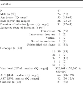
Forty seven patients were eligible for the study. The basal characteristics of the patients are shown in Table I. The predominant route of infection was blood transfu-sion (55%), with only 1 patient having a history of intra-venous drug use. A considerable proportion of patients were cirrhotic (45%) and three patients had mixed cryo-globulinemia at the time of recruitment.
Direct sequencing of the RT-PCR amplicons, corre-sponding to the 5’untranslated region (UTR) and to the NS5B coding region of the HCV RNA, derived from total RNA isolated from plasma allowed confirmation of the assigned HCV genotype previously established by the INNO-LIPA HCV II test. Genotype 1b was confirmed to be the most prevalent among the studied population (83%). This observation is in agreement with previous studies showing a high prevalence of genotype 1b among the Chilean population.35,36
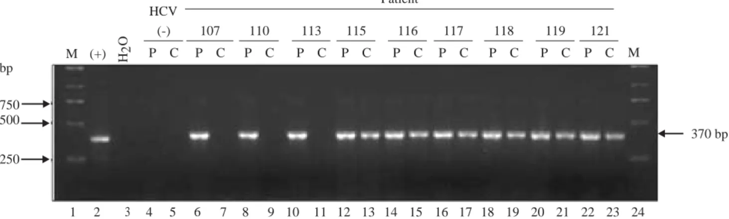
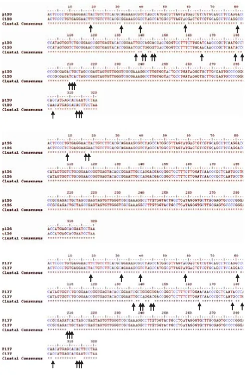
HCV RNA was detectable in PBMCs of 34 out of 47 patients (72%) (Figure 1). Sequencing of the amplicon corresponding to the 5’UTR of predominant viral quasiespecies in each biological compartment (plasma
and PBMC) showed that virus present in PBMC shared the same genotype as virus in plasma, yet in most cases nucleotidic dissimilarities were found. Our findings are exemplified in Figure 2 which shows the alignment be-tween the HCV 5’UTR sequences obtained from plasma (P) or PBMCs (C) of seven randomly selected patients, identified by their code number 23, 26, 105, 108, 120, 126, and 137.
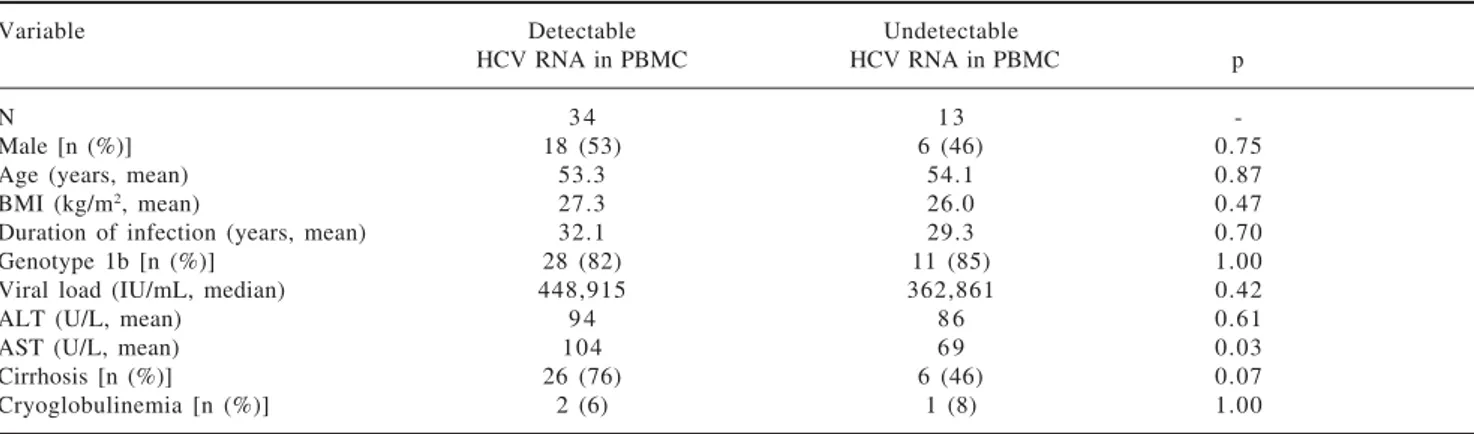
Intrigued by the fact that 72% of the studied HCV in-fected patients harbored detectable amounts of HCV RNA in both plasma and PBMCs we next sought to es-tablish if they presented dissimilar clinical features to pa-tients that showed viral RNA only in plasma (28%). Ta-ble II summarizes the clinical features of the HCV infect-ed patients includinfect-ed in this study. For the sake of comparison patients are separated based on to the pres-ence or abspres-ence of detectable viral RNA in PBMCs. Strikingly, upon analysis no statistically significant dif-ferences could be drawn when comparing the clinical fea-tures of both populations of HCV infected patients in terms of age, gender, BMI, HCV genotype, viral load or estimated duration of infection. Interestingly, however, is the observation that patients presenting detectable amounts of HCV RNA in PBMC exhibit higher AST lev-els (p = 0.03) and a trend to be more likely cirrhotic (76%) than those patients without detectable HCV RNA in PBMC (46%), p=0.07, OR 3.8, 95% CI: 0.98 to 14.
Among the studied patients recruited with cryoglobu-linemia only two (2/3) had detectable HCV RNA in PBMC. Thus, no conclusion associating
cryoglobuline-Table I. Clinical features of the 47 Chilean patients enroled.
Variable
N 4 7
Male [n (%)] 2 4 (51)
Age [years (IQ range)] 5 3 (45-63)
BMI [kg/m2 (IQ range)] 2 6 (23-28)
Duration of infection [years (IQ range)] 3 1 (22-41) Suspected route of infection [n (%)]
Transfusion 2 6 (55) Intravenous drug use 1 (2)
Vertical 1 (2) Sexual transmission 1 (2) Unidentified risk factor 1 8 (38) Genotype [n (%)]
1 b 3 9 (83)
2a 1 (2)
3a 5 (11)
4 1 (2)
5a 1 (2)
Viral load (IU/mL, median (IQ range)] 362,861 (170,365 ≥ 850.000)
ALT [(U/L, median (IQ range)] 6 4 (48-139)
AST [(U/L, median (IQ range)] 8 2 (50-125)
Cirrhosis [n (%)] 2 1 (45)
www.medigraphic.com
mia with the presence of HCV RNA in PBMCs or HCV extrahepatic replication can be drawn.
Discussion
Hepatitis C virus infection is a chronic disease that may take several decades to progress to clinically signifi-cant outcomes. Several viral, host and environmental fac-tors have been described to modulate progression of the disease.5 However, up to date no report shows that
infec-tion of tissues other than the liver by HCV could acceler-ate the progression of fibrosis or induce a more severe form of the disease.
A high chronicity rate, a high relapse rate and the presence of extrahepatic manifestations are all pieces of the yet intriguing puzzle constituted by HCV infec-tion. Extrahepatic replication of the virus can be asso-ciated to some or all of these manifestations, as several other studies have already suggested.8,17,37-39 A
correla-tion between the presence of cryoglobulinemia and more advanced liver fibrosis has been described in Greek patients,40 and at least two studies show a
possi-ble association between HCV infection of PBMCs and mixed cryoglobulinemia, but a direct relationship of viral infection of extrahepatic cells with the rate of progression or severity of the disease has not been sug-gested previously.
It has been already shown that HCV infected PBMC often contain virus variants differing from those circulat-ing in serum or plasma.41-47 Our results confirm these
pre-vious observations. The meaning of this divergence is still a matter of debate, but they strongly suggest a differ-ential tropism of HCV variants in tissues.
The present study has several limitations that hamper the interpretation of the results. First, the demonstration of HCV RNA in PBMC does not prove active virus repli-cation in these cells. It has been suggested that amplifi-cation of the negative strand of the HCV RNA, the natu-ral RNA replication intermediate, is the gold standard for demonstrating active viral replication,48 but the
tech-nique is far from well validated and still highly contro-versial. Moreover, there are several arguments that sup-port the notion that activation of PBMC is an absolute requirement for HCV replication, therefore the presence of negative strand would be expected only in mitogen activated PBMC.14,49,50 In consequence, as our study did
not include the activation of PBMCs prior to total RNA isolation we would not expect to find HCV negative strand RNA within these cells. Nevertheless, the herein presented data clearly shows that HCV RNA can be readily detected in PBMC from treatment naïve HCV pa-tients that are not injection drugs users (IDU) nor HIV-HCV coinfected, stressing that PBMC infection by HIV-HCV is not restricted to the latter populations. Additionally, we present data that favors the notion of HCV compart-mentalization as plasma and PBMC harbor different vi-rus variants belonging to different viral quasiespecies. In-terestigly, even though indirect, this later finding are an indicative of HCV infection and independent replication in PBMCs.51
A second limitation of our study is the weak statisti-cal weight of the herein driven association. We have shown a trend for a higher proportion of cirrhosis in pa-tients who had detectable HCV RNA in PBMC with an OR 3.8 (95% CI: 0.98 to 14). This association of course does not imply causality. In fact it may well represent
Figure 1. Detection of HCV RNA in plasma (P) samples and PBMCs (C) of infected patients (lanes 6 through 23). Sample 1 lanes 4 and 5 corresponds to plasma and PBMC collected from a non-HCV infected donor (negative control). Lane 2 corresponds to the amplicon gene-rated when in vitro transcribed HCV 1b RNA is used as template (positive control). Lane 3 corresponds to the RT-PCR water control (ne-gative control). Lanes 1 and 24 correspond to the molecular weight marker.
Numbers in the upper row represent individual patients. P: Plasma
C: Cells
Lane 2: Positive control (Bartenschlager’s replicon) Lane 3: Negative control (water)
M (+) H2
O
HCV
(-) 107 110 113 115 116 117 118 119 121
P C P C P C P C P C P C P C P C P C P C M
370 bp bp
750 500
250
1 2 3 4 5 6 7 8 9 10 11 12 13 14 15 16 17 18 19 20 21 22 23 24
www.medigraphic.com
the opposite, that cirrhosis may be a factor predisposing to viral infection of extrahepatic sites. Progression of fi-brosis is multifactorial, therefore it is important to ac-count for possible confounding factors such as age, route of infection, genotype, viral load and others. The small sample size precluded formal multivariable analy-sis of data, but none of these factors were independent-ly found to be associated to detection of HCV RNA in PBMCs in our study. A more accurate way to search for this association would be to examine the rate of
pro-gression of fibrosis, rather than the mere presence or ab-sence of cirrhosis.
Notwithstanding the highlighted limitations of our re-port, this observational study does however raises a num-ber of intriguing question that are worth further investi-gatation, i. e. the possible role of hepatitis C virus infec-tion and replicainfec-tion in PBMC as a key factor influencing the natural history of the disease. The mechanism by which infection of PBMC could result in an accelerated progression of fibrosis remains completely speculative. It
www.medigraphic.com
ESTE DOCUMENTO ES ELABORADO PORMEDI-GRAPHIC
is tempting to hypothesize that lymphotropic HCV strains could derange immunologic responses of CD4 cells or other immune cells important to keep hepatic HCV replication under control.
In summary, we have shown evidence of HCV specific infection of PBMC, with detection of RNA in these cells belonging to different quasiespecies from those found in the plasma of the same patients. Moreover, our data strongly suggest that PBMC infection by HCV is not re-stricted, as previously suggested, to IDU, liver transplant recipients, nor HCV-HIV co-infected patients. In addi-tion, we have also found that HCV extrahepatic infection could be associated with more advanced liver disease. In order to be confirmed, this finding needs to be addressed with further studies with higher number of patients and a prospective design.
Acknowledgements
We thank Dr. Ralf Bartenschlager, University of Heidelberg, Germany, for kindly providing HCV related reagents. We appreciate the help of Nurse Molque Velas-co during sample Velas-collection. We thank Chanell Pasquot for her administrative assistance.
References
1. Lauer GM, Walker BD. Hepatitis C virus infection. N Engl J Med
2001; 345: 41-52.
2. Gumber SC, Chopra S. Hepatitis C: a multifaceted disease. Re-view of extrahepatic manifestations. Ann Intern Med 1995; 123: 615-20.
3. Agnello V, De Rosa FG. Extrahepatic disease manifestations of HCV infection: some current issues. J Hepatol 2004; 40: 341-52. 4. Zignego AL, Ferri C, Pileri SA, Caini P, Bianchi FB. Extrahepatic manifestations of Hepatitis C Virus infection: a general overview and guidelines for a clinical approach. Dig Liver Dis 2007; 39: 2-17. 5. Chen SL, Morgan TR. The natural history of hepatitis C virus
(HCV) infection. Int J Med Sci 2006; 3: 47-52.
6. Tsai SL, Chen YM, Chen MH, et al. Hepatitis C virus variants circumventing cytotoxic T lymphocyte activity as a mechanism of chronicity. Gastroenterology 1998; 115: 954-65.
7. Hughes CA, Shafran SD. Chronic hepatitis C virus management: 2000-2005 update. Ann Pharmacother 2006; 40: 74-82.
8. Pham TN, MacParland SA, Mulrooney PM, Cooksley H, Naoumov NV, Michalak TI. Hepatitis C virus persistence after spontaneous or treatment-induced resolution of hepatitis C. J Virol 2004; 78: 5867-74.
9. de Maddalena C, Zehender G, Bianchi Bosisio A, et al. HCV-RNA detection using different PCR methods in sera, cryoglobulins and peripheral blood mononuclear cells of patients with mixed cryo-globulinemia. Clin Exp Rheumatol 1995; 13 Suppl 13: S119-22. 10. Di Liberto G, Roque-Afonso AM, Kara R, et al. Clinical and therapeutic implications of hepatitis C virus compartmentaliza-tion. Gastroenterology 2006; 131: 76-84.
11. Ferri C, Monti M, La Civita L, et al. Infection of peripheral blood mononuclear cells by hepatitis C virus in mixed cryoglobuline-mia. Blood 1993; 82: 3701-4.
12. Laskus T, Radkowski M, Wang LF, Jang SJ, Vargas H, Rakela J. Hepatitis C virus quasispecies in patients infected with HIV-1: correlation with extrahepatic viral replication. Virology 1998; 248: 164-71.
13. Lerat H, Berby F, Trabaud MA, et al. Specific detection of hepa-titis C virus minus strand RNA in hematopoietic cells. J Clin Invest 1996; 97: 845-51.
14. Moldvay J, Deny P, Pol S, Brechot C, Lamas E. Detection of hepatitis C virus RNA in peripheral blood mononuclear cells of infected patients by in situ hybridization. Blood 1994; 83: 269-73. 15. Radkowski M, Wang LF, Vargas HE, Rakela J, Laskus T. Detec-tion of hepatitis C virus replicaDetec-tion in peripheral blood mono-nuclear cells after orthotopic liver transplantation. Transplanta-tion 1998; 66: 664-6.
16. Rodriguez-Inigo E, Casqueiro M, Navas S, Bartolome J, Pardo M, Carreno V. Fluorescent «in situ» hybridization of hepatitis C virus RNA in peripheral blood mononuclear cells from patients with chronic hepatitis C. J Med Virol 2000; 60: 269-74. 17. Zehender G, de Maddalena C, Monti G, et al. HCV genotypes in
bone marrow and peripheral blood mononuclear cells of patients with mixed cryoglobulinemia. Clin Exp Rheumatol 1995; 13 Suppl 13: S87-90.
18. Manzin A, Solforosi L, Candela M, et al. Hepatitis C virus infec-tion and mixed cryoglobulinaemia: assessment of HCV RNA copy numbers in supernatant, cryoprecipitate and non-liver cells.
J Viral Hepat 1996; 3: 285-92.
19. Goutagny N, Fatmi A, De Ledinghen V, et al. Evidence of viral replication in circulating dendritic cells during hepatitis C virus infection. J Infect Dis 2003; 187: 1951-8.
20. Morsica G, Bernardi MT, Novati R, Uberti Foppa C, Castagna A, Lazzarin A. Detection of hepatitis C virus genomic sequences in the cerebrospinal fluid of HIV-infected patients. J Med Virol
1997; 53: 252-4.
21. Laskus T, Radkowski M, Bednarska A, et al. Detection and analysis of hepatitis C virus sequences in cerebrospinal fluid. J Virol 2002; 76: 10064-8.
Table II. Comparison of patients who had detectable and undetectable HCV RNA (5’ UTR) in PBMC.
Variable Detectable Undetectable
HCV RNA in PBMC HCV RNA in PBMC p
N 3 4 1 3
-Male [n (%)] 18 (53) 6 (46) 0.75
Age (years, mean) 53.3 54.1 0.87
BMI (kg/m2, mean) 27.3 26.0 0.47
Duration of infection (years, mean) 32.1 29.3 0.70
Genotype 1b [n (%)] 28 (82) 11 (85) 1.00
Viral load (IU/mL, median) 448,915 362,861 0.42
ALT (U/L, mean) 9 4 8 6 0.61
AST (U/L, mean) 104 6 9 0.03
Cirrhosis [n (%)] 26 (76) 6 (46) 0.07
www.medigraphic.com
22. Bagaglio S, Cinque P, Racca S, et al. Hepatitis C virus populations in the plasma, peripheral blood mononuclear cells and cerebrospi-nal fluid of HIV/hepatitis C virus-co-infected patients. Aids 2005; 19 Suppl 3: S151-65.
23. Takamatsu K, Okayasu I, Koyanagi Y, Yamamoto N. Hepatitis C virus propagates in salivary glands. J Infect Dis 1992; 165: 973-4.
24. Arrieta JJ, Rodriguez-Inigo E, Ortiz-Movilla N, et al. In situ detection of hepatitis C virus RNA in salivary glands. Am J Pathol
2001; 158: 259-64.
25. Carrozzo M, Quadri R, Latorre P, et al. Molecular evidence that the hepatitis C virus replicates in the oral mucosa. J Hepatol
2002; 37: 364-9.
26. Nowicki MJ, Laskus T, Nikolopoulou G, et al. Presence of hepa-titis C virus (HCV) RNA in the genital tracts of HCV/HIV-1-coinfected women. J Infect Dis 2005; 192: 1557-65.
2 7 . Sansonno D, De Vita S, Cornacchiulo V, Carbone A, Boiocchi M, Dammacco F. Detection and distribution of hepatitis C virus-related proteins in lymph nodes of pa-tients with type II mixed cryoglobulinemia and neoplastic or non-neoplastic lymphoproliferation. Blood 1996; 88: 4 6 3 8 - 4 5 .
28. Rodriguez-Inigo E, Casqueiro M, Bartolome J, et al. Hepatitis C virus RNA in kidney biopsies from infected patients with renal diseases. J Viral Hepat 2000; 7: 23-9.
29. Widell A, Mansson S, Persson NH, Thysell H, Hermodsson S, Blohme I. Hepatitis C superinfection in hepatitis C virus (HCV)-infected patients transplanted with an HCV-(HCV)-infected kidney. Trans-plantation 1995; 60: 642-7.
30. Laskus T, Radkowski M, Wang LF, Vargas H, Rakela J. Search for hepatitis C virus extrahepatic replication sites in patients with acquired immunodeficiency syndrome: specific detection of nega-tive-strand viral RNA in various tissues. Hepatology 1998; 28: 1398-401.
31. Beld M, Penning M, van Putten M, et al. Low levels of hepa-titis C virus RNA in serum, plasma, and peripheral blood mono-nuclear cells of injecting drug users during long antibody-undetectable periods before seroconversion. Blood 1999; 94: 1183-91.
32. Meltzer M, Franklin EC, Elias K, McCluskey RT, Cooper N. Cryoglobulinemia—a clinical and laboratory study. II. Cryoglobulins with rheumatoid factor activity. Am J Med 1966; 40: 837-56.
33. Chomczynski P, Sacchi N. Single-step method of RNA isolation by acid guanidinium thiocyanate-phenol-chloroform extraction.
Anal Biochem 1987; 162: 156-9.
34. Lohmann V, Korner F, Koch J, Herian U, Theilmann L, Bartenschlager R. Replication of subgenomic hepatitis C virus RNAs in a hepatoma cell line. Science 1999; 285: 110-3. 35. Soza A, Arrese M, Gonzalez R, et al. Clinical and
epidemiologi-cal features of 147 Chilean patients with chronic hepatitis C. Ann Hepatol 2004; 3: 146-51.
36. Soza A, Lopez-Lastra M. [Hepatitis C in Chile: burden of the disease]. Rev Med Chil 2006; 134: 777-88.
37. Gong GZ, Lai LY, Jiang YF, He Y, Su XS. HCV replication in PBMC and its influence on interferon therapy. World J Gastroenterol 2003; 9: 291-4.
38. Galli M, Zehender G, Monti G, et al. Hepatitis C virus RNA in the bone marrow of patients with mixed cryoglobulinemia and in subjects with noncryoglobulinemic chronic hepatitis type C. J Infect Dis 1995; 171: 672-5.
39. Sansonno D, Tucci FA, Lauletta G, et al. Hepatitis C virus pro-ductive infection in mononuclear cells from patients with cryoglobulinaemia. Clin Exp Immunol 2007; 147: 241-8. 40. Siagris D, Christofidou M, Tsamandas A, Lekkou A,
Thomopoulos K, Labropoulou-Karatza C. Cryoglobulinemia and progression of fibrosis in chronic HCV infection: cause or effect?
J Infect 2004; 49: 236-41.
41. Shimizu YK, Igarashi H, Kanematu T, et al. Sequence analysis of the hepatitis C virus genome recovered from serum, liver, and peripheral blood mononuclear cells of infected chimpanzees. J Virol 1997; 71: 5769-73.
42. Radkowski M, Wang LF, Vargas H, Wilkinson J, Rakela J, Laskus T. Changes in hepatitis C virus population in serum and peripheral blood mononuclear cells in chronically infected patients receiving liver graft from infected donors. Transplantation 2001; 72: 833-8. 43. Laskus T, Radkowski M, Wang LF, Nowicki M, Rakela J. Un-even distribution of hepatitis C virus quasispecies in tissues from subjects with end-stage liver disease: confounding effect of viral adsorption and mounting evidence for the presence of low-level extrahepatic replication. J Virol 2000; 74: 1014-7.
44. Bartolome J, Castillo I, Quiroga JA, Navas S, Carreno V. Detec-tion of hepatitis C virus RNA in serum and peripheral blood mononuclear cells. J Hepatol 1993; 17 Suppl 3: S90-3. 45. Okuda M, Hino K, Korenaga M, Yamaguchi Y, Katoh Y, Okita
K. Differences in hypervariable region 1 quasispecies of hepatitis C virus in human serum, peripheral blood mononuclear cells, and liver. Hepatology 1999; 29: 217-22.
46. Roque-Afonso AM, Ducoulombier D, Di Liberto G, et al. Compartmentalization of hepatitis C virus genotypes between plasma and peripheral blood mononuclear cells. J Virol 2005; 79: 6349-57.
47. Roque Afonso AM, Jiang J, Penin F, et al. Nonrandom distribu-tion of hepatitis C virus quasispecies in plasma and peripheral blood mononuclear cell subsets. J Virol 1999; 73: 9213-21. 48. Blackard JT, Kemmer N, Sherman KE. Extrahepatic replication
of HCV: insights into clinical manifestations and biological con-sequences. Hepatology 2006; 44: 15-22.
49. Pham TN, Macparland SA, Coffin CS, Lee SS, Bursey FR, Michalak TI. Mitogen-induced upregulation of hepatitis C virus expression in human lymphoid cells. J Gen Virol 2005; 86: 657-66. 50. MacParland SA, Pham TN, Gujar SA, Michalak TI. De novo
infection and propagation of wild-type Hepatitis C virus in hu-man T lymphocytes in vitro. J Gen Virol 2006; 87: 3577-86. 51. Navas S, Martin J, Quiroga JA, Castillo I, Carreno V. Genetic