Annals of Hepatology 6(4) 2007: 242-250
242
medigraphic.com
Annals of Hepatology 2007; 6(4): October-December: 242-250
Annals of Hepatology
Original Article
Dipeptidyl peptidase IV (DDP IV) in NASH patients
Yasemin H Balaban;1 Petek Korkusuz;2 Halis Simsek;3 Hale Gokcan;4 Gokhan Gedikoglu;5 Asli Pinar;6
Gulsen Hascelik;7 Esin Asan;2 Erhan Hamaloglu;8 Gonca Tatar3
1Department of Internal Medicine, Division of Gastroenterology,
Abant Izzet Baysal University, Bolu - Turkey.
2Department of Histology & Embryology, Hacettepe University,
Ankara - Turkey.
3Department of Internal Medicine, Division of Gastroenterology,
Hacettepe University, Ankara - Turkey.
4Department of Internal Medicine, Division of Gastroenterology,
Baskent University, Ankara - Turkey.
5Department of Pathology, Hacettepe University, Ankara
-Turkey.
6Children’s Hospital Clinical Chemistry Laboratory, Hacettepe
University, Ankara - Turkey.
7Department of Microbiology and Clinical Microbiology,
Hacettepe University, Ankara – Turkey.
8Department of General Surgery, Hacettepe University, Ankara –
Turkey.
Addrees for correspondence: Yasemin H Balaban, MD Abant Izzet Baysal University Izzet Baysal Faculty of Medicine Department of Internal Medicine Division of Gastroenterology Bolu - Turkey
Tel: +90 0533 234 4232 Fax: +90 0374 253 4559 E-mail: [email protected] [email protected]
Support for work
The study was supported by Hacettepe University, Scientific Research Foundation (02 02 101 030).
The abstract of the study was presented during 12nd United European Gastroenterology Week UEGW) (September 25-29, 2004) Prague, Czech Republic.
Manuscript received and accepted: 6 July and 16 October 2007
ABSTRACT
Objective(s): Non-alcoholic steatohepatitis (NASH) is a chronic liver disease with unknown etiology. The insu-lin resistance, immune mechanisms and oxidative stress are the main factors in its pathogenesis. Dipepti-dyl peptidase IV (DPPIV) or CD26 is a protein with en-docrine and immune functions. This study aimed to eli-cudate the changes related to DPPIV in NASH patients. Methods: Serum and urinary DPPIV activities were measured in 31 NASH patients and 17 healthy controls. The liver biopsies of 29 patients were immunolabeled for CD26. Results: The mean age of patients were 46 ±
11 years and 14 (45%) of them were female. The serum DPPIV activity was higher in patients (57.3 ± 7.8 U/L) than controls (43.6 ± 10.6 U/L) (p < 0.0001), and corre-lated with the histopathological grade (p = 0.038, r = 0.373) and hepatosteatosis (p = 0.018, r = 0.423) but not with stage (p = 0.286), class (p = 0.286) or CD26 stain-ing (p = 0.743). The urinary DPPIV activity was simi-lar in patients (1.52 ± 0.94 U/mmol creatinine) and con-trols (1.37 ± 0.68 U/mmol creatinine) (p = 0.861). Three acinar zones of liver had equal CD26 expression (p = 0.076). The intensity of CD26 immunostaining was cor-related with histopathological grade (p = 0.001) and hepatosteatosis (p = 0.003) but no correlation with stage or class could be detected (p = 0.610 and 0.956, respectively). In Conclusions: The serum DPPIV activ-ity and the staining intensactiv-ity of CD26 in liver are cor-related with histopathologic grade of NASH and hepa-tosteatosis. DPPIV can be proposed as a novel candi-date with several potential functions in NASH pathogenesis.
Key words: CD26, dipeptidyl peptidase IV, non-alco-holic steatohepatitis, metabolic syndrome.
Introduction
Nonalcoholic steatohepatitis (NASH) is defined as he-patic steatosis associated with inflammation and fibrosis in the absences of significant alcohol consumption.1
NASH is the most commune cause of liver enzyme eleva-tion in the populaeleva-tions which have been attacked by cur-rent epidemics of obesity, diabetes and so metabolic syn-drome.2-4 The prognosis of NASH has been debated but
nearly half of the patients have progressive fibrosis and one in five of them develop cirrhosis.5 The recurrence of
NASH after liver transplantation and the development of nonalcoholic fatty liver disease (NAFLD) in a quarter of transplanted patients for cryptogenic cirrhosis support believe that NASH is a metabolic disease. Although high prevalence and relatively poor prognosis of NASH ur-gently necessitate effective treatments, there is currently no drug which can improve the prognosis of NASH.
Dipeptidyl peptidase IV (DPP IV; E.C 3.4.14.5) is syn-onym with CD26, adenosine deaminase binding protein (ADAbp), and T cell activation protein. DPPIV is a
cell-Artemisa
medigraphic.com
surface dipeptidase that activates or inactivates the pep-tides by removing proline or alanine from second posi-tion of N-terminal. DPPIV inactivates incretins which im-prove insulin resistance and β cell function. Apart from this enzymatic activity, DPPIV has function as a receptor and interacts with several proteins such as adenosine deaminase, HIV gp 120 protein, fibronectin, collagen, chemokine receptor CXCR4, and CD45. DPPIV also serves as a co-stimulatory molecule to activate T cells. DPPIV is primarily expressed on T lymphocytes, endot-helial cells and epitendot-helial cells. Its soluble form can be detected in plasma and body fluids. The change in serum DPPIV level is associated with autoimmune diseases, in-fections, cancers, depression and also liver diseases.6-8
DPPIV inhibitors are new group of oral antidiabetic agents which have been shown to improve insulin resis-tance in prediabetic people and patients with type 2 dia-betes.9-11 Experimental animal studies and clinical
stud-ies have firmly established that DPPIV inhibition im-prove glucose tolerance and β cell function without any adverse effect on normal physiology, therefore, DPPIV is a drug target and DPPIV inhibitors have been approved by FDA for the combination or monotherapy of type 2 diabetes.9-16 Since NASH is strongly associated with
insu-lin resistance and DPPIV inhibitors improve both hepatic and peripheral insulin sensitivity, DPPIV inhibitors are also novel candidates for the treatment of NASH. To the best of authors’ knowledge this is the first study in the literature investigating the changes related to DPPIV in NASH patients. The liver, serum and urine samples have been investigated in order to elucidate the rational of treatment with DPPIV inhibitors in NASH.
Methods
The study protocol has been approved by the institu-tional ethic committee of Hacettepe University. Informa-tion was given to all patients before the biopsy about the specimens’ use in an experimental study and written con-sent was obtained. All procedures were in full compli-ance with the Helsinki Declaration of Human Rights. Thirty one patients with clinical diagnosis of NASH were recruited from the gastroenterology policlinic of Hac-ettepe University Hospital. After exclusion of viral, au-toimmune, alcoholic, metabolic and toxic causes of liver disease, patients were given advice on diet and exercise. Those patients with malignancy, diabetes or using hepa-totoxic drugs were not included in order to eliminate concomitant factors effecting liver enzyme or DPPIV lev-els. Anthropometric measurements (weight, height, waist and hip circumference) were done. All the patients under-went 75 mg Oral Glucose Tolerance Test (OGTT). Blood and urine samples were collected from 31 NASH patients and 17 healthy volunteers whose body mass index (BMI), serum transaminase levels and abdominal ultrasonogra-phy were normal. The fasting serum samples were used
for measurements of liver enzymes, glucose, insulin, C-peptide and lipids. Homeostasis Model Assessment of In-sulin Resistance (HOMA-IR) was calculated as inIn-sulin (pmol/L) × glucose (μmol/L)/22.5). HOMA-IR values equal to or greater than 3.0 were considered to be indicative of insulin resistance.
Biochemical analyses
Serum measurements for liver enzymes, glucose, lip-ids, and urine measurements for creatinine were assayed immediately with Roche/Hitachi Modular Analytics (To-kyo, Japan). Reagents and calibrators from the same man-ufacturer (Roche Diagnostics, Mannheim, Germany) were used for each assay, and instrument operations and cali-brations were performed as the instructions from manu-facturer.
DPPIV measurements were performed on samples which were kept frozen at -70°C. In order to minimize of the analytical variability, all serum and urine specimens were analyzed at the same day. Determination of DPPIV activities in serum and urine were assayed according to the colorimetric method of Nagatsu.17 This method uses
the chromogenic substrate glycyl-L-proline-p -nitroanil-ide. Briefly, 50 μL of serum was incubated with 200 μL of 71 mM glycine/NaOH buffer (pH 8.7) and 50 μL of 3 mM glycylproline p-nitroanilide at 37°C for 30 min. The reaction was stopped by addition of the 2.7 mL acetate buffer (1 M, pH 4.5). The absorption at 385 nm was mea-sured in order to detect p-nitroanilide. For calculations,
p-nitroanilide was used as a standard and dipeptidyl pep-tidase from porcine kidney was used as a control. One unit of activity per liter was defined as the enzyme activ-ity which produces 1 mol of p-nitroanilide in 1 min un-der the assay conditions. Urinary DPPIV activity was cor-rected with urinary creatinine. All chemicals were ob-tained from Sigma Chemical Co.
Pathological and immunohistological evaluation
All patients with persistently more than two fold eleva-tions of transaminases after 3 months of life style chang-es, underwent liver biopsy. The ultrasound guided liver biopsies were done by using 14 G, 15 cm true-cut needle. Conventional pathological examination was performed on 2 cm of liver specimens that were paraffin-embedded, cut and stained with haematoxylin & eosin. Histopathological classification for NASH was made according to the Brunt’s criteria.18 Approximately 0.5 cm biopsy material
medigraphic.com
were fixed in cold acetone for 10 minutes and air-dried for at least 30 minutes. Then, they were incubated with mono-clonal Ig G antibody against human CD26 (#MCA1317, clone number M-A261, mouse anti-human Ig G1, Serotec, UK) for 60 minutes at room temperature in humidity cham-bers. After washing with 0.01 M phosphate buffered sa-line at pH = 7.4, a group of sections were covered with FITC conjugated anti-mouse Ig G (1:20) (#F0261, Dako, USA) containing 0.2% bovine serum albumin and 1% nor-mal human serum. Other groups of slides were covered with 3-3’ diaminobenzidine tetrahydrochloryde (DAB) conjugated anti-mouse screening kit (Serotec, UK) for peroxidase activity following the manifacturers’ instruc-tions. All antibodies were diluted in a background reduc-ing buffer solution in 0.05 M TrisHCl containreduc-ing 0.1% tween (#S3022, Dako, USA). A counterstaining with pro-pidium iodide (Anchor, USA) was performed for FITC-la-beled sections; and with Mayers’ haematoxylin for DAB-labeled sections. Control staining was performed by omit-ting the initial primary antibody staining step and using a control mouse Ig G. Frozen thymus sections were used as positive control following manufacturers’ instructions. Stained sections were examined in a random order and blinded fashion and scored by two investigators. For each observer all sections were evaluated in one sitting using the same microscope at the same magnification. The reported histological score is the average of these observations. Each section was graded for immune reac-tion on hepatocytes for each acinar zone (1, 2, and 3) on a scale of 0 to +++. 0 was given to no immune reactivity, + to weak but continuous reactivity, ++ for moderate but continuous reactivity, and +++ to intense but continuous immunostaining of hepatocytes for each zone. Data were documented with a Leica DMR microscope (Germany); images were captured via Leica DC500 digital camera (Germany).
Statistical methods
Statistical analyses were done by using SPSS 10.0 program. The statistical significance was accepted as p values less than 0.05. Independent samples t test was used for parametric measurements, and χ2 test,
Mann-Whitney U test or Kruskal Wallis test were used for non-parametric variables. The related variable groups were compared by Friedman test. The associations for ordinal variable were analyzed by Kendall’s tau-c test.
Results
1. Characteristics of patients with NASH
Out of 31 patients with NASH, 14 (%45) were female and the mean age was 46 ± 11 (Table I). Obesity, espe-cially central obesity was present in most of the patients; 12 (38.9%) had BMI > 30 kg/m2 and 28 (90.3%) had
in-creased waist to hip ratio. The mean fasting insulin level and HOMA-IR were 84.8 ± 40.8 pmol/L and 2.8 ± 1.4, re-spectively. The evidence of insulin resistance was detect-ed as impairdetect-ed fasting glucose 5 (16.1%), impairdetect-ed OGTT 6 (19.4%), increased fasting insulin level 10 (32.1%) or high HOMA-IR index 13 (41.9%).
There was no patients with simple hepatosteatosis and the class of NAFLD were of class 2, 3, 4 in 6.5%, 64.5% and 29.0% patients, respectively (Table II). The liver dis-ease was graded as mild (45.2%), moderate (22.6%) or se-vere (32.3%) according to the intensity of portal inflam-mation, hepatosteatosis, ballooning degeneration and lobular inflammation. The majority of the patients had mild portal inflammation (80.6%), mild hepatosteatosis (54.8%), ballooning degeneration (93.5%) and lobular inflammation (96.8%). The stage of fibrosis was mild to moderate in all patients, except one with severe fibrosis.
2. Immunohistological evaluation of CD26
All of the NASH patients expressed CD26 of vari-able intensity on liver parenchymal cells (hepato-cytes), stromal fibroblasts, and some capillary
endot-Table I. Characteristics of NASH Patients.
NASH (n = 31)
Female: Male 45:55
Age (yr)* 46 ± 11
Obesity
Normal (BMI < 25.0 kg/m2) 16.1%
Overweight (BMI 25.0-29.9 kg/m2) 45.2%
Obese (BMI ≥ 30 kg/m2) 38.7%
Central obesity
Waist circumference (≥ 102 cm for men or
≥ 88 cm for women)† 45.2%
Waist/hip ratio (≥ 0.9 cm for men or
≥ 0.8 cm for women)†† 90.3%
Insulin resistance
Impaired fasting glucose (110-125 mg/dL) 16.1% Impaired OGTT (postload
glucose 140-200 mg/dL) 19.4%
Increased fasting insulin (>100 pmol/L) 32.1%
HOMA-IR ≥ 3 41.9%
Hypertension (≥ 130/85 mmHg) 25.8% Hypertriglyceridemia (≥ 150 mg/dL) 45.2% Low HDL (< 40 mg/dL for men or
< 50 mg/dL for women) 19.4%
LDL (mg/dL)* 126.2 ± 37.6
AST (U/L) * 49.1 ± 20.8
ALT (U/L) * 75.9 ± 24.1
GGT (U/L) * 99.1 ± 82.6
CRP (mg/L) * 0.6 ± 0.5
ALT, alanine aminotransferase; AST, aspartate aminotransferase; BMI, body mass index; CRP, c-reactive protein; GGT, gama-glutamyl transpeptidase; HDL, high density lipoprotein; HOMA-IR, homeostasis model assessment of insulin resistance; LDL, low density lipoprotein; OGTT, oral glucose tolerance test.
* Data are presented as mean ± SD.
† female vs. male p=0.052.
medigraphic.com
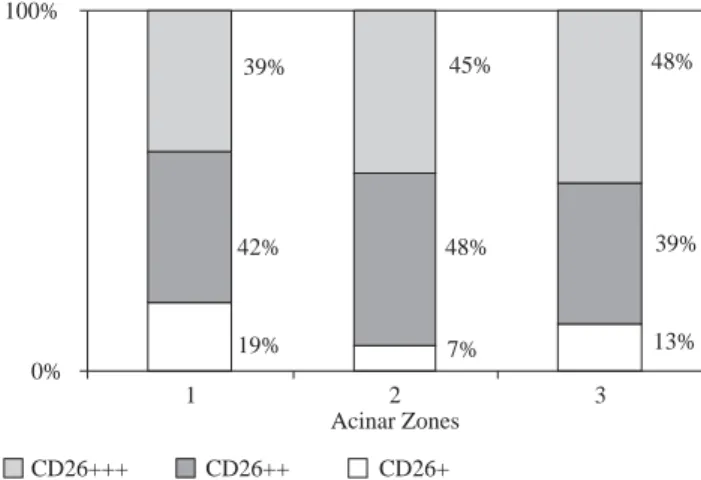
helia. A more diffuse and strong immunolabeling was observed on hepatocytes (Figure 1). CD26 immunore-activity was strictly located on the apical (biliary) side of hepatocyte in all NASH specimens (Figure 2). Im-munoreaction was not present at the basolateral (sinu-soidal) compartment in any of the cases. Hepatocytes expressed CD26 in all acinar zones. Though the inten-sity of CD26 immunostaining was higher in zone 3 when compared to that of the zone 1 in several sub-jects, the difference was not statistically significant re-garding the zonal distribution pattern (p = 0.076, Fig-ure 3). This may probably relate to the limited number of patients.
There was correlation between CD26 immunore-activity with histolopathological grade of NASH (p = 0.001) and hepatosteatosis (p = 0.003) by kendal’s tau test. Patients with higher grade or more sever ste-Table II. Histopathological features of patients.
Number Percentage
Class of NAFLD
1/2/3/4 0/2/20/9 0.0/6.5/64.5/29.0 Grade of NASH
Mild/moderate/severe 14/7/10 45.2/22.6/32.3 i. Portal inflammation
Absent/mild/moderate/sever 5/25/1/0 16.1/80.6/3.2/0.0 ii. Hepatosteatosis
Mild/moderate/sever 17/4/10 54.8/12.9/32.3 iii. Ballooning degeneration
Absent/persent 2/29 6.5/93.5
iv. Lobular inflammation
Absent/Present 1/30 3.2/96.8
Stage of NASH
1/2/3 22/8/1 71.0/25.8/3.2
NAFLD, nonalcoholic fatty liver disease; NASH, nonalcoholic steatohepatitis.
Figure 1. (A, B) Low magnifica-tion micrographs showing the zo-nal expression pattern of CD26 in NASH livers. (C-F) CD26 immu-noreactivity is obviously observed on the biliary (apical) side of the hepatocytes at higher magnifica-tions (arrows). PS: Portal space; CV: Central vein; PV: Portal vein branch; Z1, 2, 3: Zone 1, 2, 3. All micrographs are FITC labeled, Nuclei are counterstained with propidium iodide in A and E.
A
B
C
D
E
F
200x 200x
400x 400x
1000x 400x
CV
Z3 Z2 Z1
Z1 Z2
Z3 CV
PS
Z3
Z2
Z1
medigraphic.com
atosis had poorer CD26 immunoreactivity (Figure 4). Statistically significant correlation was not de-tected between CD26 immune reaction or portal in-flammation (p = 0.713), ballooning degeneration (p = 0.158), lobular inflammation (p = 0.318), stage (p = 0,610) and class (p = 0.956) parameters of NASH
(Table III).
medigraphic.com
3. Serum DPPIV enzyme activity
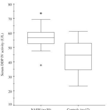
Serum DPPIV activitywas significantly higher in pa-tients with NASH (57.3 ± 7.8 U/L) than in controls (43.6 ± 10.6 U/L) (p < 0.000, Figure 5). Serum DPPIV activity correlated with grade (p = 0.038, r = 0.373) and steatosis (p = 0.018, r = 0.423). But there was no association with zonal DPPIV staining (p = 0.743), stage (p = 0.286) or class (p = 0.286). Serum DPPIV activity was not associat-ed with clinical (sex, age, anthropometric measurements) or laboratory (liver enzymes, lipid levels, fasting glu-cose, OGTT, HOMA, CRP) parameters, except for body mass index (p = 0.045, r = 0.363).
4. Urinary DPPIV activity
Urinary DPPIV activity was similar in patients with NASH (1.52 ± 0.94 U/mmol creatinine) and controls (1.37 ± 0.68 U/mmol creatinine) (p < 0.63, Figure 6). There was no association with zonal CD26 staining (p = 0.861), grade (p = 0.311), stage (p = 0.541) or class (p = 0.541).
Discussion
The presented data suggest that the high serum DPPIV activity is an indicator of NASH. The serum activity and staining intensity of DPPIV correlate with hepatosteato-sis and grade indicating the necro-inflamatory activity in the liver.
DPPIV may act by several possible mechanisms in NASH pathogenesis: First, it might be regulating the in-sulin resistance of liver which determines the steatosis in liver. Second, DPPIV might direct the immune response towards proinflammatory Th1 type rather than anti-in-flammatory Th2 type which subsequently may initiate hepatic inflammation. Third, DPPIV might control the fi-brogenesis in the liver by mediating the interaction of ex-tracellular matrix proteins with cells of immune system and hepatocytes. These possible mechanisms will be dis-cussed in details.
The insulin resistance is believed to be main patholo-gy leading to NASH, therefore the treatment of NASH has been targeted to regulate insulin sensitivity by either life style modification or drugs.20 Incretins; namely glucagon
like peptide-1 (GLP-1) and glucose-dependent insulino-tropic polypeptide (GIP) are hormones stimulating glu-cose-dependent insulin secretion. Incretins improve β
cell function and survival in an insulin dependent
man-19% 7% 13%
42% 48% 39%
39% 45% 48%
0% 100%
1 2 3
Acinar Zones
CD26+++ CD26++ CD26+
Figure 3. The distribution of CD26 immunostaining intensity in acinar zones.
0% 10% 20% 30% 40% 50% 60% 70% 80% 90% 100%
1 2 3
Grade of NASH
0% 10% 20% 30% 40% 50% 60% 70% 80% 90% 100%
1 2 3
Hepatosteatosis
A
B
CD26+++ CD26++ CD26+ CD26+++ CD26++ CD26+ Figure 4. The intensity of CD26
im-munostaining according grade of NASH (A) and hepatosteatosis (B).
Table III. Association of CD26 immunostainingwith histopathological parameters.
Zone 1* Zone 2* Zone 3* Overall*
Grade 0.002 0.000 0.004 0.001
Hepatosteatosis 0.035 0.001 0.000 0.003 Portal inflammation 0.400 0.740 0.600 0.713 Ballooning degeneration 0.327 0.791 0.174 0.158 Lobular inflamation 0.421 0.314 0.327 0.318
Stage 0.726 0.476 0.750 0.610
Class 0.933 0.481 0.917 0.956
medigraphic.com
ner; while as an insulin independent function they cause hepatic and peripheral energy disposal which in turn in-crease insulin sensitivity. DPPIV degredates incretins and so regulates endocrine arm of entero-insulin axis which relays presence of nutrients in intestine to endocrine pan-creas. Experiments on DPPIV mutated animal models have been demostrated that DPPIV mutation is compati-ble with life.15 Inhibition of DPPIV stimulates insulin
se-cretion, inhibits glucagon release, slows gastric empty-ing, promotes satiety and reduces body weight resulting in improved glucose tolerance.12-14 In human studies, oral
DPPIV inhibitors have been shown to prevent the transi-tion from prediabetic to diabetic state as well as shown to be effective in treatment of diabetes without causing hy-poglycemia or another serious side effect including hepa-totoxicity.9-11,15 In this study, serum DPPIV activity of
NASH patients were increased without any correlation with parameters of insulin resistance. Therefore, other mechanisms might cause the beneficial effects of DPPIV inhibition in NASH patients.
CD26, which is identical to DPPIV, is considered as a general marker for cellular activation in the immune sys-tem. Antigenic stimulations induce CD26 expression on T cells, B cells, NK cells as well as on a subpopulation of dendritic cells.6-7,21 Since antigen presentation without
costimulatory signal induces anergy or tolerance in T cells, co-stimulatory signals are essential for T cell acti-vation. The association of ADA expressing dendritic cells and CD26 expressing T cell functions as a costimu-latory signal during engagement between T cell antigen receptor and CD3 complex.22 The costimulatory
poten-tial of T cells is directly related to the amount of CD26 on its surface. CD26 is three to six folds higher on Th1 cells than Th2 cells.23 Thus CD26 promotes an
augment-ed T cell activation for a Th1 type and pro-inflammatory cytokine production, such as IFN-γ, IL-2, IL-6, IL-8, and TNF-α resulting in enhanced immune response. The per-centage of CD26 positive peripheral lymphocytes has been found to be lower in primary biliary cirrhosis pa-tients comparing to healthy controls, and CD26 positive lymphocyte number has been normalized after immune modulation with ursodeoxycholic acid treatment.24
In-flammation and fibrosis are the basic pathologies differ-entiating simple steatosis and NASH. TNF-α is a well known pro-inflammatory mediator and it has been sug-gested to be most critical cytokine inducing liver injury in NASH.25 DPPIV has a potential effect on inflammation
pathway by mediating TNF-α synthesis in NASH pa-tients. The CD26 profile of lymphocytes in NASH should be investigated for determining the immunological role of CD26.
DPPIV also functions as a receptor providing the in-teraction between activated T cells, hepatocytes, hepatic satellite cells (HSC) and extracellular matrix proteins such as collagen and fibronectin.6,26 Additionally,
fibro-blast activation protein (FAP) has been revealed to be ex-hibited DPPIV activity and its expression is limited to ac-tivated fibroblasts and HSC at sites of tissue injury and remodeling.7 The role of DPPIV on necroinflammatory
signal for the fibrosis in liver has been supported by the studies with thiazolidinediones (TZD) which are orally available, reversible DPPIV inhibitors. TZD retards the fibrosis in the liver by restoring PPARγ activity on HSC. During liver fibrosis reduced expression of PPARγ caus-Figure 6. Urinary DPPIV activity in NASH patients and controls.
medigraphic.com
ESTE DOCUMENTO ES ELABORADO PORMEDI-GRAPHIC
es activation of HSC which leads to transforming growth factor (TGF) β1 induced collagen synthesis. Besides de-creasing the insulin resistance, TZD have been shown to inhibit collagen and fibronectin synthesis in toxic and cholestatic models of liver fibrosis in rats.27 TZD can
nor-malize the liver enzymes levels; improve metabolic pa-rameters and liver histopathology in NASH patients.20,28
The potential of DDPIV inhibitors to improve liver fibro-sis without causing hepatotoxicity makes them bether candidate than TZD for the treatment of NASH.
CD26 expression patterns differ in rat and human liver tissue. Biliary side of hepatocytes and brush border of cholangiocytes are equally stained by OX-61 in tree aci-nar zones of adult rat liver.8,29 However, CD26 expression
with TaI, 1F7 and TS145 is restricted to acinar zones 2 and 3 in normal human liver.30 Several theories have been
proposed for zonal distribution of CD26 in human, such as different metabolic demand and “microenvironment heterogeneity”; hepatocyte differentiation and “the steaming liver”; and preferential zonal excretion of an un-known substrate together with DPPIV into the bile.30 CD26
expression is distorted in cirrhosis so that zonal expres-sion is lost and all hepatocytes become CD26 positive. The changing pattern of DPPIV expression has been pro-posed to be important for altered extracellular matrix (ECM) - parenchymal cell interaction that leads to loss of tissue architecture in development of cirrhosis. Addition-ally, CD26 expression alters from biliary side to basolater-al side of hepatocytes during severe liver injury or basolater- al-lograft rejection.7,30 M-A261 clone was used for the
evalu-ation of CD26 distribution in the present study. In view of the fact that CD26 immunoreactivity was present in all zones at only biliary side of hepatocytes, we thought that metabolic changes in NASH or necro-inflamation initiated fibrogenesis may be altering the DPPIV/CD26 expression pattern in liver. This is supported by the finding that the intensity of staining was correlated with hepatosteatosis and the grade but not with stage. The absence of correla-tion between CD26 immunostaining and stage of NASH might be due to statistical reasons since, the liver fibro-sis was mild in majority of patients. Alternatively, though the intensity of CD26 immunostaining was higher in zone 3 than zone 1 in several patients, the dif-ference was not statistically significant by using non-parametric tests regarding the zonal distribution pat-tern. This may most likely relate to the limited number of patients. Ideally study for CD26 expression patter should include sufficient number of liver biopsies with “normal” histology, simple hepatosteatosis and steato-hepatitis.
The serum DPPIV activity invariably increases in cir-rhosis and several liver diseases such as intra and extra hepatic cholestasis, primary biliary cirrhosis, toxic or al-coholic hepatitis and chronic hepatitis C. This increase is correlated with the prognosis of liver diseases.7,31-33
Addi-tionally, the serum DDPIV activity is lower in women,
elderly, obese people and diabetics, all of which are asso-ciated with increased insulin resistance. The reduced se-rum DDPIV activity in these conditions has been thought as an adaptive response for impaired insulin response of body. The lowered DDPIV activity reduces the degrada-tion of incretins, thus incretin-mediated glucose-depen-dent insulin secretion is enhanced.34-35 In this study,
se-rum DDPIV activity was higher in NASH patients when compared to controls and the activity was correlated with BMI. However, age or sex of patients did not have any ef-fect on DDPIV activity. Similar to the intensity of CD26 staining in liver, serum DPPIV activity was correlated with hepatosteatosis and grade but not with stage. These changes of DDPIV in NASH might result either primarily from underlying liver disease or secondary to insulin re-sistance. The origin of soluble DDPIV in the serum is not understood but the possible sources are endothelial, epi-thelial or T cells. The uncertainty about the source of in-creased serum DPPIV activity makes a weak point in this study. The cellular origen of the correlation between tis-sue and serum DPPIV activity remains to be determined with feature studies.
Serum DPPIV level is strongly correlated with serum direct bilirubin level and so it is a valuable marker of cholestasis both in adults and children. Nevertheless, uri-nary DPPIV excretion is increased only in pediatric liver disease.31 As a possible explanation for this discrepancy
between adults and children, the restricted biliary excre-tion of DPPIV in adults has been proposed. This study also supported the biliary excreation of DDP IV, since urinary DPPIV activity in NASH patients were similar to controls in spite of increased serum activity.
In conclusion, NASH is a disease affecting significant proportion of the populations and has an unknown pathogenesis. The diagnosis of NASH is based on clini-cal exclusion of other liver diseases and demostrating the characteristic histopathological findings. The current studies on NASH have been aimed to develop markers detecting patients with high risk of progressive fibrosis and to develop effective treatments. If the feature studies confirm that the alterations related to DPPIV is a conse-quence of liver injury specific to NASH, the serum DPPIV activity could be used to differentiate simple steatosis from steatohepatitis and DDPIV inhibitors could be a novel candidate in NASH treatment.
References
1. Neuschwander-Tetri B, Caldwell SH. Nonalcoholic steatohepatitis: Summary of an AASLD single topic conference. Hepatology 2003; 37: 1202-1219.
2. Cheng D. Prevalence, predisposition and prevention of type II diabetes. Nutr Metab (Lond) 2005; 2: 29.
3. Medina J, Fernandez-Salazar LI, Garcia-Buey L, Moreno-Otero R. Approach to the pathogenesis and treatment of nonalcoholic steatohepatitis. Diabetes Care 2004; 27: 2057-2066.
medigraphic.com
a feature of the metabolic syndrome. Diabetes 2001; 50:1844-1850.
5. Ong JP, Younossi ZM. Is hepatocellular carcinoma part of the natural history of nonalcoholic steatohepatitis. Gastroenterol-ogy 2002; 123: 373-378.
6. Lambeir AM, Durinx C, Scharpe S, De Meester I. Dipeptidyl-peptidase IV from bench to bedside: an update on structural properties, functions and clinical aspects of the enzyme DPP IV. Crit Rev Clin Lab Sci 2003; 40: 209-294.
7. McCaughan GW, Gorrell MD, Bishop GA, Abbott CA, Shackel NA, McGuinness PH, Levy MT, et al. Molecular pathogenesis of liver disease: an approach to hepatic inflamation, cirrhosis and liver transplant tolerence. Immunol Rev 2000; 174: 172-191. 8. Ogata S, Misými Y, Ikehara Y. Primary structure of rat liver
dipeptidyl peptidase IV deduced from its cDNA and identifica-tion of the NH2-terminal signal sequence as the membrane-an-choring domain. J Biol Chem 1989; 264: 3596-3601.
9. Ahren B, Ladin-Olsson M, Jansson PA, Svensson M, Holmes D, Schweizer A. Inhibition of dipeptidyl peptidase-4 glycemia, sus-tains insuline levels, and reduces glucagone levels in type 2 dia-betes. J Clin Endocrinol Metab 2004; 89: 2078-2084. 10. Kendall DM, Kim D, Maggs D. Incretin mimetics and dipeptidyl
peptidase-IV inhibitors: a review of emerging therapies for type 2 diabetes. Diabetes Technol Ther 2006; 8: 385-396.
11. Deacon CF. Therapeutic strategies based on glucagon-like pep-tide 1. Diabetes 2004; 53: 2181-2189.
12. Pospisilik JA, Stafford SG, Demuth HU, McIntosh CHS, Pederson RA. Long-term treatment with dipeptidyl peptidase IV infibitor improves hepatic and periphereal insulin sensitivity in the VDF zucker rat. A euglycemic-hyperinsulinemic clamp study. Diabe-tes 2002; 51: 2677-2683.
13. Reimer MK, Holst JJ, Ahren B. Long-term inhibition of dipeptidyl peptidase IV improves glucose tolerance and preserves islent function in mice. Eur J Endocrinol 2002; 51: 2677-2683. 14. Takasaki K, Nakajima T, Ueno K, Nomoto Y, Higo K. Effects of
combination treatment with dipeptidyl peptidase IV inhibitor and sulfonylurea on glucose level in rats. J Pharmacol 2004; 95: 291-293. 15. Yasuda N, Nagakura T, Yamazaki K, Inoue T, Tanaka I. Improve-ment of high fat-diet-induced insulin resistance in dipeptidyl pep-tidase IV-deficient Fischer rats. Life Sci 2002; 71: 227-238. 16. Barnett A. DPP-4 inhibitors and their potential role in the
management of type 2 diabetes. Int J Clin Pract 2006; 60: 1454-1470.
17. Nagatsu T, Hino M, Fuyamada H, Hayakawa T, Sakakibara S, Nakagawa Y, Takemoto T. New chromogenic substrates for x-prolyl dipeptidyl-aminopeptidase. Anal Biochem 1976; 74: 466-476.
18. Brunt EM, Janney CJ, Di Bisceglie AM, Neuschwander-Tetri BA, Bacon BR. Non-alcoholic steatohepatitis: a proposal for grading and staging the histologic lesions. Am J Gastroenterol 1999; 94: 2467-2474.
19. Tokgozoglu L, Can I, Korkusuz P, Asan E, Ozer N, Deminci M. Correlation of tissue selectin expression and hemodynamic pa-rameters in rheumatic mitral valve disease. J Heart Valve Dis 2006; 15: 671-678.
20. Portincasa P, Grattagliano I, Palmieri VO, Palasciano G. Current pharmacological treatment of nonalcoholic fatty liver. Curr Med Chem 2006; 13: 2889-2990.
21. Gines S, Mariño M, Mallol J, Canela EI, Morimoto C, Callebaut C, Hovanessian A, et al. Regulation of epithelial and lymphocyte cell adhesion by adenosine deaminase-CD26 interaction. Biochem J 2002; 361: 203-209.
22. Pacheco R, Martinez-Navio JM, Lejeune M, Climent N, Oliva H, Gatell JM, Gallart T, et al. CD26 adenosine deaminase, and ad-enosine receptors mediate costimulatory signals in the immuno-logical synapse. PNAS 2005; 102: 9583-9588.
23. Boonacker EP, Wierenga EA, Smits HH, Van Noorden CJF. CD26/ DPPIV signal transduction function, but not proteolytic activity, is directlyrelated to its expression level on human Th1 and Th2 cell lines as detected with living cell cytochemistry. J Histochem Cytochem 2002; 50: 1169-1177.
24. Kürktschiev D, Subat S, Adler D, Schentke KU. Immunomodulating effect of ursodeoxycholic acid therapy in patients with primary biliary cirrhosis. J Hepatology 1993; 18: 373-377.
25. Czaja MJ. Liver injury in the setting of steatosis: crosstalk be-tween adipokine and cytokine. Hepatology 2004; 40: 19-22. 26. Dang NH, Torimoto Y, Schlossman SF, Morimoto C. Human
CD4 helper T cell activation: functional involvement of two dis-tinct collagen receptors, 1F7 and VLA integrin family. J Exp Med 1990; 172: 649-652.
27. Gali A, Crabb DW, Ceni E, Salzano R, Mello T, Svegliati-Baroni G, et al. Antidiabetic thiazolidinediones inhibit collagen synthe-sis and hepatic stellate cell activation in vivo and in vitro. Gastro-enterology 2002; 122: 1924-1940.
28. Belfort R, Harrison SA, Brown K, Darland C, Finch J, Hardies J, et al. A placebo-controlled trial of pioglitazone in subjects with non-alcoholic steatohepatitis. N Engl J Med 2006; 355: 2297-2307.
29. McCaughan GW, Wickson JE, Creswick PF, Gorrell MD. Identi-fication of the bile canalicular cell surface molecule GP110 as the ectopeptidase dipeptidyl peptidase IV: an analysis of tissue distri-bution, purification and N-terminal amino acid sequence. Hepatology 1990; 11: 534-544.
30. Matsumoto Y, Bishop GA, McCaughan GW. Altered zonal ex-pression of CD26 antigen (dipeptidyl peptidase IV) in human cirrhotic liver. Hepatology 1992; 15: 1048-1053.
31. Perner F, Gyuris T, Rakoczy G, Salvary E, Gorog D, Szalay F, et al. Dipeptidyl peptidase activity of CD26 in serum and urine as a marker of cholestasis: Experimental and clinical evidence. J Lab Clin Med 1999; 134: 56-67.
32. Maes M, Lin A, Bonaccorso S, Vandoolaeghe E, Song C, Goossens F, et al. Lower activity of serum peptidases in abstinent alcohol-dependent patients. Alcohol 1999; 17: 1-6.
33. Fierneisz G, Lakatos PL, Hungarian Viral Hepatitis Study Group, and Szalay F. Serum dipeptidyl peptidase IV (DPP IV, CD26) activity in chronic hepatitis C. Scand J Gastroenterol 2001; 8: 877-880.
34. Durinx C, Neels H, Van der Auwera JC, Naelaerts K, Scharpe S, De Meester I. Reference values for plasma dipeptidyl-peptidase IV activity and their association with other laboratory param-eters. Clin Chem Lab Med 2001; 39: 155–159.