Is intestinal oxidative stress involved in patients
with compensated liver cirrhosis?
Kirsten E. Pijls,*,*** Daisy M.A.E. Jonkers,*,*** Montserrat Elizalde,*,*** Marie-Jose Drittij-Reijnders,**,*** Guido R. Haenen,**,*** Aalt Bast**,*** Ad A.M. Masclee,*,*** Ger H. Koek*,***
* Division Gastroenterology-Hepatology, Department of Internal Medicine, Maastricht University Medical Center, the Netherlands. ** Department of Toxicology, Maastricht University Medical Center, the Netherlands. *** School for Nutrition, Toxicology and Metabolism (NUTRIM), Maastricht University Medical Center, the Netherlands.
A B S T R A C T A B S T R A C T A B S T R A C T A B S T R A C T A B S T R A C T
Background. Background.Background. Background.
Background. Liver cirrhosis is associated with intestinal epithelial barrier dysfunction, which may be affected by oxidative stress. Studies in cirrhotic rats provided evidence for intestinal oxidative stress, but studies in cirrhotic patients are scarce. We have shown intestinal barrier dysfunction in patients with compensated cirrhosis. Aim.Aim.Aim.Aim. The present study aimed to investigate whether oxidativeAim. stress occurs in the intestinal mucosa of compensated cirrhotic patients and may contribute to barrier dysfunction. Material and Material and Material and Material and Material and methods.
methods.methods. methods.
methods. Oxidative stress was studied in duodenal and sigmoid biopsies from 15 cirrhotic patients and 22 controls by analyzing transcription of genes involved in glutathione and uric acid metabolism using quantitative real-time polymerase chain reaction. Protein levels of glutathione and glutathione disulphide were measured and the glutathione/glutathione disulphide ratio was calculated as mar-ker of oxidative stress. In addition, intestinal myeloperoxidase and fecal calprotectin were determined. Results.Results.Results.Results. Gene transcriptionResults. of glutathione synthetase and glutathione reductase were significantly different in duodenal and sigmoid biopsies of cirrhotic patients vs. controls, but no alterations were found for other genes nor for glutathione, glutathione disulphide, glutathione/glutathione disulphide ratio and intestinal myeloperoxidase and fecal calprotectin concentrations. Conclusion.Conclusion.Conclusion.Conclusion.Conclusion. This study did not find indications for oxi-dative stress and low-grade inflammation in the small and large intestine of stable compensated cirrhotic patients. Although these preliminary findings need further validation, we found intestinal oxidative stress not to be a major mechanism contributing to epithelial barrier dysfunction in patients with compensated cirrhosis.
Key words. Key words.Key words. Key words.
Key words. Intestine. Epithelial barrier. Glutathione. Inflammation.
May-June, Vol. 15 No. 3, 2016: 402-409
INTRODUCTION
Cirrhosis is the end stage of chronic liver diseases and is associated with a high morbidity and mortality. Increas-ing evidence indicates that the gastrointestinal (GI) tract plays a role in the development of liver cirrhosis and its complications. Cirrhosis has been associated with both structural and functional alterations in the GI tract, such as
edema of lamina propria,1 distended intercellular spaces,2
impaired motility,3 changes in microbiota composition4
and dysfunction of the epithelial barrier. Several studies have shown an increased intestinal permeability in patients with liver cirrhosis.5-7 This may facilitate translocation of
bacteria and bacterial products such as endotoxin into the
systemic circulation,8 and thereby contribute to the
devel-opment of cirrhotic complications, such as spontaneous bacterial peritonitis.9 Epithelial barrier function is
regulat-ed by intercellular tight junctions (TJ) and adherens
junc-tions (AJ). Recently, Assimakopoulos, et al.10 and Du
Plessis, et al.,11 found alterations in the expression of TJ
proteins in duodenal biopsies of compensated and decom-pensated cirrhotic patients.
Oxidative stress is a potential mechanism underlying intestinal epithelial barrier dysfunction as it can induce di-rect epithelial cell damage and disrupt TJ and/or AJ
func-tion and structure.12-15 Possible factors causing oxidative
The Official Journal of the Mexican Association of Hepatology, the Latin-American Association for Study of the Liver and
the Canadian Association for the Study of the Liver
Manuscript received: Manuscript received: Manuscript received: Manuscript received:
Manuscript received: June 05, 2015. Manuscript accepted:Manuscript accepted:Manuscript accepted: August 28, 2015.Manuscript accepted:Manuscript accepted:
damage in the intestine of patients with cirrhosis include ingested alcohol,16 changes in intestinal microbiota,4,17,18
intestinal inflammation11,19 and disturbed microcirculation
of the intestinal mucosa secondary to portal
hyperten-sion.1,20,21 In addition, systemic inflammation and
oxida-tive stress as well as oxidaoxida-tive stress inducers produced in the liver, may be transferred to the intestine via blood and
bile.15,22 These factors together with a decreased
antioxi-dant status in cirrhotic patients23 may promote intestinal
oxidative damage.
Few studies in rats with carbon tetrachloride (CCl4
)-induced cirrhosis have provided evidence for oxidative stress in both the small and large intestine, by e.g. in-creased xanthine oxidase (XO) activity, inin-creased malond-ialdehyde (MDA) levels, and alterations in antioxidant status, such as low levels of reduced glutathione (GSH)
and high levels of glutathione disulphide (GSSG).13,24-26
So far, one recently published study has assessed mucosal proliferation, apoptosis and oxidative stress in duodenal biopsies of patients with cirrhosis and found a significantly increased intestinal lipid peroxidation (i.e. lipid hydroperoxides) as well as increased plasma endotoxin concentrations.27
In a previous study we have investigated the intestinal epithelial barrier function in patients with compensated liver cirrhosis in order to find out whether an increased intestinal permeability may be a risk factor for the pro-gression towards decompensated cirrhosis and observed an increased small intestinal permeability in a subgroup of patients with alcohol-related cirrhosis and an increased large intestinal permeability in the whole group of com-pensated cirrhotic patients.28
We hypothesized that intestinal oxidative stress is in-volved in the epithelial barrier dysfunction of patients with compensated liver cirrhosis. Aim of the present study was therefore to investigate the occurrence of oxidative stress not only in the mucosa of the small, but also of the large in-testine in patients with compensated cirrhosis and to com-pare data with those obtained in healthy controls.
MATERIAL AND METHODS
Patients and study design
A subgroup of compensated cirrhotic patients (i.e. without clinically evident complications, including as-cites, variceal hemorrhage, hepatic encephalopathy and/ or jaundice) and a control group of healthy volunteers participating in a prior prospective case-control study on intestinal permeability were available for analyses of in-testinal oxidative stress. The design and clinical details
of the study have been described elsewhere.28 Briefly, 15
cirrhotic patients and 22 controls underwent a
gastrodu-odenoscopy and/or sigmoidoscopy after an overnight fast without prior bowel cleansing. Mucosal biopsies were obtained from standardized locations: the second seg-ment of the duodenum and the sigmoid approximately 20 cm from the anal sphincter, respectively. Biopsies for gene transcription and protein expression were snap fro-zen in liquid nitrogen and stored at -80 °C until further analyses. Biopsies for histological evaluation of haema-toxylin and eosin (H&E) stained sections by one experi-enced pathologist, were fixed in 4% formaldehyde and embedded in paraffin.
In addition, subjects collected a fresh fecal sample in a sterile container. Aliquots were stored within 12 h after defecation at -80 °C until further analysis.
The study has been approved by the Medical Ethics Committee of Maastricht University Medical Center (MUMC), conducted according to the revised version of the Declaration of Helsinki (October 2008, Seoul) and registered at the US National Library of Medicine (http:// www.clinicaltrials.gov, NTC01081236). All subjects (pa-tients and healthy volunteers) gave their written informed consent before participation.
Transcription of oxidative stress-related genes
Transcription of genes involved in glutathione (GSH) and uric acid metabolism, i.e. glutamate-cysteine ligase, cat-alytic subunit (GCLC), glutamate-cysteine ligase, modifier subunit (GCLM), glutathione synthetase (GS), glutathione peroxidases (GPX1, GPX2 and GPX3), glutathione reduct-ase (GR), and xanthine dehydrogenreduct-ase (XDH), in mucosal biopsies were determined by qRT-PCR. Total RNA was isolated from the frozen biopsies using TRIzol reagent (Invitrogen, Carlsbad, USA) and purified with the RNe-asy Mini Kit (Qiagen, Venlo, the Netherlands). The con-centration of purified RNA was measured with the NanoDrop. Finally, 500 ng total RNA was used as a template for the cDNA reaction, which was synthesized using the iScriptcDNA Synthesis Kit (Bio-Rad, Veenendaal, the Netherlands). The cDNA was diluted to
a concentration of 4 ng/μL. Each reaction contained 5 μL
cDNA template solution, 12.5 μL iQ SYBR Green
Supermix (Bio-Rad), 1 μL forward and reverse primers
(10 μM) and 5.5 μL sterile water. Primer sequences have
GSH/GSSG ratio and
myeloperoxidase in mucosal biopsies
Frozen biopsies for oxidative stress analyses were ground with a mortar and pastel cooled in liquid nitrogen,
and resuspended in 220 μL ice-cold milliQ. Two hundred
μL from this suspension was added to 20 μL of an acidic
buffer (13% 5-Sulfosalicilic acid, 100 mmol/LHCl in PBS). After centrifugation, the supernatant was used to de-termine the concentrations of GSH and GSSG using the
method described by Julicher, et al.29 Frozen biopsies
for myeloperoxidase (MPO) analyses were ground with a mortar and pastel cooled in liquid nitrogen, and
resus-pended in 100 μL ice-cold PBS containing 10 μL/mL
Protease Inhibitor Cocktail (Sigma-Aldrich, Zwijndrecht, the Netherlands). After centrifugation for 20 min (10.000 rpm, 4 °C), the supernatant was stored at -80 °C until fur-ther analysis. MPO in supernatant was determined using an ELISA Kit (HBT, Uden, the Netherlands) according to the manufacturer’s instructions. Total protein content in the supernatants was quantified using the BCA Protein Assay Kit (PierceTM, Rockford IL, USA).
GSH and GSSG were expressed as nmol/mg protein in biopsies and the GSH/GSSG ratio was calculated as a
marker of oxidative stress. MPO was expressed as μg/g
protein in biopsies.
Fecal calprotectin
Approximately 100 mg of wet weight feces was diluted 50 times in extraction buffer (0.1 mol/L Tris, 0.15 mol/L
NaCl, 1 mol/L Urea, 10 mmol/L CaCl2 • 2H2O, 0.1 mol/L
Citric acid, 5 g/L BSA, pH; 8.0).30 Samples were shaken
for 30 min and subsequently centrifuged for 20 min (10.000 rpm, 4 °C). Supernatants were used for analysis of
calpro-tectin using a standard ELISA Kit (HBT) according to the
manufacturer’s instructions. Data were expressed as μg/g
feces.
Statistical analysis
Statistical analyses were performed using SPSS version 20.0. Data were tested for normality by the Kolmogorov-Smirnoff test. Subsequently, continuous variables were presented as median (range) and compared between groups using the Mann-Whitney U test for non-paramet-ric data. Dichotomous variables were compared with the
χ2 test. A P <0.05 was considered statistically significant
using a two-tailed test.
RESULTS
Patients
Duodenal and/or sigmoid biopsies were obtained from 15 compensated cirrhotic patients (i.e. 12 duodenal and 13 sigmoid biopsies) and from 22 healthy controls (i.e. 22 du-odenal and 22 sigmoid biopsies). Characteristics of sub-jects are given in Table 2. No significant differences with regard to age, sex or BMI were observed between cirrhotic patients and controls. Serum alanine transaminase (ALT) and Gamma-glutamyltranspeptidase (GGT) levels were significantly higher in cirrhotic patients compared to con-trols (P = 0.004 and P < 0.001, respectively). Further-more, 11 and 4 patients were classified as Child-Pugh class A and B, respectively. None of the patients had clinically evident complications, i.e. ascites, variceal hemorrhage, hepatic encephalopathy and/or jaundice. Cause of liver cir-rhosis was alcohol-related in 4 patients, autoimmune-re-lated in 3 patients, cryptogenic in 4 patients, chronic viral
Table 1. Primer sequences of housekeeping genes and genes involved in glutathione and uric acid metabolism.
Sequence ID Forward primer Reverse primer
18S rRNA M10098 5’-GTAACCCGTTGAACCCCATT-3’ 5’-CCATCCAATCGGTAGTAGCG-3’
GAPDH NM_002046 5’-TGCACCACCAACTGCTTAGC-3’ 5’-GGCATGGACTGTGGTCATGAG-3’
GCLC NM_001498 5’-TGGAAGTGGATGTGGACACC-3’ 5’-GTCTTGCTTGTAGTCAGGATGG-3’
GCLM NM_002061 5’-GGCACAGGTAAAACCAAATAGTAAC-3’ 5’-CAAATTGTTTAGCAAATGCAGTCA-3’
GS NM_000178 5’-AAGACACTCGTGATGAACAAGC-3’ 5’-GGAGAGGAATGACAAATACAGAGG-3’
GPX1 NM_000581 5’-CCGACCCCAAGCTCATCACC-3’ 5’-GATGTCAGGCTCGATGTCAATGG-3’
GPX2 NM_002083 5’-ATCCTGAACAGTCTCAAGTATG-3’ 5’-TGGGTCATCATAAGGGTAGG-3’
GPX3 NM_002084 5’-ACATGCCTACAGGTATGCGTGATTG-3’ 5’-TGGAGTGGAGAACTGGAGAGAAAGG-3’
GR NM_000637 5’-CAGGGACTTGGGTGTGATGAAATGC-3’ 5’-GAGGTAGGGTGAATGGCGACTGTG-3’
XDH NM_000379.3 5’-CCTCTTCCTGGCTGCTTCTATCTTC-3’ 5’-TGACACACAGGGTGGTGAACTTG-3’
infection in 1 patient, hereditary hemochromatosis in 1 pa-tient and multifactorial in 2 papa-tients.
Drug therapy as part of the standard medical care was given to all patients, including among others glucocorticos-teroids/immunosuppressives (n = 2), non-steroid anti-inflammatory drugs (NSAIDs) (n = 1) and colchicine (n = 1).
Transcription of oxidative stress-related genes
Gene transcription of GR was significantly up-regulated in duodenal biopsies of cirrhotic patients vs. controls [2.11 (0.39 - 4.44) vs. 1.28 (0.51 - 2.48); P = 0.047], whereas no sig-nificant differences were found for GCLC, GCLM, GS, GPX1, GPX2, GPX3 and XDH between both groups (Table 3).
In sigmoid biopsies, gene transcription of both GS and GR was significantly down-regulated in cirrhotic patients compared to controls [0.68 (0.15 - 2.45) vs. 1.24 (0.43 - 3.64);
P = 0.013 and 0.90 (0.18 - 2.99) vs. 2.07 (0.48 - 10.88);
P = 0.022]. No significant differences in gene transcrip-tion were found for GCLC, GCLM, GPX1, GPX2, GPX3 and XDH (Table 3).
GSH/GSSG ratio and MPO in mucosal biopsies
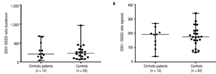
Concentrations of GSH and GSSG, and the GSH/ GSSG ratio did not differ significantly between cirrhotic patients and controls in duodenal nor in sigmoid bi-opsies (Table 4 and Figure 1A and 1B).
Table 2. Characteristics of subjects.
Cirrhotic patients (n=15) Controls (n=22) P-value
Age (years) 63 (18-72) 47 (19-78) 0.377
Sex (M / F) 10 / 5 12 / 10 0.461
BMI (kg/m2) 25.9 (19.6-39.0) 25.0 (18.1-29.8) 0.467
ALT (U/L)* 30.0 (9.0-130.0) 19.5 (10.0-32.0) 0.004
GGT (U/L)* 47.0 (17.0-194.0) 19.5 (6.0-40.0) <0.001
Child Pugh class (A/B/C) 11/ 4/ 0 -
-Child-Pugh score 5.0 (5.0-9.0) -
-MELD-score 8.0 (6.0-15.0) -
-M: male. F: female. BMI: body mass index. ALT: alanine transaminase. U/L: units per liter. GGT: gamma-glutamyltranspeptidase. MELD-score: Model for End-Stage Liver Disease-score. Continuous values are presented as medians (range).* Significant difference, P < 0.05.
Table 3. Transcription of genes involved in glutathione and uric acid metabolism.
Cirrhotic patients Controls P-value
Duodenal biopsies
GCLC (12 vs. 22) 1.17 (0.28-5.57) 0.60 (0.13-1.85) 0.140
GCLM (12 vs. 22) 1.40 (0.21-5.57) 0.91 (0.24-2.73) 0.207
GS (12 vs. 22) 0.99 (0.11-3.08) 0.79 (0.18-1.66) 0.387
GPX1 (12 vs. 22) 0.95 (0.25-3.28) 1.14 (0.25-2.85) 0.449
GPX2 (12 vs. 22) 0.85 (0.18-5.57) 0.67 (0.18-1.91) 0.207
GPX3 (12 vs. 22) 1.91 (0.14-4.83) 1.00 (0.28-3.38) 0.061
GR (12 vs. 22)* 2.11 (0.39-4.44) 1.28 (0.51-2.48) 0.047
XDH (12 vs. 22) 1.14 (0.10-3.43) 0.92 (0.21-1.71) 0.564
Sigmoid biopsies†
GCLC (13 vs. 19) 0.97 (0.34-4.00) 1.68 (0.41-4.38) 0.058
GCLM (13 vs. 19) 0.68 (0.20-3.09) 0.67 (0.11-13.35) 0.774
GS (13 vs. 19)* 0.68 (0.15-2.45) 1.24 (0.43-3.64) 0.013
GPX1 (13 vs. 19) 1.19 (0.24-7.33) 1.54 (0.74-3.74) 0.291
GPX2 (13 vs. 19) 0.56 (0.11-2.28) 0.96 (0.22-2.70) 0.173
GPX3 (13 vs. 18) 1.38 (0.13-5.47) 0.81 (0.15-5.04) 0.447
GR (13 vs. 19)* 0.90 (0.18-2.99) 2.07 (0.48-10.88) 0.022
XDH (13 vs. 18) 1.82 (0.13-7.64) 0.64 (0.09-9.08) 0.378
All data are normalized expression ratios, presented as medians (range).* Significant difference. P < 0.05. † Good quality RNA could only be obtained from 19
Table 4. Glutathione and glutathione disulphide in mucosal biopsies
Cirrhotic patients Controls P-value
Duodenal biopsies (n = 12 vs. n = 20)
GSH (nmol/mg protein) 43.3 (36.2-56.3) 42.4 (28.5-54.9) 1.000
GSSG (nmol/mg protein) 0.22 (0.05-1.13) 0.18 (0.04-0.66) 0.785
Sigmoid biopsies (n = 10 vs. n = 22)
GSH (nmol/mg protein) 29.4 (25.2-34.4) 30.3 (26.0-42.9) 0.223
GSSG (nmol/mg protein) 0.17 (0.11-0.74) 0.18 (0.09-0.64) 0.714
All values are medians (range). GSH: glutathione. GSSG: glutathione disulphide.
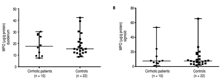
To further support the oxidative stress data, local intes-tinal inflammation was assessed by analysis of MPO levels
in the mucosal biopsies.31 The concentrations of MPO
were also not significantly different between both groups in duodenal [17.57 (7.51-30.21) μg/g protein vs. 15.35
(8.48-42.32) μg/g protein; P = 0.968; Figure 2A] nor in sigmoid
biopsies [7.56 (1.16-53.55) μg/g protein vs. 7.61
(1.87-65.57) μg/g protein; P = 1.000; Figure 2B].
Fecal calprotectin
The intestinal inflammation marker fecal calprotectin did also not differ significantly between cirrhotic patients
(n = 15) and controls (n = 22) [(6.37 (0.08-23.46) μg/g
feces vs. 1.95 (0.13-29.63) μg/g feces; P = 0.216)].
Histology of mucosal biopsies
Microscopically only minor morphological changes were observed in duodenal biopsies of cirrhotic patients and controls and did not differ significantly between the groups, such as a (slightly) increased number of
intra-epithelial lymphocytes (n = 2 vs. n = 2,
respec-tively), mild edema of the lamina propria (n = 2 vs. n = 3,
respectively) and mild chronic inflammation (n = 3 vs.
n = 2, respectively). None of the subjects had villous atro-phy or signs of acute inflammation. In sigmoid biopsies, only mild edema of the lamina propria was observed in both groups (n = 4 vs. n = 4, respectively).
DISCUSSION
We investigated the occurrence of oxidative stress in both duodenal and sigmoid mucosal biopsies of patients with compensated liver cirrhosis. Differences were only found for gene transcription of GS and GR in duodenal and sigmoid biopsies between compensated cirrhotic pa-tients and healthy controls, but no alterations were found for other genes nor for protein levels of GSH, GSSG and GSH/GSSG ratio. The intestinal inflammation markers MPO and (fecal) calprotectin were also not different between both groups.
The present study was initiated to investigate whether intestinal oxidative stress occurs in patients with
Figure 1. Figure 1. Figure 1. Figure 1.
Figure 1. Glutathione (GSH) / glutathione disulphide (GSSG) ratio in mucosal biopsies of cirrhotic patients and controls. A.A.A.A.A. Glutathione (GSH) / glutathione disulphide (GSSG) ratio in duodenal biopsies. B.B.B.B. Glutathione (GSH) / glutathione disulphide (GSSG) ratio in sigmoid biopsies. Data are presented as scatterB. dot plots displaying median with range (P > 0.05).
GSH/ GSSG
ratio
duodenum
1,500
1,000
500
0
Cirrhotic patients Controls
(n = 12) (n = 20)
Cirrhotic patients Controls
(n = 10) (n = 22)
GSH / GSSG
ratio
sigmoid
400
300
200
100
0 A
A A A
compensated liver cirrhosis and thereby could be an un-derlying mechanism contributing to the epithelial barrier
dysfunction observed in our previous study,28 which
po-tentially may increase the risk of progression towards de-compensated cirrhosis.
The transcription of genes involved in the metabolism of GSH and uric acid, which are both important antioxi-dants and protect cells against oxidative damage, were ana-lyzed. GSH reduces hydrogen peroxide and lipid hydroperoxide to less reactive oxygen species (ROS), and in this process GSH is converted into GSSG. Uric acid is produced from hypoxanthine and xanthine by the enzymes
XO and XDH. In vitro, uric acid is a powerful scavenger of
ROS32 and by being preferentially oxidized as a substrate
for oxidation by haem protein or H2O2 systems, it can
protect against oxidative damage.33 As marker of oxidative
stress, the GSH/GSSG ratio was determined in both duo-denal and sigmoid biopsies.
In the duodenal mucosa, we observed that the gene tran-scription of GR, the enzyme that recycles GSSG into GSH, was up-regulated in cirrhotic patients. Protein levels of GSH, GSSG and the GSH/GSSG ratio were not altered. These findings may point to an enhancement of repair proc-esses for oxidative stress. In the sigmoid mucosa, gene tran-scription of GR as well as GS, the latter being one of the enzymes involved in the synthesis of GSH, was down-regu-lated in cirrhotic patients, which might indicate a decreased formation of GSH. However, protein levels of GSH, GSSG and the GSH/GSSG ratio were not altered. It should be noted that the differences in gene transcription in both duo-denum and sigmoid did not remain statistically significant after correcting for multiple testing and therefore should be interpreted with care. In addition, no alterations were found for the other genes investigated. Thereby the above findings indicate that there is no clear evidence for
oxida-tive stress in the intestine of patients with compensated liv-er cirrhosis. This is furthliv-er supported by lack of diffliv-erences in intestinal MPO and fecal calprotectin concentrations between cirrhotic patients and controls. Moreover, we observed no clear morphological abnormalities in the duo-denal and sigmoid biopsies.
The lack of changes in oxidative stress and inflamma-tory parameters is not likely to be attributed to drug therapy as patients used almost no drugs with antioxidant and/or anti-inflammatory effects and no antioxidant sup-plements.
Alcohol is an important cause of liver cirrhosis and can disturb intestinal epithelial barrier function by inducing
intestinal oxidative damage.16, 34 In our previous study,
small intestinal permeability was found to be increased in a subgroup of patients with alcohol-related cirrhosis, but-the small number of patients with alcohol-related cirrho-sis (n = 4) in the present study precluded us from performing additional analyses to investigate whether this subgroup may also be more susceptible for intestinal oxi-dative stress.
In contrast to the present findings in humans with com-pensated liver cirrhosis, some animal studies did provide evidence of oxidative stress in the small and large intestine
of rats with carbon tetrachloride (CCl4)-induced liver
cir-rhosis, although part of them had ascites.13,24-26 Published
data on the occurrence of intestinal oxidative stress in cir-rhotic patients are scarce. Only one other recent study has investigated intestinal oxidative stress in duodenal biopsies of cirrhotic patients by measuring lipid hydroperoxides and found no difference in the levels of lipid hydroperoxides between patients with compensated cirrhosis and healthy controls.27 However, the investigators did find significantly
increased levels of lipid hydroperoxides in patients with decompensated cirrhosis when compared to those
Figure 2. Figure 2. Figure 2.
Figure 2. Figure 2. Myeloperoxidase (MPO) concentrations in mucosal biopsies of cirrhotic patients and controls. AAAA... Myeloperoxidase (MPO) concentrations inA duodenal biopsies. BBBBB... Myeloperoxidase (MPO) concentrations in sigmoid biopsies. Data are presented as scatter dot plots displaying median with range (P > 0.05).
MPO
(
μ
g/g protein)
duodenum
50
40
30
20
10
0
Cirrhotic patients Controls
(n = 10) (n = 22)
Cirrhotic patients Controls
(n = 10) (n = 22)
MPO
(
μ
g/g protein)
sigmoid
80
60
40
20
0 A
A A A
with compensated cirrhosis and healthy controls. There-fore, we cannot exclude that intestinal oxidative stress does occur in patients with decompensated cirrhosis.
The strength of our study is that we investigated the oc-currence of oxidative stress not only in the mucosa of the small, but also of the large intestine in patients with com-pensated cirrhosis. As we previously did find a clear in-creased large intestinal permeability, it would be interesting to investigate also other potential causative fac-tors contributing to barrier dysfunction, such as changes in microbiota composition.
Some limitations of the present study should also be taken into account.The GSH/GSSG ratio was analyzed as this ratio is a marker of oxidative stress and gives insight into the body’s anti-oxidant defense capacity.35, 36 Because
of the limited number of intestinal biopsies available per subject, we were not able to measure other markers of ox-idative stress, such as MDA and 4-hydroxynonenal (4-HNE). Our findings are supported by unaltered in-flammatory parameters and intestinal morphology. Although we cannot completely exclude the presence of intestinal oxidative stress, the current findings indicate that oxidative stress seems not to be a major factor in these patients. Furthermore, the intestinal biopsies could only be obtained from a rather small group of patients due to the invasiveness of sampling and the complexity of the pa-tient population. Therefore, our results are preliminary and subgroup analyses of cirrhotic patients for example with regard to etiology or drug therapy were not possible. Although we cannot exclude the effect of etiology or drug therapy, nor the possibility that other markers of oxidative stress would be increased in cirrhotic patients, we believe that these factors are not important confounders in this study. Furthermore, inclusion of a larger group of patients is not very likely to change our results as no trend to sig-nificance was observed in both oxidative stress and in-flammatory parameters between cirrhotic patients and controls.
In conclusion, in stable compensated cirrhosis, there were no indications for the occurrence of oxidative stress and low-grade inflammation in both the small and large intestine. Although these preliminary findings need fur-ther validation, we found intestinal oxidative stress not to be a major mechanism contributing to epithelial barrier dysfunction observed in patients with compensated cir-rhosis.
ABBREVIATIONS
• 4-HNE: 4-hydroxynonenal.
• AJ: adherens junction.
• ALT: serum alanine transaminase.
• CCl4: carbon tetrachloride.
• GAPDH: glyceraldehyde-3-phosphate dehydrogenase. • GCLC: glutamate-cysteine ligase, catalytic subunit. • GCLM: glutamate-cysteine ligase, modifier subunit.
• GGT: gamma-glutamyltranspeptidase.
• GI tract: Gastrointestinal tract.
• GPX1, GPX2 and GPX3: glutathione peroxidases 1, 2 and 3.
• GR: glutathione reductase.
• GS: glutathione synthetase.
• GSH: glutathione.
• GSSG: glutathione disulphide. • H&E: haematoxylin and eosin.
• MDA: malondialdehyde.
• MUMC: Maastricht University Medical Center. • NSAIDs: non-steroid anti-inflammatory drugs.
• ROS: reactive oxygen species.
• TJ: tight junction.
• XDH: xanthine dehydrogenase.
• XO: xanthine oxidase.
REFERENCES
1. Misra V, Misra SP, Dwivedi M, Gupta SC. Histomorphometric study of portal hypertensive enteropathy. Am J Clin Pathol
1997; 108: 652-7.
2. Such J, Guardiola JV, de Juan J, Casellas JA, Pascual S, Aparicio JR, Sola-Vera J, et al. Ultrastructural characteris-tics of distal duodenum mucosa in patients with cirrhosis.
Eur J Gastroenterol Hepatol 2002; 14: 371-6.
3. Chesta J, Defilippi C. Abnormalities in proximal small bowel moti-lity in patients with cirrhosis. Hepatology 1993; 17: 828-32. 4. Chen Y, Yang F, Lu H, Wang B, Lei D, Wang Y, Zhu B, et al.
Characterization of fecal microbial communities in patients with liver cirrhosis. Hepatology 2011; 54: 562-72.
5. Campillo B, Pernet P, Bories PN, Richardet JP, Devanlay M, Aussel C. Intestinal permeability in liver cirrhosis: relations-hip with severe septic complications. Eur J Gastroenterol Hepatol 1999; 11: 755-9.
6. Zuckerman MJ, Menzies IS, Ho H, Gregory GG, Casner NA, Crane RS, Hernandez JA. Assessment of Intestinal Per-meability and Absorption in Cirrhotic Patients with Ascites Using Combined Sugar Probes. Dig Dis Sci 2004; 49: 621-6. 7. Norman K, Pirlich M, Schulzke JD, Smoliner C, Lochs H, Va-lentini L, Buhner S. Increased intestinal permeability in mal-nourished patients with liver cirrhosis. Eur J Clin Nutr 2012; 66: 1116-9.
8. Bellot P, Frances R, Such J. Pathological bacterial transloca-tion in cirrhosis: pathophysiology, diagnosis and clinical im-plications. Liver Int 2013; 33: 31-9.
9. Wiest R, Garcia-Tsao G. Bacterial translocation (BT) in cirr-hosis. Hepatology 2005; 41: 422-33.
10. Assimakopoulos SF, Tsamandas AC, Tsiaoussis GI, Karatza E, Triantos C, Vagianos CE, Spiliopoulou, et al. Altered intesti-nal tight junctions’ expression in patients with liver cirrhosis: a pathogenetic mechanism of intestinal hyperpermeability. Eur J Clin Invest 2012; 42: 439-46.
12. Rao RK, Basuroy S, Rao VU, KarnakyJr KJ, Gupta A. Tyrosi-ne phosphorylation and dissociation of occludin-ZO-1 and E-cadherin-beta-catenin complexes from the cytoskeleton by oxidative stress. Biochem J 2002; 368: 471-81.
13. Ramachandran A, Prabhu R, Thomas S, Reddy JB, Pulimood A, Balasubramanian KA. Intestinal mucosal alterations in ex-perimental cirrhosis in the rat: role of oxygen free radicals.
Hepatology 2002; 35: 622-9.
14. Sheth P, Basuroy S, Li C, Naren AP, Rao RK. Role of phos-phatidylinositol 3-kinase in oxidative stress-induced disrup-tion of tight juncdisrup-tions. J Biol Chem 2003; 278: 49239-45. 15. Assimakopoulos SF, Gogos C, Labropoulou-Karatza C. Could
antioxidants be the “magic pill” for cirrhosis-related compli-cations? A pathophysiological appraisal. Med Hypotheses
2011; 77: 419-23.
16. Banan A, Choudhary S, Zhang Y, Fields JZ, Keshavarzian A. Ethanol-induced barrier dysfunction and its prevention by growth factors in human intestinal monolayers: evidence for oxidative and cytoskeletal mechanisms. J Pharmacol Exp-Ther 1999; 291: 1075-85.
17. Forsythe RM, Xu DZ, Lu Q, Deitch EA. Lipopolysaccharide-induced enterocyte-derived nitric oxide induces intestinal monolayer permeability in an autocrine fashion. Shock 2002; 17: 180-4.
18. Bajaj JS, Hylemon PB, Ridlon JM, Heuman DM, Daita K, White MB, Monteith P, et al. Colonic mucosal microbiome differs from stool microbiome in cirrhosis and hepatic encephalopa-thy and is linked to cognition and inflammation. Am J Physiol-Gastrointest Liver Physiol 2012; 303: G675-85.
19. Saitoh O, Sugi K, Lojima K, Matsumoto H, Nakagawa K, Ka-yazawa M, Tanaka S, et al. Increased prevalence of intesti-nal inflammation in patients with liver cirrhosis. World J Gastroenterol 1999; 5: 391-6.
20. Iwao T, Toyonaga A, Ikegami M, Oho K, Sumino M, Harada H, Sakaki M, et al. Reduced gastric mucosal blood flow in pa-tients with portal-hypertensive gastropathy. Hepatology
1993; 18: 36-40.
21. Xu WH, Wu XJ, Li JS. Influence of portal pressure change on intestinal permeability in patients with portal hypertension.
Hepatobiliary Pancreat Dis Int 2002; 1: 510-4.
22. Ramachandran A, Balasubramanian KA. Intestinal dysfunc-tion in liver cirrhosis: Its role in spontaneous bacterial perito-nitis. J Gastroenterol Hepatol 2001; 16: 607-12.
23. Zuwala-Jagiello J, Pazgan-Simon M, Simon K, Warwas M. Elevated advanced oxidation protein products levels in pa-tients with liver cirrhosis. Acta Biochim Pol 2009; 56: 679-85. 24. Chiva M, Soriano G, Rochat I, Peralta C, Rochat F, Llovet T, Mirelis B, et al. Effect of Lactobacillus johnsonii La1 and antioxidants on intestinal flora and bacterial translo-cation in rats with experimental cirrhosis. J Hepatol 2002; 37: 456-62.
25. Chiva M, Guarner C, Peralta C, Llovet T, Gomez G, Soriano G, Balanzo J. Intestinal mucosal oxidative damage and
bac-terial translocation in cirrhotic rats. Eur J Gastroenterol He-patol 2003; 15: 145-50.
26. Natarajan SK, Ramamoorthy P, Thomas S, Basivireddy J, Kang G, Ramachandran A, Pulimood AB, et al. Intestinal mu-cosal alterations in rats with carbon tetrachloride-induced cirrhosis: changes in glycosylation and luminal bacteria.
Hepatology 2006; 43: 837-46.
27. Assimakopoulos SF, Tsamandas AC, Tsiaoussis GI, Karatza E, Zisimopoulos D, Maroulis I, Kontogeorgou E, et al. Intesti-nal mucosal proliferation, apoptosis and oxidative stress in patients with liver cirrhosis. Ann Hepatol 2013; 12: 301-7. 28. Pijls KE, Koek GH, Elamin EE, de Vries H, Masclee AA,
Jo-nkers DM. Large intestine permeability is increased in pa-tients with compensated liver cirrhosis. Am J Physiol Gastrointest Liver Physiol 2014; 306: G147-53.
29. Julicher RH, Sterrenberg L, Haenen GR, Bast A, Noordhoek J. The effect of chronic adriamycin treatment on heart kidney and liver tissue of male and female rat. Arch Toxicol 1988; 61: 275-81.
30. van der Sluijs Veer G, van den Hoven B, Russel MG, van den Bergh FA. Time-resolved fluorimetric immunoassay of calprotectin: technical and clinical aspects in diagnosis of inflammatory bowel diseases. Clin Chem Lab Med 2006; 44: 292-8.
31. de Haan JJ, Lubbers T, Hadfoune M, Luyer MD, Dejong CH, Buurman WA, Greve JW. Postshock intervention with high-lipid enteral nutrition reduces inflammation and tissue dama-ge. Ann Surg 2008; 248: 842-8.
32. Ames BN, Cathcart R, Schwiers E, Hochstein P. Uric acid provides an antioxidant defense in humans against oxidant-and radical-caused aging oxidant-and cancer: a hypothesis. Proc Natl Acad Sci U S A 1981; 78: 6858-62.
33. Halliwell B, Gutteridge JMC. Free radicals in biology and medicine: New York: Oxford University Press Inc.; 1999. 34. Banan A, Fields JZ, Decker H, Zhang Y, Keshavarzian A.
Nitric oxide and its metabolites mediate ethanol-induced microtubule disruption and intestinal barrier dysfunction.
J Pharmacol Exp Ther 2000; 294: 997-1008.
35. Sen SK, Packer L, Hanninen O. Handbook of Oxidants and Antioxidants in Exercise: Elsevir Science B.V.; 2000. 36. Koek GH, Liedorp PR, Bast A. The role of oxidative stress in
non-alcoholic steatohepatitis. Clin Chim Acta 2011; 412: 1297-305.
Correspondence and reprint request:
Kirsten E. Pijls, M.D.
Division Gastroenterology-Hepatology. Department of Internal Medicine. Maastricht University Medical Center. PO Box 5800, 6202 AZ Maastricht, the Netherlands.