SUMMARY
Recent theories postulate that memory can be divided into multiple brain memory systems. Although memory systems depend on the function of variables based on the level of analysis and are subserved by different neural substrates, current definitions of memory systems have categorized them as psychological and biological entities. Under such context, the studies of different memory systems have shown that complex interactions take place during perfor-mance of any memory task. Such interactions among multiple memory systems are based on dynamic interactive independent neural networks which make possible the better understanding of how memory systems work in the brain of mammals. Both behavioral and electrophysiological studies over the last decades demonstrate that learning and memory are encoded through activity dependent changes of the strength of synaptic connections between neurons, as experimentally demonstrated by Long-Term Potentiation (LTP) in mammalian synapses. LTP is a form of synaptic plasticity, and is considered as an accepted cellular model for stabilization of synapses involved in the expression of several neurobiological phenomena. Most of the understanding of the neurochemical, pharmacological, and molecular mechanisms involved in LTP induction, expression, and maintenance, have been demonstrated through the involvement of glutamate neurotransmission system, as well as through the different glutamate recep-tor subtypes, known to be expressed widely in different neural networks of the brain of mammals.
Key words: Glutamate receptors, N-Methyl-D-Aspartate, Metabotropic receptors.
RESUMEN
El fenómeno de la memoria se define como un proceso de adquisición, almacenamiento y recuperación de infor-mación. En términos operacionales, el fenómeno de la memoria se infiere como un evento neurobiológico resul-tado de alteraciones en el comportamiento del sujeto, cau-sado por experiencias previas no dependientes de modi-ficaciones de los órganos efectores sensoriales. En este contexto, algunas teorías recientes postulan que la memo-ria puede dividirse en múltiples sistemas de memomemo-ria fun-cional en el cerebro de los mamíferos. Si bien estos siste-mas de memoria funcional dependen de múltiples varia-bles sujetas al grado de análisis del experimentador, asi-mismo están regulados por diferentes circuitos neuronales enlazados entre sí. Las definiciones más recientes de es-tos sistemas de memoria funcional postulan que eses-tos sis-temas se enmarcan ya sea como entidades psicológicas, al considerar que los sistemas de memoria operan como módulos especializados que poseen tanto la capacidad de procesar diferentes tipos de información como de rea-lizar tareas operacionales y almacenar información en lap-sos cortos o largos, o sea como entidades biológicas, si se define que los sistemas de memoria operan mediante cir-cuitos neuronales y conexiones neurales complejas que, en conjunto, permiten operar un tipo particular de infor-mación y procesar el almacenamiento de inforinfor-mación dentro del mismo circuito neuronal u otro distinto. Por ejemplo, el lóbulo temporal medial (LTM) que contiene la estructura nerviosa del hipocampo y sus interconexiones con los diferentes campos corticales resultan ser cruciales para estructurar y consolidar la memoria de tipo declara-tivo. En este contexto, diversos trabajos en el campo de la neuropsicología y la neurobiología de la memoria han mostrado que los diferentes sistemas de memoria operan según complejas interacciones durante la ejecución de tareas de aprendizaje y memorización en el cerebro de
U
NDERSTANDING
THE
NEUROBIOLOGICAL
MECHANISMS
OF
LEARNING
AND
MEMORY
: M
EMORY
SYSTEMS
OF
THE
BRAIN
,
LONG
TERM
POTENTIATION
,
AND
SYNAPTIC
PLASTICITY
.
P
ART
III
A
Philippe Leff*, Maura Matus*, Adriana Hernández*, Mayra Medécigo*, Carlos Torner**, Benito Antón*
*Laboratorio de Neurobiología Molecular y Neuroquímica de Adicciones. Instituto Nacional de Psiquiatría Ramón de la Fuente. Calzada México-Xochimilco # 101. San Lorenzo Huipulco, 14370. México D.F. pleff@imp.edu.mx
**Atención a la Salud, CBS, Universidad Autónoma Metropolitana-Xochimilco. Calzada del Hueso 1100, Col. Villa Quietud, 04960 México, D.F.
los mamíferos. Estas interacciones entre los múltiples sis-temas de memoria funcional dependen de complejas interacciones dinámicas de sistemas o sustratos neuronales independientes que posibilitan una mejor comprensión de la forma en que trabaja nuestra memoria en el cerebro. Además, diversos trabajos experimentales de naturaleza tanto conductual como electrofisiológica en las últimas décadas demuestran que tanto el aprendizaje como la memoria se codifican mediante cambios dependientes de la actividad entre las conexiones nerviosas, tal como com-probó el descubrimiento del fenómeno de Potenciación de Largo Plazo (LTP, por sus siglas en inglés) en las sinapsis de las neuronas del hipocampo en el cerebro de los ma-míferos. El fenómeno de LTP, considerado una forma de expresión de plasticidad sináptica, también se ve como un modelo celular que favorece la estabilidad de la activi-dad sináptica y su expresión en múltiples eventos neurobiológicos. En este contexto, diversos estudios del fenómeno de LTP tanto in vitro como in vivo, con dife-rentes métodos experimentales y de registro, han demos-trado que el fenómeno de LTP ocurre en múltiples regio-nes del cerebro, como son la neocorteza, la amígdala y en estructuras que conforman el sistema nervioso perifé-rico de los mamíferos. Más aún, estudios recientes mues-tran que el fenómeno de LTP puede inducirse en tejidos neurales de animales invertebrados, como ocurre en la unión neuromuscular y en sinapsis específicas de dife-rentes estructuras nerviosas del cerebro de los artrópo-dos. En su gran mayoría, los eventos neuroquímicos,
neurofarma-cológicos y moleculares que participan en la inducción, mantenimiento y expresión del fenómeno de LTP se ba-san en la actividad de un sistema de transmisión particu-lar, como es el sistema de neurotransmisión glutamatérgica mediado a través de la activación de diferentes subtipos de receptores glutamatérgicos, como los receptores ionotrópicos tipo NMDA (NMDA glutamate receptors, por sus siglas en inglés), los receptores ionotrópicos tipo no-NMDA (Non-no-NMDA glutamate receptors, por sus siglas en inglés) y los receptores metabotrópicos que se encuen-tran ampliamente distribuidos y se expresan de manera funcional en diferentes circuitos neuronales y sinapsis en el SNC de los mamíferos.
Palabras clave: Receptores de glutamato, N-metil-D-aspartato, receptores metabotrópicos.
MEMORY SYSTEMS OF THEBRAIN
Memory can be defined as a function of many variables depending on the levels of analysis (Kim and Baxter, 2001). Memory has been typically attributed to a process of information acquisition, storage, and retrieval, but in operational terms, memory has been inferred from alterations in behavior that are caused by some prior experience not dependent on the modifications of the responsivity of sensory effector organs (Rescorla, 1988). Moreover,
hipoccampal memory interacting with other network systems might acquire information represented as associative strength, either independently, synergistically, or competitively (see Kim and Baxter, 2001, for detailed information). Thus, when memory systems (such as the hippocampal formation) act independently, only one system is able to acquire information (acquires associative strength) under a learning experience or situation. For instance, if hippocampal memory system is required to acquire a certain kind of learning information, lesions to this structure might obliterate the learning acquisition, while lesions to other systems will not influence the process of learning in the former memory system (Kim and Baxter, 2001). Synergistic interactions between memory systems functionally characterizes that at least two-memory systems are required for the acquirement of some degree of information. Thus, lesions impinge on either one of the memory systems, which might result in an impairment of the learning processing or com-plete deficit or absent of acquisition of information, when both memory systems or neural structures confining them are exposed to combined injuries (Kim and Baxter, 2001). Moreover, competitive interactions allows one of the memory system to acquire high levels of information (over normal levels) if other memory system results to be damaged; nevertheless, in such model, the intact memory system will acquire much more of the information, that potentially will be distributed between both systems (see Kim and Baxter, 2001, for more descriptive details of such interacting models of memory systems in the brain). Although much of the conceptualized framework have emerged from lesion studies performed in experimental animals, complex results might be expected to occur from the interaction of such memory systems (Kim and Baxter, 2001) as will be described below. For instance, in the classical (Pavlovian) eye-blink conditioning, it has been demonstrated that the cerebellum is essential for mammals to learn a relationship between conditioned (CS) and unconditioned stimuli (US) (Kim and Thompson, 1997; McCormick et al., 1982). This simple form of associative learning seems to engage several brain structures, which presumably are involved in the performance of different aspects of the conditioning response (Thompson and Kim, 1996). For example, hippocampal neurons
exhibit unit-firing patterns that emulate the amplitude time-course events of the conditioned response during delay eye-blink conditioning, favoring the idea that the hipoccampal formation is implicated in the development of such reflex response, even though this neural structure is not required for the development of such delay conditioning response. Thus, besides the implication of the hippocampus in the delayed eye-blink conditioning response, several studies have demonstrated that other conditioning-related events altered the hipoccampal physiology in experimental animals. Thus, it is possible to argue that during conditioning-related response, hipoccampal neurons process information in a similar context as the formation of the CS-US association in the cerebellum, interacting or interfering with this association process, in such a way, that an apparent competition between both neural structures seems to occur in standard delay conditioning (Kim and Baxter, 2001). Several experimental evidences support this view, as shown that manipulations that altered hipoccampal physiology facilitate acquisition of delay eye-blink conditioning (see in Kim and Baxter, 2001, table 1). Moreover, experimental evidences have demonstrated that although both cerebellum and hippocampus are essential for acquisition and development of eye-blink conditioning response, a minor alteration to the conditioning procedure eventually alters the interaction between both neural structures, such as when a brief time interval separates the CS and US (Solomon et al., 1986). These results demon-strate that multiple memory systems are engaged in the development of even simple classical conditioning tasks (Kim and Baxter, 2001).
2001), and infusion of glutamate into the hippocampus or the caudate induced an hipoccampal dependent strategy to persist in contrast to a caudate-dependent response strategy (Packard, 1999). These findings supports the idea that multiple memory systems in the brain are recruited in normal learning tasks and behavioral performance, where each structure– relationship memory system encodes different patterns of the learned task (Thompson and Kim, 1996; Kim and Baxter, 2001). Such behavioral conducted experiments revealed that hipoccam-pal memory systems interfere with the natural operation of other memory systems whether this interactions are competitive or synergistic depends on the structure-relationship memory system used to consolidate a behavioral task response. For instance, neocortical areas and hipoccampal formation interactions might operate synergistically based on that sensory information conveyed by cortical efferents into the hippocampus subserves spatial cognition (Aggleton et al, 2000; Kim and Baxter, 2001).
LONG-TERM POTENTIATION
One of the crucial functional properties of the brain is memory. Memory is part of our daily functions of our lives, which allow us to accomplish numerous tasks, such as recalling personal experiences, learning facts and knowledge about our environment, as well as recognizing people, objects, and even acquiring skills and habits (Eichenbaum et al, 1999). Memory is not a unitary monolithic entity, reflecting a single faculty of the mind and brain. Several converging evidences from psychology and neuroscience have emerged concerning the existence of multiple memory systems than can be dissociated from one another (Eichenbaum et al, 1999). Moreover, the concept that the brain uses multiple memory systems (see below) comes from recent physiological and behavioral experimental evidences, but most of the historical background that led to this criteria emerged from several philosophers, thinkers, and neurologists from the 19th century; such as Gall, founder of the phrenological movement, who focused on the notion that each specialized faculty of the mind is concerned with particular contents (from a complete review see Zola-Morgan, 1995; Gall, 1835); De Biran, who distinguished three kinds of memory such as a
representative memory, a mechanical memory and a sensitive memory (see Maine de Biran, 1929); the french psychologist T. Ribot (1881), who viewed that brain contains memory centers specialized to handled different kinds of information, such as the cortical auditory, vi-sual, and motor centers, handling each one different forms of memory (for a complete review see Eichenbaum et al, 1999). Over the past 20th century, different disciplines in the neuroscience field have focused on the learning and memory processing in the brain. As such, several hypothesis proposed that learning and memory could be encoded via activity dependent changes in the strength of synaptic connections between neurons, initially postulated by D. Hebb, 1949 (Hebbian postulate), who advanced the concepts that underlie the conditions that cause synapses to change, which resulted essential for the experiments that demonstrated the mechanisms of LTP (Beggs et al., 1999).
formation, due to the intrinsic circuitry is a relatively simple structure, where major cells within the laminar organization in this brain area remains intact in transverse slides (Dingledine, 1984; Barrionuevo and Brown, 1983). Such tissue preparations have resulted to be useful to obtain both extracellular and intracellular recordings as well (Beggs et al., 1999). Moreover, one of the most common synapse studied during LTP formation and maintenance in the mammalian brain, is the Schaffer collateral/commissural input to the pyramidal cells in the CA1 region within the hipoccampal formation (see figure 1) (Beggs et al., 1999). Nonetheless, several studies have been focused on the circuitry formed by the mossy fiber input arising from the granule cells of the dentate gyrus to the pyramidal neurons of the CA3 region in this same brain region (Beggs et al., 1999). For example, LTP can be induced after presynaptic tetanic stimulation of mossy
fibers with short train of stimuli (e. g., 10 trains at 100 Hz) in transverse brain slices of the hipoccampal formation, while postsynaptic cell was depolarized under current clamp conditions. Under such experimental procedures defined as pulse” paradigm used to reveal “paired-pulse” facilitation (PPF) in the mossy fiber/CA3 circuitry, these showed potentiation of the excitatory postsynaptic current (EPSC) amplitude recorded in response to the application of set of paired-pulses. Thus, LTP induction at least in these
in vitro preparations of the hipoccampal formation was observed to last for an hour or more (Barrionuevo et al., 1986; Xiang et al., 1994), with almost no decrement of the EPSC amplitude, but in vivo studies have reported longer periods of weeks or even months to obtain similar electrophysiological recording responses (Beggs et al., 1999).
Most of the synapses in the hipoccampal Figure 1. Structure and organization of the hipoccampal formation.- The hipoccampal formation is located in the posteromedial border of the hemispheres. It extends from the rostromedially located septum to the ventrolaterally located amygdaloid area. Although the hipoccampal region is structurally divided into the area dentate and the hippocampus proper, the hipoccampal formation includes the subicular region, retrospenial area and the entorhinal cortex. From the hipoccampal structure depicted above, the major hipoccampal neuronal circuit is described as follows: the entorhinal cortex (EC) sends the perforant pathway fibers (PP) to the granule cells of the dentate gyrus (DG) as well as to the pyramidal cells of the CA3 region. Major axonal output of the DG-granule cells (G), the mossy fibers (MF), make synaptic contacts with principal neurons of the CA3 field. While CA3 pyramidal cells project recurrent axon fibers into the same CA3 region, they also emit axon fibers into CA1 hipoccampal subfield through the Schaffer collaterals (SC), where CA1 region sends projecting axons back to the EC and to the medial septal complex (not shown). Both DG-granule cells, CA1and CA3 pyramidal cells receive cholinergic afferents through the septo-hypocampal pathway (SHP). Most of the principal neurons confined in each hipoccampal region are arranged in a dense continue layer; the stratum pyramidale (p) containing the pyramidal cell layer divides the CA1 and CA3 subfields into the stratum laconosum (L)/stratum oriens layer (o), which contains the basal dendrites of the pyramidal cells; the alveus/stratum radiatum (A)/stratum lacunosum moleculare (lm) contains the apical pyramidal dendrites, as shown in the figure. Several classes of neurons have been shown to be confined in the different cell layers that structure the hippocampal formation. Thus, confined in the dentate gyrus (DG) of the hilus fasciae dentate (H) are gabaergic polymorphic cells, mossy cells, chandelier cells, granule, basket, and molecular (m) associated pathway cells; interneurons have been described in the Stratum radiatum/stratum oriens layer; chandelier and pyramidal cells, in the CA3 hipoccampal subfield; interneurons in the lacunosum-moleculare layer; and both chandelier, pyramidal and basket cells, in the CA1 hipoccampal subfield (for more details see Walaas, 1983)(figure adapted from Ascoli et al., 1998, and modified by author of the present article).
References.
formation that have shown to exhibit LTP are characterized by several “classical properties” (cooperativity, associativity, input sensitivity, and spatiotemporal specificity) (Brown et al., 1990; Bliss et al., 1993) which are out of scope to describe them in this review (for more information see Beggs et al., 1999). They reflect different expressions of the same underlying mechanisms responsible for the induction of different forms of LTP. Briefly, LTP induction in the SCh/com synaptic input to pyramidal neurons in the CA1 region of the hippocampus requires strong high-frequency stimulation (tetanic). However it does not need weak stimulation to recruit enough axons (cooperativity) to induce LTP, neither the association between a weak stimulation of an afferent input (e. g., Schaffer collateral/commisure input to CA1 or the perforant pathway input to dentate gyrus of hipoccampal formation) nor a strong stimulation of same afferent inputs for LTP to be induced
i. e., LTP can be induced only if a weak input (small number of stimulated afferents recruited), is associated with a strong input (large number of stimulated afferents recruited) and this response is restricted to the afferent inputs receiving tetanic stimulation (input specificity) at the same time (spatial and temporal specificity) (Beggs et al., 1999). Although most of the research focused in LTP requires explanations of how this process occurs physiologically in the CNS system, including the underlying neurochemical and molecular mechanisms involved in, conventionally LTP has been divided in three correlated mechanisms defined as induction, expression, and maintenance, describing each step, the initial events that trigger the specific plastic synaptic modifications in neural pathways, the expression of the final synaptic enhancement, and the enduring over time of the enhancement of the synaptic strength defined as maintenance (Beggs et al., 1999).
Much of the understanding of the neurochemi-cal, pharmacologineurochemi-cal, and molecular mechanisms involved in LTP induction, and that ultimately leads to LTP expression and maintenance, has been through the study of the glutamate neurotransmission system and their receptors. By far, most of the neurotransmission system shown to participate in LTP formation involve glutamate as preferential neurotransmitter, although exceptions have been recently reported (Beggs et al., 1999). Therefore, in order to understand the neurochemical and molecular
events occurring during LTP it is crucial to rela-te LTP with glutamarela-te and their receptors as well (Beggs et al, 1999).
a ) Glutamate receptors subtypes. Glutamate receptors (GluRs) known to be widely distributed in the nervous system are responsible for mediating major excitatory
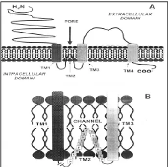
Figure 2. - Schematic representation of a structural model of a Non-NMDA (ionotropic) glutamate receptor. (A) Shows the structural conformation of one of the subunits of a typical ionotropic glutamate receptor conformed by four hydro-phobic membrane- spanning domains (TM1-TM4), as evidenced by recent molecular and biochemical studies. The TM2 membrane-spanning segment forms a “hook” that does not transverse the membrane comple-tely, and extends right back into the cytoplasm, sharing some similarities with the P segment (pore forming domain) of the voltage-activated K+ channels. Both NH2-terminus
synaptic transmission in the brain and spinal cord (Waxham, 1999). Most of the neural systems where LTP has been identified used glutamate as neurotransmitter (Beggs et al., 1999). Initial pharmacological studies performed early in the 70´s suggested that glutamate receptors were not homogenous in the CNS, as they could be distinguished and separated in different glutamate receptors subtypes after demonstrating that different synthetic agonists, namely N-methyl-D-aspartate or NMDA; α -amino-3-hydroxy-5-methyl-isoxazoleproprionic acid or AMPA;
kainate and quisqualate, in the presence of competitive antagonists were able to bind distinctively different types of membrane receptors (Watkins et al., 1990). These agonists (used extensively to characterize the glutamate receptor family) led to the initial identification of ionotropic (membrane bound protein complexes that combine to form an ion permeable pore or ion channel through the membrane) and metabotropic glutamate receptors (receptors composed by a single
transmembrane polypeptide chain containing an extracellular binding domain for the preferential ligand-agonist and an intracellular binding domain that couples and activates upon receptor activation, GTP-binding proteins or G proteins (Waxham, 1999). For instance, the pharmacological agonist quisqualate is unique in that it shows a binding profile to both ionotropic and metabotropic glutamate receptor subtypes (Hollmann and Heinemann, 1994). In same context, ionotropic glutamate receptors [see figure 2 (a, b), and figure 3] were defined as either NMDA or non-NMDA receptors subtypes, depending on the capability of the NMDA agonist to bind with high affinity and selectively to such receptors subtypes (Watkins et al., 1990; Hollmann and Heinemann, 1994, Waxham 1999).
b ) Non-NMDA glutamate receptors and LTP.
a molecular mass of 99.8 kDa, after its transfection and expression in Xenopus oocytes, producing a functional and stable glutamate-activated channel, named GluR-K-1 (Hollman et al., 1989). Similar reports describ-ing the isolation and functional expression of a new family of glutamate receptor subunits (termed Glu-R1-Glu-R4) demonstrated that the Non-NMDA-glutamate receptor form a native pentameric complex in the brain, structured with four predicted membrane-spanning segments (TM1-TM4) (figure 2a) and a large extracellular domain expressing a total molecular mass of around 600 kDa (Boulter et al., 1990; Keinanen et al., 1990; Nakanishi et al., 1990; Blackstone et al., 1992; Wenthold et al., 1992). Thus, these results demonstrated that the molecular complex of this receptor is twice the molecular size of the reported for the nAChR (see Waxham , 1999). Moreover, functional expression of these cloned cDNAs encoding such glutamate receptors showed that they were capable to produce inward currents after its functional and stable expression either in oocytes or Hek-293 cells and after application of Non-NMDA agonist AMPA or Kainate. Similar studies showed that both GluR1 and GluR3 subunits when expressed alone or in combination in these cells produced functionally channels with large inward currents exhibiting channels permeable exclusively to the calcium ion, situation that did not occur when co-expression was performed in the presence of the GluR2 subunit (Hollmann and Heinemann, 1994). Therefore, the expression of this glutamate receptor in the presence of the GluR2 subunit induced channels impermeable to Ca2+ (Hollmann and Heinemann, 1994). This structure-activity relationship studies of the Non-NMDA glutamate receptor led to the hypothetical proposition that two types of receptors were expressed in neurons, as several electrophysiological studies showed in embryonic hipoccampal neurons describing the presence of a glutamate receptor imper-meable to Ca2+, and another one permeable to the same ion (Hollmann and Heinemann, 1994). Actually, it has been shown that glutamate receptors have the ability to acquire different properties producing different intracellular responses depending on the structural conformation of their protein
subunits expressed (Waxham, 1999). For instance, several studies have demonstrated that these receptors are able to switch from a Ca2+ impermeable to a Ca2+ permeable channel through the exchange of a single amino acid (Arg→Gln) on the TM2 loop of the GluR-2 subunit, or replacing of Arg-to-Gly at GluR3 and GluR4 subunits, a conversion that allows a fast rate of receptor recovery from desensitized state (see figure 2b)(Lomeli et al., 1994). This molecular event depends on the molecular mechanism in which the mRNAs of different GluR subunits are edited and spliced inside the neuron, enhancing the functional expression of glutamate receptor subtypes (see Waxham, 1999, for more details). In this context, molecular studies on the analysis of the mRNAs encoding the different GluR subunits have shown that each subunit can be expressed in one of two splice variants, as defined in terms as flip and flop (Sommer et al., 1990). For example, this flip and flop variants are represented by small segments that will determine the nature of the TM4 transmembrane domain in all four GluR subunits favoring the expression of a GluR-receptor channel with different properties. These flip-flop expressing versions in glutamate receptors are widely expressed in the brain with some exceptions (e. g., CA3 pyramidal cells in rat hippocampus contain GluRs deficient in flop-version-mo-dules, while CA1 pyramidal and dentate granule cells express GluRs with high abundant flop-version-modules) (Waxham, 1999). For instance, it has been shown that glutamate receptors expressing the “flop-version-modules” exhibit greater magnitudes of desensitization after application of glutamate, and express steady-state currents than receptors expressing the “flip-version-modules” (e. g., CA3 and dentate granule cells) (Waxham, 1999).
NMDA-indepen-dent forms of LTP induction, besides the demonstration that same synapses respond to the classical Hebbian form of LTP mediated by NMDA receptors (Johnston et al., 1992). In such context, high frequency tetanic stimulation of hipoccampal synapses was able to induce LTP in the presence of the competitive antagonist DL-APV. Tetanic stimulation was able to release glutamate from presynaptic terminals and competitively unblocked the binding of competitive antagonist when applied in high concentration (200 mM)(Grover and Teyler, 1995). Although the onset of the NMDA-R-independent LTP resulted to be slow (20-30 min), this form of LTP showed input specificity (see below) and could be prevented by L-type calcium channel blockers (e. g., nifedipine)(Grover and Teyler, 1995). These results, in addition to others, have demonstrated that NMDA-R-independent form of LTP is not only restricted to the CA1 or CA3 hipoccampal region but also exists in several neocortical areas (Beggs et al., 1999). Based on these experimental results, is possible to suggest that both NMDA-R-dependent and NMDA-R independent forms of LTP could be potentially co-expressed in same brain regions, where different types of synaptic inputs and/or same inputs could be recruited, impinging on same postsynaptic neuron. Given such potential possibilities, one could tentatively suggest that NMDA-R antagonists (APV) may not be expected to block all forms of LTP when applied in behavioral studies (see above)(Beggs et al., 1999).
c ) Ligand-gated ion channels coupled to NMDA glutamate receptors and its role in LTP. NMDA receptors have been shown to be involved partially in several neurobiological functions such as neural development, learning and memory, as well as neuronal damage induced by brain injury. The significance between the functional expressions of this ionotropic re-ceptor subtype to neuron function is mainly due to several functional properties of this receptor (Waxham, 1999). Under such context, one property, known as associativity, defines that a sequence of events must first occur in order to allow Ca2+ ions to permeate through the channel-membrane receptor (second property). Thus, as an initial step, the binding of glutamate will alter its recep-tor conformation as to facilitate a membrane
depolarization state, and then produce a calcium ions influx through the receptor-channels. This natural behavior of this recep-tor is merely due because at the physiological membrane resting potential, this receptor shows a Mg2+-dependent blockage (see figu-re 3) (Ascher & Novak, 1988). The intra-cellular increase of calcium, brought by re-ceptor-channel Ca2+ influx, produce an activation of several neuron processes that ultimately modify the properties of the neuron (Waxham, 1999). Is worth to note that high levels of Ca2+ might be toxic to neurons, thus, hyperactivity of NMDA receptors have been postulated to promote a variety of neurodege-nerative disorders (Waxham, 1999). Further-more, pharmacological studies have demon-strated that although the specific ligand agonist for this receptor is NMDA (see above); glutamate is one order of magnitude more potent for activating this receptor. In addition, further studies have demonstrated that potent antagonists of such receptor-channels, such as the hallucinogenic compound phencycli-dine (PCP) and the non competitive antagonist MK-801, effectively block the NMDA-recep-tor-ion channel when the receptor is in an open-state to allow access to intra-channel-binding sites (open-channel blockers) and prevent NMDA-R dependent LTP induction (Waxham, 1999, Beggs et al., 1999). More-over, such antagonists result to be trapped when channel is closed, and therefore they are difficult to be washed out from cell or tissue preparations (see figure 3).
Recently, four new protein subunits of this receptor subtype have been identified and cloned (NMDAR2A-2D), with the difference that these protein subunits do not structurally conform a membrane-receptor channel when expressed by themselves, unless they coexpressed with NMDAR1 receptors (Kutsuwada et al., 1992; Meguro et al., 1992). Functional studies revealed that these receptors are relevant for modulating recep-tor activity when mixed as heteromeric forms with NMDAR1 (Waxham, 1999; Monyer et al., 1992). From the molecular characterization of the cloned cDNA of the NMDAR2 subunit, it was revealed that the C-terminus of this subunit is relatively large as compared to the NMDAR1 receptor subunit (C-terminus) suggesting that this domain possibly interacts with other intracellular proteins, potentially used to target this receptor subunit to specific domains of the neuron (Ehlers et al., 1995; Komau et al., 1995). The biophysical
properties of the NMDA receptor have been shown to be complex, because different levels of single-channel conductances can be registered after combining different protein-receptor subunits. Moreover, neurochemical and biophysical studies have shown that Ca2+ influx through the NMDA receptor induce the binding of Ca2+-calmodulin, producing a significant decrease in ion influx (Ehlers et al., 1996). Studies performed to reveal the anatomical distribution of these protein-re-ceptor subunits have shown the neural expression of these receptors in restricted areas of the brain when compared to NMDAR1 (with exception of the NMDAR2A, which display a widely distribution throughout the brain) (Waxham, 1999).
NMDA receptors have been shown to be involved in LTP induction at specific hippocampal synapses. Due that NMDA receptors are permeable to Ca2+, postsynaptic Ca2+ has been shown to play a crucial role for induction of the NMDA receptor-dependent form of LTP. Channel-receptor permeability depends on several presynaptic and postsynaptic conditions that ultimately will activate the channel-receptor opening. First, channel opening requires that glutamate (or any ligand agonists) released by presynaptic activity (presynaptic condition) binds to NMDA binding site, localized in the extracellular domain of the channel-receptor (Beggs et al., 1999). Second, as Mg2+ physiologically blocks the channel-receptor at the usual resting membrane potential, induced depolarization of the postsynaptic membrane where NMDA-receptors are located will unblock the ionic channel, and the enhanced ionic conductance through the channel will allow Ca2+ permeability only when presynaptic release of glutamate is paired with postsynaptic depolarization (postsynaptic condition)(figure 3).
Several studies have shown that ionotropic NMDA-R and AMPA-R receptors have different biophysical properties that make them unique in their role of induction of specific forms of LTP. For instance, AMPA-receptors do not exhibit a voltage-dependent blocking effect by magnesium ions, and the ionic conductance mediated by this channel-receptor is actually voltage independent. Not surprising, it could be assumed that glutamate could activate both ionic-channel receptors Figure 4. – Schematic representation of the structure of
meta-botropic (glutamate) receptor. Metabotropic glutamate receptors are comprised within a large family of G-coupled protein receptors (over 250 cloned members) containing seven membrane-spanning (α -helical) domains that share structural homology with several other well-characterized metabotropic receptors. The figure depicts a structural model of a metabotropic glutamate receptor (mGluR) consisting of seven transmembrane segments (TM1-TM7) which traverse the membrane lipid bilayer, giving rise to three extracelular loops and the initial NH2-termini domain, and three intracellular loops and the COOH terminal segment. Specific amino acids codified within the TM3, TM5, and TM5 membrane are crucial for binding of ligand agonists (not shown). A disulfide bond (S-S) between two cysteine residues is localizedbetween the 2nd and 3rd extracellular
loops, favoring the structural stabilization of the protein receptor. In a similar context, specific group of amino acids codified at the 3rd and the COOH terminal tail are
(NMDA and AMPA) co-localized in same dendritic spines (Beggs et al., 1999; Magee & Johnston, 1997). In such context, one would be able to scope that sequence of concurring events defined by presynaptic and post-synaptic activity, mediated by an initial significant amount of presynaptic glutamate released, binding of same neurotransmitter to channel-receptor ligand agonist-binding sites at postsynaptic membrane, and release of Mg2+ block by concurrent postsynaptic depolarization, will enhance Ca2+ inflow into dendritic spines confined in the postsynaptic neurons (Beggs et al., 1999). Thus, based on computational models of NMDA-receptor behavior in LTP, (Holmes and Levy, 1990) Ca2+ influx into dendritic spines and the resultant increased of intracellular concen-tration of Ca2+ in critical regions of same dendritic spines (close to NMDA-R), will activate calcium dependent enzymes (e. g., CAM-kinase II) that play a crucial role in LTP induction (Beggs et al., 1999). Furthermore, correlation between molecular events can be visualized with the physical properties of LTP (see above); for instance, active synapses releasing glutamate will result in an initial binding of the neurotransmitter to NMDA-R, which will cause a Ca2+ influx into dendritic spines on postsynaptic neurons, only when the synaptic input is strong enough to cause a depolarization on the postsynaptic membrane, resulting in an input specific LTP. Moreover, calcium permeability into post-synaptic neurons will relieve the blocking effect of Mg2+, enhancing another property of LTP, defined as cooperativity (see above, Beggs et al., 1999). Although depolarization itself is mostly mediated by voltage-independent-AMPA-R, activity of a weak input will not be enough to depolarize the postsynaptic membrane and to unblock the binding of Mg2+ from the channel-receptor. Therefore, a strong synaptic input on same postsynaptic cell will then be necessary to relieve the Mg2+ blocking effect (Beggs et al., 1999). Depolarization of postsynaptic cell by both synaptic inputs will result in two other properties of LTP, such as, associativity and spatio-temporality specificity (Beggs et al., 1999).
Computational models based on the gating properties of the NMDA-R have been developed and used to evaluate much of what
has been already investigated, regarding the properties of NMDA-receptor dependent form of LTP. Computer analysis of formal models of this neurobiological process have revealed that NMDA-R alone is not sufficient to account for the classical properties of LTP, due to the intrinsic characteristics of the dendritic spines that play a main role in LTP induction, as well as the activity of second messenger signals mediated by changes of intracellular concentration of calcium ions ([Ca2+]i) known to perform a number of functions that allow the functional operation of this neural mechanism (for details see Beggs et al., 1999, Zador et al., 1990; Holmes and Levy, 1990; Martin et al., 2000). Moreover, several studies using computer simulations, have shown that propagation of antidromically action potentials mediated by Na+ ions (spikes generated in the soma and back-propagating into dendrites) have little effect, unless antidromic Na+ -dependent-spikes activate voltage gated calcium channels (VGCCs), which will cause a pronounced effect in inducing or at least will effectively participate in LTP induction in active synapses (Beggs et al., 1999).
the receptor polypeptide extend to the extracellular and intracellular space, respectively (Waxham, 1999). Several studies concerning proposed models for metabotropic receptors have assumed that binding of ligand-agonists to receptor-binding sites (which in the case for the mGLuR, the binding site for glutamate resides at the N-terminal extra-cellular domain) induced an inactive to an active conformational change of such receptors which enhance the coupling of Gs/ Go proteins at the third intracellular loop and the C-terminus of same polypeptide chain (see Waxham, 1999, for more details). More-over, molecular studies have confirmed that coupling of G-proteins to metabotropic receptors increases the binding affinity of such receptors for ligand agonists (Waxham, 1999). Molecular studies using DNA recombination technology have identified and characterized eight different mGLuRs, which overall comprise a large family of heterogeneous protein receptors which vary in size (from 854-1179 amino acids), where the N- and C-terminus domains are unusually large as compared with several other identified and molecular characterized G-protein-coupled membrane receptors (Waxham, 1999). Moreover, same studies have shown, for instance, that class I mGluRs subtypes (mGLuR1 and mGLuR5) activate G-protein-coupled to phospholipase C (PLC) and phospholipase A2, as demonstrated after stably transfection and functional expression of both mGluRs subtypes cDNAs into eukaryotic cells. Activation of PLC induces the enzymatic breakdown of membrane phospholipids to produce dyacylglycerol (DAG) and inositol 1,4,5-triphosphate (IP3) (Beggs et al., 1999; Bashir et al., 1993; Aramori, et al., 1992). While DAG released will be responsible for modulating channel activity after functional activation of protein kinase C (PKC); IP3 will induce the release of Ca2+ from intracellular stores. Such metabolic process does not lead to a fast increase of Ca2+ concentration, as does opening of membrane voltage-gated calcium channels (Beggs et al., 1999; Bashir et al., 1993). Conversely, Class II mGluRs subtypes, represented by mGluR2 and mGluR3, are G-protein-coupled receptors that mediate inhibition of the intracellular signaling cascade pathway formed by adenylate cyclase-cAMP
and several phosphorilating proteins (e. g., PKA, PKC) (Beggs et al., 1999) which will result in a significant decreased of cAMP and reduced activity of several intracellular processes.
In addition, activation of mGluR1 has been implicated with long-term depression in the cerebellum, as well as in synaptic plasticity events in many areas of the brain (Waxham, 1999). Concerning the role of such mGluRs in long-term potentiation in the hippocampus, several studies have demonstrated the implication of mGluR1 in LTP induction in the Sch/com input to the hippocampal CA1 region (Waxman, 1999; Bashir et al., 1993). These experiments have shown that LTP induction could be blocked by application of the mGluR-antagonist, α -methyl-4-carboxy-phenylglycine (MCPG), in hippocampal synapses that were not exposed to previous high frequency stimulation (HFS), but not in those that received prior exposure to same HFS. Such results led to hypothetical postulation that mGluR may act as a “molecular switch” (Beggs et al., 1999; Bortolotto et al., 1994) that need to be activated for LTP induction. Although extensive experimental work has been performed to enroll mGluRs in LTP induction, several issues concerning the implication of the different mGluRs in the different process or events taking place in LTP (e.g., LTP induction, expression and maintenance) still remains elusive. Moreover, one important aspect that still needs to inquire for refers to the molecular mechanisms involved in the neural distribution and quantitative cell expression of each of the mGLuRs subtypes if implicated in LTP. Finally, according to their brain distribution and parti-cular cellular expression of these glutamate receptors subtypes, the next search would then be focused in the roles each receptor display in neuronal function.
Acknowledgments
REFERENCES
1. AGGLETON JP, VANN SD, OSWALD CJ, GOOD, M.: Identifying cortical inputs to the rat hippocampus that subserve allocentric spatial processes: a simple problem with complex answer. Hippocampus, 10:466-474, 2000. 2. ARAMORI I, NAKANISHI S: Signal transduction and pharmacological characteristics of a metabotropic glutamate receptor, mGluR1, in transfected CHO cells. Neuron, 8:757-765, 1992.
3. ASHER P, NOWAK L: The role of divalent cations in the N-methyl-D-aspartic acid responses of mouse cen-tral neurones in culture. J Physiol, 399:247-266, 1988. 4. BARRIONUEVO G, KELSO S, JOHNTON D, BROWN
TH: Conductance mechanisms responsible for long-term-potentiation in monosynaptic and isolated excitatory synaptic inputs to the hippocampus. J Neurophysiol, 55:540-550, 1986.
5. BASHIR ZI, BORTOLOTO ZA, DAVIES CH, BERRET-TA N, IRVING AJ: Induction of LTP in the hippocampus needs synaptic activation of glutamate metabotropic receptors. Nature, 363:347-50, 1993.
6. BEGGS JM, BROWN TH, BYRNE JH, CROW T, LeDOUX JE, LeBAR KS, THOMPSON RF: Learning and memory: Basic mechanism. In: Fundamental Neuroscience. Zigmond MJ, Bloom FE, Landis SC, Roberts JL, Squire LR (eds.). Chapter 55. Academic Press, pp.1411-54, San Diego, 1999.
7. BLACKSTONE CD, MOSS SJ, MARTIN LJ, LEVEY AI, PRICE DL, HUGANIR RL: Biochemical characterization and localization of a non- N-methyl-D-aspartate glutamate receptor in rat brain. J Neurochem, 58:1118– 1126, 1992.
8. BLISS TVP, COLLINGRIDGE GL: A synaptic model of memory: long-term potentiation in the hippocampus. Nature, 361:31-39, 1993.
9. BLISS TVP, LOMO T: Long lasting potentiation of synaptic transmission in the dentate area of the anaesthetized rabbit following stimulation of the perforant path. J Physiol, 232:331-56, 1973.
10. BORTOLOTTO ZA, BISHIR ZI, DAVIES CH, COLLIN-GRIDGE GL: A molecular switch activated by metabotropic glutamate receptors regulates induction of long–term potentiation. Nature, 368:740-43, 1994. 11. BOULTER J, HOLLMANN M, O’SHEA-GREENFIELD A,
HARTLEY M, DENERIS E, MARON C, HEINE-MANN: Molecular cloning and functional expression of glutamate receptor subunit genes. Science, 249:1033-1037, 1990.
12. BROWN TH, GANONG AH, KAIRISS EW, KEENAN CL: Hebbian synapses: biophysical mechanisms and algorithms. Ann Rev Neurosci, 13:475-512, 1990. 13. DEUTCH AR, ROTH RH: Neurotransmitters. In:
Funda-mental Neuroscience. Zigmond MJ, Bloom FE, Landis SC, Roberts JL, Squire LR (eds). Chapter 8, pp. 193-234, Academic Press, San Diego, 1999.
14. EHLERS MD, ZHANG S, BERNHARDT JP, HUGANIR RL: Inactivation of NMDA receptors by direct interaction of calmodulin with the NR1 subunit. Cell, 84:745-755, 1996.
15. EICHENBAUM HB, CAHIL LF, GLUCK MA, HASSEL-MO ME, KEIL FC et al.: Learning and memory: System analysis. In: Fundamental Neuroscience. Zigmond MJ, Bloom FE, Landis SC, Roberts JL, Squire LR (eds). Chapter 56, pp. 1455-86, Academic Press, San Diego, 1999.
16. GROVER LM, TEYLER TJ: Different mechanisms may be required for maintenance of NMDA receptor– dependent and independent forms of long-term poten-tiation. Synapse, 19(2):121-133, 1995.
17. HARRIS MK: How multiple synapses boutons could preserve input specificity during an interneuronal spread of LTP. Trends Neurosci, 18(8):365-369, 1995.
18. HOLLMAN M, O’SHEA-GREENFIELD A, ROGERS SW, HEINEMANN S: Cloning by functional expression of a member of the glutamate receptor family. Nature, 342: 643-648, 1989.
19. HOLLMAN M, HEINEMANN S: Cloned glutamate receptors. Ann Rev Neurosci, 17:31-108, 1994. 20. HOLMES WR, LEVY WB: Insights into associative
long-term potentiation from computational models of NMDA receptor-mediated calcium influx and intracellular calcium changes. J Neurophysiol, 63:1148-1168, 1990. 21. JOHNSTON D, WILLIAMS D, JAFFE D, GRAY R: NMDA
receptors–independent long-term potentiation. Annu Rev Physiol, 54:489-505, 1992.
22. KEINANEN K, WISDEN W, SOMMER B, WERNER P, HERB A, VERDOORN TA, SAKMANN B, SEEBURG PH: A family of AMPA– selective glutamate receptors. Science, 249:556-560, 1990.
23. KIM JJ, LEE HJ, HANS JS, PACKARD MG et al.: Amygdala is critical for stress-induced modulation of hipoccampal long-term potentiation (LTP) and learning. J Neurosci, 21(14):5222-28, 2001.
24. KIM JJ, BAXTER MG: Multiple brain-memory systems: the whole does not equal the sum of its parts. Trends Neurosci, 24(6):324-330, 2001.
25. KIM JJ, THOMPSON RF: Cerebelar circuits and synaptic mechanisms involved in classical eyeblink conditioning. Trend Neurosci, 20:177-181, 1997. 26. KOBILKA B: Adrenergic receptors as models for G
protein-coupled receptors. Ann Rev Neurosci, 15:87-114, 1992.
27. KUTSUWADA T, KASHIWABUCHI N, MORI H, SAKIMURA K et al.: Molecular diversity of the NMDA receptor channels. Nature, 358:36-41, 1992.
28. LeDOUX JE: Emotion circuits in the brain. Annu Rev Neurosci, 23:155-184, 2000.
29. LOMELI H, MOSBACHER J, MELCHER T, HOGER T et al.: Control of kinetic properties of AMPA receptors channels by nuclear RNA editing. Science, 266:1709-1713, 1994.
30. MAGEE JC, JOHNSTON D: A synaptically controlled, associative signal for Hebbian plasticity in hippocampal neurons. Science, 275:209-13, 1997.
31. MARTIN SJ, GRIMWOOD PD, MORRIS GM: Synaptic plasticity and memory: An evaluation of the hypothesis. Annu Rev Neurosci, 23:649-711, 2000.
32. McCORMICK DA et al.: The engram found? Role of the cerebellum in classical conditioning of nictitating membrane and eyelid responses. Bull Psychon Soc, 18:103-105, 1982.
33. MCDONALD RJ, WHITE NM: Parallel information processing in the water maze: evidence for independent memory systems involving dorsal striatum and hippocampus. Behav Neural Biol, 61:260-270, 1994. 34. McGAUGH JL: Memory – a century of consolidation.
Science, 287:248-251, 2000.
35. MEGURO H, MORI H, ARAKI K, KUSHIKA E, KUTSUWADA T et al.: Functional characterization of a heteromeric NMDA receptor channel expressed from cloned cDNAs. Nature, 357:70-74, 1992.
KOBILKA BK: Arrangement of transmembrane domains in adrenergic receptors: Similarity to bacteriorhodopsin. J Biol Chem, 271:2387-2389, 1996.
37. MONYER H, SPRENGEL R, SCHOEPFEER R, HERB A et al.: Heteromeric NMDA receptors: molecular and functional distinction of subtypes. Science, 256:1217-1221, 1992.
38. MORIYOSHI K, MASU M, ISHII T, SHIGEMOTO R, MIZUNO N, NAKANISHI S: Molecular cloning and characterization of the rat NMDA receptor. Nature, 354:31-37, 1991.
39. NAKANISHI N, SHNEIDER NA, AXEL R: A family of glutamate receptor genes: Evidence for the formation heteromultimeric receptors with distinct channels properties. Neuron, 5:569-581, 1990.
40. PACKARD MG: Glutamate infused posttraining into the hippocampus or caudate-putamen diferentially strengthens place and response learning. Proc Natl Acad Sci (USA), 96:12881-12886, 1999.
41. PITKANEN A, SAVENDER V, LeDOUX JE: Organiza-tion of intra-amygdaloid circuitries in the rat. An emerging framework for understanding functions of the amygdala. Trends Neurosci, 20:517-23, 1997. 42. RESCOLA RA: Behavioral studies of Pavlovian
conditio-ing. Annu Rev Neurosci, 11:329-352, 1988.
43. SOLOMON PR, VANDER-SCHAAF ER, THOMPSON RF, WEISZ DJ: Hippocampus and trace conditioning of the rabbit´s classically conditioned nictitating membrane response. Behav Neurosci, 100:729-744, 1986. 44. SOMMER B, KEINANEN K, VERDOORN TA, WISDEN
W et al.: Flip and flop: A cell- specific functional swit-ch in glutamate-operated swit-channels of the CNS. Science, 249:1580-1585, 1990.
45. SQUIRE LR: Memory and brain. Oxford University Press. Oxford, 1987.
46. STRADER CD, FONG TM, GRAZIANO MP, TOTA MR: The family of G-protein-coupled receptors. FASEB J, 9:745-754, 1995.
47. THOMPSON RF, KIM JJ: Memory systems in the brain and localization of a memory. Proc Natl Acad Sci (USA), 93:13438-13444, 1996.
48. WATKINS JC, KROGSGAARD-LAERSEN P, HONO-RE T: Structure activity relationships in the development of excitatory aminoacid receptor agonists and competitive antagonists. Trends Pharmacol Sci, 11:25-33, 1990.
49. WAXHAM MN: Neurotransmitter receptors. In: Funda-mental Neuroscience. Zigmond MJ, Bloom FE, Landis SC, Roberts JL, Squire LR (eds). Chapter 9. Academic Press, pp. 235-267, San Diego, 1999.
50. WENTHOLD RJ, YOKOTANI N, DOI K, WADA K: Immunochemical characterization of the non-NMDA glutamate receptor using subunit-specific antibodies: Evidence for a hetero-oligomeric structure in rat brain. J Biol Chem, 267:501-507, 1992.
51. XIANG Z, GREENWOOD AC, KAIRISS EW, BROWN TH: Quantal mechanisms of long-term potentiation in hippocampal moss-fiber synapses. J Neurophysiol, 71: 2552-2556, 1994.
52. ZADOR A, KOCH C, BROWN TH: Biophysical model of a Hebbian synapse. Proc Natl Acad Sci (USA), 87(16):6718-6722, 1990.