Percutaneous ethanol injection before
liver transplantation in the hepatocellular carcinoma
Fernanda Branco,* Concepció Brú,** Ramon Vilana,** Luis Bianchi,** Angelo Alves de Mattos***
Radiology Department, CDI Hospital Clinic and Liver Unit, Digestive Disease Institute of Hospital Clinic I Provincial of Barcelona.
* Research Fellow in the Department of Ultrasound and BCLC group Hospital Clinic of Barcelona. Liver Unit, Digestive Disease Institute. University of Barcelona.
Postgraduate Program of Hepatology. Fundação Faculdade Federal de Ciências Médicas de Porto Alegre, Brazil. ** Radiology Department, CDI Hospital Clinic. University of Barcelona. BCLC Group.
*** MD, Professor of Hepatology. Complexo Hospitalar Santa Casa de Porto Alegre, Brazil. Postgraduate Program of Hepatology. Fundação Faculdade Federal de Ciências Médicas de Porto Alegre, Brazil.
ABSTRACT
Background/objectives. The study evaluates the outcome of patients who performed orthotopic liver transplanta-tion (LT) as treatment for hepatocellular carcinoma (HCC), with percutaneous ethanol injectransplanta-tion (PEI) while on the wai-ting list, verifying the effectiveness of this treatment in producing tumor necrosis and avoiding dropout and identifying treatment-related complications. Material and methods. Medical records of 97 patients on the waiting list for LT at Hospital Clinic of Barcelona were examined. Sixty-two (56.3%) patients had been treated with PEI (group 1); 35 (31.8%) had not received any anti-tumor therapy before LT (group 2). Results. Complete necrosis of the tumor was observed in 38/59 (64.3%) patients. The presence of additional nodules in the explant and the diameter of the main tu-mor of group 1 was significantly lower than in group 2 (p = 0.002). Dropout related to tutu-mor progression occurred in 4.8% and 8.5%, and tumor recurrence in 5% and 6.2% for groups 1 and 2, respectively. Major complications were not evidenced after 421 PEI sessions and there was no tumor implant in the needle traject. Conclusions. In conclusion, the percutaneous treatment of HCC with PEI is a safe and effective method before the LT.
Key words. Hepatocellular carcinoma. Liver transplantation. Percutaneous treatment. Percutaneous ethanol injec-tion. Dropout.
Correspondence and reprint request: Branco F, MD Dr. Florêncio Ygartua 60/405
Moinhos de Vento C.P. 90430-010
Porto Alegre, Rio Grande do Sul, Brazil Phones.: 55 51 3264-8490/55 51 9129-5699 Fax: 55 51 3214-8238
E-mail: fsba@terra.com.br mailto:fsba@terra.com.br
Manuscript received: June 9, 2009. Manuscript accepted: August 10, 2009.
INTRODUCTION
Hepatocellular carcinoma (HCC) is the fifth most common tumor in the world and the third cause of cancer-related mortality.1 Cirrhosis underlies HCC
in more than 80% of affected individuals.2 The only
options that can achieve long term control for HCC are surgical resection, liver transplantation (LT) and percutaneous ablation.3
Liver transplantation (LT) is the treatment of choice for patients with early HCC in advanced cirr-hosis. However, when it is analyzed in an intention-to-treat manner, the existence of dropouts significantly decreases the long-term outcome of LT.
Dropout for tumor progression or death during the waiting list period varies according to the series analyzed and is around 11% and 38% for the wai-ting list of six months and one year, respectively.4
Locoregional treatment with Percutaneous Ethanol Injection (PEI) and Radiofrequency Ablation (RFA) can be considered during the waiting time period, since these techniques are able to delay tumor progression and decrease the risk of list exclusion.5 According to
the American Association for the Study of the Liver Diseases guidelines,3 PEI is a safe and highly effective
treatment for small hepatocellular carcinomas. PEI in-duces local tumor necrosis as a result of cellular de-hydration, protein denaturation, and chemical occlusion of tumor vessels. The major advantages of ethanol ablation are its low cost and low rate of com-plications. Ethanol injection remains a widely used and effective option for patients waiting for LT. The main limitation of this technique is the presence of fi-brous septa inside the lesion, which limits the sprea-ding of ethanol; most of the times, multiple sessions are needed to achieve a complete response.
and some tumors located near to the main biliary tree, abdominal organs or heart represent contra-indications, however are not absolute contraindi-cations, for its application.6 Tumor seeding can be
detected in approximately 10% of the cases of sub-capsular nodules treated with RFA.7 Poor
differen-tiation degree can be other risk factor of neoplastic seeding after RFA.8 In order to decrease the risk of
neoplastic seeding the tract ablation must be routi-nely practiced at completion of tumor RFA. Consi-dering that RFA treatment is much more expensive than PEI, many institutions use PEI as the prima-ry option of percutaneous treatment in patients with HCC. Accessibility to the lesion and the costs may influence the decision of using either RFA or PEI.
Nevertheless, some questions related to treatment with PEI in the pretransplant period remain. For instance: does it reduce the risk of dropout during the waiting list for LT? Despite being a local treat-ment, does percutaneous ablation increase the risk of intrahepatic seeding? Does it increase the risk of tumor recurrence in the posttransplant period? And, finally, are treatment-related complications signifi-cant?
In the literature, there are no randomized contro-lled trials comparing any treatment option and no treatment at all on waiting lists; thus, there is no strong evidence showing that a given intervention is effective to prevent tumor progression and exclusion from the list.
Some studies9-12 have analyzed the treatment
im-pact in patients on the waiting list for LT; however, the samples in these studies were not homogeneous
because they used different types of treatment, such as PEI, RFA and Transarterial Chemoembolization (TAE), which makes it difficult to evaluate the real benefits of these individual interventions.
We conducted a retrospective study in order to analyze the outcome of a group of patients treated with PEI before LT in relation to complications of the percutaneous treatment and efficacy of PEI in the production of tumor necrosis. We compared this group to another group who did not receive any antitumor treatment during the pre-LT period. The following end points were compared: recurrence, dropout and survival.
MATERIAL AND METHODS
The medical records of 97 patients with HCC who were on the waiting list for LT between August 2001 and January 2005 were analyzed; all patients were visited at the Ultrasound Unit of Hospital Clinic of Barcelona. Sixty-two (56.3%) patients performed HCC percutaneous treatment guided by ultrasound with PEI (group 1) and 35 (31.8%) did not receive any treatment before LT (group 2). Mean follow-up period after LT was 23.5 months (1-46 months) in group 1 and 36.5 months (1-46 months) in group 2. Patients with neoplastic nodules who received RFA, TACE or multiple different modalities of treatment were excluded from the study.
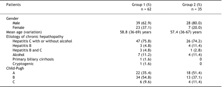
Patients’ characteristics, etiology and severity of liver disease are shown in table 1.
All patients met the Milan criteria, i.e., they ei-ther had a single tumor of 5 cm or less or three tu-mors of 3 cm or less, without any evidence of
Table 1. Patient characteristics.
Patients Group 1 (%) Group 2 (%)
n = 62 n = 35
Gender
Male 39 (62.9) 28 (80.0)
Female 23 (37.1) 7 (20.0)
Mean age (variation) 58.8 (36-69) years 57.4 (36-67) years
Etiology of chronic hepathopathy
Hepatitis C with or without alcohol 47 (75.8) 26 (74.2)
Hepatitis B 3 (4.8) 4 (11.4)
Hepatitis B and C 3 (4.8) 1 (2.8)
Alcohol 7 (11.2) 4 (11.4)
Primary biliary cirrhosis 1 (1.6) 0
Cryptogenic 1 (1.6) 0
Child-Pugh
A 22 (35.4) 18 (51.4)
B 34 (54.8) 13 (37.1)
vascular invasion or extrahepatic spread. In total, 79 HCC nodules were treated with PEI.
The HCC diagnosis was confirmed with biopsy in 55/62 (88.7%) in group 1 and in 21/35 (60%) in group 2. In 21 (21.6%) other cases (seven patients in group 1 and 14 in group 2) the HCC diagnosis was based on noninvasive methods according to the European Association for the Study of the Liver2
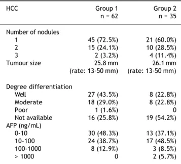
and defined as intense arterial uptake with contrast washout in the venous/delayed phase in two availa-ble dynamic modalities (contrast ultrasound, com-puted tomography, and magnetic resonance imaging). Hepatocellular carcinomas characteristics are shown in table 2.
PEI protocol and imaging follow-up
All the proceedings were performed in inpatients. All tumors were treated by board-certified abdomi-nal interventioabdomi-nal radiologists (R.V, L.B). In all ca-ses, PEI was performed with local anesthesia with lidocaine 2%, using a 22-gauge needle, a 5-mL syrin-ge and absolute ethanol, with real-time ultrasound (US) guidance using a 4.0 MHz sector-probe (Acu-son Sequoia 512). Gray-scale imaging was used for continuous monitoring at the local area of ablation during the ethanol injection. Sedation with fentanyl IV was reserved to cases in which the nodule was superficial and it was performed under the control of an anesthesiologist.
Injections were repeated in different tumor areas until they appeared completely hyperechoic (Figure
1). The amount of ethanol used varied according to the size of the lesion and the compliance of the pa-tient (mean 2-3 mL per session). The procedure was repeated for up to four or five sessions per week com-pleting one treatment cycle. A mean of 6.79 sessions (range: 1-21 sessions) was performed in each patient.
Posttreatment imaging control was repeated with contrast-enhanced US (Sonovue, Bracco, Italy) and contrast-enhanced dual-phase computed tomography (CT) or gadolinium-enhanced magnetic resonance imaging (MRI) at the end of the first month and every three months after ablation until the LT. Com-plete response was defined as the absence of enhanced tumoral areas showing complete tissue necrosis (Fi-gure 2). Any focus of abnormal enhancing tissue,
Table 2. Hepatocellular carcinomas characteristics.
HCC Group 1 Group 2
n = 62 n = 35
Number of nodules
1 45 (72.5%) 21 (60.0%)
2 15 (24.1%) 10 (28.5%)
3 2 (3.2%) 4 (11.4%)
Tumour size 25.8 mm 26.1 mm
(rate: 13-50 mm) (rate: 13-50 mm)
Degree differentiation
Well 27 (43.5%) 8 (22.8%)
Moderate 18 (29.0%) 8 (22.8%)
Poor 1 (1.6%) 0
Not available 16 (25.8%) 19 (54.2%)
AFP (ng/mL)
0-10 30 (48.3%) 13 (37.1%)
10-100 24 (38.7%) 17 (48.5%)
100-1000 8 (12.9%) 3 (8.5%)
> 1000 0 2 (5.7%)
Figure 1. Hyperechogenic nodule during ethanol injec-tion.
within or along the margin of the ethanol injection zone, was considered a residual tumor, and then a new cycle of treatment was performed (Figure 3).
Explanted livers
All explanted liver specimens were fixed in forma-lin and processed using routine protocol, consisting of 0.5-1 cm sections on transverse planes. The liver graft was investigated for HCC nodules, the presen-ce of satellite nodules (i.e., small tumors localized < 1 cm from the main HCC), and microvascular inva-sion. Complete histological response was defined as the absence of tumor cells in the treated zone.
A written informed consent was obtained from all patients before PEI.
Statistical methods
Statistical analysis was performed using SPSS statistical software 13.0. Continuous variables were analyzed using either Student’s t-test or ANOVA. Categorical variables were analyzed using Fisher’s test. Survival was analyzed using the log-rank test. Statistical significance was considered when p < 0.05 and CI = 95%.
RESULTS
Seventy-nine HCC nodules in 62 liver transplan-tation candidates were treated with PEI.
Mean time on the waiting list was eight months (range: 1-15 months). Exclusion from the list
du-ring the waiting period for LT happened in 3/62 (4.8%) patients from group 1 and in 3/35 (8.5%) pa-tients from group 2, all due to tumor progression which exceeded the limits established by the Milan criteria. Proportionally there were more dropouts in group 2; however, the difference was not statistically significant (p = 0.46).
Of the 59 transplanted patients in group 1, the mean maximal diameter of treated nodules was 24.1 mm (range: 5-55 mm) at the latest available imaging follow-up (mean: 2.8 months) before transplantation.
Complications after PEI treatment
In a total of 421 PEI sessions, no major complica-tions were detected. Minor complicacomplica-tions occurred in 10 (2.37%) cases. Segmentary or partial portal thrombosis happened in seven cases; self-limited he-moperitoneum occurred in one case, and transami-nases increased (more than 10-fold the normal level) with no clinical relevance in other two cases.
No deaths or dropouts occurred as a result of the treatment. No evidence of tumor seeding in the nee-dle track was reported during laparotomy or follow-up related to biopsy or PEI.
Characteristics of tumor on explanted livers
Fifty-nine explanted livers were analyzed in group 1 and thirty-two in group 2. In group 1, complete tumor necrosis without histological evidence of via-ble carcinoma or just a minimum quantity of cells was observed in 38/59 (64%) patients (Figure 4). In 14 patients (23%), there were less than 50% of via-ble cells, considering the total diameter of the
le-Figure 3. Control CT after PEI treatment showing a via-ble tumor.
sion, and in seven patients (12%) there was more than 50% of viable tumor.
The mean maximal diameter of the tumor in ex-planted liver was 21.6 mm in group 1 and 32.2 mm in group 2 (p = 0.03).
Microvascular invasion or satellite nodules were present in 23/59 in group 1 patients (38.9%) and in 19/32 in group 2 patients (59.3%). The presence of additional HCC nodules in the explant of group 1 patients was significantly lower when compared to group 2 patients (p = 0.002).
When the number of tumors presents in the ex-planted livers was compared to the number radiolo-gically verified before LT, it was shown that the patients were understaged in 19/59 (32.2%) cases in group 1 and in 21/32 (65.5%) cases in group 2. The-re was not any case of upstaging.
HCC recurrence after LT
Three patients from group 1 (5.1%) and two from group 2 (6.2%) showed tumor recurrence after LT (p = 0.81).
In group 1, the mean time of follow-up was 23.5 months, with a minimum of one month for a patient who died 30 days after LT for neoplastic dissemina-tion during surgery (tumor adhesion to the dia-phragm). Recurrence was intra-hepatic in the other two patients, 16 and 29 months after LT. Recurren-ces were all confirmed with nodule biopsy. The lon-gest follow-up in group 1 was 46 months.
In the first case, two nodules were treated with 12 sessions of PEI; in the second case, one nodule was treated with five sessions; and in the last case, two nodules were treated with nine sessions. Two patients died, and one was alive until the end of the follow up.
In all recurrence cases, explants showed at least four nodules; the largest tumor in each case had 65 mm of diameter in the first case, 35 mm of diameter in the second case, and 80 mm of diameter in the third case. All cases had microvascular invasion and satellite nodules in their explant. Then, it is correct to state that all the recurrence cases had been un-derstaged.
In group 2, recurrence was diagnosed in two ca-ses (6.2%) one and 19 months after LT. In the first case, the explant showed nine nodules; the largest had 50 mm, and death took place one month after LT due to disseminated HCC. In the second case, five tumors were seen in the explant, the largest had 22 mm; this patient was alive until the end of the fo-llow-up. Both cases presented microvascular inva-sion and satellite nodules in their explants.
Survival rate after liver transplantation
In the follow-up period, 11/59 (18.6%) patients from group 1 died. Five of these patients died of postoperative complications. One died of lung can-cer; one of lymphoma; one of lung Aspergillus infec-tion; and one of hepatorenal syndrome after HCV recurrence; deaths occurred 36, 7, 18 and 36 mon-ths after transplantation, respectively. The two other died from HCC recurrence one and 29 months after LT. Intention to treat analysis from the time of entering in the waiting list showed that 14/62 (22.5%) patients died; of these, 5/62 (8%) died from HCC progression. In group 2 transplanted patients, 7/32 (21.8%) died until the end of follow-up. Four died of infectious diseases at 26, 7, 13 and three months after LT; two died of complications related to the surgical procedure, and one died of tumor progression. The intention to treat analysis showed death in 10/35 (28.5%) patients and tumor progres-sion in 4/35 (11.4%). No statistically significance was found in survival between the two groups (p = 0.79). After LT, estimated 3-year survival of group 1 and 2 was 67.7% and 77.4%, respectively (p = 0.29), and there was a decrease to 64.4% and 70.7% in the 3-year survival in groups 1 and 2, respectively, when dropout cases were considered (p = 0.48).
DISCUSSION
For patients with early HCC in the setting of ad-vanced cirrhosis, the best treatment option is liver transplantation. This treatment provides excellent outcomes if its indication is restricted to patients with early stage disease as defined by the Milan cri-teria.13 However, the main problem is the lack of
or-gans due to the scarce number of donations. Even more, transplant lists are filled with severely sick patients and the velocity of donations does not fo-llow patients’ inclusion; therefore, in many centers, the average time on the waiting list for LT is over one year. During this time, tumors of patients with HCC may progress, and any tumor growth increases the risk of microvascular invasion and satellite no-dules, impeding LT. Hence, intention-to-treat survi-val is significantly reduced when the waiting time is too long (more than six months); as a result, most groups treat HCC before LT.14
The radiofrequency ablation has emerged as the most effective method for local tumor destruction, but the results of the studies do not provide signifi-cant improvement in survival favoring RFA over PEI.6,16-18 The present study has special importance
for centers that perform PEI as the primary option of percutaneous treatment, considering the high cost of radiofrequency, sometimes unfeasible for many institutions.
PEI is a highly effective treatment for HCC sma-ller than 3 cm and provides an initial complete res-ponse in more than 80% of cases.19,20 The rate of
initial complete responses is an independent predic-tor of survival in HCC patients treated with PEI, and tumor size has been considered the main factor to determine the efficacy of this treatment.21
The effectiveness of percutaneous treatments for HCC depends on the induction of necrosis of the tu-mor after the procedure and absence of local recu-rrence. However, the main objective of PEI for patients on the waiting list for LT is to control tu-mor growth, thus avoiding complications and pre-venting exclusion from the waiting list.
The rate of dropout of the waiting list due to ad-vanced disease is around 25%.4 The evaluation of
the dropout rate must consider the kind of selec-tion of patients for LT used. Therefore, Castroagu-din,et al.22 analyzed the explant of 19 patients with
HCC < 5 cm in diameter treated with PEI before LT, and in two cases (10.5%) dropouts occurred due to tumoral progression. Using the TNM classi-fication, including patients with advanced tumors (> 5 cm), dropout rate was 5/41 (12%) on a nine-month waiting list in another study using multimo-dality treatments,11 on the other hand, there was
no dropout considering only patients classified as T1. We described 4.8% of dropout in our series of treated patients following the Milan criteria, with a mean time of eight months on the waiting list. Al-though there was no significant difference between groups, lower dropout rate was seen in the treated group.
Severe complications of PEI can be detected in 2.2% of cases,23 as well as tumor seeding.24 In the
present study, no tumor seeding and no major com-plications were observed after 421 sessions of PEI. It is important to make it clear that this study in-cluded only BCLC stage A cirrhotic patients, once BCLC stages B, C and D patients are not candida-tes for curative therapies, such as LT. Despite ha-ving early stage tumors, more than 50% of the patients of the present study were Child-Pugh class B and almost 10% of the sample was Child-Pugh
class C, which denotes an important liver dysfunc-tion.
Moreover, in the present study, we demonstrated not only the safety of percutaneous treatment with PEI, but also the efficacy of this therapeutic method in the control of hepatic neoplastic disease, since significantly more nodules were seen in the explant of the non-treated group, and the diameter of the main nodule was smaller in the treated group. This finding could be related to a possible beneficial effect of the treatment in the control of tumor pro-gression, slowing down the appearance of satellite nodules.
Tumor recurrence after LT is not uncommon; mean recurrence rates are 10-20% in five years. In this study, 3/59 patients in group 1 (5%) and 2/32 patients in group 2 (6%) presented HCC recurrences in the posttransplant follow-up. The presence of mi-crovascular invasion and satellite nodules in the ex-plant of all recurrence cases confirms that these findings are the most important predictors of recu-rrence as previously reported.25 Besides, the liver
ex-plant of the five patients who presented with tumor recurrence after LT demonstrated that their neo-plastic disease exceeded Milan criteria; therefore, these patients should have been excluded from the waiting list if their radiological studies had detected the actual number and size of the tumors.
Studies of explant pieces have demonstrated that image methods performed in the pre-LT period not only fail to diagnose small tumors (< 20 mm), but also underestimate the diameter of the main nodu-le.10,22,26 In the present study, 32% of patients in
group 1 and 65% of patients in group 2 had more HCC nodules in their explant than previously diag-nosed by CT. This should be considered when one discusses the expansion of Milan criteria to include patients with more and larger tumors, since this cri-teria consider both the dimensions and the number of HCCs based on image methods and not on explant findings.27-29
We verified that the survival rate after LT was lo-wer when dropout cases due to tumor progression during the waiting period were included in the analysis. In the present study, mean survival based on intention-to-treat analysis was 37 months for group 1 and 48 months for group 2; no statistical di-fference was found. When we considered the analy-sis based on intention-to-treat, we found that 5/62 (8%) patients treated with PEI died from HCC pro-gression. In the Markov model5 applied to the
increased the seven-year survival in all waiting times before LT. Yao, et al.10 have show that
pre-operative loco-regional therapy, utilizing a multimo-dality treatment regimen, may confer a survival benefit after LT in patients with T2 and T3 HCC, when compared to a group without treatment (5-year recurrence-free survival 93.8% vs. 80.6%; p = 0.04).
We believe that, since this was a retrospective, case-controlled study, some of our finding could be the result of inherent flaws that could have caused a selection bias, such as a shorter time of follow-up for group 1 in relation to group 2 (23.5 months x 36.5 months) and a larger number of patients from group 2 who had their tumors understaged, when comparing their diagnosis before the LT and the ex-plant. Even so, the presented data may help to guide the decision of using PEI in patients with HCC befo-re LT.
In conclusion, although no difference was found between the survival rates of the two groups, we can conclude that percutaneous treatment of HCC with PEI is a safe and effective method in a BCLC stage A cirrhotic population, who met Milan crite-ria, since a significantly smaller number of additio-nal nodules in the explant of the treated group and a smaller diameter of the main nodule were detected. PEI was confirmed as a possible bridge to liver transplantation, since the rate of dropout and tumor recurrence in explanted liver was low. Furthermore, no tumor seeding and no major complications were observed during pretransplant period. Finally, fur-ther studies are needed to evaluate the influence of tumor necrosis after PEI on dropout rates and on overall survival of these patients.
ABBREVIATIONS
• AASLD. American Association for the Study of
Liver Disease.
• AFP.Alfafetoprotein.
• BCLC.Barcelona Clinic Liver Cancer .
• HCC.Hepatocellular carcinoma.
• EASL. European Association for the Study of
the Liver.
• PEI.Percutaneous ethanol injection. • TAE. Transarterial Chemoembolization. • RF. Radiofrequency.
• MRI.Magnetic Resonance Imaging. • CT.Computed Tomography.
• LT. Liver transplantation. • US. Ultrasound.
• HCV. Hepatitis C virus.
REFERENCES
1. Bosch FX, Ribes J, Borras J. Epidemiology of primary liver cancer. Semin Liver Dis 1999; 19(3): 271-85.
2. Bruix J, Sherman M, Llovet JM, Beaugrand M, Lencioni R, Burroughs AK, et al. Clinical management of hepatocellular carcinoma: Conclusions of the Barcelona-2000 EASL Confe-rence. J Hepatol 2001; 35: 421-30.
3. Bruix J, Sherman M. Management of hepatocellular carci-noma. Hepatology 2005; 42(5): 1208-36.
4. Llovet JM, Fuster J, Bruix J. Intention-to-treat analysis of sur-gical treatment for early hepatocellular carcinoma: resection versus transplantation. Hepatology 1999; 30(6): 1434-40. 5. Llovet JM, Mas X, Aponte JJ, Fuster J, Navasa M,
Chris-tensen E, et al. Cost effectiveness of adjuvant therapy for hepatocellular carcinoma during the waiting list for li-ver transplantation. Gut 2002; 50(1): 123-8.
6. Shiina S, Teratani T, Obi S, Sato S, Tateishi R, Fujishima T, et al. A randomized controlled trial of radiofrequency ablation with ethanol injection for small hepatocellular carcinoma. Gastroenterology 2005; 129(1): 122-30. 7. Llovet JM, Castellano L, De Girolamo V, Di Santolo SS,
Ma-rone A, Del Vecchio BC, et al. Tumor dissemination after radiofrequency ablation of hepatocellular carcinoma. He-patology 2001; 34(3): 609-10.
8. Imamura J, Tateishi R, Shiina S, Goto E, Sato T, Ohki T, et al. Neoplastic seeding after radiofrequency ablation for hepatocellular carcinoma. Am J Gastroenterol 2008; 103(12): 3057-62.
9. Moreno Planas JM, Lopez MJ, Gomez CA, Rubio GE, Perez AR, Boullosa GE, et al. Efficacy of hepatocellular carcinoma locoregional therapies on patients waiting for liver trans-plantation. Transplant Proc 2005; 37(3): 1484-5.
10. Yao FY, Kinkhabwala M, Laberge JM, Bass NM, Brown R, Jr., Kerlan R, et al. The impact of pre-operative loco-re-gional therapy on outcome after liver transplantation for hepatocellular carcinoma. Am J Transplant 2005; 5(4 Pt 1): 795-804.
11. Fisher RA, Maluf D, Cotterell AH, Stravitz T, Wolfe L, Luketic V, et al. Non-resective ablation therapy for hepa-tocellular carcinoma: effectiveness measured by inten-tion-to-treat and dropout from liver transplant waiting list. Clin Transplant 2004; 18(5): 502-12.
12. Porrett PM, Peterman H, Rosen M, Sonnad S, Soulen M, Markmann JF, et al. Lack of benefit of pre-transplant lo-coregional hepatic therapy for hepatocellular cancer in the current meld era. Liver Transpl 2006; 12(4): 665-73. 13. Mazzaferro V, Regalia E, Doci R, Andreola S, Pulvirenti A,
Bozzetti F, et al. Liver transplantation for the treatment of small hepatocellular carcinomas in patients with cirrho-sis. N Engl J Med 1996; 334(11): 693-9.
14. Lu DS, Yu NC, Raman SS, Lassman C, Tong MJ, Britten C, et al. Percutaneous radiofrequency ablation of hepatocellu-lar carcinoma as a bridge to liver transplantation. Hepa-tology 2005; 41(5): 1130-7.
15. Majno P, Giostra E, Morel P, Hadengue A, Mentha G. Mana-gement of hepatocellular carcinoma in the waiting list be-fore liver transplantation. J Hepatol 2005; 42(Suppl. 1): S134-S143.
16. Lencioni RA, Allgaier HP, Cioni D, Olschewski M, Deibert P, Crocetti L, et al. Small hepatocellular carcinoma in cirrho-sis: Randomized comparison of radio-frequency thermal ablation versus percutaneous ethanol injection. Radiology
2003; 228(1): 235-40.
ther-mal ablation, percutaneous ethanol injection, and percu-taneous acetic acid injection to treat hepatocellular car-cinoma of 3 cm or less. Gut 2005; 54(8): 1151-6.
18. Brunello F, Veltri A, Carucci P, Pagano E, Ciccone G, Mo-retto P, et al. Radiofrequency ablation versus ethanol injection for early hepatocellular carcinoma: A randomi-zed controlled trial. Scand J Gastroenterol 2008; 43(6): 727-35.
19. Sala M, Llovet JM, Vilana R, Bianchi L, Sole M, Ayuso C, et al. Initial response to percutaneous ablation predicts sur-vival in patients with hepatocellular carcinoma. Hepatolo-gy 2004; 40(6): 1352-60.
20. Taniguchi M, Kim SR, Imoto S, Ikawa H, Ando K, Mita K, et al. Long-term outcome of percutaneous ethanol injection therapy for minimum-sized hepatocellular carcinoma.
World J Gastroenterol 2008; 14(13): 1997-2002.
21. Vilana R, Bruix J, Bru C, Ayuso C, Sole M, Rodes J. Tumor size determines the efficacy of percutaneous ethanol in-jection for the treatment of small hepatocellular carcino-ma. Hepatology 1992; 16(2): 353-7.
22. Castroagudin JF, Delgado M, Martinez SM, Abdulkader I, Potel J, Tome S, et al. Prospective histopathological analysis of hepatocellular carcinoma treated with percu-taneous ethanol injection in patients on the waiting list for liver transplantation. Transplant Proc 2005; 37(3): 1477-9.
23. Ebara M, Okabe S, Kita K, Sugiura N, Fukuda H, Yoshikawa M, et al. Percutaneous ethanol injection for small
hepato-cellular carcinoma: Therapeutic efficacy based on 20-year observation. J Hepatol 2005; 43(3): 458-64.
24. Ishii H, Okada S, Okusaka T, Yoshimori M, Nakasuka H, Shi-mada K, et al. Needle tract implantation of hepatocellular carcinoma after percutaneous ethanol injection. Cancer
1998; 82(9): 1638-42.
25. Sala M, Fuster J, Llovet JM, Navasa M, Sole M, Varela M, et al. High pathological risk of recurrence after surgical re-section for hepatocellular carcinoma: An indication for sal-vage liver transplantation. Liver Transpl 2004; 10(10): 1294-300.
26. Burrel M, Llovet JM, Ayuso C, Iglesias C, Sala M, Miquel R, et al. MRI angiography is superior to helical ct for detec-tion of hcc prior to liver transplantadetec-tion: An explant co-rrelation. Hepatology 2003; 38(4): 1034-42.
27. Bruix J, Fuster J, Llovet JM. Liver transplantation for hepatocellular carcinoma: Foucault pendulum versus evidence-based decision. Liver Transpl 2003; 9(7): 700-2.
28. Mazzaferro V, Llovet JM, Miceli R, Bhoori S, Schiavo M, Mariani L, et al. Predicting survival after liver transplan-tation in patients with hepatocellular carcinoma beyond the Milan criteria: A retrospective, exploratory analysis.
Lancet Oncol 2009; 10(1): 35-43.