Utility and limitations of APRI and FIB4 to predict staging
in a cohort of nonselected outpatients with hepatitis C
Ana Cláudia de Oliveira,*,** Ibrahin El-Bacha,** Mônica V. Vianna,** Edison R. Parise**
* Department of Medicine, Federal University of São Carlos (UFSCar), Brazil. ** Gastroenterology Division, Department of Internal Medicine, Federal University of São Paulo (UNIFESP), Brazil.
A B S T R A C T A B S T R A C T A B S T R A C T A B S T R A C T A B S T R A C T
Background. Background.Background. Background.
Background. Chronic hepatitis C(CHC) staging is important for therapeutic decision-making. Identification of noninvasive markers can provide alternatives to liver biopsy. Aim. Aim. Aim. Aim. Aim. To assess the value of APRI and FIB4 for CHC fibrosis staging in a cohort of nonse-lected outpatients from a referral center in Sao Paulo, Brazil. Material and methods. Material and methods. Material and methods. Material and methods. Material and methods. Medical records of 798 adult outpatients were analyzed retrospectively. For calculations of APRI and FIB4, the original descriptions were considered, and markers were com-pared with degree of liver injury. Results. Results. Results. Results. Overall, 49.3% of participants were female, and mean age was 56.9 Results. ± 12.5 years. Geno-type 1 was predominant (71.7vs. 23.7% genoGeno-type 3); 64% had significant fibrosis, 44% had advanced fibrosis, and 28% had cirrhosis. The areas under the receiver operating curve for significant fibrosis, advanced fibrosis, and cirrhosis, respectively, were 0.809, 0.819, and 0.815 for the APRI marker and 0.803, 0.836 and 0.852 for FIB4. Using the recommended cut off values, approxi-mately 30-40% of the patients could not be classified. In the remainder, either APRI or FIB4 alone correctly diagnosed 80-85% of cases. Concomitant or consecutive use of both APRI and FIB4 increased the number of the cases correctly diagnosed only slightly, but also increased the number of patients not classified within the cutoff values. Conclusions. Conclusions. Conclusions. Conclusions. In conclusion, use of the APRI or Conclusions. FIB4 markers for detection of hepatic fibrosis may be a viable alternative at referral centers for treatment of CHC in low- and middle-income countries. Despite relatively good accuracy, a significant number of patients could not be assessed by these methods.
Key words. Key words.Key words. Key words.
Key words. Chronic hepatitis C. Noninvasive serum markers. Hepatic fibrosis. Low-middle income country. Accuracy.
May-June, Vol. 15 No. 3, 2016: 326-332
INTRODUCTION
Chronic hepatitis C virus infection (CHC)currently af-fects 185 million people worldwide.1 It is the leading
cause of cirrhosis of the liver and hepatocellular carcino-ma, the leading indication for liver transplantation in the United States and possibly worldwide, and an indisputably serious public health issue.2,3
Due to high rates of progression to chronic disease and progressive fibrosis,4,5 histological assessment of the
de-gree of liver injury is an important part of the decision-making process in evaluating the prognosis of hepatitis C infection and indicating antiviral treatment.
The diagnosis of hepatic fibrosis is primarily morpho-logical, and is established by histological examination of hepatic biopsy specimens. Although it is a relatively safe and reliable procedure, hepatic biopsy is not devoid of
risk6 and is subject to sampling error and inter- and
intra-observer variation in the assessment of fibrosis.7-9
In developing countries, management of hepatitis C and access to medicines must take into account the financial burden of disease control programs. Both medicines and diagnostic methods are costly, and the promotion of their rational use and prioritization of access to the neediest cases are urgently required. The World Health Organiza-tion (WHO) recently proposed that low- and middle-in-come countries implement the use of AST-to-platelet ratio index (APRI) and Fibrosis-4(FIB4) values for the staging of hepatitis C virus disease, in view of their con-venience, easy access, and accuracy.10 The APRI is an
indi-rect biochemical marker of fibrosis,which takes into account the serum level of aspartate aminotransferase (AST) and platelet count for disease staging, with good accuracy, as originally described by Wai, et al.11 The FIB412,13 The Official Journal of the Mexican Association of Hepatology,
the Latin-American Association for Study of the Liver and the Canadian Association for the Study of the Liver
Manuscript received: Manuscript received: Manuscript received: Manuscript received:
Manuscript received: June 18, 2015. Manuscript accepted:Manuscript accepted:Manuscript accepted: August 03, 2015.Manuscript accepted:Manuscript accepted:
also uses data routinely available in clinical practice, name-ly AST, alanine aminotransferase (ALT), platelet count, and patient age; it is non-inferior to the APRI for detec-tion of overall fibrosis and believed to be superior to the APRI for identification of advanced fibrosis (F3-F4). These tests have the advantage of being easy to perform, even at the bedside, and carry no additional costs.
The present study will assess the value of the noninva-sive APRI and FIB4 tests as a strategy for identification of hepatic fibrosis in a cohort of nonselected patients seen at a referral center for hepatitis C care in São Paulo, Brazil.
MATERIAL AND METHODS
A retrospective analysis was conducted of the medical records of adult patients from the Chronic Liver Disease Clinic of the Gastroenterology Division, Department of Internal Medicine, Federal University of São Paulo, a re-ferral center for patients with hepatitis C in the city of São Paulo, Brazil. The inclusion criterion was a diagnosis of hepatitis C virus infection confirmed by polymerase chain reaction (PCR)detection of HCV-RNA in peripheral blood (Amplicor-HCV, Roche Diagnostics). HCV geno-typing was performed by sequencing of the 5' non-coding region, and viral load was measured by quantitative RT-PCR (Amplicor, Roche).
Patients with hepatitis B virus or HIV coinfection, chronic liver disease of other etiologies, hepatocellular carcinoma, history of antiviral therapy, history of liver transplantation, current immunosuppressant therapy, alco-hol intake > 20g/day (male or female), or insufficient tis-sue for liver biopsy analysis were excluded. All eligible patients, except those with clinical, sonographic, and/or
endoscopic manifestations of cirrhosis underwent percu-taneous liver biopsy. Specimens were embedded in paraffin and stained with hematoxylin and eosin (H&E), reticulin, and Masson’s trichrome. On specimen analysis, structural injury was assessed using the METAVIR classi-fication.14
ALT and AST levels were measured by automated ki-netic methods in blood collected after a 12-h fast and ex-pressed as IU/mL. The platelet count was measured by an automated analyzer and expressed as platelets/mm3. The
mean interval between measurement of biochemical pa-rameters and liver biopsy was less than 3 months.
The APRI11and FIB413 were calculated using the
fol-lowing formulas, as originally reported:
• APRI = AST(*ULN)/ platelet count (*109/L) x 100.
• FIB4 = AST(IU/mL) x age / platelet count (*109/L) x
ALT (IU/mL)1/2.
The study protocol was approved by the Federal Uni-versity of São Paulo Research Ethics Committee.
Statistical analysis
Receiver operating curves (ROC) were used to ana-lyze the accuracy of the noninvasive APRI and FIB4 tests for disease staging. The discriminant ability of each test was obtained by calculating the sensitivity, specificity, positive predictive value, and negative pre-dictive value. P-values ≤ 0.05 were deemed statistically significant. All data were analyzed using the SPSS statis-tical package, version 17.0 (SPSS Inc., Chicago, IL, USA).
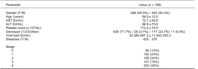
Table 1. Sample profile.
Parameter Value (n = 798)
Gender (F:M) 396 (49.3%) / 402 (50.3%)
Age (years) 56.9 ± 12.5
AST (IU/mL) 72.7 ± 55.6
ALT (IU/mL) 86.9 ± 75.9
Platelet count (x 103/mL) 173.3 ± 74.5
Genotype (1/2/3/other) 536 (71.7%) / 28 (3.7%) / 177 (23.7%) / 7 (0.9%) Viral load (IU/mL) 32,360,067.2 ± 11,542,030.3
Steatosis (Y:N) 423 : 375
Stage
0 95 (12%)
1 193 (24%)
2 158 (20%)
3 127 (16%)
4 225 (28%)
Source of the curve APRI FIB4
RESULTS
A total of 798 patients with chronic hepatitis C were as-sessed. Of these, 396 (49.3%) were female, with a mean age of 56.9 ± 12.5 years. Regarding HCV genotype, 536 (71.7%) were infected with genotype 1 and 177 (23.7%) with genotype 3. The mean AST, ALT, and platelet count-values were 72.7 ± 55.6IU/mL, 86.9 ± 75.9IU/mL, and 173.3 ± 74.5 platelets/mm3, respectively. On histological
examination of biopsy specimens, 288 patients (36%) had no fibrosis or early-stage fibrosis (F0-F1) and the remain-ing 64% had significant fibrosis (≥ F2), 28% of whom had cirrhosis (F4). Portal/periportal activity was grade ≥ 2in 520 patients (76.2%). Steatosis was present in 423 patients (63%). The demographic characteristics and biochemical and viral parameters of the sample are presented in table 1. Areas under the ROC (AUROCs) for the evaluated tests at each degree of fibrosis (significant, advanced, and cirrhosis) are shown in table 2 and figure 1.
Table 2. Diagnostic accuracy of the APRI and FIB-4 tests for different stages of hepatic fibrosis.
Fibrosis TEST AUROC (95%CI) P
Significant (≥ F2) APRI 0.809 (0.776-0.841) <0.001 FIB-4 0.803 (0.771-0.836) <0.001 Advanced (F3-F4) APRI 0.819 (0.788-0.851) <0.001 FIB-4 0.836 (0.805-0.866) <0.001
Cirrhosis (F4) APRI 0.815 (0.781-0.849) <0.001
FIB-4 0.852 (0.821-0.883) <0.001
AUC: area under the curve. 95% CI: 95% confidence interval.
Figure 1. Figure 1.Figure 1.
Figure 1.Figure 1. AUROC curve for significant fibrosis (≥ F2) for APRI [AUROC = 0.809 (0.776 - 0.841, p < 0.001) and FIB-4 [AUROC 0.803 (0.771 - 0.836, p < 0.001). Diagnosis segments are produced by ties.
Sensitivity
1.0
0.8
0.6
0.4
0.2
0.0
0.0 0.2 0.4 0.6 0.8 1.0
1-Specificity
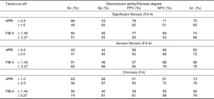
Table 3. Discriminant ability of the APRI and FIB-4 tests for identification of significant fibrosis (F2-F4), severe fibrosis (F3-4) and cir-rhosis (F4).
Tests/cut-off Discriminant ability/Fibrosis degree
Sn (%) Sp (%) PPV (%) NPV (%) Ac (%)
Significant fibrosis (F2-4)
APRI ≤ 0.5 88 53 78 71 75
≥ 1.5 49 92 92 51 65
FIB-4 ≤ 1.45 85 56 77 68 74
≥ 3.27 51 93 93 52 66
Severe fibrosis (F3-4)
APRI ≤ 0.5 93 42 56 88 65
≥ 2.0 47 92 82 69 72
FIB-4 ≤ 1.45 91 46 57 86 66
≥ 3.27 65 88 80 76 78
Chirrosis (F4)
APRI ≤ 1.0 83 68 51 91 73
≥ 2.0 56 87 63 72 78
FIB-4 ≤ 1.45 95 40 39 95 56
≥ 3.27 74 81 61 88 79
Sn: sensitivity. Sp: specificity. PPV: positive predictive value. NPV: negative predictive value. Ac: accuracy.
Measures of discriminant ability for the APRI and FIB4 tests for each stage of hepatic fibrosis, with optimal cutoff points, are shown in table 3.
With these data, and using the combined cutoff values to rule out (negative predictive value, NPV) or confirm (positive predictive value, PPV) the degree of fibrosis of interest, we were able to assess the diagnostic accuracy of the tests employed and the percentage of patients not evaluated within the defined cutoff points for the different degrees of fibrosis.
On average, approximately one-third of patients would require another method for assessment of hepatic fibrosis (Table 3), as their values were not located between the given cutoff points. After exclusion of these unclassified patients, on average, 80% of diagnoses were correct, with equivalence of the evaluated tests for significant fibrosis. Whereas FIB4 performed better for advanced fibro-sis, APRI lower and upper values performed better for diagnosis of cirrhosis, despite the better AUROC obtained for the FIB4 test.
In an attempt to increase the reliability of these tests, we evaluated their use sequentially, as in the SAFE biopsy algo-rithm,15 and in combination, as in the Fibropaca algorithm.16
Sequential use of the APRI and FIB4 tests did not pro-vide any advantage. For significant fibrosis, for instance, we used an APRI cutoff of ≥ 1.5, which had a PPV of 92%; however, approximately 50% of patients with grade F2 or higher had values below this cutoff point. An attempt at se-quential “rescue” use of the FIB4 in these patients pro-duced a modest increment in the number of correctly classified cases (to 55 from 51%) and modestly reduced the rate of unclassified patients (to 34 from 38%).
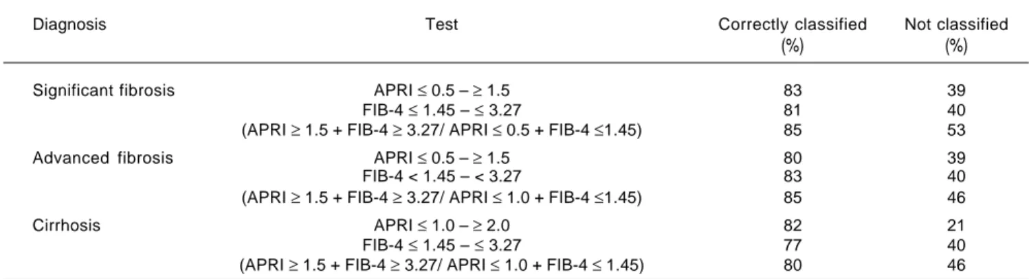
Combined application of the tests improved their dis-criminant ability for the different degrees of hepatic fibro-sis and reduced the number of incorrectly diagnosed cases. However, a greater number of cases were excluded from analysis, as the obtained values fell outside the cutoff range (Table 4).
DISCUSSION
Chronic hepatitis C virus infection is characterized by high rates of progression to chronic liver disease, with progressive hepatic fibrosis, potentially leading to cirrho-sis and its complications.4,5 For many years, liver biopsy
has been regarded as the gold standard for structural evalu-ation of hepatic tissue, despite several studies that have demonstrated its “imperfect” status due to its limitations. The accuracy of liver biopsy is directly related, among other factors, to the size of the tissue fragment obtained and to the experience of the examiner.7-9 Furthermore, it
is invasive, costly, and not entirely risk-free. Consequent-ly, its performance is refused by many patients and impos-sible in others.
Within this context, the identification of noninvasive markers of liver fibrosis has been the object of extensive research, not in an attempt to replace liver biopsy alto-gether but in the hope of restricting its use to specific cas-es. The use of noninvasive markers for assessment of liver fibrosis is considered preferable to invasive testing in the recent World Health Organization Guidelines for the Screening, Care and Treatment of Persons with Hep-atitis C Infection.10 The APRI, FIB4, Fibrotest®, and transient
elastography were all recommended in this guideline; however, for low- and middle-income countries, the APRI and FIB4 were specifically recommended for their very low cost, their ease of access, their use of tests routinely ordered in clinical practice (AST, ALT, and platelet count), and their substantial accuracy for identification of fibrosis and cirrhosis.
In its original report, the APRI11 had an AUROC
of 0.80 and 0.89 for identification of significant fibrosis (Ishak 3-6) and 0.88 and 0.94 for identification of cirrho-sis (Ishak 5-6). Using the high (APRI > 2) and low (APRI < 0.5) cutoff points, this test had a discriminant ability of 81% for presence and absence of cirrhosis. Likewise, it was able to predict significant fibrosis in 51% of patients. Table 4. Percentage of cases with significant fibrosis (F2-F4), advanced fibrosis (F3-F4), and cirrhosis (F4) classified with the APRI and FIB-4 tests.
Diagnosis Test Correctly classified Not classified
(%) (%)
Significant fibrosis APRI ≤ 0.5 – ≥ 1.5 83 39
FIB-4 ≤ 1.45 – ≤ 3.27 81 40
(APRI ≥ 1.5 + FIB-4 ≥ 3.27/ APRI ≤ 0.5 + FIB-4 ≤1.45) 85 53
Advanced fibrosis APRI ≤ 0.5 – ≥ 1.5 80 39
FIB-4 < 1.45 – < 3.27 83 40 (APRI ≥ 1.5 + FIB-4 ≥ 3.27/ APRI ≤ 1.0 + FIB-4 ≤1.45) 85 46
Cirrhosis APRI ≤ 1.0 – ≥ 2.0 82 21
FIB-4 ≤ 1.45 – ≤ 3.27 77 40
In a previous study by our group analyzing a cohort of se-lected patients, we found similar results for identification of significant fibrosis (F2-F4) and cirrhosis (F4).17 Later
studies assessing the accuracy of this method for predic-tion of fibrosis reported less robust results. In two recent meta-analyses including over 8,000 patients, the APRI ex-hibited very similar diagnostic performance for identifica-tion of significant fibrosis (AUROC = 0.77 and 0.76) and cirrhosis (AUROC = 0.83 and 0.82).18,19
The FIB4 was originally assessed in a cohort of patients with HIV/HCV coinfection,12 with an AUROC of 0.74 and
0.71 to predict fibrosis (F4-6 and Ishak F2-6), and later validated in individuals with HCV monoinfection,13 with
an AUROC of 0.85 for METAVIR F3-F4 fibrosis. In a large U.S. cohort, the FIB4 had an AUROC of 0.83 for identification of advanced fibrosis (F3-4). Of the 981 pa-tients with a FIB4 score >2.0, 87.9% had grade >F2.20
The objective of the present series was to assess the di-agnostic value of these noninvasive tests in a real-world setting, namely, in the daily practice of the outpatient liver disease clinic of a tertiary referral center. Toward this end, liver enzyme measurements and platelet counts were col-lected retrospectively from patient records. There was no specific research protocol for determination of the param-eters of interest.
Under these conditions, the AUROCs obtained for the APRI and FIB4 tests for detection of significant fibrosis, advanced fibrosis, and cirrhosis were approximately 0.80 (Table 2, Figure 1), a value quite close to those previously reported in the literature, which demonstrates the good reproducibility of these tests even in real-world clinical practice.
As recommended by WHO, our analyses used the estab-lished combination of low and high cutoff values for these biomarkers for detection of fibrosis, thus avoiding the crea-tion of new indices that might hinder practical applicacrea-tion of the evaluated tests. These cutoff values (0.5, 1.5, and 2.0 for the APRI and 1.45 and 3.27 for the FIB4) were thus used for detection of significant fibrosis (≥ F2), advanced fibrosis (F3-F4), and cirrhosis (F4). As shown in table 3, the APRI and FIB4 had similar diagnostic ability, except for de-tection of cirrhosis, in which the APRI proved superior. Strikingly, approximately 30% of patients could not be clas-sified with either of these tests. Therefore, we chose to combine the two in an attempt to increase the number of correctly diagnosed cases. A similar strategy was employed by Sebastiani, et al., who used the APRI and Fibrotest se-quentially in the SAFE biopsy study.15 In the present study,
however, sequential use of the APRI and FIB4 tests did not result in a significant reduction in the number of undiag-nosed cases, as also reported by Crisan, et al.,21 which shows
that these biomarkers have very similar diagnostic profile. When using both tests concomitantly, we observed
im-provement in discriminant ability for the different stages of fibrosis, as well as a reduction in the number of misdiag-nosed cases, as compared with either tests in isolation or both tests sequentially. However, this was offset by a signif-icant loss in the number of patients covered; approximately 50% of patients in our sample remained unclassified with this strategy.
The combination of these serum markers and mechani-cal methods, particularly FibroScan® transient elastogra-phy, would probably increase their diagnostic efficacy and reduce the number of patients with an indeterminate re-sult. This hypothesis is based on the fact that nearly half of all patients with significant fibrosis and an APRI <1.5 had METAVIR grade 3 or 4 fibrosis – precisely the patient population in which elastometry is most indicated.22,23
Such an increase in diagnostic accuracy when combining mechanical methods and biomarkers has already been re-ported in published studies.24-26
Some limitations of the present study must be stressed, including its retrospective design and its setting (specialty care centers for patients with chronic hepatitis), which justifies the high percentage of patients with cirrhosis and advanced fibrosis. The retrospective design might have an impact on the sensitivity and specificity of the evaluated tests, as well as on the selected cases. The attrition rate of our study due to missing data was only approximately 1%, as the blood tests used for calculation of the APRI and FIB4 scores are performed as part of these patients’ rou-tine care. On the other hand, the present analysis sought to portray a real-world patient care environment, with no in-terference from especially designed protocols for collec-tion of biochemical parameters. We believe referral centers are the most appropriate setting for application of these tests, as they are the venues where treatment-related decisions are made; however, their implementation in primary care settings, where a lower prevalence of ad-vanced cases is to be expected, would also be of the utmost importance.
the limitations of these methods, isolated use of the APRI or FIB4 tests should be considered for low-and middle-income countries or in settings where access to referral centers is difficult.
ABBREVIATIONS
• Ac: accuracy.
• ALT: alanine aminotransferase. • APRI: AST-to-platelet ratio index. • AST: aspartate aminotransferase.
• AUROC: area under the receiver operating curve. • CHC: chronic hepatitis C.
• FIB4: fibrosis 4.
• H&E: hematoxylin and eosin. • HCV: hepatitis C virus.
• HCV-RNA: HCV ribonucleic acid. • HIV: human immunodeficiency ví+irus. • NPV: negative predictive value.
• PCR: polymerase chain reaction. • PPV: positive predictive value. • Sn: sensitivity.
• Sp: specificity.
• WHO: World Health Organization.
REFERENCES
1. Mohd Hanafrah K, Groeger J, Flaxman AD, Weirsma ST. Glo-bal epidemiology of hepatitis C virus infection: new esti-mates of age-specific antibody to HCV seroprevalence. Hepatology 2013; 57: 1333-42.
2. Luer GM, Walker BD. Hepatitis C virus infection. N Eng J Med 2001; 345: 41-52.
3. Kim WR, Brown RS Jr, Terrault NA, El-Serag H. Burden of liver disease in the United States: summary of workshop. Hepatology 2002; 36: 227-42.
4. Serfaty L, Aumaitre H, Chazouileires O, Bonnand AM, Ros-morduc O, Poupon RE, Poupon R. Determinants of outcome of compensated hepatitis C virus-related cirrhosis. Hepatol-ogy 1998; 27: 1435-40.
5. Dienstag JL. The role of liver biopsy in chronic hepatitis C. Hepatology 2002; 5(Suppl. 1): S152-S160.
6. Garcia-Tsao G, Boyer JL. Outpatient liver biopsy: how safe is it? Ann Int Med 1993; 118: 150-3.
7. Regev A, Berho M, Jeffers LJ, Milikowski C, Molina EG, Pyr-sopoulos NT, Feng ZZ, et al. Sampling error and intraobserv-er variations in livintraobserv-er biopsy in patients with chronic HCV infection. Am J Gastroenterol 2002; 97: 2614-8.
8. Colloredo G, Guido M. Sonzoqni A, Leandro G. Impact of liv-er biopsy size on histological evaluation of chronic viral hep-atitis: the smaller, the milder the disease. J Hepatology 2003; 39: 239-44.
9. Bedossa P, Dargere D, Paradis V. Sampling variability of liver fibrosis in chronic hepatitis C. Hepatology 2003; 38: 1449-57.
10. Guideline for the screening, care and treatment of persons with hepatitis C infection. World Health Organization, April 2014.http://www.who.int/hiv/pub/hepatitis/hepatitis-c-guide-lines/en/.
11. Wai CT, Greenson JK, Fontana RJ, Kalbfleisch JD, Marrero JA, Conjeevaran HS, Lok AS. A simple noninvasive index can predict both significant fibrosis and cirrhosis in patients with chronic hepatitis C. Hepatology 2003; 38: 518-26. 12. Vallet-Pichard A, Mallet V, Nalpas B, Verkarre V, Nalpas A,
Dhalluin-Venier V, Fontaine H, et al. FIB4: an inexpensive and accurate marker of fibrosis in HCV infection: Compari-son with liver biopsy and fibrotest. Hepatology 2007; 46: 32-6.
13. Sterling RK, Lissen E, Clumick N, Sola R, Correa MC. Devel-opment of a simple noninvasive index to predict significant fi-brosis in patients with HIV/HCV coinfection. Hepatology 2006; 43: 1317-25.
14. Bedossa P, Poynard T. An algorithm for the grading of activi-ty in chronic hepatites C. The METAVIR Cooperative Study Group. Hepatology 1996; 24: 289-93.
15. Sebastiani G, Halfon P, Castera L, Pol S, Thomas DL, Mangia A, Di Marco V, et al. SAFE biopsy: a validated method for large-scale staging of liver fibrosis in chronic hepatitis C. Hepatology 2009; 49: 1821-7.
16. Sebastiani G, Halfon P, Castera L, Mangias A, Di Marco V, Pirisi M, Voiculescu M, et al. Comparison of three algorithms of non-invasive markers of fibrosis in chronic hepatitis C. Aliment Pharmacol Ther 2012; 35: 92-104.
17. Parise ER, Oliveira AC, Figueredo-Mendes C, Lanzoni V, Martins J, Nader H, Ferraz ML. Noninvasive serum markers in the diagnosis of structural liver damage in chronic hepati-tis C virus infection. Liver Inter 2006; 26: 1095-9.
18. Lin ZH, Xin YN, Dong QJ, Wang Q, Jiang XJ, Zhan SH, Sun Y,et al. Performance of aspartate aminotransferase-to-platelet ratio index for staging of hepatitis C related-fibrosis: an updated meta-analysis. Hepatology 2011; 53: 726-36. 19. Shaheen AA, Myer RP. Diagnostic accuracy of the
aspar-tate aminotransferase-to-platelet ratio index for the predic-tion of hepatitis C-related fibrosis: a systemic review. Hepatology 2007; 46: 912-21.
20. Holberg SD, Lu M, Rupp LB, Lamerato LE, Moorman AC, Vi-jayadeva V, Boscarino JA, et al. Noninvasive serum fibrosis marker for screening and staging chronic hepatitis C virus patients in a large US cohort. Clin Infection Dis 2013; 57: 240-6.
21. Crisan D, Radu C, Lupsor M, Sparchez Z, Grigorescu MC, Grigorescu Mircea. Two or more synchronous combination of noninvasive tests to increase accuracy of liver fibrosis assessment in chronic hepatitis C; results from a cohort of 446 patients. Hepat Mon 2012; 12: 177-84.
22. Castera L. Transient elastography and other noninvasive tests to assess hepatic fibrosis in patients with viral hepati-tis. J Viral Hepat 2009; 16: 300-14.
23. Castera L, Vergniol J, Foucher J, Le Bail B, Chanteloup E, Haaser M, Darriet M, et al. Prospective comparison of tran-sient elastography, Fibrotest, APRI, and liver biopsy for the assessment of fibrosis in chronic hepatitis C. Gastroenter-ology 2005; 128: 343-50.
24. Friedrich-Rust M, Ong MF, Martens S, Sarrazin C, Bojunga J, Zeuzem S, Herrmann E, et al. Performance of transient elas-tography for the staging of liver fibrosis: a meta-analysis. Gastroenterology 2008; 134: 960-74.
25. Foucher J, Chanteloup E, Vergniol J, Castera L, Le Bail B, Adhoute X, Bertet J, et al. Diagnosis of cirrhosis by transient elastography (FibroScan): a prospective study. Gut 2006; 55: 403-08.
Correspondence and reprint request:
Ana Cláudia de Oliveira M.D.,Ph.D.
Gastroenterology Division, Department of Internal Medicine. Federal University of São Paulo.
Rua Botucatu, 740, 2º andar. São Paulo, SP 04023-900. Brazil. Tel.: +55-19-992930513