Liver transplantation:
A new risk factor for intestinal intussusceptions
Sven Pischke,* Wursthorn Karsten,* Johannes Hadem,* Sebastian Schmidt,* Karl Heiringhoff Heinz,** Fabian Helfritz,** Christian P. Strassburg,* Joachim Lobers,* Lars Zender,* Oktay Tutarel,***
Jochen Wedemeyer,** Michael P. Manns,* Heiner Wedemeyer,* Kinan Rifai,* Michael Gebel*
* Hannover Medical School, Gastroenterology, Hepatology and Endocrinology. ** Hannover Medical School, Surgery. *** Hannover Medical School, Cardiology.
ABSTRACT
Background. Intestinal intussusception in adults is associated with chronic inflammatory bowel disease, co-eliac disease, abdominal tumors or previous abdominal surgery but most often of unknown origin. Aim. The aim of our study was to evaluate circumstances and identify risk factors for intussusceptions. Methods. All 65,928 abdominal ultrasound examinations performed at our tertiary medical center between January 2001 and June 2008 were analyzed retrospectively for the diagnosis “intussusception”. After identifying indivi-duals with sonographically proven intussusception we analyzed various patients’ characteristics including age, gender and underlying disease as well as sonographic findings such as localization of the intussuscep-tion, absence or presence of ascites and lymph nodes. Results. We identified 32 cases of intussusceptions [mean age 45 years (range 18 to 88); 18 patients were male]. Twelve patients (38%) had a history of abdomi-nal surgery including 8 patients who had undergone liver transplantation (2 patients with primary sclero-sing cholangitis, 1 patient with cystic fibrosis, 1 patient with sarcoidosis, 1 patient with hepatocellular carcinoma and HCV infection, 1 patient with autoimmune hepatitis, 1 patient with Crigler-Najar-syndrome and one patient with echinococcus). A hepaticojejunostomy had been performed in 4 of the patients after liver transplantation. Liver transplanted patients were significantly overrepresented in the intussusception group compared with the overall cohort of patients undergoing abdominal ultrasound examination (25% vs. 8%, Chi-Square-test, p = 0.0023). Conclusion. In our retrospective study liver transplantation, in particular with hepaticojejunostomy, was identified as a new major risk factor for intestinal intussusceptions.
Key words. Ultrasound. Intussusception. Liver transplantation. Risk factor. Hepaticojejunostomy.
Correspondence and reprint request: Sven Pischke, MD Hannover Medical School,
Carl-Neuberg Strasse 1, 30625 Hannover, Germany
E-mail: [email protected]
Manuscript received: October 12, 2010. Manuscript accepted: November 23, 2010. INTRODUCTION
Intestinal intussusception in adults as a cause of abdominal pain has been found in 0.53% of cases in a prospective study by Maconi, et al.1 Intestinal
in-tussusception is frequently considered to being of idiopathic origin, In addition it has been associated with chronic inflammatory bowel disease, Meckel di-verticula, prior abdominal surgery and gastrointesti-nal tumors.1-5 In children as well as in adults
various infectious causes for intestinal intussuscep-tions have also been described. Adenoviruses, ente-ral tuberculosis, rotaviruses, Clostridium difficile
infection or Yersiniosis6-11 have been associated
with intussusceptions. Also vaccination against ro-taviruses was associated with intestinal intussuscep-tion.9 Interestingly, biliary infection with
adenoviruses is a typical reason for intussusceptio-ns in childhood,6,12,13 but seems not important for
intussusceptions in adults.
Most previous studies investigating risk factors for intestinal intussusceptions have been focused on surgical aspects of therapy.14,15 Studies analyzing
the diagnostic use of ultrasound examinations are rare. A study of 41 cases of intussusception evalua-ted CT scans as the diagnostic gold standard for in-testinal intussusception.16 However, in Europe
ultrasound is regarded as the diagnostic tool of choice1 (Figure 1). Prerequisites for the reliable
diagnosis of intussusceptions by ultrasound are that high-frequency-sonography probes with > 6 MHZ are used and that an experienced physician is per-forming the examination (e.g. DEGUM certificate Grad II).17 These prerequisites are based on the
In a recent case report we described the case of a liver transplanted patient with repeated intussuscep-tions in context of intestinal CMV-infection.18
Howe-ver, the risk for liver transplanted patients to develop intussusceptions has never been examined systematically. Here, we examined the characteris-tics of patients with sonographically diagnosed in-tussusception in a retrospective analysis.
OBJECTIVE
The aim of our study was to identify risk factors associated with intestinal intussusception in adults.
PATIENTS AND METHODS
The study was performed at Hannover Medical School, a tertiary center with a large transplanta-tion program, performing more than 140 liver transplantations per year over the last decade.19
Our liver transplant outpatient clinic follows near-ly 2,000 liver transplanted patients. The section for ultrasound within the Department of Gastroentero-logy, Hepatology and Endocrinology at Hannover Medical School is a highly specialized unit perfor-ming around 8,000 ultrasound examinations each year.
For our analysis we searched a database of ultra-sound examination reports for the term “intussuscep-tion” including every ultrasound examination report between January 1, 2001 and June 30, 2009. A total of 65,928 ultrasound examinations were investigated. Only cases with sonographically documented
intus-susception were included in this study. Required so-nographic criteria for intussusception were:
• A multilayered lesion with concentric circles (onion sign) in the transversal plane.
• In case of involvement of the mesentery an echo-rich crescent open towards the ante-mesenteric side (donut sign).
• An echo-rich layer between two multilayered structures (the sandwich sign) in the longitudi-nal plane.20
Suspicious cases of intussusception without ade-quate sonographic proof of the disorder were exclu-ded (n = 11).
For all patients with intussusceptions, the follo-wing characteristics were analyzed: age, gender, un-derlying disease, localization of intussusception, history and type of former abdominal surgery and/or former liver transplantation, the presence of ascites and enlarged intra-abdominal lymph nodes.
In total, 5,257 ultrasound examinations (~ 8%) were performed on patients after liver transplanta-tion.19 In most of them liver transplantation
inclu-ded duct-to-duct biliary reconstruction, while approximately 8% of all liver transplanted patients had a primary hepaticojejunostomy due to primary sclerosing cholangitis (PSC). 3% of liver transplant recipients received a secondary hepaticojejunostomy due to bile duct stenosis. Overall, 11% of all liver transplanted patients at Hannover Medical School received a hepaticojejunostomy. In our study cohort we focussed on ultrasound records of the 982 exami-nations (1.5%) that have been performed on patients with a history of hepaticojejunostomy. 572 examina-tions (0.9%) were in patients with Whipple Opera-tion, 538 (0.8%) in patients with a history of colonic resection and 295 (0.4%) in patients with gastrec-tomy.
Statistical analysis was performed by Chi-Square-test. A p-value of < 0.05 was considered to be statis-tically significant.
RESULTS
Of the 65,928 ultrasound examinations we identi-fied 32 patients with sonographically proven intus-susceptions. This represents 0.05% of all ultrasound examinations.
was located at the ileocoecal region (41%). Six pa-tients had multiple intussusceptions at more than one location (19%), four of these six patients also had enlarged lymph nodes. Two cases had the intus-susception directly at the ileocoecal valve.
In the majority of patients with intussusception (22/32, 69%) enlarged abdominal lymph nodes could be identified.15 of them were located in the mesente-rium (47%) and 9 at the hepatoduodenal ligament (28%). Two patients had enlarged lymph nodes at both locations and one had enlarged lymph nodes at the ligamentum hepatoduodenale as well as along the abdominal part of the aorta.
There was no statistically significant gender di-fference with regard to intestinal intussusceptions in our cohort. Eighteen male and 14 female pa-tients presented with intussusceptions. Both sexes were represented evenly in our group of liver transplanted patients which included 4 men and 4 women (Table 1).
In twelve patients with intussusception (38%) ul-trasound examination revealed ascites, predominant-ly located between parts of the colon (n = 10) but also diffusely distributed in the whole abdomen (n = 1) and perihepatically (n = 1).The patient with perihe-patic ascites presented with sarcoma and pleural carcinomatosis.
Seven patients had neither detectable lymph no-des nor ascites. Four of these had been liver trans-planted, two had Crohn’s disease and one patient had acute pancreatitis. Three of the four liver trans-planted patients had no history of hepaticojejunos-tomy, the remaining patient had a primary hepaticojejunostomy due to the preexisting PSC.
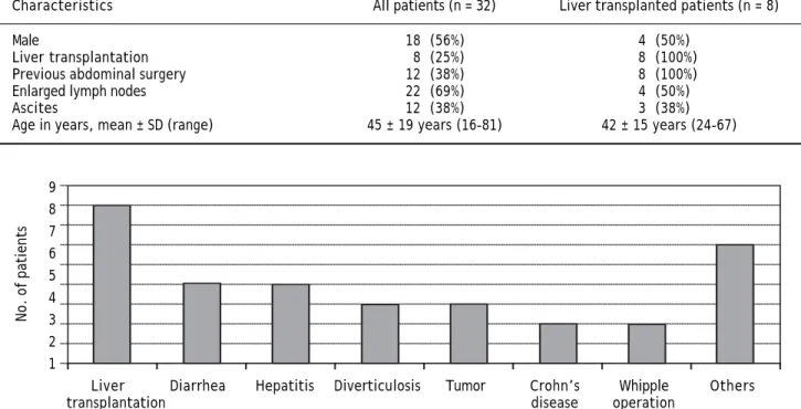
Patients with intussusception had different un-derlying diseases and previous surgical interventio-ns as shown in figure 2.
Eight of them were liver transplanted patients (25%). The age range in the group of liver trans-planted patients with intussusception was 22 and 58 years (mean 36 years).
Four patients had repeated episodes of diarrhea, three also showed signs of sigmadiverticulosis and two patients each have been diagnosed with Crohn’s disease, hepatitis of unknown origin, chronic Hepa-titis C Virus (HCV) infections or previous Whipple operations. There were single cases with pancreati-tis, primary sclerosing cholangitis (PSC), meteo-rism, hepatocellular carcinoma (HCC), anemia of unknown origin, sarcoma, previous lung transplan-tation and suspicion of pancreatic cancer.
In relation to the percentage of liver transplanted patients who underwent ultrasound examination (8%) the percentage of those with proven
intussus-Liver Diarrhea Hepatitis Diverticulosis Tumor Crohn’s Whipple Others transplantation disease operation
Figure 2. Part of liver transplanted patients in the group of all patients with intussusception.
9 8 7 6 5 4 3 2 1
No. of patients
Table 1. Characteristics of 32 patients with ultrasound-proven intussusception.
Characteristics All patients (n = 32) Liver transplanted patients (n = 8)
Male 18 (56%) 4 (50%)
Liver transplantation 8 (25%) 8 (100%) Previous abdominal surgery 12 (38%) 8 (100%) Enlarged lymph nodes 22 (69%) 4 (50%)
Ascites 12 (38%) 3 (38%)
ception was higher (25%) suggesting that liver transplantation might be a new risk factor for intestinal intussusception (p = 0.002). 4 of these 8 patients were patients with a hepaticojejunostomy (50%).
The frequency of patients with previous Whipple operation and proven intussusception compared with frequency of patients with intussusceptions in the whole study collective was not significant in Chi-Square-test (p = 0.177). Even the compari-son with the total cohort of patients with gastrec-tomy or colonic resection was not significant in Chi-Square-test (p = 0.705 and p = 0.609).
DISCUSSION
Analysing at total of > 65.000 ultrasound exami-nations at our department, sonographic evidence of intestinal intussusception was found in only 0.05% of all ultrasound investigations. Interestingly, a re-cent Italian study by Maconi, et al., found that in-tussusceptions were more frequent in that cohort.1
However, the Italian cohort differed significantly from ours since it included only patients presenting with abdominal pain and it was a prospective study with examinators focused on intussusceptions. Our study in contrary included retrospectively all abdo-minal ultrasound examinations independent of the clinical indication (Figure 1).
It is well known that intestinal intussusception in adults is a rare disorder in the general popula-tion. It becomes a more frequent diagnosis if the co-hort is narrowed to patients with abdominal pain. It has also been shown that intussusceptions are more common in HIV-infected patients21 and patients with
intestinal Karposi-sarcoma,22,23
Burkitt-Lympho-ma24 or infections.25 Patients with a history of
abdo-minal operations are at an increased risk for intussusceptions. Recently, we described a case of a liver transplanted patient with repeated intussuscep-tions in context of an intestinal CMV-infection.18
In the present study we could identify liver trans-plantation as a potential risk factor for intestinal intussusception. 25% of patients with intussuscep-tion were liver transplant recipients in our series.
Possible mechanisms causative for this correla-tion could be exposicorrela-tion to the immunosuppressive drugs or the prior abdominal surgery. While in pre-vious studies intestinal intussusception in adults is predominantly linked to benign or malign tu-mors,3,16,26 in our study patients with tumor were
the minority. A possible explanation for this discre-pancy might be that while most other studies
focus-sed on clinically severe cases of intussusception with subsequent surgery, the present study included all sonographically proven intussusceptions with and without clinical symptoms.
The frequent presence of enlarged lymph nodes and ascites can be explained as symptom of an un-derlying enteric infection. This was described pre-viously for mesenterial lymph nodes in children with transient intussusceptions in the context of gas-troenteritis.27 In addition to infectious causes,
me-chanic or inflammatory processes might also lead to intussusception.5,14,15,25
Patients with former gastrectomy or colonic re-section were not overrepresented in our group of in-tussusceptions. This could be explained by the rather special study population, recruited at Hanno-ver Medical School, a tertiary transplant center.
From the analysis of our cohort, hepaticojejunos-tomy seems to be another risk factor for intussuscep-tion. This surgical procedure was more frequent, but needs to be confirmed in future prospective studies.
Obviously, our study has several limitations. First, the number of patients identified with an in-tussusception was still relatively small. Second, the retrospective design of our study needs to be consi-dered. Third, no complete data set for all parameters analyzed was available for each single individual and the control cohort which did not allow us to perform a better designed case-control study. Thus, future studies should aim to prospectively investiga-te the frequency of intussusseceptions in liver trans-plant patients in general and especially in those with hepaticojejunostomy.
CONCLUSION
In summary, we suggest for the first time that previous liver transplantation is a special risk fac-tor for intussusceptions, in particular when a hepaticojejunostomy has been performed. The fre-quently found enlarged lymph nodes in patients with intussusception indicate the possible role of intussusception triggered by infection, especially in immunosuppressed patients like liver transplant recipients.
REFERENCES
1. Maconi G, Radice E, Greco S, Bezzio C, Bianchi Porro G. Transient small-bowel intussusceptions in adults: signifi-cance of ultrasonographic detection. Clin Radiol 2007; 62: 792-7.
3. Martin-Lorenzo JG, Torralba-Martinez A, Liron-Ruiz R, Flo-res-Pastor B, Miguel-Perello J, Aguilar-Jimenez J, Aguayo-Albasini JL. Intestinal invagination in adults: preoperative diagnosis and management. Int J Colorectal Dis 2004; 19: 68-72.
4. Korkmaz O, Yilmaz HG, Tacyildiz IH, Akgun Y. Intussuscep-tion in adults. Ulus Travma Acil Cerrahi Derg 2009; 15: 154-8.
5. Marinis A, Yiallourou A, Samanides L, Dafnios N, Anastaso-poulos G, Vassiliou I, TheodosoAnastaso-poulos T. Intussusception of the bowel in adults: a review. World J Gastroenterol
2009; 15: 407-11.
6. Calico I, Bertran Sangues JM, Elcuaz Romano RI, Casal J, Sune Gracia MJ. Viral infections associated with intestinal invagination. Enferm Infecc Microbiol Clin 1990; 8: 406-10.
7. de Steenwinkel JE, Driessen GJ, Kamphorst-Roemer MH, Zeegers AG, Ott A, van Westreenen M. Tuberculosis mi-micking ileocecal intussusception in a 5-month-old girl. Pe-diatrics 2008; 121: e1434-1437.
8. Horvath M, Szucs G, Uj M. Enteral adenovirus and infanti-le intussusception]. Orv Hetil 1996; 137: 1933-4.
9. Simonsen L, Viboud C, Elixhauser A, Taylor RJ, Kapikian AZ. More on RotaShield and intussusception: the role of age at the time of vaccination. J Infect Dis 2005; 192(Suppl. 1): S36-43.
10. Nakamura S, Yanagihara K, Izumikawa K, Seki M, Kakeya H, Yamamoto Y, Miyazaki Y, et al. Severe pulmonary tu-berculosis complicating Ileocecal intussusception due to intestinal tuberculosis: a case report. Ann Clin Microbiol Antimicrob 2008; 7: 16.
11. Bode CO, Omilabu SA. Viral isolates of intussusception in Nigerian infants. S Afr J Surg 2002; 40: 57-8.
12. Staatz G, Alzen G, Heimann G. Intestinal infection, the most frequent cause of invagination in childhood: results of a 10-year clinical study. Klin Padiatr 1998; 210: 61-4. 13. Bhisitkul DM, Todd KM, Listernick R. Adenovirus infection
and childhood intussusception. Am J Dis Child 1992; 146: 1331-3.
14. Yakan S, Caliskan C, Makay O, Denecli AG, Korkut MA. In-tussusception in adults: clinical characteristics, diagnosis and operative strategies. World J Gastroenterol 2009; 15: 1985-9.
15. Hanan B, Diniz TR, da Luz MM, da Conceicao SA, da Silva RG, Lacerda-Filho A. Intussusception in adults. Colorectal Dis 2009 (E-pub).
16. Wang N, Cui XY, Liu Y, Long J, Xu YH, Guo RX, Guo KJ. Adult intussusception: a retrospective review of 41 cases.
World J Gastroenterol 2009; 15: 3303-8.
17. Kratzer W, Pfeiffer M, Gebel M, Dietrich C, Adler G. The re-search situation in abdominal sonography in the gastroente-rology departments of university hospitals in the Federal Republic of Germany. Z Gastroenterol 2000; 38: 833-4, 836. 18. Pischke S, Tutarel O, Greten TF, Heim A, Wedemeyer J,
Her-zog P, Saddekni N, et al. CMV-enterocolitis as a cause for re-peated intestinal intussusceptions in an adult patient after liver transplantation? Z Gastroenterol 2010; 48: 688-92. 19. Schrem H TN, Becker T, Bektas H, Manns MP, Strassburg
CP, Klempnauer J. Long-term results after liver transplan-tation. Chirurg 2008; 79: 121-9.
20. Nylund K, Odegaard S, Hausken T, Folvik G, Lied GA, Viola I, Hauser H, et al. Sonography of the small intestine.
World J Gastroenterol 2009; 15: 1319-30.
21. Wood BJ, Kumar PN, Cooper C, Silverman PM, Zeman RK. AIDS-associated intussusception in young adults. J Clin Gastroenterol 1995; 21: 158-62.
22. Gulick RM. Abdominal pain in a man with HIV, TB, and KS.
AIDS Clin Care 1997; 9: 40, 44.
23. Wang NC, Chang FY, Chou YY, Chiu CL, Lin CK, Ni YH, Liu YC. Intussusception as the initial manifestation of AIDS associated with primary Kaposi’s sarcoma: a case report.
J Formos Med Assoc 2002; 101: 585-7.
24. Wetter A, Schaudt A, Lehnert T, Schmidt-Matthiesen A, Ja-cobi V, Vogl TJ. Small-bowel intussusception as a rare di-fferential diagnosis in HIV-positive patients with acute abdominal pain. Eur Radiol 2006; 16: 952-3.
25. Blazes DL, Lipscomb SJ, Schoenfeld PS, Martin GJ. Intus-susception in an HIV-infected patient: a case report and review of the literature. AIDS Read 2001; 11: 525-8. 26. Pang LC. Intussusception revisited: clinicopathologic
analysis of 261 cases, with emphasis on pathogenesis.
South Med J 1989; 82: 215-28.
27. Park NH, Park SI, Park CS, Lee EJ, Kim MS, Ryu JA, Bae JM. Ultrasonographic findings of small bowel intussusception, focusing on differentiation from ileocolic intussusception.