Fatty liver and abdominal fat relationships with high
C-reactive protein in adults without coronary heart disease
Guillermo C. Cardoso-Saldaña,* Aida X. Medina-Urrutia,* Carlos Posadas-Romero,*
Juan G. Juárez-Rojas,* Esteban Jorge-Galarza,* Gilberto Vargas-Alarcón,** Rosalinda Posadas-Sánchez*
* Departamento de Endocrinología. ** Departamento de Biología Molecular. Instituto Nacional de Cardiología Ignacio Chávez, Mexico City, Mexico. ABSTRACT
Background and rationale. Fatty liver (FL) and abdominal visceral fat (AVF) are strongly associated with sys-temic inflammation, however, it has not been defined if each one is independently involved, and if the in-sulin resistance is associated. To investigate if FL, AVF and inin-sulin resistance are independently or additively associated with the high-sensitivity C-reactive protein (hs-CRP) in subjects without coronary ar-tery disease we included 491 men and 553 women. Material and methods. All had anthropometric and plasma biochemical measurements, FL and AVF assessments by computed tomography. Results. The FL prevalence was 35.6% in men and 28.0% in women, p < 0.01. The prevalence of obesity, metabolic syndrome and homeostasis model assessment of insulin resistance (HOMA-IR) was significantly higher in FL compared to non FL subjects. FL and AVF accounted for 21 and 17%, respectively, to hs-CRP plasma levels. FL, AVF ≥ P75 and HOMA-IR ≥ P75 were independently and additively associated with plasma hs-CRP. The risk of hs-CRP ≥ 3 mg/L increased progressively in men from 1.36 (0.5-3.86) through 3.58 (1.32-9.7) in those with 1 or 3 fac-tors respectively. In women from 2.25 (1.2-4.2) to 4.67 (2.3-9.4), respectively. In conclusion, both the FL
and hs-CRP ≥ 3 mg/L occur in 1 of every 3 non CAD subjects. In men, FL and AVF ≥ P75 were associated
with 3.6 times the risk of hs-CRP ≥ 3 mg/L, while in women, these factors were independently and addi-tively associated with a 4.7 times higher risk of systemic inflammation.
Key words. Visceral adiposity. Hepatic fat. Insulin resistance. Systemic inflammation.
Correspondence and reprint request: Guillermo C. Cardoso-Saldaña, Ph.D. Juan Badiano No. 1, Col. Sección XVI. C.P. 14080, Mexico City, Mexico. Tel.: (5552) 5573-2911, ext.: 1272. Fax: (5552) 5573-4687
E-mail: gccardosos@yahoo.com
Manuscript received: October 14, 2014. Manuscript accepted: December 20, 2014.
September-October, Vol. 14 No. 5, 2015: 658-665
ORIGINAL ARTICLE
INTRODUCTION
Fatty liver (FL) disease is the most common he-patic condition in adult population. Depending on the ethnic group, it affects from 24 to 42% of the overall population and from 70 to 90% of obese sub-jects.1 It is characterized by the hepatic triglyceride
(TG) infiltration in individuals with less than 20 g of alcohol consumption per day and without other causes of liver disease.2 Several studies have shown
that FL is associated with cardiometabolic risk factors.3 -5
A recent study showed that FL identified by computed tomography was associated
independ-ently of visceral fat, with diabetes, hypertension, impaired fasting glucose, metabolic syndrome, high TG levels, as well as low concentrations of serum high-density lipoprotein cholesterol (HDL-C) and adiponectin.6 In another study that used
the hyperinsulinemic-euglycemic clamp, increased insulin resistance in liver, adipose tissue and muscles was observed in obese subjects with FL compared to obese subjects without fatty liver.7 In
addition, recent studies have shown association of FL with coronary artery disease (CAD) even after adjusting for traditional risk factors.8,9
Several reports have shown the association of FL with C-reactive protein,11-14 however, the possible
in-volvement of abdominal visceral fat (AVF) and insu-lin resistance, two pro-inflammatory factors closely related to FL, has not been considered in those stud-ies. The main objective of this study was to investi-gate whether fatty liver is associated with the C-reactive protein concentration, independently of AVF, and insulin resistance, in men and women. Furthermore, we investigated whether the coexist-ence of increased AVF and insulin resistance increases that association.
MATERIAL AND METHODS
The Genetics of Atherosclerotic Disease Study (GEA) was designed at the National Institute of Cardiology Ignacio Chávez, Mexico, to examine the genetic basis of CAD and to investigate the relation-ship between traditional and emerging risk factors with atherosclerosis in the adult Mexican mestizo population. The GEA study included 1,500 patients with premature CAD and a control group of 1,600 individuals with no personal or family history of premature CAD. The present study included 1,044 individuals of the control group, aged 30 to 75 years, without diabetes mellitus, alcohol consump-tion less than 20 g/day, no acute or chronic inflam-matory processes, and no history or clinical evidence of renal failure or liver disease.2
Partici-pants were recruited from blood bank-donors, and through brochures posted in social service centers. The GEA study was approved by the Ethics commit-tee of the National Institute of Cardiology Ignacio Chávez and conducted according to the Declaration of Helsinki. All participants signed an informed consent form.
Subjects
Questionnaires were applied to participants to obtain information regarding demographics, person-al history of cardiovascular risk factors, physicperson-al activity, alcohol consumption, and medications. Weight was measured in kilograms (kg) and height was measured in centimeters (cm), using a calibrat-ed scale and a wall-mountcalibrat-ed height rod. Body mass index (BMI) was calculated using the weight (kg)/ height (m2) formula. Waist circumference was
meas-ured with a fiberglass tape, at a midpoint between the lower margin of the last rib and the iliac crest with a participant breathing out gently. After at least 5 min of rest, systolic and diastolic blood
pres-sures were measured three times in a sitting posi-tion. The average of the last two consecutive meas-urements was used for the analysis.
Samples
After 10 h of fasting and 20 minutes in a sitting position, venous blood samples were collected. Glu-cose, total cholesterol, TG and HDL-C concentra-tions were determined in plasma with enzymatic methods (Roche Diagnostics GmbH, Mannheim, Germany) using a Hitachi 902 auto analyzer (Hitachi LTD, Tokyo, Japan).15 Low-density
lipo-protein cholesterol (LDL-C) was calculated based on the Friedewald’s equation.16 The reproducibility and
precision of lipids and lipoproteins measurements were periodically assessed by the Lipid Standardiza-tion Program of the Center for Disease Control and Prevention (LSP-CDC, Atlanta, GA. USA). The intra and inter-assay coefficients of variation were less than 3%. The serum insulin concentration was determined by radioimmunoassay (Human insulin RIA kit; Millipore, Cat. HI-14K St. Charles, Missouri, USA). The intra and inter-assay coefficients of variation were 2.1% and 6.8%, respectively. Insulin resistance was estimated using the homeostasis model assessment of insulin resistance (HOMA-IR) (Insulin IU/mL X Glucose mmol/22.5).17
High-sensi-tivity C-reactive protein (hs-CRP) concentration was determined by immunonephelometry (Cardiophase hsCRP, SIEMENS Healthcare Diagnostics Products GmbH, Marburg, Germany) on a BN ProSpec nephelometer according to the manufacturer’s procedures. Inter and intra-assay coefficients of variation were less than 6%.
Tomography
Computed axial tomography (CAT) is a validated method to quantify AVF and abdominal subcutane-ous fat (ASF) and to identify the presence of steato-sis.18-20 In this study, these measurements were
performed using a 64-slice CAT scanner (Somaton Sensation, 64, Forcheim, Germany). Liver attenua-tion in CAT was calculated as the average of five measurements made in regions of interest of 1.0 cm2
in both lobes. Spleen attenuation measured in sever-al regions of interest was used as an internsever-al con-trol for the standardization of hepatic attenuation. The presence of hepatic steatosis was defined as the liver/spleen attenuation ratio less than 1.0 Houns-field units.21 To measure abdominal fat, a single
Cardoso-Saldaña GC, et al. , 2015; 14 (5): 658-665
660
disc space was performed. AVF and ASF areas were separated by manual tracing along the abdominal muscle wall. Total abdominal fat and AVF were quantified in cm3 and the ASF was calculated by
subtracting the AVF area from total abdominal fat.15,16
Hypertension was defined as systolic blood pres-sure ≥ 140 mmHg or diastolic blood pressure ≥ 90 mmHg and/or use of antihypertensive medications. Overweight was defined as a BMI from 25.0 to 29.9 kg/m2 and obesity as a BMI ≥ 30 kg/m2. The
pres-ence of abdominal obesity was considered when waist circumference values were ≥ 80 cm in women and ≥ 90 cm in men.25 Fasting blood glucose from
100 to 125 mg/dL was considered impaired fasting glucose, whereas values ≥ 126 mg/dL or treatment with hypoglycemic agents were used to determine the presence of diabetes mellitus. Metabolic syn-drome was defined based on the presence of 3 or more of the following factors:23
• Central obesity.
• Triglycerides ≥ 150 mg/dL.
• HDL cholesterol < 40 mg/dL in men and < 50 mg/dL in women.
• Fasting glucose ≥ 100 mg/dL.
• Systolic blood pressure ≥ 130 mmHg and/or diastolic blood pressure ≥ 85 mmHg.
According to its association with increased cardi-ovascular risk, hs-CRP was considered high when it was ≥ 3 mg/dL.24 Physical activity was measured
us-ing the Baecke questionnaire.25 Total activity was
obtained from the sum of the work exercise and lei-sure time activities. This questionnaire, has been validated in adult populations and provides reliable information.
In order to set the cut-off points to other coro-nary risk factors in our study population, a group of individuals with BMI < 30, no hypertension, hyper-triglyceridemia, hypoalphalipoproteinemia or diabe-tes mellitus was selected. The 75th percentile (P75) for risk factors in the sub-sample was as follows: in-sulin = 15.20 μU/mL in men, and 16.97 μU/mL in women; HOMA-IR= 3.38 in men, and 3.66 in women; ASF = 221.7 cm3 in men and 335.5 cm3
in women; AVF = 151.5 cm3 in men and 122.0 cm3 in
women.
Statistical analysis
Continuous variables with normal distribution were expressed as mean ± SD, and categorical
variables were expressed as percentage. Variables with normal distribution were compared using the Student’s t-test and variables with asymmetrical dis-tribution with the Mann-Whitney U test. Chi-square test was applied to compare the prevalence of coro-nary risk factors between the groups. The independ-ence of the associations was assessed with a multivariate linear or logistic regression analysis [standardized β coefficient or odds ratio (OR)] and a 95% confidence interval (95% CI). Two-tailed p val-ues < 0.05 were considered statistically significant. All analyses were performed using the statistical package SPSS version 15.0 (SPSS Chicago, II).
RESULTS
The prevalence of FL in the study population was 31.6% (35.6% in men and 28.0% in women, p < 0.01). In subjects with FL compared to participants with-out FL, the prevalence of abdominal obesity, defined by waist circumference (93 vs. 70%, p < 0.001 in men and 94 vs. 80%, p < 0.001 in women) or AVF ≥
P75 (86 vs. 59%, p < 0.001 in men and 81 vs. 43%, p < 0.001 in women), was markedly increased. The high prevalence of abdominal obesity was associated with high rates of metabolic syndrome (55.3 vs.
38.1%, p < 0.001 in men and 48.4 vs. 23.2%, p < 0.001 in women) and insulin resistance (86.8
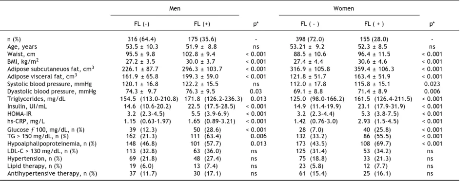
vs. 53.1%, p < 0.001 in men and 86.7 vs. 45.2%, p < 0.001 in women), in participants with and with-out FL, although significantly higher in subjects of both genders with intra-hepatic fat excess. Com-pared to subjects with non FL, all adiposity meas-urements were significantly higher in the subjects with FL (Table 1).
Overall, FL was associated with a more adverse metabolic profile (Table 1), characterized in both genders by higher blood pressure, triglycerides, in-sulin, HOMA-IR and hs-CRP values, as well as by significantly higher prevalence of impaired fasting glucose, increased triglycerides and low HDL-C. No significant differences were observed in age or the prevalence of increased LDL-C, hypertension, and the percentage of subjects under pharmacological treatment for these conditions in both groups. Fig-ure 1 shows that compared to subjects without risk factors, subjects with FL, AVF ≥ P75 or HOMA-IR ≥
P75, particularly those of female gender, had a high-er prevalence of hs-CRP ≥ 3 mg/L. The independence of the associations of FL, AVF ≥ P75 and HOMA-IR
gen-661
Fatty liver and abdominal fat relationships with high C-reactive protein.
, 2015; 14 (5): 658-665
Table 2. Association between fatty liver, adipose visceral fat ≥ P75 and HOMA-IR ≥ P75 with the natural logaritm of hs-CRP in multivariate linear regression analyses.
Fatty liver AVF ≥ P75 HOMA-IR ≥ P75
β coefficients (95%CI) β coefficients (95%CI) β coefficients (95%CI)
Men
Model 1 0.21 (0.12 - 0.30) 0.20 (0.12 - 0.30) 0.16 (0.07- 0.25)
Model 2 0.19 (0.10 - 0.30) 0.16 (0.60 - 0.30) 0.09 (-0.01 -0.20)
Model 3 0.17 (0.06 - 0.30) 0.12 (0.02 - 0.20) 0.01 (-0.09 - 0.13)
Women
Model 1 0.27 (0.20 - 0.40) 0.31 (0.25 - 0.43) 0.26 (0.19 - 0.30)
Model 2 0.19 (0.10 - 0.30) 0.25 (0.17- 0.38) 0.18 (0.08 - 0.30)
Model 3 0.13 (0.04 - 0.30) 0.19 (0.10 - 0.32) 0.08 (-0.02 - 0.21)
AVF: adipose visceral fat. HOMA-IR: homeostasis model assessment of insulin resistance. Model 1: unadjusted. Model 2: ajusted by age, hypertension, glucose. HDL-C: high density lipoprotein cho-lesterol, triglycerides, total kilocalories and physical activity. Model 3: model 2 plus AVF and HOMA-IR.
FL (-) FL (+) p* FL ( - ) FL ( + ) p*
n (%) 316 (64.4) 175 (35.6) - 398 (72.0) 155 (28.0)
-Age, years 53.5 ± 10.3 51.9 ± 8.8 ns 53.21 ± 9.2 52.3 ± 8.5 ns
Waist, cm 95.5 ± 9.8 102.8 ± 9.4 < 0.001 88.5 ± 10.6 96.4 ± 11.5 < 0.001
BMI, kg/m2 27.2 ± 3.5 30.0 ± 3.7 < 0.001 27.4 ± 4.4 30.6 ± 4.6 < 0.001
Adipose subcutaneuos fat, cm3 226.1 ± 87.7 296.3 ± 103.7 < 0.001 316.9 ± 105.8 359.4 ± 106.3 < 0.001 Adipose visceral fat, cm3 161.9 ± 65.8 199.3 ± 59.0 < 0.001 121.8 ± 51.7 163.4 ± 51.9 < 0.001
Systolic blood pressure, mmHg 120.1 ± 16.8 122.2 ± 15.5 ns 112.0 ± 17.8 115.8 ± 15.1 0.023
Dyastolic blood pressure, mmHg 74.3 ± 9.7 76.3 ± 9.5 0.03 69.1 ± 8.8 71.4 ± 8.9 0.006
Triglycerides, mg/dL 154.5 (113.0-210.8) 171.8 (126.2-236.3) 0.013 125.0 (98.0-166.2) 161.5 (126.4-211.5) < 0.001 Insulin, UI/mL 14.6 (10.6-20.2) 22.5 (17.5-28.5) < 0.001 14.9 (11.4-19.9) 23.1 (17.9-31.9) < 0.001
HOMA-IR 3.2 (2.3-4.5) 5.5 (3.9-6.9) < 0.001 3.2 (2.3-4.4) 5.3 (3.8-7.5) < 0.001
hs-CRP, mg/L 1.15 (0.63-1.97) 1.65 (0.89-3.21) < 0.001 1.42 (0.76-3.0) 2.93 (1.5-4.5) < 0.001
Glucose ≥ 100, mg/dL, n (%) 39 (12.3) 50 (28.6) < 0.001 28 (7.0) 40 (25.8) < 0.001
TG > 150 mg/dL, n (%) 162 (21.3) 111 (63.4) 0.006 132 (33.2) 86 (55.5) < 0.001
Hypoalphalipoproteinemia, n (%) 148 (46.8) 101 (57.7) 0.013 173 (43.5) 108 (69.7) < 0.001
LDL-C > 130 mg/dL, n (%) 113 (32.8) 63 (36.0) ns 125 (31.4) 53 (34.2) ns
Hypertension, n (%) 69 (21.8) 48 (27.4) ns 75 (18.8) 33 (21.3) ns
Lipid therapy, n (%) 19 (6.0) 13 (7.4) ns 23 (5.8) 12 (7.7) ns
Antihypertensive therapy, n (%) 37 (11.7) 30 (17.1) ns 61 (15.4) 25 (16.1) ns
Cardoso-Saldaña GC, et al. , 2015; 14 (5): 658-665
662
der using unadjusted and adjusted models (Table 2). All three factors were related to increased hs-CRP concentrations in both genders for model 1. After adjustment for traditional risk factors, model 2, the association of hs-CRP with FL and AVF ≥ P75 re-mained significant whereas the significance of the HOMA-IR association was lost in men. In model 3, the analysis of the association of ln hs-CRP with FL was further adjusted for AVF ≥ P75 and HOMA-IR ≥
P75, while the association of the protein with AVF ≥
P75 was adjusted for FL and HOMA-IR ≥ P75. In this full adjusted model, the presence of FL was inde-pendently and significantly associated with an in-crease in ln hs-CRP concentrations in men (β [95% CI]: 0.17 [0.06-0.30]; p < 0.001) which correspond to a 21% higher average hs-CRP level, and in wom-en (β [95% CI]: 0.13 [0.04-0.30]; p < 0.01) which correspond to a 17% higher average hs-CRP level. The AVF ≥ P75 association with ln hs-CRP in both genders was also significant and independent;
how-Figure 1. Prevalence of hs-CRP ≥ 3 mg/L in subjects with-out (-) or with (+) fatty liver (FL), adipose visceral fat (AVF) ≥ P75 (AVF: men = 151.5 cm3, women = 122 cm3) or HOMA-IR ≥ P75 (men = 3.38, women = 3.66).
60
50
40
30
20
10
0
% hs-CRP
≥
3 mg/L
(-) (+) (-) (+) (-) (+) (-) (+) (-) (+) (-) (+)
FL AVF HOMA-IR FL AVF HOMA-IR
≥ p75 ≥ p75 ≥ p75 ≥ p75
Men Women
Figure 2. Prevalence of hs-CRP (≥ 3 mg/L) associated with the presence of fatty liver, adipose visceral fat ≥ P75 (AVF: men = 151.5 cm3, women = 122 cm3) or HOMA-IR ≥ P
75 (men = 3.38, women = 3.66) and combination of two or three cardiometabolic risk factors. Trend in men p = 0.005, in women p < 0.001.
60
50
40
30
20
10
0
% hs-CRP
≥
3 mg/L
0 1 2 3
Number of cardiometabolic risk factors Men
Women
Table 3. Association of fatty liver, adipose visceral fat ≥ P75 and HOMA-IR ≥ P75, with high hs-CRP (≥ 3 mg/L) in multivariate logistic regression analyses.
Risk factors Men Women
(n)*
Unadjusted, OR (95% CI) Ajusted,† OR(95% CI) Unadjusted, OR(95% IC) Ajusted,† OR(95% IC)
0** Reference Reference Reference Reference
1 1.26 (0.5 - 3.0) 1.36 (0.5 - 3.86) 2.25 (1.27 - 3.96) 2.25 (1.20 - 4.2) 2 2.04 (0.92 - 4.5) 2.03 (0.75 - 5.5) 3.29 (1.93 - 5.61) 2.61 (1.4 - 4.9) 3 3.16 (1.44 - 6.43) 3.58 (1.32 - 9.7 ) 6.20 (3.54 - 10.9) 4.67 (2.3 - 9.4)
* Factors: fatty liver, abdominal visceral fat ≥ P75 and HOMA-IR ≥ P75. ** Without fatty liver, abdominal visceral fat ≥ P75, HOMA-IR ≥ P75. † Ajusted by age, hypertension, glucose. HDL-C: high density lipoprotein cholesterol, triglycerides, total kilocalories and physical activity.
ever, ln hs-CRP was more strongly associated in women than in men (p < 0.01) (Table 2).
Other analyses were conducted to individually and jointly examine the association of FL, AVF ≥
DISCUSSION
This study analyzes for the first time the influ-ence of AVF, FL and HOMA-IR on hs-CRP in Mexi-can mestizo population. Both adipose stores and HOMA-IR had an independent and additive effect on the inflammation degree. FL and increased AVF were associated with hs-CRP concentration in both men and women independently of traditional risk factors and insulin resistance. The effect of FL was more evident in men while AVF was in women. These results suggest that, independent of insulin resistance, both adipose stores can predict an in-creased systemic inflammation. The combination of FL, AVF ≥ P75 and HOMA-IR ≥ P75 was associated with a gradual increase in the risk of hs-CRP ≥ 3 mg/L, which was 3.58 and 4.67 times higher in men and women in which all three factors were present, respectively.
It has been previously reported that compared to obese subjects with no liver impairment, obese sub-jects with steatosis or steatohepatitis had increased hs-CRP and other markers of inflammation.5 Also,
serum C reactive protein was found associated with liver steatosis and visceral fat accumulation in type 2 diabetes mellitus patients but not in non-diabetic subjects.26 Another study in 2,388
Brazil-ian subjects, showed that hepatic steatosis, obesity, central obesity (with a crude estimation of waist circumference) and metabolic syndrome are inde-pendently and additionally associated with the like-lihood of having hs-CRP ≥ 3 mg/L.10 In a study of
Mexican subjects in which AVF was not deter-mined, those with FL also showed a significant in-crease in ultrasensitivity CRP, after adjustment for traditional cardiovascular risk factors.14 Our data
confirm and extend the findings from those studies by demonstrating that FL is associated with hs-CRP even after controlling for visceral abdominal adiposity and insulin resistance. Although increas-es visceral abdominal adiposity and insulin rincreas-esist- resist-ance are closely related to hepatic steatosis,27,28 the
observation that AVF and HOMA-IR are associat-ed, independently from one another and together with systemic inflammation, and that they do not affect the association of FL with hs-CRP, could be interpreted as evidence that insulin resistance and AVF contribute to the risk of systemic inflamma-tion by different but closely related pathophysiolog-ical mechanisms. Indeed previous evidence has shown that insulin-resistant adipose tissue plays a key role in the development of the metabolic and histological abnormalities present in obese subjects
with FL.29,30 In physiological conditions, the
adi-pose tissue is the main source of free fatty acids for the liver synthesis of triglycerides. However, when the adipose tissue is insulin-resistant, releases ex-cessive amounts of fatty acids that accumulate in the liver along with other compounds such as dia-cylglycerols and ceramides, which trigger proin-flammatory cytokine production.31,32 Oxidative
stress, produced by the lipotoxic effect related to the accumulation of saturated fatty acids in the liv-er, is another inductive factor of inflammation.33,34
Together, these abnormalities could explain the di-rect and independent link of FL with hs-CRP, and HOMA-IR observed in our study.
Women of Latin American descent have one of the highest average hs-CRP concentrations.35-37
Epidemi-ologic and clinical trials have consistently shown that compared to men, women have higher hs-CRP concentrations, which are unrelated to age, BMI, eth-nicity and hormone replacement therapy. The present study confirms that plasma hs-CRP concentration is higher in women than in men.38,39 The reason for
this gender-based difference is unknown but some plausible explanations have been suggested:
• Because hormone replacement therapy increases CRP concentrations,40 it is possible that
endog-enous estrogen stimulates CRP synthesis.
• The larger total body fat reservoir in women could raise the synthesis of proinflammatory cytokines such as IL-6, that leads to increased production of CRP by the liver.41
The higher levels of subcutaneous abdominal fat and hs-CRP we observed in women support the lat-ter hypothesis.
Study strengths
Cardoso-Saldaña GC, et al. , 2015; 14 (5): 658-665
664
method for differential quantification of the subcu-taneous and visceral central adiposity stores.18,19
Study limitations
Due to the cross sectional nature of the study, it is not possible to establish causal or temporal rela-tionships between cardiometabolic risk factors and increased hs-CRP. Nevertheless, the findings sug-gest that FL is not only a cardiovascular risk mark-er, but also a factor with atherogenic effect mediated by systemic inflammation. It has been suggested that at least two hs-CRP concentration measure-ments in metabolically stable subjects are needed to appropriately determine inflammatory status.42 In
this study hs-CRP was determined only once; how-ever a great care was taken to avoid sampling of subjects with acute or chronic inflammatory condi-tions. The diagnosis of FL was established by CAT scans, but was not confirmed by liver biopsies. How-ever, a significant correlation has been demonstrat-ed between the liver attenuation images on CAT and the histological grade of steatosis.43 Although
insu-lin resistance was not assessed by more sophisticat-ed approaches, the HOMA-IR index has proven to be a reliable measure of insulin sensitivity in non-dia-betics subjects.17
In conclusion, both steatosis and hs-CRP ≥ 3 mg/dL are present in 1 of 3 non-diabetic subjects free of clinical cardiovascular disease. FL, AVF ≥ P75 and HOMA-IR ≥ P75 are independently and additively as-sociated in both genders with approximately twice the risk of increased hs-CRP values. The gender-based differences in the association of FL and the cardiometabolic risk factors with the systemic inflammatory status, suggest that hs-CRP related cardiovascular risk is greater in women than men.
ABBREVIATIONS
• AVF: abdominal visceral fat.
• BMI: body mass index.
• CAD: coronary artery disease.
• CAT: computed axial tomography.
• FL: fatty liver.
• GEA: Genetics of Atherosclerotic Disease Study.
• HDL-C: high density lipoprotein cholesterol.
• HOMA-IR: homeostasis model assessment of
in-sulin resistance.
• hs-CRP: high sensitivity C reactive protein.
• LDL-C: low density lipoprotein cholesterol.
• SAF: subcutaneous abdominal fat.
• TG: triglycerides.
SUPPORTING
This study was supported by CONACYT grant No. SALUD 2010-2 150537.
ACKNOWLEDGEMENTS
We acknowledge to healthy volunteers and patients from the Instituto Nacional de Cardiología Ignacio Chávez, as well as the staff of the Endo-crinology Department.
REFERENCES
1. Thakur ML, Sharma S, Kumar A, Bhatt SP, Luthra K, Guleria R, Pandey RM, et al. Nonalcoholic fatty liver disease is as-sociated with subclinical atherosclerosis independent of obesity and metabolic syndrome in Asian Indians.
Athero-sclerosis 2012; 223: 507-11.
2. Brunt EM. Nonalcoholic steatohepatitis: definition and pa-thology. Semin Liver Dis 2001; 21: 3-16.
3. Bhatia LS, Curzen NP, Calder PC, Byrne CD. Non-alcoholic fatty liver disease: a new and important cardiovascular risk factor? Eur Hearth J 2012; 33: 1190-200.
4. Ampuero J and Romero-Gómez M. Influencia de la en-fermedad por hígado graso no alcohólico en la enen-fermedad cardiovascular. Gastroenterol Hepatol 2012; 35: 585-93. 5. Assy N, Djibre A, Farah R, Grosovski M and Marmor A.
Presence of coronary plaques in patients with nonalcohol-ic fatty liver disease. Radiology 2010; 254: 393-400. 6. Speliotes EK, Massaro JM, Udo Hoffmann U, Vasan RS,
Meigs JB, Sahani DV, Hirschhorn JN, et al. Fatty Liver is Associated With Dyslipidemia and Dysglycemia Independ-ent of Visceral Fat: The Framingham Heart Study.
Hepa-tology 2010; 51: 1979-87.
7. Fabbrini E, Magkos F, Mohammed BS, Pietka T, Abumrad NA, Patterson BW, Okunade A, et al. Intrahepatic fat, not visceral fat, is linked with metabolic complications of obesity. Proc Natl Acad Sci USA 2009; 106: 15430-5. 8. Liu J, Musani SK, Bidulescu A, Carr JJ, Wilson JG, Taylor HA,
Fox CS. Fatty liver, abdominal adipose tissue and athero-sclerotic calcification in African Americans: the Jackson Heart Study. Atherosclerosis 2012; 224: 521-5.
9. Sung KC, Wild SH, Kwag HJ, Byrne CD. Fatty liver, insulin resistance, and features of metabolic syndrome: relation-ship with coronary artery calcium in 10,153 people.
Diabe-tes Care 2012; 35: 2359-64.
10. Lavie CJ, Milani RV, Verma A, O’Keefe JH. C-reactive pro-tein and cardiovascular diseases—is it ready for prime-time? Am J Med Sci 2009; 338: 486-92.
11. Haukeland JW, Damas JK, Konopski Z, Løberg EM, Haaland T, Goverud I, Torjesen PA, et al. Systemic inflammation in nonalcoholic fatty liver disease is characterized by ele-vated levels of CCL2. J Hepatol 2006; 44: 1167-74. 12. Ndumele CE, Nasir K, Conceiçao RD, Carvalho JA,
Blumen-thal RS, Santos RD. Hepatic steatosis, obesity, and the metabolic syndrome are independently and additively as-sociated with increased systemic inflammation.
Athero-scler Thromb Vasc Biol 2011; 31: 1927-32.
13. Targher G, Bertolini L, Rodella S, Lippi G, Franchini M, Zop-pini G, Muggeoso M, et al. NASH Predicts plasma inflamma-tory biomarkers independently of visceral fat in men.
14. Lizardi-Cervera J. Association among C-reactive protein, fatty liver disease, and cardiovascular risk. Dig Dis Sci 2007; 52: 2375-9.
15. Sugiuchi H, Uji Y, Okabe H, Irie T, Uekama K, Kayahara N, Miyauchi K. Direct measurement of high-density lipopro-tein cholesterol in serum with polyethylene glycol-modified enzymes and sulfated alpha-cyclodextrin. Clin Chem 1995; 41: 717-23.
16. DeLong DM, DeLong ER, Wood PD, Lippel K, Rifkind BM. A comparison of methods for the estimation of plasma low-and very low-density lipoprotein cholesterol. The Lipid Re-search Clinics Prevalence Study. JAMA 1986; 256: 2372-7. 17. Matthews DR, Hosker JP, Rudenski AS, Naylor BA, Treach-er DF, TurnTreach-er RC. Homeostasis model assessment: insulin resistance and beta-cell function from fasting plasma glu-cose and insulin concentrations in man. Diabetologia 1985; 28: 412-9.
18. Sjöström L, Kvist H, Cederblad A, Tylén U. Determination of total adipose tissue and body fat in women by comput-ed tomography, 40K, and tritium. Am J Physiol 1986; 250: E736-E745.
19. Kvist H, Chowdhury B, Grangard U, Tylén U, Sjöström L. Total and visceral adipose-tissue volumes derived from measure-ments with computed tomography in adult men and women: predictive equations. Am J Clin Nutr 1988; 48: 1351-61. 20. Ricci C, Longo R, Gioulis E, Bosco M, Pollesello P, Massutti
F, Croce LS, et al. Non invasive in vivo quantitative as-sessment of fat content in human liver. J Hepatol 1997; 27: 108-13.
21. Piekarski J, Goldberg HI, Royal SA, Axel L, Moss AA. Differ-ence between liver and spleen CT numbers in the normal adult: its usefulness in predicting the presence of diffuse liver disease. Radiology 1980; 137: 727-9.
22. Sánchez-Castillo CP, Velázquez-Monroy O, Berber A, Lara-Esqueda A., Tapia-Conyer R, James WP. Encuesta Nacional de Salud (ENSA) 2000 Working Group. Anthropometric cut-off points for predicting chronic diseases in the Mexican National Health Survey 2000. Obes Res 2003; 11: 442-51. 23. Alberti KG, Eckel RH, Grundy SM, Zimmet PZ, Cleeman JI,
Do-nato KA, Fruchart JC, et al. Harmonizing the metabolic syn-drome: a joint interim statement of the International Diabetes Federation Task Force on Epidemiology and Preven-tion; National Heart, Lung, and Blood Institute; American Heart Association; World Heart Federation International Atherosclerosis Society; and International Association for the Study of Obesity. Circulation 2009; 120: 1640-5. 24. Pearson TA, Mensah GA, Alexander RW, Anderson JL,
Can-non RO 3rd, Criqui M, Fadl YY, et al. Markers of inflamma-tion and cardiovascular disease: applicainflamma-tion to clinical and public health practice: a statement for healthcare profes-sionals from the Centers for Disease Control and Preven-tion and the American Heart AssociaPreven-tion. CirculaPreven-tion 2003; 107: 499-511.
25. Baecke JA, Barema J, Frijters JE. A short questionnaire for the measurement of habitual physical activity in epi-demiological studies. Am J Clin Nutr 1982; 36: 936-42. 26. Iwasaky T, Nakajima A, Yoneda M, Terauchi Y.
Relation-ship between the serum concentrations of C-reactive pro-tein and parameters of adiposity and insulin resistance in patients with type 2 diabetes mellitus. Endocrine J 2006; 53: 345-56.
27. Lemieux I, Pascot A, Prud’homme D, Alméras N, Bogaty P, Nadeau A, Bergeron J, et al. Elevated C-reactive protein another component of the atherotrombotic profile of ab-dominal obesity. Atheroscler Thromb Vasc Biol 2001; 21: 961-7.
28. Sobhonslidsuk A, Jongjirasiri S, Thakkinstian A, Wisedopas N, Bunnag P, Puavilai G. Visceral fat and insulin resistance as predictor of non-alcoholic steatohepatitis. World J
Gastroenterol 2007; 13: 3614-8.
29. Cusi K. Role of insulin resistance and lipotoxicity in nonal-coholic steatohepatitis. Clin Liver Dis 2009; 13: 545-63. 30. Larter CZ, Chitturi S, Heydet D, Farrell GC. A fresh look at
NASH pathogenesis. Part 1: the metabolic movers. J
Gas-troenterol Hepatol 2010; 25: 672-90.
31. Kolak M, Westerbacka J, Velagapudi VR, Wagsäter D, Yetukuri L, Makkonen J, Rissanen A, et al. Adipose tissue inflammation and increased ceramide content character-ize subjects with high liver fat content independent of obesity. Diabetes 2007; 56: 1960-8.
32. Pagadala M, Kasumov T, McCullough AJ, Zein NN, Kirwan JP. Role of ceramides in nonalcoholic fatty liver disease.
Trends Endocrinol Metab 2012; 23: 365-71.
33. Fon Tacer K, Rozman D. Nonalcoholic Fatty liver disease: focus on lipoprotein and lipid deregulation. J Lipids 2011: 783976. doi: 10.1155/2011/783976.
34. Park S, Kim M, Paik JK, Jang YJ, Lee SH, Lee JH. Oxidative stress is associated with C-reactive protein in nondiabet-ic postmenopausal women, independent of obesity and in-sulin resistance. Clin Endocrinol (Oxf) 2013; 79: 65-70. 35. Lakoski SG, Gender and C-reactive protein: data from the
multiethnic study of atherosclerosis (MESA) cohort. Herat
J 2006;152: 593-8.
36. Onat A, Ceyhan K, Basar O, Erer B, Toprak S, Sansoy V. Meta-bolic syndrome: major impact on coronary risk in a population with low cholesterol levels-a prospective and cross-sectional evaluation. Atherosclerosis 2002; 165: 285-92.
37. Han TS, Sattar N, Williams K, Gonzalez-Villalpando C, Lean ME, Haffner SM. Prospective study of C-reactive protein in relation to the development of diabetes and metabolic syndrome in the Mexico City Diabetes Study. Diab Care 2002; 25: 2016-21.
38. Thorand B, Baumert J, Döring A, Herder C, Kolb H, Rath-mann W, Giani G, et al. Sex differences in the relation of body composition to markers of inflammation.
Atheroscle-rosis 2006; 184: 216-24.
39. Clark CR, Ridker PM, Ommerborn MJ, Huisingh CE, Coull B, Buring JE, Berkman LF. Cardiovascular inflammation in healthy women: multilevel associations with state-level prosperity, productivity and income inequality. BMC
Pub-lic Health 2012; 12: 211. doi: 10.1186/1471-2458-12-211.
40. Modena MG, Bursi F, Fantini G, Cagnacci A, Carbonieri A, Fortuna A, Rossi R. Effects of hormone replacement thera-py on C-reactive protein levels in healthy postmenopausal women: comparison between oral and transdermal admin-istration of estrogen. Am J Med 2002; 113: 331-4. 41. Rutter MK, Meigs JB, Sullivan LM, D’Agostino RB Sr, Wilson
PW. C-reactive protein, the metabolic syndrome, and pre-diction of cardiovascular events in the Framingham Off-spring Study. Circulation 2004;110: 380-5.
42. Pearson TA, Mensah GA, Alexander RW, Anderson JL, Can-non RO 3rd, Criqui M, Fadl YY, et al. Centers for Disease Control and Prevention; American Heart Association. Markers of inflammation and cardiovascular disease: appli-cation to clinical and public health practice: A statement for healthcare professionals from the Centers for Disease Control and Prevention and the American Heart Associa-tion. Circulation 2003; 107: 499-511.
43. Park SH, Kim KW, Lee SW, Yoon SE, Park SW, Ha HK, Lee MG, et al. Macrovesicular hepatic steatosis in living liver donors: use of CT for quantitative and qualitative assess-ment. Radiology 2006; 239: 105-12.