Otras secciones de este sitio:
☞ ☞ ☞ ☞
☞ Índice de este número
☞ ☞ ☞ ☞
☞ Más revistas
☞ ☞ ☞ ☞
☞ Búsqueda
Others sections in this web site:
☞ ☞ ☞ ☞
☞ Contents of this number
☞ ☞ ☞ ☞
☞ More journals
☞ ☞ ☞ ☞ ☞ Search
Article:
Intrahepatic cholestasis of pregnancy: Changes in maternal-fetal bile acid balance and improvement by ursodeoxycholic acid
Copyright © 2002: Mexican Association of Hepatology
ANNALS OF HEPATOLOGY
Number 1 January-March 2002
Volume 1
Annals of hepatology
1 Centro de Patogénese Molecular, Faculdade de Farmácia, University
of Lisbon, Portugal.
Abbreviations: ICP, intrahepatic cholestasis of pregnancy; UDCA, ursodeoxycholic acid.
Address for correspondence:
Dora Maria Tuna de Oliveira Brites, Ph.D., Centro de Patogénese Molecular, Faculdade de Farmácia, Av. das Forças Armadas, 1600-083 Lisboa, Portugal. Phone: 7946450; Fax: +351-21-7946491; e-mail: dbrites@ff.ul.pt.
Abstract
Intrahepatic cholestasis of pregnancy (ICP) is a dis-ease characterized by generalized pruritus and bio-chemical cholestasis that appears typically during the last trimester of gestation. The most predictive and accu-rate markers for diagnosis and follow-up of ICP are increased total bile acid levels (above 11,0 µµµµµmol/L), en-hanced cholic acid percentage (above 42%) and de-creased glycine/taurine bile acid ratio (below 1.0). Al-though essentially benign for the mother, evidence as-sociates ICP with fetal poor prognosis resulting from increased transfer of bile acids from mother to fetus, who showed reduced ability to eliminate bile acids across the placenta. Those conditions lead to an accu-mulation of bile acids in the cord blood serum, meconi-um and amniotic fluid that may account for a dimin-ished fetal well-being and sudden intra-uterine death by ICP. Ursodeoxycholic acid (UDCA) treatment was shown to reduce the bile acid content in the fetal com-partment, while restoring the ability of the placenta to carry out vectorial transfer of these compounds to-wards the mother, decreasing bile acid levels in mater-nal serum and its passage to the fetus. In addition, UDCA administered to the mother also lowers the amount of bile acids present in colostrum without ei-ther increasing the UDCA concentration or causing major changes in lithocholic acid levels, further sup-porting the safety of UDCA in late pregnancy. There-fore, it is tempting to indicate UDCA as a first choice therapy for ICP as much as relevant aspects of fetal outcome may also be improved. This review focuses on the altered bile acid profiles in maternal and fetal compartments during ICP and its recovery by UDCA administration. Further elucidation of the precise mech-anisms of action of UDCA and its therapeutic potential
in improving fetal prognosis could result in the ap-proval of UDCA for ICP treatment.
Key words: Cholestasis, Pregnancy, Ursodeoxycholic acid, Bile acid.
Introduction
Cholestasis, whether resulting from hereditary or ac-quired liver diseases, is one of the most common manifes-tations of impaired bile flow. Intrahepatic cholestasis of pregnancy (ICP) is a condition of unknown etiology char-acterized by generalized pruritus and biochemical cho-lestasis, which occurs predominantly during the last tri-mester of pregnancy.1,2 However, appearance of ICP as early as the 10th week of pregnancy has been reported.3 Pruritus generally starts in palms and soles. A mild jaun-dice may be noticed in 20% of patients after the onset of itching4 while a slight elevation in the concentration of conjugated bilirubin (17.1 ± 1.7 µmol/L) is observed in 90% of ICP women.5 Among serum liver enzymes, alan-ine aminotransferase is the most sensitive test,6,7 with 2 to 10 times increased values8 and its efficiency was referred to be over 90%,5 while γ-glutamiltranspeptidase is the least elevated. A more specific biochemical parameter of ICP is the rise of serum bile acids1,6,9 with levels com-prised between 12.4 and 219.4 µmol/L5 as compared to the upper normal limit in late gestation of 11 µmol/L.10 Shortly after delivery, symptoms disappear and biochemi-cal parameters of liver cell function return to normal.11
The incidence of ICP is high in Chile (14%)4 and Boli-via (9.2%),12 but less common in Europe where preva-lence rates of 1 to 1.5% have been described for Portugal, Sweden, Poland and Finland.12,13 Prevalence is higher in twin than in single pregnancies and several clinical and experimental observations evidenced a primary role of es-trogens12,14,15 and progesterones7,16,17 in ICP. Genetic pre-disposition also plays a key role in the pathogenesis of ICP and incidence increases within the same family.4 Cur-rent research indicates that mutations of the MDR3 gene encoding the canalicular phosphatidylcholine translocase (Figure 1) may in some cases predispose to ICP,18 justi-fying the raised γ-glutamiltranspeptidase in 20 to 40% of the patients.19,20 However, other characteristics of ICP Concise Review
Intrahepatic cholestasis of pregnancy:
Changes in maternal-fetal bile acid balance
and improvement by ursodeoxycholic acid
D Brites. / Intrahepatic cholestasis of pregnancy 21
edigraphic.com
suggest that exogenous factors may superimpose on the hormonal and genetic factors. This is indicated by seasonal changes in the appearance of the disease, with a higher number of cases in January, pointing to a greater incidence during the winter.5,21 Recent studies also link ICP to low serum selenium levels.22,23 In conclusion, ICP seems to result from combined and multivariate effects.
Although essentially benign to the mother, quality of life can be impaired due to itching, and urinary tract infec-tion, postpartum hemorrhage, and fat and vitamin K mal-absorption are common complications during ICP.11,24,25 The most serious consequences of ICP are increased fetal distress, premature deliveries and perinatal mortality and morbidity24,26,27 (Table I). Meconium staining of amniotic fluid is considered a sign of poor prognosis for the fe-tus.27,28 Interruption of pregnancy is considered mandatory and urgent, mainly in an already mature fetus, when fetal distress is detected and there is risk of sudden death in utero.4 Therefore, ICP should be considered a high-risk condition, and an early and accurate identification of high-risk pregnancies together with an appropriate med-ical intervention might improve fetal outcome.
Several treatments, such as cholestyramine, phenobarbi-tal, dexamethasone, S-adenosyl-L-methionine, and epome-diol failed to improve pruritus, biochemical abnormalities, or fetal prognosis during ICP.29-35 The most efficacious cur-rent medical management that improves maternal condition and might prevent the perinatal complications of ICP is ursodeoxycholic acid (UDCA) administration.36-42 Thus, as soon as ICP is diagnosed, UDCA administration coupled with close maternal-fetal surveillance is indicated.
Bile acids in fetal pathophysiology
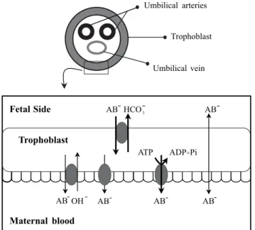
Bile acids easily go through the placenta to fetal com-partments and also to the amniotic fluid. Usually, there are higher concentrations of bile acids in fetal than in mater-nal serum and the main transfer of bile acids across the placenta occurs towards the mother43,44 (Figure 2). B. Dur-ing ICP, in addition to the increased levels of bile acids, the efficiency of the ATP-independent transport mech-anisms is enhanced (Figure 2). Provided they are not uni-directional, both changes may facilitate the passage of bile acids from the mother to the fetus, and hence counterbal-ance or even overcome bile acid flux across the placenta in the physiological direction, which is mediated in part by ATP-independent systems and whose efficiency is re-duced during ICP.45 Furthermore, the same authors have shown that vectorial bile acid transfer from fetus-to-moth-er is also impaired, thus contributing to an accumulation of bile acids in the fetal compartment. This accumulation may be associated with the occurrence of increased fetal distress by ICP. Actually, Laatikainen and Tulenheimo46 found a correlation between total serum bile acids and the incidence of meconium and fetal distress. In contrast, others were unable to find a direct correlation between bile acids in any compartment and fetal distress.47 Never-theless, total bile acids in maternal serum greater than 50 µmol/L, or superior to 25 µmol/L in fetal cord serum, are associated with diminished well-being47 and levels partic-ularly elevated are referred to cause sudden fetal death.7
Autopsy specimens in cases of intrauterine fetal loss from ICP are consistent with death from acute intrauterine anoxia.27 Meconium and bile acids, especially cholic acid, have been indicated to induce vasoconstriction of human placental chorionic veins in vitro,48 as well as causing a-cute umbilical vein constriction.49,50 Thus, there is some experimental evidence that bile acids are implicated in the mechanisms triggering fetal asphyxia in pregnancies com-plicated by ICP. In addition, it was recently shown that taurocholate (0.3 and 3 mM), the main bile acid during ICP, causes a decrease in the rate of contraction of rat car-diomyocytes and loss of synchronous beating.51 This data corroborates a direct role of bile acids in the sudden intra-uterine death by ICP.
Therefore, the decrease in bile acid levels induced by UDCA therapy in ICP patients, besides reversing maternal symptoms, may also improve fetal outcome. Palma et al.52 confirmed that patients who received UDCA had had their deliveries closer to term and less frequent fetal distress as compared to non-treated patients.
Relevance of UDCA as a therapeutic option for ICP
During the three last decades, several clinical studies have established UDCA as a promising therapeutic option in a variety of cholestatic liver diseases, including prima-Figure 1. Hepatocanalicular transport systems. This schematic
represen-tation shows two adjacent hepatocytes comprising a bile canaliculus. Bile salts are excreted into the bile via a bile salt export pump (BSEP). The canalicular conjugate export pump (MRP2) appears to be responsi-ble for ATP-dependent elimination of amphipathic anionic conjugates and reduced glutathione (GSH). The MDR1 gene product (MDR1) is responsible for elimination of hydrophobic cationic compounds while
MDR3 P-glycoprotein (MDR3) mediates canalicular secretion of
ry biliary cirrhosis53 benign recurrent intrahepatic choles-tasis54 and progressive familial intrahepatic cholestasis;55 improvements in clinical symptoms and biochemical indi-ces of liver function consistently occur following UDCA administration. Although not fully understood, the mech-anisms of action of UDCA include protection against in-jury of bile ducts by hydrophobic bile acids,12 replacement of hepatotoxic bile acids,56 immune modulation,57 cyto-protective mechanisms by preventing apoptosis,58,59 chole-retic activity and stimulated secretion of potentially hep-atotoxic compounds by the liver.60 It also inhibits intestinal absorption of more cytotoxic bile acids.61,62 It was proposed that the mechanisms of action of UDCA in ICP were sim-ilar to those observed in other cholestatic liver diseases but protection against estrogen-induced cholestasis in rats couldn’t be attributed to choleretic activity or lipid mem-brane protection.63 Beneficial effect may be partly due to stimulation of bile secretory function, as also occurs with sulphated progesterone metabolites in ICP women during UDCA therapy.64 Recently, it was proposed that taurine-conjugated UDCA stimulates organic anion secretion of cholestatic hepatocytes by inducing hepatobiliary ex-ocytosis and insertion of the transport protein MRP2 in the canalicular membrane65 (Figure 1). In addition to the beneficial effect on the functionality of the maternal liver, UDCA therapy restores the ability of the placenta to carry out vectorial bile acid transfer,45 thus contributing to pre-vent an excessive accumulation of bile acids in the fetal compartment during ICP.
This highly hydrophilic bile acid reduced pruritus, ami-notransferases, and serum levels of total bile acids when administered to patients with ICP.3,36,66,67 Patients usually receive UDCA at oral applications of 450 mg/day,12,66 12-16 mg/kg/day36,64 or 1.5 to 2 g/day (20-25 mg/kg/day),42 in three or four daily divided doses. The drug is well tolera-ted and seems to be completely safe for the mothers or their babies.42,52 Pregnancy outcome and perinatal progno-sis were improved in ICP patients treated with UDCA compared to placebo.41,52
UDCA normalizes bile acid profile in maternal serum
The most specific and sensitive biochemical test of ICP is serum bile acid levels, which may reach values 100 times above normal.1 Concentrations of total bile acids in normal pregnant women are consistently lower than 11.0 µmol/L,10 while values from 12.3 to 219.4 µmol/L5 or even 290 mmol/L7 are encountered in women with ICP. Evaluation of serum total bile acids is recommended, not only to diagnose but also to monitor ICP.1,7,9,68 The upper reference limits for both cholic and chenodeoxycholic acids in healthy pregnant women vary from 1.5 µmol/L6 to 4.2 µmol/L.5 In contrast, cholic acid values may increase up to 176 and 170 µmol/L5 during ICP, while chenodeoxy-cholic acid represent 3 to 4 times less (Table II). Tauro-cholic acid is the predominant species presenting values between 2.4 and 119.8 µmol/L, accounting for 38.1 ± 1.9% of the total bile acids as compared to 23.6 ± 1.4% for glycocholic acid (concentrations from 2.0 to 55.8 µmol/L).5 This shift towards a more extensive conjugation with taurine (glycine/taurine bile acid ratio of 0.8 ± 0.1, as compared to 1.4 ± 0.1 in normal pregnancies) is pointed as a marker of ICP.5,69 In healthy women, either pregnant or not, cholic acid is never higher than 45% of serum total bile acids, but accounts for 60 to 70% during cholesta-sis.5,70 Therefore cholic/chenodeoxycholic acid ratio is always greater than one.6,16,33,67,71
As previously noticed the beneficial effect of UDCA therapy may be associated with its ability to promote chang-es in the hydrophobic-hydrophilic balance of the bile acid pool, by increasing hydrophilicity.72 In a study with 15 ICP patients treated with UDCA (14 mg/kg/day), the concentra-tion of total bile acids decreased significantly (P<0.01) from 68.4 ± 16.1 µmol/L at baseline to 20.8 ± 5.1 µmol/L during therapy,67 in agreement with reports by others.3,37,52,66 Individually, the most significant alteration is the reduction (P<0.01) in serum cholic acid concentration from 45.9 ± 11.4 µmol/L at baseline to 6.4 ± 1.8 µmol/L with UDCA administration (67) (Table II). The decrease in cholic acid Table I. Perinatal outcome in both control and cholestatic pregnancies.
Control (25) Control* (28) ICP (13) ICP (25) ICP (27) ICP (28)
(n=42) (n=79) (n=117) (n=44) (n=83) (n=79)
Spontaneous labor 3(7.9%) 37(46.8) 17(38.6%) 27(33%) 60(75.9)
Fetal distress 1 (1.3%) 22(18.8%) 12(14%) 6 (7.6%)
Preterm delivery 6 (7.6%) 4 (3.4%) 11 (14%)
Small for gestational age 7(15.8%) 3 (3.8%) 2(1.7%) 8(18.4%) 6 (7.6%)
Meconium passage 6 (7.6%) 19(16.2%) 37(45%) 35 (44.3%)
Stillbirth 0 0 1 2 2
Neonatal death 0 0 2 1 0
Low birth weight 3(7.9%) 14(31.6%)
(<2500 g)
D Brites. / Intrahepatic cholestasis of pregnancy 23
edigraphic.com
may be clinically relevant since it has been reported to cause fetal distress,48,73 as previously mentioned.
This change in the composition of the bile acid pool is accompanied by a significant increase in the concentration and proportion of UDCA from 0.6 ± 0.2 µmol/L and 1.4 ± 0.6% to 5.9 ± 1.9 µmol/L and 24.7 ± 2.3%, respectively, at baseline and during treatment.67 These results resem-bled those of 0.1-4.8 µmol/L obtained in another set of patients under similar dosage (12-16 mg/kg/day).64 Howev-er, serum conjugated UDCA levels may reach concentra-tions as high as 16.5 ± 1.8 µmol/L when dosages of 1.5 to 2g/day are used.42 Experimental studies have not shown any teratogenic or adverse effects of UDCA on pre- or post-natal development in rats.40,74
Only a small proportion of total bile acids is detected in the unconjugated fraction,70 even during UDCA admin-istration, despite its increase from 7.2 ± 1.3% at baseline to 12.8 ± 1.6% during treatment.67 Since UDCA may be converted to lithocholic acid, known to induce growth re-tardation and malformations in rat embryos,75 some con-cern has been expressed regarding its safety in pregnancy
and the treatment is contraindicated in France.7 Neverthe-less, data has shown that the serum lithocholic acid con-centration is maintained during UDCA administration (1.7 ± 0.5 µmol/L at baseline, and 1.2 ± 0.2 µmol/L during therapy), despite an increase in its proportion (7.4 ± 1.3% vs 3.3 ± 0.5%, P<0.01).67 Therefore, although under con-tinued evaluation, UDCA might be the first-line therapy for ICP.
Passage of bile acids into colostrum decreases follo-wing UDCA administration
Infant exposure to high levels of bile acids may be partic-ularly risky, since enterohepatic circulation is immature at birth,76 ileal and hepatocyte transport mechanisms are im-paired, and serum bile acid concentrations are increased,77-79 conditions that may aggravate neonatal complications in ICP. Excretion of bile acids in colostrum is enhanced follow-ing ICP. In fact, when bile acid excretion in colostrum col-lected from 16 lacting ICP women, within 72 h postpartum was compared to five lacting healthy women, elevation of total bile acids levels (23.3 ± 14.8 µmol/L vs 0.7 ± 0.2 µmol/L, P<0.01) and of cholic acid concentrations (19.0 ± 13.1 µmol/L vs 0.6 ± 0.2 µmol/L, P<0.01) were found (Ta-ble II).80 The low levels of cholic acid in colostrum from control lacting women are similar to those reported in other studies, even for breast milk.81,82 Cholic acid predominates either in colostrum from normal or ICP lacting women.80,81 It is worthwhile to mention, however, that values of total bile acids as higher as 100 µmol/L may arise in colostrum of ICP patients and be absorbed by breast-feeding infants.80 Therapy diminishes the excretion of total bile acids in co-lostrum to 5.7 ± 2.5 µmol/L and cholic acid is the most re-duced one (3.6 ± 1.5 µmol/L). Accumulations of UDCA (0.3 ± 0.2 µmol/L) and lithocholic acid (0.01 ± 0.01 µmol/ L) in colostrum are irrelevant and toxicity is not expected to occur in nursing infants.
Altered patterns of bile acids in meconium are un-changed by UDCA therapy
Data on meconium bile acid composition in neonates from ICP patients is scant. In a recent study it was report-ed a considerable meconium elevation of total bile acids (13.5 ± 5.1 µmol/g vs 2.0 ± 0.5 µmol/g)and cholic acid (8.4 ± 4.1 µmol/g vs 0.8 ± 0.3 µmol/g)83 in newborns from women with ICP, indicating placental transfer of these compounds from mother-to-fetus (Table II). This is rein-forced by the presence of deoxycholic and lithocholic acids in meconium, which are supposed to be of maternal origin since they are products of bacterial metabolism and fetal colon is sterile. Thus, these secondary bile acids must reach the fetus by transfer across the placenta.84,85 Continued ingestion by the fetus of amniotic fluid may also contribute to the increase of bile acids in meconium, due to its high content in cholic acid.6,47,86
Figure 2. Transport systems of bile acids across the trophoblast
In addition to cholic, chenodeoxycholic, deoxycholic and lithocholic acids, significant amounts of unusual bile acids were equally identified,83,87,88 resembling composi-tion of fetal gallbladder bile.89,90 Their increased levels in meconium during ICP, besides reflecting hepatic immatu-rity and/or activation of liver enzymes less important in later life, may be caused by swallow of amniotic fluid en-riched in bile acids as a consequence of ICP. Since the presence of these unusual bile acids is scarce in the mater-nal blood, it is assumed that they derive from the fetus.91 Interestingly, during ICP there is a decrease in the rate of the chenodeoxycholic acid (from about 30% to less than 20%) and in that of the hyocholic acid (from 17% to 7%),83 which is the predominant unusual bile acid in me-conium.87,92
In contrast to the preferential conjugation with taurine in normal meconium,92,93 an equal representation of gly-cine and taurine conjugates occur in ICP, which may be due to a restrained maternal-to-fetus transport across the placenta for taurine conjugates.83 However, this finding does not take place in amniotic fluid or blood cord serum (see below). Regarding the sulphate conjugated fraction, and similarly to normal pregnancies, it also represents a large proportion of the total bile acids in meconium of ba-bies from ICP women.83,94
Most importantly, UDCA administration appears not to influence major bile acid concentrations in meconium in-cluding that of its toxic metabolite, lithocholic acid. Cho-lic acid percentage, instead of decreasing, is slightly in-creased in meconium (79.2 ± 4.1% vs 74.0% ± 4.4%), fol-lowing UDCA therapy.83 The accumulation of cholic acid
in meconium may result both from the inability of the fe-tus to excrete meconium (in normal conditions) and the diminished intestinal absorption, greatly reducing the ben-eficial effects of the therapy at this level. Nevertheless, it must be emphasized that in order to restrain the placental passage of bile acids and the accumulation of these com-pounds in meconium, it is important to decrease bile acid levels in maternal serum and thus UDCA treatment must be initiated as soon as the diagnosis of ICP is made.
UDCA reduces the levels of bile acids in amniotic fluid
Bile acids in amniotic fluid may originate from the mother, the fetus, or both.86 Levels of primary bile acids are dramatically elevated in the amniotic fluid during ICP, indicating the maternal compartment as the first source. In addition, the presence of unusual bile acids, from which the more abundant are polyhydroxylated bile acids, sug-gest that fetal liver under conditions of higher bile acid level attempts to excrete bile acids into urine by increasing their polarity.91
Whether bile acids reach the amniotic fluid by diffu-sion through umbilical cord or placental membranes or by fetal urine and meconium remains to be established. Inter-estingly, both cholic and chenodeoxycholic acids are more elevated in the amniotic fluid from normal pregnan-cies than in the maternal serum.47
During ICP, cholic acid elevation is the main contrib-utor to the increased content of amniotic fluid in primary bile acids (Table II) with mean values from 12.5 ± 4.7 Table II. Concentrations of primary bile acids in maternal serum, amniotic fluid, umbilical cord serum, colostrum and meconium from both control and
cholestatic pregnant women and their babies, when untreated or treated with ursodeoxycholic acid.
Control Intrahepatic cholestasis of pregnancy
Untreated‡ (42) Treated‡** (42) Untreated Treated
Maternal serum (at diagnosis) Cholic
Quenodeoxycholic Maternal serum (at delivery)
Cholic
Quenodeoxycholic Amniotic fluid
Cholic
Quenodeoxycholic Umbilical cord serum
Cholic Quenodeoxycholic Colostrum Cholic Quenodeoxycholic Meconium Cholic Quenodeoxycholic
Data are µmol/g for meconium and µmol/L for the other biological fluids; **, ursodeoxycholic acid at 1.5 to 2 g/day; ‡, relative to bile acid conjugated species; *, Brites D, unpublished data. §P<0.10, ¥P<0.05 and †P<0.01 from control; ƒP<0.05 and £P<0.01 from untreated patients; ºP<0.01 from patients at diagnosis. References are indicated in parentheses.
(n=38)* 2.2 ± 0.2 2.2 ± 0.1 (n=17)* 3.9 ± 1.3 0.4 ± 0.2 (n=17)* 3.1 ± 0.8 3.3 ± 0.4 (n=5) (79) 0.6 ± 0.2 0.19 ± 0.02 (n=8) (83) 0.8 ± 0.3 0.3 ± 0.1
(n=10) 20.0 ± 3.1
5.6 ± 0.6 (n=10) 20.3 ± 2.3
5.4 ± 0.5 (n=9) 17.9 ± 27.5 18.5 ± 20.9
(n=9) 21.9 ± 5.6 18.9 ± 2.1
(n=20) 18.5 ± 1.9
5.8 ± 0.8 (n=20) 10.5 ± 1.9º£
3.0 ± 0.7º£ (n=15) 4.9 ± 12.4£
4.8 ±7.7£ (n=20) 6.0 ± 0.9£ 5.2 ± 0.95£
(n=39)* 42.7 ± 6.2† 11.3 ± 1.6† (n=9)* 12.5 ± 4.7§
1.9 ± 0.7 (n=9)* 14.0 ± 2.9¥
6.9 ± 3.7 (n=9) (79) 19.0 ± 13.1†
2.7 ± 2.1 (n=8) (83) 8.4 ± 4.1† 1.0 ± 0.3¥
(n=15) (67) 45.9 ± 11.4 14.7 ± 3.7 (n=15) (67)
6.4 ± 1.8º£ 4.8 ± 1.3ºƒ (n=17)* 8.2 ± 2.1¥
D Brites. / Intrahepatic cholestasis of pregnancy 25
edigraphic.com
µmol/L (95) to 49.9 ± 19.0 µmol/L,86 4 to 70 times higher than those in the controls (3.9 ± 1.3 µmol/L and 0.58 ± 0.17, respectively). Contribution of meconium contamina-tion may justify the highest values reported. It is possible that cholic acid is carried more easily than chenodeoxy-cholic acid to the amniotic fluid,47 although chenodeoxy-cholic, deoxycholic and even lithocholic acid concentra-tions also increase by 3- to 5-fold (former) by ICP.95
In a previous study (Brites D, unpublished data), treat-ment with UDCA did not induce any significant changes in concentration of all the major bile acids in amniotic flu-id, despite the decrease in cholic acid (12.5 ± 4.7 µmol/L to 8.2 ± 2.1 µmol/L) (Table II). UDCA concentration in the amniotic fluid enhanced significantly (4.5 ± 1.4 µmol/ L, after treatment; 0.49 ± 0.25 µmol/L, controls; 0.37 ± 0.13 µmol/L, ICP women; P<0.01). In addition, in an un-usual case of severe cholestasis of pregnancy,3 admin-istration of 1g/day UDCA originated an elevation in the proportion of UDCA that reached 30.4% of total bile ac-ids, whereas lithocholic acid did not surpass 4%. Total bile acid concentration in amniotic fluid of 27.3 µmol/L must represent a beneficial consequence of UDCA thera-py, since the patient presented at entrance 198.0 µmol/L of serum total bile acids. Other reports42 using dosages of 1.5 to 2g/day of UDCA obtained a significant reduction in conjugated cholic acid concentration from 17.9 ± 27.5 µmol/L in untreated patients to 4.9 ± 12.4 µmol/L in treat-ed ones (Table II). In contrast to previous works, the lev-el of 0.8 ± 2.4 µmol/L found for conjugated UDCA in amniotic fluid taken from patients treated with such high doses, immediately before or at the time of delivery, agrees with the existence of only 0.1 µmol/L obtained in another study38 after a week of treatment (600 mg daily in two divided doses) and indicate that accumulation of UDCA probably depends on the severity of each case. Additionally, cholic acid percentage that reveals to be similar in both control and non-treated patients (78.0 ± 4.1% and 76.9 ± 5.1%, respectively) shows a marked de-crease during therapy (56.2 ± 4.4%, P<0.01).96 The rise in chenodeoxycholic acid proportion from non-treated ICP patients relatively to control (14.1 ± 3.7% vs 6.8 ± 1.2%) seems to be adjusted by treatment (7.1 ± 0.7%). Litho-cholic acid proportion is maintained but, although not significant, 2-fold elevated concentrations are observed by UDCA therapy (0.51 ± 0.23 µmol/L vs 0.25 ± 0.08 µmol/L in non-treated ICP patients).95 The beneficial ef-fects expected from the decrease in cholic and chenodeox-ycholic acids and the increase in UDCA levels probably overcome the potential toxic effect due to the moderate elevation of lithocholic acid concentration.
Bile acid levels diminish in umbilical cord blood when UDCA is given to ICP women
Concentrations of total bile acids in umbilical cord serum of premature and term neonates range from slightly
to 2-3 times higher than in maternal blood69,89,97-99 and in-crease even more in fetus of an early gestational age.100 Primary bile acids predominate and the cholic/cheno-deoxycholic acid ratio is close to 1.0.71,97 The elevation of both cholic and chenodeoxycholic acids in the fetal blood as compared to maternal serum suggests immaturity of liv-er function during the fetal pliv-eriod,47 as seen during the first months after birth.100 On the other hand, the placenta also seems to maintain this concentration difference.47
Levels of primary bile acids increase up to 2 and 5 times in the cord blood serum during ICP, although elevation in maternal serum is even more notorious.47,97 Mean ratio between levels of cholic and chenodeoxycholic acids aug-ment to 2.097 reflecting, although to a less extent, the typ-ical elevation of this ratio in the ICP women. Serum total bile acids are greatly increased in babies from ICP com-plicated gestations (26.0 ± 12.2 µmol/L, P<0,01) when compared with values from those of healthy mothers (11.8 ± 4.7 µmol/L).101 Cholic acid levels of 14.0 ± 2.9 µmol/L obtained in the same study are close to the value of 21.9 ± 5.9 µmol/L reported for another group of 9 babies42 (Ta-ble II). The higher cholic acid proportion (51.8 ± 7.8%), in contrast with that of chenodeoxycholic acid (25.2 ± 8.3%), appears to result from its greater distribution in maternal serum, which is considered a characteristic fea-ture in ICP patients. The diminished glycine/taurine ratio found in the fetal bile acid pool (0.8 ± 0.2) also seems to be determined by an equivalent ratio in maternal serum.101 UDCA therapy induces an increase in the fetal serum proportion of this bile acid (from 1.9 ± 2.0% to 12.5 ± 8.4%, P<0.05), accompanied by a significant decrease in total bile acid concentration (14.8 ± 2.7 µmol/L, P<0.05), and in cholic acid proportion (39.4 ± 13.3%, P<0.05), and by a trend to normalization in the glycine/taurine ratio (1.2 ± 0.4, P<0.10).99 UDCA concentration increases from 0.57 µmol/L in control, and from 0.40 µmol/L in non-treated patients, to 1.83 µmol/L in ICP women receiving UDCA. In contrast, a more recent report42 indicates a low-er accumulation of conjugated UDCA (0.9 ± 0.14 µmol/ L), despite the high-dosage of 1.5 to 2.0 g/day used. The authors justify the differences based on an increase in the mother’s bile acid secretion by such high-dose UDCA treatment strategy. Both studies show that important im-provements are achieved for cholic and chenodeoxycholic acids (Table II). Therefore, improved fetal prognosis dur-ing maternal treatment with UDCA, in the course of ICP, is probably related with the UDCA capability to normal-ize the bile acid profile in the umbilical cord serum.
Conclusion
infants, respectively, with cholic acid representing the major species. Whether bile acids reach the fetus through umbilical cord or placental membranes, remains to be established. To the accumulation of bile acids in fetal serum, meconium and amniotic fluid contribute the enhanced transplancental systems of bile acid transport towards the fetus on one hand, and the reduced ability of the fetus to eliminate bile acids on the other hand. UDCA therapy restores the maternal-fetal bile acid bal-ance during ICP, is not harmful to the fetus and above all may hamper stillbirths and preterm labors. It is hoped that research in this field yields insights that will result in the approval of UDCA as the first line treatment op-tion for ICP patients.
Acknowledgements: The author wishes to express his appreciation to Drs. Joaquina Poeiras, Nuno Oliveira, M Conceição Cardoso, Cecília MP Rodrigues, JJG Marín, MA Serrano, MY El-Mir, Helena van-Zeller, Alexandra Brito and Rui Silva who participated in the realization of the publications in which the findings are described in this review. Studies from the author’s research Unit were fund-ed by Fundação para a Ciência e Tecnologia (Portugal).
Bibliografía
1. Heikkinen J, Mäentausta O, Ylöstalo P, Jänne O. Changes in serum bile acids concentrations during normal pregnancy, in patients with intrahepatic cholestasis of pregnancy and in pregnant women with itching. Br J Obstet Gynaecol 1981; 88: 240-5.
2. Reyes H. The enigma of intrahepatic cholestasis of pregnancy: lessons from Chile. Hepatology 1982; 2: 87-96.
3. Brites D, Rodrigues CM, Cardoso MC, Graça LM. Unusual case of severe cholestasis of pregnancy with early onset, improved by ursodeoxycholic acid administration. Eur J Obstet Gynecol Reprod Biol 1998; 76: 165-8. 4. Reyes H. The spectrum of liver and gastrointestinal disease seen in cholestasis of pregnancy. In: Riely CA, Abell TL, eds.
Gastroenterology Clinics of North America. Gastrointestinal and Liver
Problems in Pregnancy. Philadelphia: WB Saunders, 1992: 905-21. 5. Brites D, Rodrigues CM, van-Zeller H, Brito A, Silva R. Relevance of serum bile acid profile in the diagnosis of intrahepatic cholestasis of pregnancy in an high incidence area: Portugal. Eur J Obstet Gynecol
Reprod Biol 1998; 80: 31-8.
6. Heikkinen J. Serum bile acids in the early diagnosis of intrahepatic cholestasis of pregnancy. Obstet Gynecol 1983; 61: 581-7.
7. Bacq Y, Sapey T, Brechot MC, Pierre F, Fignon A, Dubois F.
Intrahepatic cholestasis of pregnancy: a French prospective study.
Hepatology 1997; 26: 358-64.
8. Davidson KM. Intrahepatic cholestasis of pregnancy. Semin Perinatol 1998; 22: 104-11.
9. Shaw D, Frohlich J, Wittmann BAK, Willms M. A prospective study of 18 patients with cholestasis of pregnancy. Am J Obstet Gynecol 1982; 142: 621-5.
10. Carter J. Serum bile acids in normal pregnancy. Br J Obstet Gynaecol 1991; 98: 540-3.
11. Van Dyke RW. The liver in pregnancy. In: Zakim D, Boyer TD, eds.
Hepatology. A Textbook of Liver Disease. Volume 2. Philadelphia:
WB Saunders, 1990: 1438-59.
12. Lammert F, Marschall HU, Glantz A, Matern S. Intrahepatic cholestasis of pregnancy: molecular pathogenesis, diagnosis and management. J Hepatol 2000; 33: 1012-21.
13. Läatikainen T, Tulenheimo A. Maternal serum bile acid levels and fetal distress in cholestasis of pregnancy. Int J Gynaecol Obstet 1984; 22: 91-4.
14. Davies MH, Ngong JM, Yucesoy M, Acharya SK, Mills CO, Weaver JB, Waring RH, Elias E. The adverse influence of pregnancy upon sulphation: a clue to the pathogenesis of intrahepatic cholestasis of pregnancy? J Hepatol 1994; 21: 1127-34.
15. Leslie KK, Reznikov L, Simon FR, Fennessey PV, Reyes H, Ribalta J. Estrogens in intrahepatic cholestasis of pregnancy. Obstet Gynecol 2000; 95: 372-6.
16. Meng LJ, Reyes H, Axelson M, Palma J, Hernandez I, Ribalta J, Sjovall J. Progesterone metabolites and bile acids in serum of patients with intrahepatic cholestasis of pregnancy: effect of ursodeoxycholic acid therapy. Hepatology 1997; 26: 1573-9.
17. Sjövall J, Reyes H. Bile acids and progesterone metabolites in intrahepatic cholestasis of pregnancy. Ann Med 2000; 32: 94-106. 18. Jacquemin E. Role of multidrug resistance 3 deficiency in pediatric
and adult liver disease: one gene for three diseases. Semin Liver Dis 2001; 21: 551-62.
19. Jacquemin E, Cresteil D, Manouvrier S, Boute O, Hadchouel M. Heterozygous non-sense mutation of the MDR3 gene in familial intrahepatic cholestasis of pregnancy. Lancet 1999; 353: 210-1. 20. Dixon PH, Weerasekera N, Linton KJ, Donaldson O, Chambers J,
Egginton E, Weaver J, et al. Heterozygous MDR3 missense mutation associated with intrahepatic cholestasis of pregnancy: evidence for a defect in protein trafficking. Hum Mol Genet 2000; 9: 1209-17. 21. Laatikainen T, Ikonen E. Fetal prognosis in obstetric hepatosis. Ann
Chir Gynecol Fenn 1975; 64: 155-64.
22. Kauppila A, Korpela H, Makila UM, Yrjanheikki E. Low serum selenium concentration and glutathione peroxidase activity in intrahepatic cholestasis of pregnancy. Br Med J 1987; 294: 150-2. 23. Reyes H, Baez ME, Gonzalez MC, Hernandez I, Palma J, Ribalta J,
Sandoval L, Zapata R. Selenium, zinc and copper plasma levels in intrahepatic cholestasis of pregnancy, in normal pregnancies and in healthy individuals, in Chile. J Hepatol 2000; 32: 542-9.
24. Reyes H, Gonzalez MC, Ribalta J, Aburto H, Matus C, Schramm G, Katz R, et al. Prevalence of intrahepatic cholestasis of pregnancy in Chile. Ann Intern Med 1978; 88: 487-93.
25. Johnston WG, Baskett TF. Obstetric cholestasis - a 14 year review.
Am J Obstet Gynecol 1979; 133: 299-301.
26. Reyes H, Radrigan ME, Gonzalez MC, Latorre R, Ribalta J, Segovia N, Alvarez C, et al. Steatorrhea in patients with intrahepatic cholestasis of pregnancy. Gastroenterology 1987; 93: 584-90.
27. Fisk NM, Storey GNB. Fetal outcome in obstetric cholestasis. Br J
Obstet Gynaecol 1988; 95: 1137-43.
28. Alsulyman OM, Ouzounian JG, Ames-Castro M, Goodwin TM. Intrahepatic cholestasis of pregnancy: perinatal outcome associated with expectant management. Am J Obstet Gynecol 1996; 175: 957-60. 29. Laatikainen T. Effect of cholestyramine and phenobarbital on pruritus
and serum bile acid levels in cholestasis of pregnancy. Am J Obstet
Gynecol 1978; 132: 501-6.
30. Heikkinen J, Maentausta O, Ylostalo P, Janne O. Serum bile acid levels in intrahepatic cholestasis of pregnancy during treatment with phenobarbital or cholestyramine. Eur J Obstet Gynecol Reprod Biol 1982; 14: 153-62.
31. Frezza M, Pozzato G, Chiesa L, Stramentinoli G, Di Padova C. Reversal of intrahepatic cholestasis of pregnancy in women after high dose S-adenosyl-L-methione administration. Hepatology 1984; 4: 274-8. 32. Almasio P, Bortolini M, Pagliaro L, Coltorti M. Role of
S-adenosyl-L-methionine in the treatment of intrahepatic cholestasis [Supplement]. Drugs 1990; 40: 111-23.
33. Ribalta J, Reyes H, Gonzalez MC, Iglesias J, Arresse M, Poniachik J, Molina C, et al. S-Adenosyl-L-methionine in the treatment of patients with intrahepatic cholestasis of pregnancy: a randomized, double-blind, placebo controlled study with negative results. Hepatology 1991; 13: 1084-9.
34. Hirvioja ML, Tuimala R, Vuori J. The treatment of intrahepatic cholestasis of pregnancy by dexamethasone. Br J Obstet Gynaecol 1992; 99: 109-11.
D Brites. / Intrahepatic cholestasis of pregnancy 27
edigraphic.com
36. Palma J, Reyes H, Ribalta J, Iglesias J, Gonzalez MC, Hernandez I,Alvarez C, et al. Effects of ursodeoxycholic acid in patients with intrahepatic cholestasis of pregnancy. Hepatology 1992; 15: 1043-7. 37. Marpeau L, Chazouillière D, Rhimi Z, Poupon R, Pigne A, Barrat J. Pregnancy-associated idiopathic intrahepatic cholestasis. Hypotheses of physiopathology: a therapeutic case report. Fetal Diagn Ther 1991; 6: 120-5.
38. Mazzella G, Rizzo N, Salzetta A, Iampieri R, Bovicelli L, Roda E. Management of intrahepatic cholestasis in pregnancy. Lancet 1991; 338: 1594-5.
39. Brites D, Poeiras J, Rodrigues C. Colestase intra-hepática da gravidez. Etiopatogénese, prognóstico e terapêutica. Acta Med Port 1994; 7: 181-8. 40. Floreani A, Paternoster D, Grella V, Sacco S, Gangemi M, Chiaramonte M. Ursodeoxycholic acid in intrahepatic cholestasis of pregnancy. British J Obstet Gynaecol 1994; 101: 64-5.
41. Davies MH, da Silva RCMA, Jones SR, Weaver JB, Elias E. Fetal mortality associated with cholestasis of pregnancy and the potential benefit of therapy with ursodeoxycholic acid. Gut 1995; 37: 580-4. 42. Mazzella G, Nicola R, Francesco A, Patrizia S, Luciano B, Anna M,
Giuliana S, et al. Ursodeoxycholic acid administration in patients with cholestasis of pregnancy: effects on primary bile acids in babies and mothers. Hepatology 2001; 33: 504-8.
43. Colombo C, Roda A, Roda E, Buscaglia M, dell’Agnola CA, Filippetti P, Ronchi M, Sereni F. Correlation between fetal and maternal serum bile acid concentrations. Pediatr Res 1985; 19: 227-31.
44. Monte MJ, Rodriguez-Bravo T, Macias RI, Bravo P, el-Mir MY, Se-rrano MA, Lopez-Salva A, Marin JJG. Relationship between bile acid transplacental gradients and transport across the fetal-facing plasma membrane of the human trophoblast. Pediatr Res 1995; 38: 156-63. 45. Serrano MA, Brites D, Larena MG, Monte MJ, Bravo MP, Oliveira N, Marin JJ. Beneficial effect of ursodeoxycholic acid on alterations induced by cholestasis of pregnancy in bile acid transport across the human placenta. J Hepatol 1998; 28: 829-39.
46. Laatikainen T, Tulenheimo A. Maternal serum bile acid levels and fetal distress in cholestasis of pregnancy. Int J Gynaecol Obstet 1984; 22: 91-4.
47. Heikkinen J, Ylöstalo P, Mäentausta O, Jänne O. Bile acids in mater-nal serum, umbilical cord serum and amniotic fluid of healthy women, women with pruritus and patients with intrahepatic cholestasis of pregnancy. J Obstet Gynaecol 1983; 4: 17-20.
48. Sepulveda WH, Gonzalez C, Cruz MA, Rudolph MI. Vasoconstrictive effect of bile acids on isolated human placental chorionic veins. Eur
J Obstet Gynecol Reprod Biol 1991; 42: 211-5.
49. Altshuler G, Hyde S. Meconium-induced vasocontraction: a potential cause of cerebral and other fetal hypoperfusion and of poor pregnancy outcome. J Child Neurol 1989; 4: 137-42.
50. Altshuler G, Arizawa M, Molnar-Nadasdy G. Meconium-induced umbilical cord vascular necrosis and ulceration: apotential link between the placenta and poor pregnancy outcome. Obstet Gynecol 1992; 79: 760-6.
51. Williamson C, Gorelik J, Eaton BM, Lab M, de Swiet M, Korchev Y. The bile acid taurocholate impairs rat cardiomyocyte function: a proposed mechanism for intra-uterine fetal death in obstetric cholestasis. Clin Sci 2001; 100: 363-9.
52. Palma J, Reyes H, Ribalta J, Hernandez I, Sandoval L, Almuna R, Liepins J, Lira F, Sedano M, Silva O, Toha D, Silva JJ. Ursodeoxycholic acid in the treatment of cholestasis of pregnancy: a randomized, double-blind study controlled with placebo. J Hepatol 1997; 27: 1022-8.
53. Poupon RE, Balkau B, Eschwege E, Poupon R. A multicenter, controlled trial of ursodiol for the treatment of primary biliary cirrhosis. UDCA-PBC Study Group. N Engl J Med 1991; 324: 1548-54. 54. Maggiore G, de Giacomo C. Efficacy of ursodeoxycholic acid in
preventing cholestatic episodes in a patient with benign recurrent intrahepatic cholestasis. Hepatology 1992; 16: 504.
55. Jacquemin E, Hermans D, Myara A, Habes D, Debray D, Hadchouel M, Sokal EM, Bernard O. Ursodeoxycholic acid therapy in pediatric patients with progressive familial intrahepatic cholestasis. Hepatology 1997; 25: 519-23.
56. Lazaridis KN, Gores GJ, Lindor KD. Ursodeoxycholic acid ‘mechanisms of action and clinical use in hepatobiliary disorders’. J
Hepatol 2001; 35: 134-46.
57. Calmus Y, Gane P, Rouger P, Poupon R. Hepatic expression of class I and class II major histocompatibility complex molecules in primary biliary cirrhosis: effect of ursodeoxycholic acid. Hepatology 1990; 11: 12-5.
58. Rodrigues CM, Steer CJ. The therapeutic effects of ursodeoxycholic acid as an anti-apoptotic agent. Expert Opin Investig Drugs 2001; 10: 1243-53.
59. Rodrigues CM, Fan G, Ma X, Kren BT, Steer CJ. A novel role for urso-deoxycholic acid in inhibiting apoptosis by modulating mitochondrial membrane perturbation. J Clin Invest 1998; 101: 2790-9.
60. Beuers U, Boyer JL, Paumgartner G. Ursodeoxycholic acid in cholestasis: potential mechanisms of action and therapeutic applications. Hepatology 1998; 28: 1449-53.
61. Stiehl A, Raedsch R, Rudolph G. Acute effects of ursodeoxycholic and chenodeoxycholic acid on the small intestinal absorption of bile acids. Gastroenterology 1990; 98: 424-8.
62. Marteau P, Chazouilleres O, Myara A, Jian R, Rambaud JC, Poupon R. Effect of chronic administration of ursodeoxycholic acid on the ileal absorption of endogenous bile acids in man. Hepatology 1990; 12: 1206-8.
63. Bouchard G, Yousef IM, Tuchweber B. Influence of oral treatment with ursodeoxycholic and tauroursodeoxycholic acids on estrogen-induced cholestasis in rats: effects on bile formation and liver plasma membranes. Liver 1993; 13: 193-202.
64. Meng LJ, Reyes H, Palma J, Hernandez I, Ribalta J, Sjovall J. Effects of ursodeoxycholic acid on conjugated bile acids and progesterone metabolites in serum and urine of patients with intrahepatic cholestasis of pregnancy. J Hepatol 1997; 27: 1029-40.
65. Beuers U, Bilzer M, Chittattu A, Kullak-Ublick GA, Keppler D, Paumgartner G, Dombrowski F. Tauroursodeoxycholic acid inserts the apical conjugate export pump, Mrp2, into canalicular membranes and stimulates organic anion secretion by protein kinase C-dependent mechanisms in cholestatic rat liver. Hepatology 2001; 33: 1206-16. 66. Floreani A, Paternoster D, Melis A, Grella PV. S-adenosylmethionine
versus ursodeoxycholic acid in the treatment of intrahepatic cholestasis
of pregnancy: preliminary results of a controlled trial. Eur J Obstet
Gynecol Reprod Biol 1996; 67: 109-13.
67. Brites D, Rodrigues CMP, Oliveira N, Cardoso MC, Graça LM. Correction of maternal serum bile acid profile during ursodeoxycholic acid therapy in cholestasis of pregnancy. J Hepatol 1998; 28: 91-8. 68. Lunzer M, Barnes P, Byth K, O’Halloran M. Serum bile acid
concentrations during pregnancy and their relationship to obstetric cholestasis. Gastroenterology 1986; 91: 825-9.
69. Laatikainen T, Lehtonen P, Hesso A. Biliary bile acids in uncomplicated pregnancy and in cholestasis of pregnancy. Clin Chim
Acta 1978; 85: 145-50.
70. Bacq Y, Myara A, Brechot MC, Hamon C, Studer E, Trivin F, Metman EH. Serum conjugated bile acid profile during intrahepatic cholestasis of pregnancy. J Hepatol 1995; 22: 66-70.
71. Sjovall K, Sjovall J. Serum bile acid levels in pregnancy with pruritus (bile acids and steroids 158). Clin Chim Acta 1966; 13: 207-11. 72. Hofmann AF. Bile acid hepatotoxicity and the rationale of UDCA
therapy in chronic cholestatic liver disease: some hypotheses. In: Paumgartner G, Stiehl A, Barbara L, Roda E, editors. Strategies for
the Treatment of Hepatobiliary Diseases. Dordrecht: Kluwer
Academic, 1990: 13-34.
73. Roszkowski I, Pisarek-Miedzinska D. Jaundice in pregnancy. II. Clinical course of pregnancy and delivery and condition of neonate.
Am J Obstet Gynecol 1968; 101: 500-3.
74. Ward A, Brogden RN, Heel RC, Speight TM, Avery GS. Ursodeoxycholic acid: a review of its pharmacological properties and therapeutic efficacy. Drugs 1984; 27: 95-131.
75. Zimber A, Zusman I. Effects of secondary bile acids on the intrauterine development in the rat. Teratology 1990; 42: 215-24.
77. Lester R. Physiologic cholestasis. Gastroenterology 1980; 78: 864-5. 78. de Belle RC, Vaupshas V, Vitullo BB, Haber LR, Shaffer E, Mackie GG, Owen H, et al. Intestinal absorption of bile salts: immature development in the neonate. J Pediatr 1979; 94: 472-6.
79. Suchy FJ, Balistreri WF, Heubi HE, Searcy JE, Levin RS. Physiologic cholestasis: elevation of the primary serum bile acid concentrations in normal infants. Gastroenterology 1981; 80: 1037-41.
80. Brites D, Rodrigues CMP. Elevated levels of bile acids in colostrum of patients with cholestasis of pregnancy are decreased following ursodeoxycholic acid therapy. J Hepatol 1998; 29: 743-51. 81. Forsyth JS, Ross PE, Bouchier IAD. Bile salts in breast milk. Eur J
Pediatr 1983; 140: 126-7.
82. Forsyth JS, Donnet L, Ross PE. A study of the relationship between bile salts, bile salt-stimulated lipase, and free fatty acids in breast milk: normal infants and those with breast milk jaundice. J Pediatr
Gastroenterol Nutr 1990; 11: 205-10.
83. Rodrigues CMP, Marín JJG, Brites D. Bile acid patterns in meconium are influenced by cholestasis of pregnancy and not altered by ursodeoxycholic acid treatment. Gut 1999, 45: 446-452.
84. Lester R, Pyrek Jst, Little JM, Adcock EW. Nature of bile acids in the fetus and newborn infant. J Ped Gastroenterol Nutr 1983; 2: S197-206. 85. Sewell RB, Hardy KJ, Smallwood RA, Hoffman NE. Fetal bile salt metabolism: placental transfer of dihydroxy bile salts in sheep. Am J
Physiol 1982; 243: G172-5.
86. Heikkinen J, Mäentausta O, Tuimala R, Ylöstalo P, Jänne O. Amniotic fluid bile acids in normal and pathologic pregnancy. Obstet Gynecol 1980; 56: 60-4.
87. Back P, Walter K. Developmental pattern of bile acid metabolism as revealed by bile acid analysis of meconium. Gastroenterology 1980; 78: 671-6.
88. Murai T, Mahara R, Kurosawa T, Kimura A, Tohma M. Determination of fetal bile acids in biological fluids from neonates by gas chromatography-negative ion chemical ionization mass spectrometry.
J Chromatogr B Biomed Sci Appl 1997; 28: 13-22.
89. Colombo C, Zuliani G, Ronchi M, Breidenstein J, Setchell KDR. Biliary bile acid composition of the human fetus in the early gestation.
Pediatr Res 1987; 21: 197-200.
90. Setchell KD, Dumaswala R, Colombo C, Ronchi M. Hepatic bile acid metabolism during early development revealed from the analysis of human fetal gallbladder bile. J Biol Chem 1988; 263: 16637-44. 91. Shoda J, Mahara R, Osuga T, Tohma M, Ohnishi S, Miyazaki H,
Tanaka N, Matsuzaki Y. Similarity of unusual bile acids in human umbilical cord blood and amniotic fluid from newborns and in sera
and urine from adult patients with cholestatic liver diseases. J Lipid
Res 1988; 29: 847-58.
92. Jönsson G, Midtvedt A-C, Norman A, Midtvedt T. Intestinal microbial bile acid transformation in healthy infants. J Pediatr Gastroenterol
Nutr 1995; 20: 394-402.
93. Sharp HL, Peller J, Carey JB. Primary and secondary bile acids in meconium. Pediatr Res 1971; 5: 274-9.
94. Laatikainen TJ, Lehtonen PJ, Hesso AE. Fetal sulfated and nonsulfated bile acids in intrahepatic cholestasis of pregnancy. J Lab Clin Med 1978; 92: 185-93.
95. Brites D, El-Mir MY, Oliveira N, Marín JJG. Amniotic fluid bile acid changes in the course of ursodeoxycholic acid therapy in intrahepatic cholestasis of pregnancy [Abstract]. J Hepatol 1997; 26: 164A. 96. Brites D, El-Mir MY, Rodrigues CMP, van-Zeller H, Marín JJG. Bile
acid composition of amniotic fluid and maternal serum in cholestasis of pregnancy and effect of ursodeoxycholic acid [Abstract]. J Hepatol 1998; 28: 125A.
97. Laatikainen TJ. Fetal bile acid levels in pregnancies complicated by maternal intrahepatic cholestasis. Am J Obstet Gynecol 1975; 122: 852-6.
98. Barbara L, Lazzari R, Roda A, Aldini R, Festi D, Sama C, Morselli AM, Collina A, Bazzoli F, Mazzella G, Roda E. Serum bile acids in newborns and children. Pediatr Res 1980; 14: 1222-5.
99. Barnes S, Berkowitz G, Hirschowitz BI, Wirtschafter D, Cassady G. Postnatal physiologic hypercholemia in both premature and full-term infants. J Clin Invest 1981; 68: 775-82.
100.Heikura S, Simila S, Finni K, Maentausta O, Janne O. Cholic acid and chenodeoxycholic acid concentrations in serum during infancy and childhood. Acta Paediatr Scand 1980; 69: 659-62.
101.Brites D, Oliveira N, van-Zeller H, Rodrigues CMP, Brito A. Correction of fetal cord serum bile acid profile by maternal ursodeoxycholic acid therapy in cholestasis of pregnancy [Abstract].
J Hepatol 1997; 26: 163A.
102.Arrese M, Ananthananarayanan M, Suchy FJ. Hepatobiliary transport: molecular mechanisms of development and cholestasis. Pediatr Res 1999; 44: 1-13.
103.Kullak-Ublick GA, Beuers U, Paumgartner G. Hepatobiliary transport.
J Hepatol 2000; 32: 3-18.
104.Zollner G, Fickert P, Zenz R, Fuchsbichler A, Stumptner C, Kenner L, Ferenci P, et al. Hepatobiliary transporter expression in percutaneous liver biopsies of patients with cholestatic liver diseases. Hepatology 2001; 33: 633-46.