Annals of Hepatology 7(3) 2008: 200-211
200
www.medigraphic.com
Annals of Hepatology 2008; 7(3): July-September: 200-211
Annals of Hepatology
Concise Review
Cavoportal hemitransposition for the simultaneous
thrombosis of the caval and portal
systems - A review of the literature
Dietmar H. Borchert1
1St. Bartholomew´s Hospital, Directorate of Surgery & Anaesthesia, London, UK.
Address for correspondence: Dietmar H. Borchert, MD St. Bartholomew’s Hospital
Directorate of Surgery & Anaesthesia West Smithfield
London, EC1A 7BE Tel.: 0044 (0)20 7601 8394 Fax.: 0044 (0)20 7601 7844
[email protected] [email protected]
Manuscript received and accepted: 9 May and 16 July 2008
Abstract
Portal venous thrombosis was originally considered to be a contraindication for liver transplantation. Cur-rently, several methods exist to re-establish blood flow to the hepatic portal system. Cavoportal hemitranspo-sition is a surgical procedure that can be used in liver transplantation when the portal venous system is thrombosed and portal flow cannot be re-established from the mesenteric venous system. In cavoportal hemitransposition the blood flow from the inferior vena cava of the recipient is directed to the portal vein of the donor liver to compensate for the lost portal venous supply. This can either be done by to-end or end-to-side anastomosis. Seventy-one cases of cavoportal hemitransposition have been reported worldwide. All patients reported had been in a critical and life-threat-ening condition, presenting with either end–stage–liver disease or acute hepatic failure combined with severe vascular pathology. Of the cases reported, 32 patients died for reasons non-related to the surgical procedure. Seven of the 71 patients had Budd-Chiari syndrome complicated by thrombosis of the portal–venous tem. This means thrombosis in two different venous sys-tems at the same time, the mesenteric and main venous system. To date this «two-system» venous thrombosis of both the caval and portal system has only been
re-viewed together with the possible medical, radiological and non-transplant treatment options. This article eval-uates the available literature and reviews the evolution of the surgical procedure of cavoportal hemitransposi-tion emphasising its role in combined thrombosis of the hepatic veins and portal venous systems.
Key words: CPHT cavoportal hemitransposition, PCHT portocaval hemitransposition, CPT cavoportal transposition, BCS Budd-Chiari Syndrome, PVT portal vein thrombosis, liver disease, liver failure; transplan-tation.
Introduction
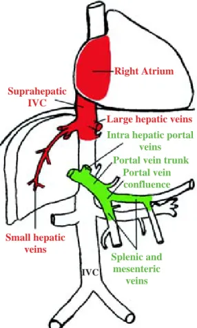
The Budd-Chiari Syndrome (BCS) is caused by hepat-ic venous outflow obstruction. This obstruction can be at different levels and present as a complete or incomplete obstruction. The venous obstruction can be continuous from or localized at the levels of the small hepatic veins, the large hepatic veins, the hepatic inferior vena cava (IVC), or the suprahepatic IVC up to the right atrium (Figure 1, on red). In 1990, Ludwig et al. classified the different types of Budd-Chiari-Syndrome and this classi-fication is seen as the standard today.1 It excludes
veno-occlusive disease (also known as sinusoidal obstruction) and hepatic outflow obstruction due to impaired func-tion of the heart.2
Large series of patients undergoing treatment for BCS have been reported, especially from Asian countries.3,4
Smaller studies on the epidemiology and treatment of BCS exist from western countries.5-7 Menon et al. had
re-cently produced an extensive review of the presentation, pathogenesis, treatment and prognosis of BCS.8 The
chronic form of BCS dominates over the acute presenta-tion (fulminant form). Thus treatment strategies vary ac-cording to the type of presentation of BCS and when as-sociated to liver failure it becomes a well-recognized in-dication for liver transplantation.9 If left untreated, the
stasis in the outflow tract of the liver may lead to in-creased pressure in the hepatic sinusoids and portal hy-pertension. This in turn may result in subsequent portal vein thrombosis.10,11
Artemisa
DH Borchert.Cavoportal hemitransposition for the simultaneous thrombosis of the caval and portal systems
www.medigraphic.com
Thrombosis of the portal venous system
Portal vein thrombosis (PVT) is a partial or complete obstruction of the trunk of the portal vein (Figure 1, on green). But similar to BCS it can occur at different levels and extend distally (in direction of flow) into the left and right hepatic portal veins and further up into the segmen-tal intrahepatic branches or it may extend proximally into the splenic vein, superior and inferior mesenteric vein. Whenever the thrombosis is not confined to the portal vein trunk the term «portal venous system throm-bosis» should be used.12
PVT has been classified surgically as «complete seg-mental», «complete and complex» and «complete and extensive» thrombosis including portal vein tributaries by Stieber et al.13 Another classification described PVT
as a spectrum of «partial thrombosis» (< 50% of vessel lumen) to «complete thrombosis including the superior mesenteric vein».14 A more simplified system described
any PVT with less than 90% occlusion as partial PVT and occlusion above 90% as complete PVT regardless of further involvement of the portal vein tributaries.15
The extent of PVT can only be reliably evaluated by thorough dissection in situ or in autopsy studies.16-18
Data from autopsy reports in general hospital settings state an overall incidence in the range of 0.05-0.5% for PVT.19 In patients with end-stage liver disease, the
inci-dence of PVT in those undergoing liver transplantation, is much higher: between 2% and 19%15,20 A study on
379 transplant patients found 39 patients with PVT. The thrombosis was extending to or beyond the confluens in two thirds of patients and a third had cavernous trans-formation of the portal vein. This study also found a high incidence of spontaneous splenorenal shunts (31%) among patients with PVT. Moreover patients with PVT have mesenteric oedema and mesenteric va-rices in up to 50% of the cases.16
Two - system thrombosis
Combined thrombosis of the inferior vena cava and portal–venous system is a rare and considered a severe condition. The combined thrombosis (BCS-PVT) can in-volve both venous systems to a varying extent and can be either acute or chronic in nature (Figure 1). Data on the prevalence of this two-system-thrombosis are sparse; however, Nonami et al. reported nine patients with Budd-Chiari syndrome in a liver transplant population of 885 patients. Of these nine, two patients had complete throm-bosis of the portal-venous-system.21 In a recent
North-In-dian study by Saxena et al., four patients out of 57 (with BCS) had complete thrombosis of both the inferior vena cava (IVC) and portal–venous system.22 Mahmoud et al.
found thirteen patients with portal–venous thrombosis out of 51 with BCS as the main diagnosis.23 A recent
multicenter study found 33 cases of combined BCS-PVT among 282 patients with the primary diagnosis of BCS. In this study 70% of patients with combined BCS-PVT were female. Patients with combined BCS-PVT tended to have a worse prognosis compared to patients with BCS only, but this was statistically not significant.24
In a histopathological study on BCS, involvement of the portal system of the liver was not a prognostic fac-tor.25 However, obstruction of the intrahepatic portal
sys-tem seems to be common in BCS and was found in all of 17 livers in one study and in 12 of 15 livers in another study from patients with BCS undergoing liver trans-plantation.10,11 Thrombosis of the portal vein system was
also found to be associated with a more acute onset and a shorter pre-transplant course.
The aetiological factors for BCS and PVT are similar with two exceptions. Both are results of acute or chron-ic thrombotchron-ic processes due to mechanchron-ic obstruction, inflammation or coagulation disorders. However, in the paediatric population infections, (especially umbilical vein sepsis and appendicitis), agenesis or atretic portal vein and previous portoenterostomy (Kasai procedure) for biliary atresia are the cause for PVT. In older pa-Figure 1. Combined thrombosis of the main venous tree (on red)
(according to Ludwig et al. classification on BCS) and portal -mesenteric system (on green), indicating different levels of throm-bosis. IVC, inferior vena cava. Figure designed and produced by Dr. D.H. Borchert © 2005.
Suprahepatic IVC
Right Atrium
Intra hepatic portal veins
Large hepatic veins
Small hepatic veins
Portal vein trunk Portal vein confluence
Splenic and mesenteric
veins
www.medigraphic.com
tients pancreatic cancer comprises the second aetiologi-cal subgroup, which is different regarding underlying pathology compared to other causes for BCS. Murad et al. also described predominance in local factors like cir-rhosis, abdominal tumours and inflammation as well as previous abdominal surgery for PVT.24 Increasingly
ac-quired or inherited coagulation disorders are found in association with other factors in BCS and PVT. Often the aetiological factors of BCS-PVT seem to be multi-factorial and the severity of the disease increases with multifactorial aetiology.
Method of review
A literature search was used to identify all cases of cavoportal hemitransposition (CPHT) using the key-words cavoportal hemitranspostition, portocaval hemi-transposition, portal vein thrombosis, Budd Chiari Syn-drome and liver transplantation. Standard publications on liver transplantation and medical management of pa-tients with liver disease were reviewed for indications and treatment options. More recent interventional radio-logical methods in treating acute thrombotic disease where retrieved from standard publication databases. A total of 23 out of 25 publications with reports on cavoportal hemitransposition only have been used to as-sess experience with this procedure.15,17,18,26-47 In one case
report the surgical procedure was started in attempt to perform a cavoportal hemitransposition, but was not fin-ished as such.31 In another review the patient had been
re-ported previously by another team.15 These reports were
excluded from the analysis. Furthermore we excluded reno-portal anastomosis in contrast to a recent review.48
We extracted demographic patient data, indication for liver transplantation, previous surgical procedures, signs of portal hypertension, postoperative complications, morbidity and mortality.
Surgical techniques
Indication for liver transplantation depends on sever-al factors. The most important factor being irreversible and progressive liver failure.49-51 The patients described
in this article presented with either acute-on-chronic liver failure complicated by vascular diseases or with acute liver failure due to vascular pathology. If the hepatic veins were thrombosed, as in BCS, and liver function was irreversibly damaged, liver transplantation was indicat-ed. In this situation, Sennings procedure (a direct hepa-toatrial anastomosis) has also been used.52 If only the
portal vein is thrombosed, there are several surgical tech-niques, which can be used to restore the blood flow to the
porta hepatis. First, a simple portal thrombectomy can be performed if possible. If the thrombus is organized and the portal vein cannot be re-canalized alternative meth-ods are used. Several shunt and bridging techniques exist
to prevent or treat developing portal hypertension.53-55 If
liver transplantation is indicated, the blood flow to the portal vein can be restored by one of several methods: anastomosis of the donor portal vein to the recipient su-perior mesenteric vein; anastomosis of the donor portal vein to the recipient splenic vein; or a venous jump graft to any suitable mesenteric tributary vein. When the por-tal venous system is only partially thrombosed, the blood flow can be increased by arterializing the portal vein.56
If the portal vein, splenic vein and mesenteric veins are all occluded, there are three feasible techniques for re-storing blood flow to the donor portal vein in liver trans-plantation. In this situation, liver transplantation can be combined with small bowel transplantation, cavoportal hemitransposition (Figure 2), or renoportal anastomo-sis.57 Suprahepatic caval and arterial anastomoses are
car-ried out in the usual manner. Delayed abdominal closure has to be anticipated in view of mesenteric oedema.
Cavoportal hemitransposition
History – Evolution of a technique
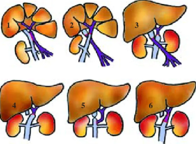
The ancestors of cavoportal hemitransposition, the so-called «Eck-Fistula», and several other shunt tech-niques, were developed since 1877 and used to treat portal hypertension, ascites and oesophageal varices due to chronic liver disease.58,59 The basic principle of
the «Eck-Fistula» is ligation of the portal vein at the he-patic hilum and anastomosis of the distal portal vein to the inferior vena cava (Fig. 2/1 dog liver). Pawlow et al. used this procedure and published their classical paper of «meat intoxication» in 1893.60 The «Eck-Fistula»
was used to study the physiology of liver regeneration and to search for the «hepatotropic factor». This was to clarify the question as to whether a hepatotropic factor exists in the portal venous blood. C.G. Child developed the technique of «portacaval transposition» in dogs in 1953 (Fig. 2/2 dog liver).61 Later in 1964 Starzl et al.,
followed in 1966 by Riddell et al., used the «portacaval transposition» or «portal diversion» in non-transplant patients with glycogen storage disease and familial hy-perlipidaemia (Fig. 2/3, these patients are not included in this review).62 This non–transplant procedure was
preceded by several experiments in animals.61,63,64 In
fur-DH Borchert.Cavoportal hemitransposition for the simultaneous thrombosis of the caval and portal systems
www.medigraphic.com
ther, it was noted by Starzl et al. that the full transposi-tion was not needed, the anastomosis between the distal portal vein and the proximal part of the inferior vena cava could safely be spared and a hemitransposition might be sufficient.65 This led to the development of
«cavoportal hemitransposition», which involves only half of the original, complete transposition of the portal vein and vena cava. The complete «cavoportal transpo-sition» can only be performed, if both the IVC and the portal venous system are patent. CPHT was introduced for patients in urgent need of a liver transplantation, who have complete thrombosis of the portal–venous system following a report by Tzakis et al. in 1998 (Fig. 2/4 and 5). In this first publication on CPHT the experi-ence of four transplant centres was summarized.18
Nomenclature
Tzakis et al. used the term cavoportal hemitransposi-tion (CPHT) in the title of their original report, but also coined the term portocaval hemitransposition together with abbreviation «PCHT». Both terms were used inter-changeably in the original article. The term portacaval hemitransposition was also used by Weeks et al. (2000), Wang et al. (2000), Pinna et al. (2001) and Lipshutz et al. (2006). In seventeen publications the authors used the term cavoportal hemitransposition. One author titled the procedure cavoportal transposition.29 Historically the
term cavoportal hemitransposition is correct, because it describes the procedure as being only half of the original
cavoportal transposition.
Current technique
Cavoportal hemitransposition is indicated in the pres-ence of complete thrombosis of the portal venous sys-tem, with or without Budd–Chiari syndrome. In cavopor-tal hemitransposition, the donor porcavopor-tal vein stump is anastomosed to the recipient inferior vena cava, above the renal veins. The lower end of the donor inferior vena cava (IVC) is unused and ligated (Fig. 2/4). If conven-tional hepatectomy precedes cavoportal hemitransposi-tion, blood flow can be restored using the donor portal vein for an end-to-end anastomosis to the recipient IVC. If preceded by piggy-back hepatectomy, cavoportal hemitransposition can be carried out using the donor por-tal vein for end-to-side anastomosis to the recipient IVC (Fig. 2/5). In one cases, an iliac vein graft was used for interposition between the donor portal vein and the re-cipient IVC to allow for the difference in size of the ves-sels (Fig. 2/6).41 In eleven cases an iliac vein graft was
used as a conduit, from the recipient IVC to the donor portal vein, in an end-to-side technique.27-29,37,41,43 In
end-to-side cavoportal hemitransposition, the recipient IVC is tied off above the anastomosis. In end-to-end anastomo-sis there may be a considerable difference in the size of the IVC and the donor portal vein.
Advantages and pitfalls (Benefits and problems)
Advantages
The technique of CPHT has developed over more than a century from the Eck-Fistula to the report of Tzakis et al. Since the first multicentre report in 1998 seventy-one cases have been reported in the international literature. Liver failure combined with complete portal venous sys-tem thrombosis has been regarded as an untreatable con-dition for a long time. CPHT is the surgical procedure that has successfully proven that liver transplantation is not contraindicated in the presence of portal vein system thrombosis. But the situations where portal venous in-flow can not be re-established by any other means are rare and in these instances CPHT is a «last resort» proce-dure.17,48 Thus the advantage of CPHT is to rescue a
pa-tient, where no other strategies are available. A corner stone of CPHT is to divert flow of the IVC completely to the donor portal vein to achieve a good portal inflow and to prevent re-thrombosis. So far in seven cases recurrence of portal vein thrombosis has been mentioned as a com-plication after CPHT.33;35;37;41;45;46 But eight patients
devel-oped thrombosis of the IVC below or above the renal veins after CPHT. If good flow through the donor portal vein is not achieved, then thrombosis of the portal vein is likely to happen as demonstrated by the case report of Ho et al. In complete thrombosis of the mesenteric venous system multivisceral transplantation is an alter-native option to CPHT. Compared to CPHT, multiviscer-Figure 2. Evolution of cavoportal hemitransposition (vena cava
on light blue, portal vein on purple, venous graft on pink, anasto-mosis on red ):1.1877, Eck – Fistula in canine liver; 2. 1953 (CG Child), cavoportal transposition in canine liver; 3. 1964 (Starzl), cavoportal transposition in glycogen storage disease; 4. 1998 (Tzakis), cavoportal hemitransposition end-to-end for PVT +/-BCS; 5. 1998 (Tzakis), cavoportal hemitransposition end-to-side;
6. 2005 (Ceulemans), cavoportal hemitransposition with interposi-tion iliac vein graft. Figure designed and produced by Dr. D.H. Borchert © 2005.
1 2 3
www.medigraphic.com
al transplantation is technically and immunologically more challenging. Another advantage of CPHT is to pre-vent extensive dissection of mesenteric veins. This can cause pancreatitis and increase damage to the mesenteric venous and arterial supply.13,16 However this is only
pos-sible if a decision is made preoperatively. This might not always be possible and in some centres extensive mesen-teric dissection is thought to be essential to find any suit-able mesenteric vein for reconnection to the portal sys-tem.17,18 In case of transplantation, CPHT can be
re-garded as a last resort procedure as well, if the vascular situation is deranged to a degree leaving no other option to re-establish portal inflow. CPHT has been used as a salvage procedure in re-transplantation in seven cases so far.17,18,29,36,37,43 CPHT does not correct or cure portal
hy-pertension, but may attenuate its severity. Authors re-ported the presence of signs of portal hypertension at least in 31 cases prior to CPHT (Table I). These signs per-sisted reportedly in 18 patients. In seven patients au-thor’s explicitly stated the disappearance of the symp-toms of portal hypertension.
Pitfalls
During recipient hepatectomy access to the portal pedi-cle can be complicated when there has been longstanding thrombosis of the portal–venous system and numerous col-laterals have developed. Four authors reported the existence of portal cavernoma, which can complicate the procedure and lead to significant bleeding.30,40,41,44 Some authors have
considered the existence of these cavernomas and collateral vessels, a contraindication for liver transplantation.14
Pa-tients undergoing CPHT presented in several cases with symptoms and signs of portal hypertension. Given the risk of bleeding from oesophageal and gastric varices, several
authors attempted to decompress the portal system or pre-vent gastro-oesophageal bleeding either before, during or following liver transplantation. In eight cases, a splenecto-my was performed during transplantation;37,39,41 in five cases
gastric devascularization was carried out; and in seven cas-es, the portal system was drained using interpositional grafts, or collaterals for shunting the venous blood back to the liver (Table II).36,37,41 In attempts to redirect venous flow,
portal hypertension persisted especially in patients were it existed already preoperatively.44
Despite refashioning the venous circulation in cavoportal hemitransposition in a non–anatomical way, no immediate problems with venous return or blood pres-sure have been reported. Even so, long-term changes in the venous system are possible after this operation. The devel-opment of a collateral venous circulation has been noted from previous experience with CPHT in animals and hu-mans.65 In their initial report, Tzakis et al. proposed
liga-tion of the right adrenal vein to prevent this from occur-ring and this has been also reported by Gerunda et al.18,30 In
the remaining twenty-one reports, no information is given about preventing the formation of collaterals to the IVC. Whether this is necessary or not remains a question of pa-tient follow-up. As partial or complete re-thrombosis of the portal venous system after CPHT has been reported in seven cases so far, embolization of the portal vein from thrombi of the lower extremities remain a threat. This has been clearly shown in the reports by Weeks et al and Shrotri et al. To date no reports exist on the preoperative evaluation of lower leg and/or pelvic thrombosis in CPHT and the use of perioperative calf compression and caval filter. Moreover pulmonary embolism is not banned due to ligation of the IVC. Instead at least three cases of upper extremities thromboembolism causing pulmonary artery emboli have been reported.18,37,39
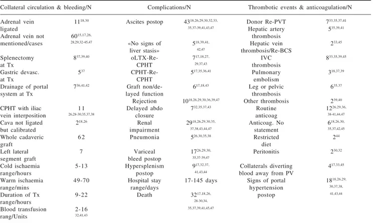
Table I. Demographic data, underlying liver disease and preoperative status.
Demographic data Underlying disease + PVT/N Pretransplant condition/N
Male/N 3415,18,26,28, HCV 918,35, Ascites pre Tx 3718,26,29,
30,33,35,37-39, 38,39,42 30,33,37-39,44,47
41-44,47,47 HBV 1 0 Encephalopathy 3026,27,33,
18,30,33, pre Tx 39,47
Female/N 3 5 41,45,47 Signs of portal 3118,26,27,29,
18,29,35-37,39, hypertension 30,32,33,37-39,
39-42,44,47,47 Other viral 6 41,42,47
sex unknown 232,34 Cryptogenic 9 Decision for CPHT 3818,26,29,
18,26,29,35,39 pre Tx 30,35,38-42
recipient-age 0.5–71 EtOH 7 Decision for CPHT 2118,30,32,
range/years 18,29,35,37,41,44 28,30,35,41,44 intraop 33,35,41,43,47
Donor age 3.5–82 Biliary atresia 817,18,32,43 Paediatric 917,32,
range/years Budd Chiari 718,29,39,40 portoenterostomy 36,43
CPHT for 7 Coagulation 4 Portosystemic shunt pre Tx 618,30,37,39
previous Oltx 17,18,27,29,37,43 disorder 39,40,43,44 Variceal bleeding pre Tx 2426,33,38,39,44,47
CPHT for 517,35,36,41 Other 9 Gastric devascular pre Tx 818,33,37
previous CPHT Hospital/ICU 2 8 Splenectomy pre Tx 1033,37,39,47
Peritonitis prior to Tx 318,27,30
DH Borchert.Cavoportal hemitransposition for the simultaneous thrombosis of the caval and portal systems
www.medigraphic.com
Patients reviewed
Demography
In the reviewed reports on CPHT, the median age of re-cipient ranged from 0.5 month to 71 years and donor age ranged from 3.5 to 82 years (Table I). In 35 cases the pa-tients were female, in 34 cases male and in two case the gender was not given.32,34 Eleven patients out of
seventy-one reported are paediatric patients. Thirty-four cases have been operated on in the USA, thirty-one in Europe, five in Asia and one in Australia. All of these patients had extensive thrombosis of the portal venous system. The majority of patients presenting with portal venous thrombosis had end stage liver disease due to viral dis-ease, hepatitis B and C, ethanol related and cryptogenic cirrhosis. Seven had combined portal vein thrombosis and Budd Chiari syndrome. In only five of the patients an underlying or associated coagulation disorder was di-agnosed.39,40,43,44 Paediatric patients presented with atretic
portal vein, portal vein agenesis or portal vein thrombo-sis after previous portoenterostomy. Several patients had surgical and endoscopic procedures for sequelae of portal hypertension before CPHT. Reflecting the severity of the underlying condition more than half of the patients have been hospitalised and on ITU prior to CPHT. Patients dis-played symptoms of end stage liver failure and portal
hy-pertension in the majority of cases and were described as moribund by several authors.
Indication and selection
The presence of thrombosis of the portal-venous sys-tem, Budd-Chiari syndrome or both at the same time, may lead to the complications of portal hypertension or either acute or chronic liver failure. Cavoportal hemi-transposition is used as a life-saving procedure when the portal venous system is thrombosed and inflow into the hepatic portal system cannot be established by any other technique. Patients in the reviewed reports presenting with thrombosis of the portal venous system or congeni-tal absence of appropriate porcongeni-tal inflow, were critically ill. In these patients, portal inflow needs to be re-estab-lished as a matter of urgency. If the liver is severely dam-aged they may undergo urgent liver transplantation. Thrombosis of the portal–venous system is technically no longer a contraindication for liver transplantation.66 In
Budd-Chiari syndrome, the venous outflow from the liv-er is compromised. When this occurs in addition to thrombosis of the portal-venous system, posthepatic liver failure results. Patients with this condition deteriorate even more rapidly and need urgent surgery to rectify the underlying cause. Thus, the main selection criteria for cavoportal hemitransposition are thrombosis of the portal Table II. Surgical technique and postoperative complications.
Collateral circulation & bleeding/N Complications/N Thrombotic events & anticoagulation/N
Adrenal vein 1118,30 Ascites postop 4318,26,29,30,32,33, Donor Re-PVT 733,35,37,41
ligated 35,37-39,41,43,47 Hepatic artery 535,39,41
Adrenal vein not 6015,17,26, thrombosis
mentioned/cases 28,29,32-45,47 «No signs of 518,39,41, Hepatic vein 233,45 liver stasis» 42,47 thrombosis/Re-BCS
Splenectomy 837,39,40 oLTX-Re- 717,18,27, IVC 833,35,39,45
at Tx CPHT 29,37,43 thrombosis
Gastric devasc. 537 CPHT-Re- 517,35,36,41 Pulmonary 318,37,39
at Tx CPHT embolism
Drainage of portal 736,41,42 Graft non/de- 617,18,43 Leg or pelvic 635,37
system at Tx layed function thrombosis
Rejection 1018,26,29,30,36,39,47 Other thrombosis 239,40
CPHT with iliac 1 1 Delayed abdo 732,35,37,43 Routine 1226,29,36,
vein interposition 26,28-30,35,37,38 closure anticoag 38-41,44,47
Cava not ligated 218,26 Renal 2918,26,29,30,35, Anticoag. No 618,26,30,
but calibrated impairment 37,38,43,44,47 statement 35,37,42,45
Whole cadaveric 6 2 Pneumonia 526,30,35,38 Restricted 244
graft diet
Left lateral 7 Variceal 1726,29,30, Peritonitis 230,32
segment graft bleed postop 35,37-39,47
Cold ischaemia 5-13 Hypersplenism 917,32,37, Collaterals diverting 417,33,45
range/hours postop 41,43,44 blood away from PV
Warm ischaemia 49-70 Hospital stay 17-145 days Signs of portal 1818,26,29,
range/mins range/days hypertension 30,37,38,
Duration of Tx 9-22 Death 3217,18,26, postop 41,43,44
range/hours 28-30,34,
Blood transfusion 2-16 35,37,39,41,45,47
rang/Units 32,41,43
www.medigraphic.com
venous system, either alone, or in combination with Budd–Chiari syndrome; and more importantly, the asso-ciated liver failure. Selection criteria in cavoportal hemi-transposition are based on individual cases and experi-ence. In most case reports, adult patients were preopera-tively selected for cavoportal hemitransposition upon findings from radiological imaging. In nine out of eleven paediatric patients the decision for CPHT was made intra-operatively. Thrombosis of the portal-venous system was commonly related to underlying viral disease (N = 25), cryptogenic cirrhosis (N = 9), alcoholic cirrhosis (N = 7), or other liver disease (N = 30) (Table I). Seven patients have undergone CPHT as a re-transplantation procedure for a failing liver graft from prior orthotopic liver trans-plantation.17,18,27,29,37,43 In five cases CPHT was followed
by CPHT as a retransplantation procedure.17,35,36,41 In at
least 38 patients, it was known prior to transplantation, that the portal–venous system was thrombosed and CPHT was planned as the surgical procedure. In 21 pa-tients, the decision to use CPHT was made intraopera-tively.
The consequences of deteriorating liver function and portal hypertension, such as ascites, encephalopathy and variceal bleeding, were not a contraindication to CPHT for most surgical teams. However, the «absence of as-cites» and «abscence of serious gastroscopic signs of por-tal hypertension» were reported by Urbani et al. as selec-tion criteria for the procedure.41 In all the other reports,
patients presented with classical features of portal hyper-tension associated with liver disease and deteriorating liver function. Ascites was reported in 37 patients, and encephalopathy in 30 patients prior to transplantation. Several patients presented with complications of portal hypertension prior to transplantation and underwent sur-gical procedures to correct these (Table I). Six patients had shunts from the portal vein to systemic veins, twen-ty-four had variceal bleeding, eight patients had gastric devascularization and ten patients underwent splenecto-my prior to transplantation. Further factors influencing selection for CPHT are preoperative renal impairment or renal failure; extensive previous abdominal surgery;18,35
previous irradiation for malignancy; spontaneous bacte-rial peritonitis;30 and a history of a coagulation
disor-der.35,39,40,44 The most important factor however is the
vas-cular status of the donor as diagnosed pre- and intraoper-atively. The size, flow and fragility of the vessel wall from any portal - mesenteric veins will lead to a decision for or against CPHT. In cases where CPHT was used as a re-transplantation procedure the previous standard ortho-topic liver transplantation either used a corrected hypo-plastic portal vein or its confluens, or thrombectomy from the portal - mesenteric system was performed and the anastomosis done in an anatomical way. In these cas-es the rcas-esult was rethrombosis, leaving CPHT as the only viable option at re-transplantation to establish adequate inflow into the graft portal system.18,27,43
Surgical differences
The technique of CPHT has been used in situations were the vascular status allowed no other reconstruction than use of the IVC to achieve adequate portal inflow to the graft. The connection between the IVC and the graft portal system can be done in mainly two ways; an end-to-end or and end-to-end-to-side anastomosis between donor portal vein and recipient suprarenal IVC. The end-to-side anas-tomosis can be done either using the donor portal vein directly or using an interposition graft (donor iliac vein). To perform the end-to-end anastomosis, the IVC has to be size adjusted to the donor portal vein or iliac vein graft. End-to-end anastomosis were used in fifty-one cases and end-to-side anastomosis in twenty cases and from these, iliac vein grafts were used in eleven cases. Numbers may slightly differ here from the true numbers of patients and operations, as this figure could not be exactly differenti-ated in the partial double reporting of Tzakis et al. and Pinna et al.
Another important step in liver transplantation with CPHT is the use of conventional hepatectomy or piggy-back technique. If the native vena cava is preserved the IVC can be tied off above the cavoportal anastomosis or calibrated with a sleeve or clip. If the IVC was left open, the donor portal vein is likely to thrombose as the flow is diverted away along the anatomical venous return to the right heart. In sixty-two cases a whole cadaveric liver graft was used for CPHT and in six cases a left lateral seg-ment for paediatric transplantation. CPHT corrects liver failure but does not reverse portal hypertension. In seven cases authors combined CPHT with drainage of the por-tal - mesenteric system. Urbani et al created surgical shunts in five of their patients and Varma et al. anasto-mosed a retroperitoneal varix to the donor portal vein to provide drainage. In the report from Varma et al. the shunt was patent one month after the operation.
Twelve authors started their patients on postoperative anticoagulation, and this was inherent to some of the liv-er transplant protocols.17,26,27,29,36,38-41,44,46,47 Another nine
authors did not mention anticoagulation at all. 18,28,30,32-35,37,42
Complications
The complications caused by thrombosis of the portal and mesenteric venous system should be divided into three groups: pre-, intra- and postoperative. The preoper-ative symptoms should be rectified by the surgical proce-dure, so the reccurrence of these symptoms postopera-tively can be used as a measure of the effectiveness of cavoportal hemitransposition as a treatment for these crit-ically ill patients.
DH Borchert.Cavoportal hemitransposition for the simultaneous thrombosis of the caval and portal systems
www.medigraphic.com
theses patients. In at least 31 of the cases, patients dis-played signs of portal hypertension preoperatively. In seven cases authors specifically reported the abscence of these symptoms. Preoperatively, surgical portosys-temic shunts were carried out in six patients; twenty-four patients had endoscopies for variceal bleeding; eight patients underwent gastric devascularization; and ten had splenectomies. Postoperatively; seventeen pa-tients had gastro-oesophageal bleeding26,29,30,35,37-39,47
and in nine patients hypersplenism developed or per-sisted.32;37;41;43;44 The majority of patients had varying
de-grees of recurrent ascites post-transplantion (N = 43)
(Table II).18,32,33,35,37-39,41,43,44,47 Only one author
men-tioned the continued need for diuretic therapy and re-stricted diet after cavoportal hemitransposition.44
Intraoperative complications include bleeding from extensive collaterals and massive bowel oedema, leading to delayed closure of the abdomen, as reported in seven cases.32,35,37,43 Postoperatively increased blood supply to
the liver can be expected immediately after CPHT. Four groups reported, explicitly, that there were no signs of congestion in the transplanted organ.18,35,41,42 This is
simi-lar to living donor liver transplantation, where there is increased portal inflow into the grafted liver lobe and congestion is usually not observed.67,68 However, in
small-for-size grafts, portal hyperperfusion can lead to early graft failure.69,70 «Portal hyperperfusion injury» is
theoretically possible in full-sized liver grafts, as report-ed elsewhere.71,72 Thrombotic events form a further
cate-gory of postoperative complications. Donor portal vein thrombosis was reported in seven cases.33,35,37,41,45 In two
cases similar symptoms for donor portal vein thrombosis have been reported. These two patients were both found to have left lower/upper quadrant pain and fever and both had apparently emboli from lower leg thrombosis to the portal vein.40,46 Hepatic artery thrombosis has been
re-ported in five cases;35,39,41 thrombosis of the inferior vena
cava in eight cases; lower limb or pelvic thrombosis in six cases;35,37 pulmonary embolism in three cases18,37,39
and other thrombotic events in two cases.39,40 Recurrence
of hepatic vein thrombosis (Re-BCS, table II) has been reported in two case leading to a situation, where the he-patic artery was the only blood supply to the liver and the blood flow reversed in the portal vein as the only vessel draining the liver.33,45 The first of these cases
re-ported in 2005 from Peking University died from recur-rent and metastatic liver disease after ten month but did not display liver dysfunction or signs of Budd-Chiari syndrome according to the authors. The second case re-ported in 2008 from Sichuan University is well and alive after one and a half years follow-up. Cavoportal hemi-transposition has challenged the understanding of liver physiology in these two cases. Re-transplantation was carried out in seven cases for previous standard orthoto-pic liver transplantation and in five cases for previous CPHT.17,18,27,29,35-37,41,43 Ten episodes of rejection have
been reported.18,26,29,30,33,36,39 The majority of patients
de-veloped varying degrees of renal impairment (N = 29) and four patients needed dialysis. Five episodes of pneumonia in the immediate post-transplant hospital stay have been documented in the reports. Nearly half of patients, published from 1998 – 2008, have died (N = 32).18,26,28-30,34,35,37,39,41,45,47 In the majority of the
re-maining patients, postoperative complications have set-tled, suggesting that CPHT can be regarded as an effective treatment for thrombosis of the portal–venous system.
Outcome of patients
The peri – and postoperative complications resulting from cavoportal hemitransposition are not unique to this procedure, but resemble the spectrum of complications from liver transplantation. The surviving patients are mainly «alive and well», including three paediatric pa-tients, as reported in the available literature. Surviving patients returned to normal activities and had normal liv-er function tests. Their conditions range from «patient re-mains at home»26 to «enjoying normal life»41 to
«excel-lent condition».18 Dietary restrictions were mentioned in
one report. The main symptoms from portal hyperten-sion, such as ascites and bleeding from gastro-oesoph-ageal varices, had resolved. The series of Ceulemans et al., showed a slow recovery from pre-transplant portal hy-pertension and prolonged hospital stay. Thirty-two out of 71 patients did not survive. Two patients died from prima-ry non-function of the liver graft , and one died of primaprima-ry dysfunction of the liver after four weeks.17,18 Other causes
of death were rejection (2),26,30 pulmonary embolism (2),18,37 sepsis/multi-organ failure (7, (18;29;35;37;41))
and cardiac failure (2).28,37 Late deaths also occured at
sev-en months, tsev-en months and one year post-transplantation. Twelve patients died within four weeks of the operation, indicating the severity of the underlying disease. In the re-port of Lipshutz et al. two paediatric patients out of seven are longterm survivors with 4 and 8 years posttransplant respectively.
Collateral circulation
In cavoportal hemitransposition, the venous flow from the IVC is diverted to the liver to substitute for the lost venous inflow from the portal–venous system. Ex-perimental studies in dogs and portacaval transposition for inherited liver disease have shown that this tech-nique is feasible. It allows for normal liver function and cure of the underlying disease without causing hepatic congestion from increased venous inflow.61,73 One of the
long-term problems with this technique is that blood flow can be diverted away from the liver again, through developing collaterals. Preexisting collateral pathways, such as the azygos system, may enlarge.63 Both Tzakis
www.medigraphic.com
ligate the right adrenal vein.18,30 In one case the caval
flow was diverted through the azygos system after six months despite intraoperative ligation of the right adre-nal vein.30 Cavoportal collateral pathways have been
re-ported in chronic obstruction of the inferior vena cava by several groups, including Dahan et al.. This group described the following collateral pathways: a) cavo – superficial – umbilical – portal, b) cavo – mammary – phrenic – hepatic capsule – portal, c) cavo – mesenteric – portal, d) cavo – renal – portal and cavo – retroperito-neal – portal and e) intrahepatic cavoportal pathways.74
This suggests that the development of collateral path-ways is unpredictable. So far, there have been no reports of problems associated with the haemodynamic changes that occur following ligation of the IVC, proximal to the cavoportal anastomosis. Similarly, there have been no negative reports of the effect of collaterals diverting venous flow away from the liver, after cavoportal hemi-transposition. In this regard the significance of the drainage of the portal system in CPHT as reported in seven cases remains to be elucidated. Bypassing venous flow to the liver can lead to symptoms similar to those prior to transplantation. Conversely persisting exten-sive collateral veins may contribute to the success of CPHT, bypassing and forwarding enough blood from the infra-anastomotic area to support cardiac output.38
Due to the relatively rare and young nature of cavopor-tal hemitransposition, no state of the art technique has been described, which would allow surgery to be car-ried out in a standardized manner. This is reflected by the different surgical approaches described in the pub-lished case reports, for example Varma et al. divided the IVC just above the right renal vein, whereas Shrotri et al. divided the IVC 7 cm above the right renal vein. Most of the authors did not report ligation of the adre-nal veins. Investigations on development of collaterals postoperatively are missing in most reports. Anatomi-cally, collateral pathways can vary considerably, espe-cially those involving the azygos and hemiazygos sys-tem.75 In a study of the anatomy of the adrenal venous
system, Monkhouse et al. found the right adrenal vein to be located at an average of 45 mm (range 0 – 85 mm) above the entry of the right renal vein.76 The right
adre-nal vein can be duplicated or triplicated, and there can be connections of accessory veins with the right renal vein and the inferior phrenic vein. The situation with the left adrenal vein is even more complicated. In 42 out of 57 cadavers, studied by Monkhouse et al., venous com-munications from the adrenal glands to the renal, lumbar and azygos/hemiazygos system were found. In another study, twenty-one different patterns of the lumbar and ayzgos vein system were reported.77 Therefore, the
devel-opment of collaterals is unpredictable, even following li-gation of the right adrenal vein. The longterm effects of diversion of venous blood away from the porta hepatis in these patients has not been assessed.
Unresolved questions
So far, no consensus has been reached about the surgi-cal technique of CPHT. This will probably be difficult to achieve, as cases are rare with unique individual situa-tions and treatment opsitua-tions depend on local expertise and knowledge. This situation is similar for the treatment of the two-system thrombosis of BCS-PVT. Despite thrombosis as the underlying clinical feature in all cases anticoagulation seems not to be an agreed postoperative treatment strategy. Still individual steps in treatment strategies are already established and might become more accepted internationally with continued research. Pinna et al. reported that none of their patients who underwent intraoperative splenectomy developed recurrent bleed-ing from gastro-oesophageal varices postoperatively. The follow-up report from the same group six years later stat-ed that splenectomy at the time of transplantation has been abandoned because of a case with lethal over-whelming post-splenectomy infection.39 As postoperative
haemorrhage is a severe complication, it may be possible to prevent postoperative bleeding further by evaluating oesophagogastric varices intraoperatively. This could be an important step towards improvement of the technique. But preventing postoperative bleeding by gastric devas-cularization or splenectomy may increase the risk of currence of portal–venous thrombosis. Two authors re-ported recurrence of portal–venous thrombosis in pa-tients who had undergone splenectomy. Several groups, including Settmacher et al., reported splenectomy as a risk factor for portal–venous thrombosis following liver transplantation.37,41,78 From a pathophysiological point of
view the development and effect of major collateral ves-sels, bypassing venous flow through the azygos system, remains another question to be answered by patient fol-low-up. Patients who develop collateral circulation need to be compared with patients where the venous flow from the IVC through the liver is completely preserved. Gerun-da et al. reported that one of their patients developed col-lateral flow through the azygos systems, even so the right adrenal vein had been ligated.30 Given the complex
seem-DH Borchert.Cavoportal hemitransposition for the simultaneous thrombosis of the caval and portal systems
www.medigraphic.com
ingly cured by liver transplantation. This is because ex-isting lower leg or pelvic thrombosis may be a threat to the donor portal system in CPHT and in view of a high incidence of multiple factors contributing to BCS-PVT, individual or local factors may have been undiagnosed and/or persisting.24 Given the two reports about
embo-lization of the donor portal vein by emboli from lower leg or pelvic thrombosis, the use of caval filters in pa-tients undergoing CPHT should be discussed.
The case reports of Wang et al. and Li et al. on com-pensatory blood supply through the hepatic artery in cases of new-onset or recurrent BCS-PVT after CPHT raise an interesting question.10,33,45 If liver function can
be maintained by arterial perfusion only in a situation where hepatic venous outflow is obstructed and the por-tal vein is hypoperfused or even draining the liver, could then liver transplantation be performed without an attempt to restore portal venous flow at all? This question should certainly be subject to experimental re-search in the future.
The geographic distribution of cavoportal hemitrans-position as a surgical procedure is confined to USA, Can-ada, Italy, France, Belgium, England, Denmark, Sweden, China and Australia. Despite reports from Asian coun-tries about significant numbers of BCS and PVT only three reports about the use of this surgical procedure are available from the eastern hemisphere (all reports to date are from China).
Emerging radiological approaches
Radiological interventional techniques are replacing surgical techniques in several areas. In many case reports transjugular portosystemic shunting (TIPS) and fragmen-tation or thrombolysis have been shown to be an effec-tive rescue procedure in acute combined BCS/PVT.79-83
Even in re-thrombosis after CPHT thrombolysis has been used as a rescue procedure in two cases.33,84 However, this
might not be possible in organized thrombosis of either of the two venous systems, or if severe liver failure is present. Even adequate radiological facilities might fail to establish the diagnosis and if radiological interven-tion is used as a first line treatment opinterven-tion, surgical back-up is needed. In the multicenter study of Murad et al. only four out thirtythree patients with combined BCS/ PVT had a TIPS inserted. In one of these patients TIPS failed and a rescue cavoportal (without transplantation) shunt was needed.24
Conclusion
So far seventy-one patients have been reported to have had CPHT as a surgical procedure worldwide. This operation has been used in situations where urgent liver transplantation was the only way to save patient life and it was not possible to re-establish blood flow to the
do-nor porta hepatis by any other method. Thrombosis of the IVC and portal–venous system is associated with a variety of underlying diseases. Despite improved diag-nostic techniques, this condition may only be discovered for the first time intraoperatively. In at least twenty-one cases, the decision to carry out CPHT was made intraop-eratively. Budd–Chiari syndrome associated with throm-bosis of the portal system is rare and is associated with rapid deterioration. CPHT has proved to be a successful alternative to combined small bowel and liver transplan-tation. The reviewed cases point towards the establish-ment of a sequential therapeutic approach. Where liver function allows, interventional radiology might be used as first-line treatment. Failure of this approach to re-es-tablish blood flow might lead to severe liver damage and failure. This would also document the severity of throm-bosis in both venous systems, and would need to be fol-lowed by a second-line treatment such as CPHT, or com-bined small bowel and liver transplantation.
Conflict of interests: None
Acknowledgements: The author was able to study two patients after CPHT in the Department of Surgery at Ad-denbrookes Hospital, Cambridge University, Cambridge, UK (one of these cases was published by Shrotri et al.).
I am very grateful for repeated discussion of the manu-script with Dr. H. Vilca-Melendez, consultant transplant surgeon at Kings College Hospital, Denmark Hill, Lon-don, UK.
References
1. Ludwig J, Hashimoto E, McGill DB, van Heerden JA. Classifi-cation of hepatic venous outflow obstruction: ambiguous ter-minology of the Budd-Chiari syndrome. Mayo Clin Proc 1990; 65(1): 51-5.
2. European Network for the vascular disease of the liver. Guide-lines for the management of patients with Budd-Chiari syndrome. www.mh-hannover.de/kliniken/gastro/vasc/guideline_BCS.pdf , 1-33. 2005.
3. Feng LS, Peng QP, Li K, et al. Management of severe Budd-Chiari syndrome: report of 147 cases. Hepatobiliary Pancreat Dis Int 2004; 3(4): 522-5.
4. Xu PQ, Ma XX, Ye XX et al. Surgical treatment of 1360 cases of Budd-Chiari syndrome: 20-year experience. Hepatobiliary Pancreat Dis Int 2004; 3(3): 391-4.
5. Jamieson NV, Williams R, Calne RY. Liver transplantation for Budd-Chiari syndrome, 1976-1990. Ann Chir 1991; 45(4): 362-5. 6. Orloff MJ, Daily PO, Orloff SL, et al. A 27-year experience with
surgical treatment of Budd-Chiari syndrome. Ann Surg 2000; 232(3): 340-52.
7. Ulrich F, Steinmuller T, Lang M, et al. Liver transplantation in patients with advanced Budd-Chiari syndrome. Transplant Proc 2002; 34(6): 2278.
8. Menon KV, Shah V, Kamath PS. The Budd-Chiari syndrome. N Engl J Med 2004; 350(6): 578-85.
9. Srinivasan P, Rela M, Prachalias A, et al. Liver transplantation for Budd-Chiari syndrome. Transplantation 2002; 73(6): 973-7. 10. Cazals-Hatem D, Vilgrain V, Genin P, et al. Arterial and portal
www.medigraphic.com
11. Tanaka M, Wanless IR. Pathology of the liver in Budd-Chiarisyndrome: portal vein thrombosis and the histogenesis of veno-centric cirrhosis, veno-portal cirrhosis, and large regenerative nodules. Hepatology 1998; 27(2): 488-96.
12. European Network for the vascular disease of the liver. Guide-lines for the management of patients with portal vein thrombosis. www.mh-hannover.de/kliniken/gastro/vasc/guideline_PVT.pdf . 2005.
13. Stieber AC, Zetti G, Todo S, et al. The spectrum of portal vein thrombosis in liver transplantation. Ann Surg 1991; 213(3): 199-206.
14. Yerdel MA, Gunson B, Mirza D, et al. Portal vein thrombosis in adults undergoing liver transplantation: risk factors, screening, man-agement, and outcome. Transplantation 2000; 69(9): 1873-81. 15. Bertelli R, Nardo B, Montalti R, et al. Liver transplantation in
recipients with portal vein thrombosis: experience of a single transplant center. Transplant Proc 2005; 37(2): 1119-21. 16. Brancatelli G, Federle MP, Pealer K, Geller DA. Portal venous
thrombosis or sclerosis in liver transplantation candidates: preop-erative CT findings and correlation with surgical procedure. Ra-diology 2001; 220(2): 321-8.
17. Lipshutz GS, Patel S, Hiatt JR, et al. Portocaval hemitransposition in pediatric liver transplant recipients: a single-center experience. Liver Transpl 2006; 12(7): 1097-103.
18. Tzakis AG, Kirkegaard P, Pinna AD, et al. Liver transplantation with cavoportal hemitransposition in the presence of diffuse por-tal vein thrombosis. Transplantation 1998; 65(5): 619-24. 19. Cohen J, Edelman RR, Chopra S. Portal vein thrombosis: a
re-view. Am J Med 1992; 92(2): 173-82.
20. Shaked A, Busuttil RW. Liver transplantation in patients with portal vein thrombosis and central portacaval shunts. Ann Surg 1991; 214(6): 696-702.
21. Nonami T, Yokoyama I, Iwatsuki S, Starzl TE. The incidence of portal vein thrombosis at liver transplantation. Hepatology 1992; 16(5): 1195-8.
22. Bhattacharyya M, Makharia G, Kannan M, et al. Inherited prothrombotic defects in Budd-Chiari syndrome and portal vein thrombosis: a study from North India. Am J Clin Pathol 2004; 121(6): 844-7.
23. Mahmoud AE, Helmy AS, Billingham L, Elias E. Poor prognosis and limited therapeutic options in patients with Budd-Chiari syn-drome and portal venous system thrombosis. Eur J Gastroenterol Hepatol 1997; 9 (5): 485-9.
24. Murad SD, Valla DC, de Groen PC, et al. Pathogenesis and treat-ment of Budd-Chiari syndrome combined with portal vein throm-bosis. Am J Gastroenterol 2006; 101(1): 83-90.
25. Tang TJ, Batts KP, de Groen PC, et al. The prognostic value of histology in the assessment of patients with Budd-Chiari syn-drome. J Hepatol 2001; 35(3): 338-43.
26. Azoulay D, Hargreaves GM, Castaing D, Bismuth H. Caval in-flow to the graft: a successful way to overcome diffuse portal system thrombosis in liver transplantation. J Am Coll Surg 2000; 190(4): 493-6.
27. Bakthavatsalam R, Marsh CL, Perkins JD, et al. Rescue of acute portal vein thrombosis after liver transplantation using a cavoportal shunt at re-transplantation. Am J Transplant 2001; 1(3): 284-7. 28. Bernardos A, Serrano J, Gomez MA, et al. Portal vein thrombo-sis: an emergency solution for blood flow in liver transplanta-tion. Transpl Int 2003; 16(8): 500-1.
29. Ceulemans B, Aerts R, Monbaliu D, et al. Liver Transplantation using cavoportal transposition: An effective treatment in patients with complete splanchnic venous thrombosis. Transplant Proc 2005; 37(2): 1112-4.
30. Gerunda GE, Merenda R, Neri D, et al. Cavoportal hemitransposition: a successful way to overcome the problem of total portosplenomesenteric thrombosis in liver transplantation. Liver Transpl 2002; 8(1): 72-5.
31. Ho MC, Hu RH, Lai HS, et al. Liver transplantation in a patient with diffuse portal venous system thrombosis. Transplant Proc 2000; 32(7): 2174-6.
32. Kumar N, Atkison P, Fortier MV, et al. Cavoportal transposition for portal vein thrombosis in a pediatric living-related liver trans-plantation. Liver Transpl 2003; 9(8): 874-6.
33. Li FG, Yan LN, Wang WT. Extensive thrombosis of the portal vein and vena cava after orthotopic liver transplantation with cavoportal hemitransposition: a case report. Transplant Proc 2008; 40(5): 1777-9.
34. Llado L, Fabregat J, Castellote J, et al. Management of portal vein thrombosis in liver transplantation: influence on morbidity and mortality. Clin Transplant 2007; 21(6): 716-21.
35. Olausson M, Norrby J, Mjornstedt L, et al. Liver transplantation using cavoportal hemitransposition - A life-saving procedure in the presence of extensive portal vein thrombosis. Transplant Proc 2001; 33(1-2): 1327-8.
36. Ozden I, Suoglu OD, Aydogan A. et al. Successful living-donor liver transplantation and retransplantation with cavoportal hemitransposition: a case report. Exp Clin Transplant 2006; 4(2): 562-6.
37. Pinna AD, Nery J, Kato T, et al. Liver transplant with portocaval hemitransposition: experience at the University of Miami. Trans-plant Proc 2001; 33(1-2): 1329-30.
38. Santaniello W, Ceriello A, Defez M, et al. Liver transplant with cavoportal hemitransposition for portal and mesenteric throm-bosis: case report. Transplant Proc 2001; 33(1-2): 1488-9. 39. Selvaggi G, Weppler D, Nishida S, et al. Ten-year experience in
porto-caval hemitransposition for liver transplantation in the pres-ence of portal vein thrombosis. Am J Transplant 2007; 7(2): 454-60.
40. Shrotri M, Sudhindran S, Gibbs P, et al. Case report of cavoportal hemitransposition for diffuse portal vein thrombosis in liver trans-plantation. Transplant Proc 2003; 35(1): 397-8.
41. Urbani L, Cioni R, Catalano G, et al. Cavoportal hemitransposition: patient selection criteria and outcome. Transplant Proc 2002; 34(8): 3331-3.
42. Varma CR, Mistry BM, Glockner JF, et al. Cavoportal hemitransposition in liver transplantation. Transplantation 2001; 72(5): 960-3.
43. Verran D, Crawford M, Stormon M, Shun A. Liver retransplantation in an infant requiring cavoportal hemi transpo-sition. Pediatr Transplant 2004; 8(4): 416-9.
44. Vincent C, Pomier-Layrargues G, Dagenais M et, al. Cure of gastric antral vascular ectasia by liver transplantation despite per-sistent portal hypertension: a clue for pathogenesis. Liver Transpl 2002; 8(8): 717-20.
45. Wang C, Zhang T, Song S, et al. Liver transplant with portocaval hemitransposition: blood supply with only hepatic artery is pos-sible? Transplant Proc 2005; 37(5): 2163-5.
46. Weeks SM, Alexander JR, Sandhu J, et al. Mechanic and pharma-cologic treatment of a saddle embolus to the portal vein after liver transplantation and portacaval hemitransposition. AJR Am J Roentgenol 2000; 175(2): 537-9.
47. Yan ML, Zeng Y, Li B, et al. Postoperative complications after liver transplantation with cavoportal hemitransposition. Hepatobiliary Pancreat Dis Int 2008; 7(3): 322-4.
48. Paskonis M, Jurgaitis J, Mehrabi A, et al. Surgical strategies for liver transplantation in the case of portal vein thrombosis—cur-rent role of cavoportal hemitransposition and renoportal anasto-mosis. Clin Transplant 2006; 20(5): 551-62.
49. Klupp J, Kohler S, Pascher A, Neuhaus P. Liver transplantation as ultimate tool to treat portal hypertension. Dig Dis 2005; 23(1): 65-71. 50. Loinaz C, Gomez R, Jimenez C, et al. Liver transplantation in patients with portal thrombosis: results in 76 patients. Transplant Proc 2002; 34(1): 248-9.
51. Wang ZF, Liu C. Liver retransplantation: indications and out-comes. Hepatobiliary Pancreat Dis Int 2004; 3(2):75-8. 52. Sauvanet A, Panis Y, Valla D, et al. Budd-Chiari syndrome with
extensive portal thrombosis: treatment with Senning’s procedure. Hepatogastroenterology 1994; 41(2): 174-6.
DH Borchert.Cavoportal hemitransposition for the simultaneous thrombosis of the caval and portal systems
www.medigraphic.com
54. Huguet C, Hannoun L, Nordlinger B, et al. Selective distalspleno-renal shunt. A report of 14 patients (author’s transl). Nouv Presse Med 1979; 8(47): 3881-4.
5 5 . Watanabe H, Shinzawa H, Saito T, et al. Successful emer-gency treatment with a transjugular intrahepatic portosystemic shunt for life-threatening Budd-Chiari syndrome with portal thrombotic obstruction. Hepatogastroenterology 2000; 47(33): 839-41.
56. Stange B, Glanemann M, Nussler NC, et al. Indication, tech-nique, and outcome of portal vein arterialization in orthotopic liver transplantation. Transplant Proc 2001; 33(1-2): 1414-5. 57. Azoulay D, Adam R, Castaing D, et al. Liver transplantation with
cavoportal or renoportal anastomosis: a solution in cases of diffuse portal thrombosis. Gastroenterol Clin Biol 2002; 26(4): 325-30. 58. Eck NV. K voprosu o perevyazkie vorotnois veni: Predvaritelnoye
soobschjenye. Voen Med J 1877; 130: 1-2.
59. Starzl TE, Porter KA, Francavilla A. The Eck fistula in animals and humans. Curr Probl Surg 1983; 20(11): 687-752. 60. Hahn M, Massen O, Nencki M, Pawlow J. Die Eck´sche Fistel zwischen
der unteren Hohlvene und der Pfortader und ihre Folgen für den Organismus. Arch Exp Pathol Pharmakol 1893; 32: 161-210. 61. Child CGI, Barr D, Holswade GR, Harrison CS. Liver
regenera-tion following portacaval transposiregenera-tion in dogs. Ann Surg 1953; 138(4): 600-8.
62. Riddell AG, Davies RP, Clark AD. Portacaval transposition in the treatment of glycogen-storage disease. Lancet 1966; 2(7474): 1146-8.
63. Meyer WH, Jr., Starzl TE. The reverse portacaval shunt. Surgery 1959; 45: 660-.
64. Silen W, Mawdsley DL, Weirich WL, Harper HA. Studies of hepatic function in dogs with Eck fistula or portacaval transposi-tion. AMA Arch Surg 1957; 74(6): 964-70.
65. Starzl TE, Putnam CW, Porter KA, et al. Portal diversion for the treatment of glycogen storage disease in humans. Ann Surg 1973; 178(4): 525-39.
66. Jamieson NV. Changing perspectives in portal vein thrombosis and liver transplantation. Transplantation 2000; 69(9): 1772-4. 67. Eguchi S, Yanaga K, Sugiyama N, et al. Relationship between portal venous flow and liver regeneration in patients after living donor right-lobe liver transplantation. Liver Transpl 2003; 9(6): 547-51.
68. Garcia-Valdecasas JC, Fuster J, Charco R, et al. Changes in portal vein flow after adult living-donor liver transplantation: does it influence postoperative liver function? Liver Transpl 2003; 9(6): 564-9.
69. Lo CM, Liu CL, Fan ST. Portal hyperperfusion injury as the cause of primary nonfunction in a small-for-size liver graft-suc-cessful treatment with splenic artery ligation. Liver Transpl 2003; 9(6): 626-8.
70. Sugimoto H, Kaneko T, Hirota M, et al. Critical progressive small-graft injury caused by intrasinusoidal pressure elevation following living donor liver transplantation. Transplant Proc 2004; 36(9): 2750-6.
71. Henderson JM, Gilmore GT, Mackay GJ, et al. Hemodynamics during liver transplantation: the interactions between cardiac output and portal venous and hepatic arterial flows. Hepatology 1992; 16(3): 715-8.
72. Paulsen AW, Klintmalm GB. Direct measurement of hepatic blood flow in native and transplanted organs, with accompanying sys-temic hemodynamics. Hepatology 1992; 16(1): 100-11. 73. Starzl TE, Putnam CW, Porter KA, Benichou J. Portacaval shunt
for glycogen storage disease and hyperlipidaemia. Ciba Found Symp 1977; (55): 311-25.
74. Dahan H, Arrive L, Monnier-Cholley L, et al. Cavoportal collat-eral pathways in vena cava obstruction: imaging features. AJR Am J Roentgenol 1998; 171(5): 1405-11.
75. Abrams HL. The vertebral and azygos venous systems, and some variations in systemic venous return. Radiology 1957; 69(4): 508-26.
76. Monkhouse WS, Khalique A. The adrenal and renal veins of man and their connections with azygos and lumbar veins. J Anat 1986; 146: 105-15.
77. Seib GA. Azygos System of Veins in American White and Ameri-can Negroes, Including Observations on Inferior Caval Venous System. Am J Phys Anthropol 1934; 19: 39-163.
78. Settmacher U, Nussler NC, Glanemann M, et al. Venous compli-cations after orthotopic liver transplantation. Clin Transplant 2000; 14(3): 235-41.
79.Novel procedure: percutaneous, via TIPS, portal vein thrombec-tomy in patients with Budd-Chiari syndrome complicated by acute portal vein thrombosis. Tel-Aviv, Israel.: 2005.
80. Leebeek FW, Lameris JS, van Buuren HR, et al. Budd-Chiari syndrome, portal vein and mesenteric vein thrombosis in a pa-tient homozygous for factor V Leiden mutation treated by TIPS and thrombolysis. Br J Haematol 1998; 102(4): 929-31. 81. Mancuso A, Watkinson A, Tibballs J, et al. Budd-Chiari
syn-drome with portal, splenic, and superior mesenteric vein throm-bosis treated with TIPS: who dares wins. Gut 2003; 52(3): 438. 82. Opitz T, Buchwald AB, Lorf T, et al. The transjugular intrahe-patic portosystemic stent-shunt (TIPS) as rescue therapy for com-plete Budd-Chiari syndrome and portal vein thrombosis. Z Gastroenterol 2003; 41(5): 413-8.
83. Pfammatter T, Benoit C, Cathomas G, Blum U. Budd-Chiari syn-drome with spleno-mesenteric-portal thrombosis: treatment with extended TIPS. J Vasc Interv Radiol 2000; 11(6): 781-4. 84. Haider HH, Froud T, Moon J, et al. Successful percutaneous