Cancer Stem Cells are Depolarized Relative
to Normal Stem Cells Derived from Human Livers
Wendy Bautista,* Jeremy Lipschitz,** Andrew McKay,** Gerald Y. Minuk*,*** *Section of Hepatology, Department of Medicine, **Department of Surgery, ***Department of Pharmacology and Therapeutics, University of Manitoba, Winnipeg, Manitoba, Canada. March-April, Vol. 16 No. 2, 2017: 297-303
INTRODUCTION
Cancer stem cells (CSC) are a malignant subpopulation of cells within a tumor that have the ability to self-repli-cate, differentiate into different cell lineages and resist
chemotherapy.1 They have been identified in many solid
organ tumors including hepatocellular carcinoma (HCC). Although uncertainty remains regarding the precise role these cells play in the pathogenesis of cancer, recent data suggest they contribute to tumor development, growth,
invasion and metastases.1 As a result, CSCs have emerged
as an important, albeit elusive therapeutic target.2
One of the principal challenges in developing safe and effective means of eradicating CSCs from tumor tissues is the difficulty in distinguishing CSCs from normal stem
cells (NSCs) present in adjacent non-tumor tissue.3 This
problem is compounded in the liver where attempts to eradicate CSCs from HCC often occur in the setting of cirrhosis where NSCs are required to maintain hepato-cyte populations and thereby, essential liver functions.4
Recently, our laboratory identified potentially important distinctions between CSCs and NSCs that could serve as
the basis for new therapeutic strategies for HCC.5
Specifi-cally, we documented that CSCs derived from the human Huh-7 and PLC malignant hepatocyte cell lines have signif-icantly depolarized (less negatively charged) resting cell membrane potential differences (PD) when compared to
the non-malignant, murine WBF-344 NSC cell line.5
Whether these differences exist in CSCs and NSCs derived from human liver tissues remains to be determined.
Gamma-aminobutyric acid (GABA) is a potent inhibi-tory neurotransmitter that plays an important role in
em-The Official Journal of the Mexican Association of Hepatology, the Latin-American Association for Study of the Liver and
the Canadian Association for the Study of the Liver
Manuscript received: Manuscript received:Manuscript received:
Manuscript received:Manuscript received: May 31, 2016. Manuscript accepted:Manuscript accepted:Manuscript accepted:Manuscript accepted:Manuscript accepted: August 01, 2016.
DOI:10.5604/16652681.1231590.
A B S T R A C T A B S T R A C T A B S T R A C T A B S T R A C T A B S T R A C T
Introduction and aim. Introduction and aim.Introduction and aim. Introduction and aim.
Introduction and aim. The inability to distinguish cancer (CSCs) from normal stem cells (NSCs) has hindered attempts to identi-fy safer, more effective therapies for hepatocellular carcinoma (HCC). The aim of this study was to document and compare cell membrane potential differences (PDs) of CSCs and NSCs derived from human HCC and healthy livers respectively and determine whether altered GABAergic innervation could explain the differences. Material and methods.Material and methods.Material and methods.Material and methods. Epithelial cell adhesion moleculeMaterial and methods. (EpCAM) positive stem cells were isolated from human liver tissues by magnetic bead separations. Cellular PDs were recorded by microelectrode impalement of freshly isolated cells. GABAA receptor subunit expression was documented by reverse transcriptase polymerase chain reaction (RT-PCR) and immunofluorescence. Results.Results.Results.Results.Results. CSCs were significantly depolarized (-7.0 ± 1.3 mV) rela-tive to NSCs (-23.0 ± 1.4 mV, p < 0.01). The depolarized state was associated with different GABAA receptor subunit expression profiles wherein phasic transmission, represented by GAGAAα3 subunit expression, was prevalent in CSCs while tonic transmis-sion, represented by GABAAα6 subunit expression, prevailed in NSCs. In addition, GABAA subunits α3, β3, γ3 and δ were strongly expressed in CSCs while GABAAπ expression was dominant in NSCs. CSCs and NSCs responded similarly to GABAA receptor agonists (ΔPD: 12.5 ± 1.2 mV and 11.0 ± 3.5 mV respectively). Conclusion.Conclusion.Conclusion.Conclusion.Conclusion. The results of this study indicate that CSCs are sig-nificantly depolarized relative to NSCs and these differences are associated with differences in GABAA receptor subunit expression. Together they provide new insights into the pathogenesis and possible treatment of human HCC.
Key words. Key words.Key words. Key words.
bryogenesis and tissue development by regulating stem
cell activity and differentiation.6,7 GABA
A receptors are
heterooligomeric, ligand-gated ion channels consisting of 16 subunits, commonly present in a pentameric
configura-tion.8 In non-synaptic cells (such as hepatocytes)
GABAergic activity is influenced by those subunits re-sponsible for “tonic” transmission (resulting in prolonged
hyperpolarization) of which α6 is considered the
proto-type, whereas in synaptic cells (such as neurons), “phasic” transmission, related to the presence of the α3 subunit, is
dominant.9
In the present study, we documented the PDs of pri-mary CSCs and NSCs derived from freshly isolated HCC and adjacent non-tumor tissues respectively. We also
doc-umented the expression of various GABAA receptor
subu-nits including α3 and α6 in the two cell populations and
the PD responses of CSCs and NSCs to GABAA receptor
agonists.
MATERIAL AND METHODS
Stem cell isolations
Stem cell isolations (both CSCs and NSCs) were per-formed as described by Kahan, et al.10 Briefly, tumor or
ad-jacent non-tumor tissues were dissected and digested in 1
μg/mL of type 4 collagenase (Sigma-Aldrich, Oakville,
Ont., Canada) solution at 37°C for 15 to 30 min. Contami-nated red blood cells were lysed with ammonium chlo-ride solution (STEM-CELL Technologies, Burlington,
Ont., Canada) on ice for 5 min. CD45+ leukocytes and
Annexin V+ apoptotic cells were removed by an
autoM-ACS-pro cell separator and magnet beads (Miltenyi Bio-tec, San Diego, CA, USA). EpCAM-positive and -negative cells were further enriched by an autoMACS-pro cell sep-arator and CD326 (EpCAM) MicroBeads (Miltenyi Bio-tec). All isolations and experiments were performed on 3-6 separate occasions.
Membrane
potential determinations (PDs)
Resting voltage membrane PDs were measured using
intracellular microelectrodes as previously described.11
Single-barreled microelectrodes were drawn on a hori-zontal puller (Brown-Flaming micropipette puller, mod-el P-87; Sutter Instruments, Novato, California, USA) from Omega-Dot borosilicate tubing [1.0 mm (outer di-ameter), 0.5 mm (inner diameter)]; (Sutter Instruments), filled with 0.5 mol/L potassium chloride and beveled to a
30° angle to a tip resistance of approximately 100 MΩ.
Electrical signals were conducted via an Ag-AgCl elec-trode connected to an Axoclamp 2B amplifier (Axon
In-struments, Foster City, Calif.). Data acquisition was con-trolled by interfacing the amplifier to a Digidata acquisi-tion card (Axon Instruments) and using the Axoscope software (version 7.0; Axon Instruments). Junction poten-tial was accounted for using JPCalc (version 2.2; Univer-sity of New South Wales, Sydney, Australia). Criteria for acceptable impalements included an abrupt negative de-flection on penetration of the cell, a stable intracellular potential for at least 2 min, and a return of the electrical potential to within 2 mV of baseline after withdrawal of the microelectrode to the bathing buffer. Microelectrode input resistance was continuously monitored during im-palement by passing a current pulse of 0.1 nA, for 150 ms, through the microelectrode every 10 s using a Winston Electronics Timer (Model A65; Winston Electronics, San Francisco, Calif.). Following impalement, voltage
deflec-tions (V) corresponding to the current pulse (I) were
used to estimate cell conductance (gce, = I/V). The ionic
composition of the standard external solution was (in
mmol/L) 150 NaCl, 5 KCl, 2 CaCl2, 1 MgCl2, 10 glucose,
and 10 N-2-hydroxyethylpiperazine-N’-2-ethansulfonic
acid (HEPES). The pH was adjusted to 7.4 with tris(hydroxylmethyl)-aminomethane (Tris)-OH. Where
indicated, the solution also contained the GABAA receptor
agonist muscimol or levetiracetam (100 μM respectively).
GABAA receptor subunit expression
Total RNA was extracted from 1 x 10⁶ CSCs or NSCs
by the commercially available TRIzol method (Invitro-gen, Carlsbad, CA, USA). Reverse-transcriptase PCR
(RT-PCR) reactions consisted of the following: 1 μg
RNA, 5x reaction buffer (Clontech, Palo Alto, CA), 0.5 mM dNTP, 0.5 units RNase inhibitor, 20 pmol oligo(dT) 18 primer, and 20 units Moloney murine leukemia virus reverse transcriptase (MMLV RT). Reactions were incu-bated at 42°C for 60 min, and terminated at 99°C for 6 min. Five microliters of the reactions were used for the PCR reaction.
The oligonucleotide primers for PCR reactions were
designed against human GABAA receptor sequences using
an Oligo 5.0 program (National Biosciences, Plymouth,
MN). The sequences of human GABAA receptor
oligonu-cleotide primers were as follows:
• Alpha3 3’-5’CCGTCTGTTATGCCTTTGTATT, 5’-3’ TGTTGAAGGTAGTGCTGGTTT.
• Alpha 6 ATTCTGTGGCTAGAAAATGCCC, 3’-5’ GCCGCAGCCGATTGTCATA.
• Beta3 3’-5’ GGAGATACCCCCTGGACGAGCA 5’-3’ GGATAGGCACCTGTGGCGAAGA.
• Epsilon 3’-5’ ATGCTTCTCCTAAACTCCGCC, 5’-3’ CGACCGTGTTATCAATGACC.
• Pi 5’-3’ CGACCGTGTTATCAATGACC, 3’-5’ CCCCAAACACAAAGCTAAAGCA.
These subunits were selected on the basis of previous reports describing discrepant expression in various tumor
and non-tumor tissues.12-18 The PCR amplification was
carried out in 30 cycles of denaturation (94°C, 45 s), an-nealing (57°C, 45 s) and elongation (72°C, 2 min) and with an additional 7-min final extension at 72°C. Finally, 10 ul of the PCR products were run on 2% agarose gels.
Immunofluorescence
Isolated CSCs and NSCs were plated on coverslips and fixed using 4% paraformaldehyde for immunostaining.
Mouse monoclonal anti-EpCAM and GABAA receptor
subunits α3, α6, β3, γ3, δ and π were employed (Chemicon, Co. Ltd., South Korea). The presence of primary antibod-ies was detected using Alexa 488 or Cy3-conjugated sec-ondary antibodies (Molecular Probes, Burlington, Ont.). For double immunofluorescence labelling, coverslips were incubated for 24 h at 4°C. Cells were then washed for 1 h at room temperature and incubated for 1 h
simultane-ously with Alexa Fluor 488-conjugated goat anti-rabbit IgG (Molecular Probes, Eugene, Oregon, USA) diluted 1:1000, in combination with Cy3-conjugated goat anti-mouse IgG (Jackson ImmunoResearch Laboratories, Westgrove Pennsylvania, USA) diluted 1:300. Prior to coverslipping, cell nuclei were counterstained with DAPI (Molecular Probes). Fluorescence was examined on a Zeiss Axioskop2 fluorescence microscope with image cap-ture using Axiovision 3.0 software (Carl Zeiss Canada, To-ronto, Ont.). Confocal analyses were performed using an Olympus Fluoview IX70 confocal microscope with image capture using Olympus Fluoview software (Markham, Ont.). Corel Draw software (Corel Corporation, Ottawa, Ont.) was used for image presentation. Immunopositive cells were quantified from at least 3 independent experi-ments by analyzing an average of 100 cells across multiple fields.
Statistics
Student t tests were performed for parametric data and
Wilcoxon rank sum tests for nonparametric data. A P value < 0.05 was considered statistically significant. The results provided represent the mean ± SEM unless otherwise stated.
RESULTS
Cellular membrane potential differences
The results of CSC and NSC PD determinations are
shown in figure 1. Relative to NSCs, CSCs were
signifi-cantly depolarized i.e. less negatively charged (-7.0 ± 1.3 vs. -23.0 ± 1.4 mV, n = 30, p < 0.01).
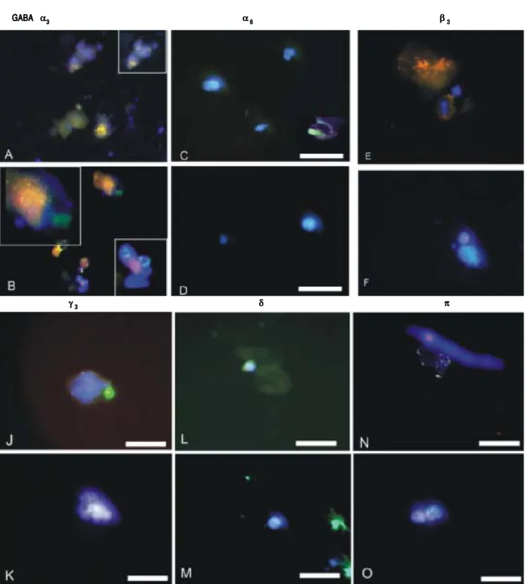
GABAA receptor subunit expression
GABAA receptor subunit expression in CSCs and
NSCs as determined by semi-quantitative RT-PCR and immunofluorescence are provided in table 1 and figure 2 respectively. Both techniques demonstrated that “phasic”
GABAAα3 expression prevailed in CSCs whereas “tonic”
GABAAα6 expression was expressed to a greater extent in
Membrane potential (mV)
0
-5
-10
-15
-20
-25
NSC CSC
*p < 0.01 n = 30
*
Figure 1. Figure 1.Figure 1.
Figure 1.Figure 1. Cell membrane potential differences (PDs) in normal stem cells (NSCs) and cancer stem cells (CSCs) derived from healthy human livers and hepatocellular carcinoma tissues respectively, as determined by microelec-trode impalements of isolated cells. The PDs of CSCs were significantly de-polarized (p < 0.01) when compared to NSCs.
Table 1. GABAA subunit expression in human liver cancer and normal stem cells.
Isolated human α3 α6 β3 γ3 δ π
liver EpCAM+
CSC ++++ + +++++ +++++ +++++ ++
NSC ++ ++ ++ + + +++++
NSCs. Other subunits selected on the basis of previous reports describing their differential expression in tumor versus adjacent non-tumor tissues also revealed different
patterns of expression in CSCs and NSCs. Specifically,
GABAAβ3, γ3 and δ expression was abundant in CSCs
whereas GABAAπ was dominant in NSCs.
Figure 2. Figure 2. Figure 2. Figure 2.
Figure 2. GABAA receptor subunit expression as documented by immunofluorescence of isolated normal stem cells (NSCs) and cancer stem cells (CSCs) de-rived from healthy human livers and hepatocellular carcinoma tissues respectively. Cell nuclei are counterstained blue with DAPI.
GABA GABA GABA GABA
GABA ααααα33333 ααααα66666 βββββ33333
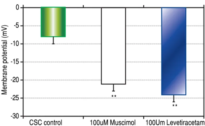
Response of stem cells to GABAA receptor agonists
Both CSCs and NSCs significantly (p < 0.01)
hyper-polarized following the addition of GABAA receptor
ago-nists muscimol and levetiracetam (Figure 3). However, the extent of hyperpolarization was similar in the two cell
populations (CSCs: Δ12.5 ± 1.2 and NSCs: Δ11.0 ± 3.5
mV).
DISCUSSION
The results of the present study indicate that primary CSCs derived from human HCC are significantly depo-larized relative to NSCs derived from the same livers. They also indicate that the differences in membrane PDs
are associated with differences in GABAA receptor
subu-nit expression which favor phasic rather than tonic GABAergic innervation and thereby, a more depolarized state for CSCs. Finally, they demonstrate that CSCs and NSCs can be hyperpolarized by the addition of specific
GABAA receptor agonists.
To date, there have been no reports documenting or comparing membrane PDs in human CSCs and NSCs de-rived from the liver or other tissues. In the only previous paper specifically designed to determine whether differ-ences in PDs exist in cancer and non-cancer stem cells, we described significantly depolarized CSCs derived from the malignant, human Huh-7 and PLC hepatocyte cell lines when compared to NSCs derived from a
non-malig-nant, rodent WBF-344 cell line.5 However, these findings
might well have related to the different species involved as significant interspecies variability has been described for
PD determinations.19,20
Unlike the paucity of data regarding PDs in stem cells, there are numerous previous reports describing differences
in PD determinations between malignant and non-malig-nant “mature” cells of the same tissue. For example, we previously documented that mature malignant hepatocytes which constitute the bulk of HCC tissue are significantly
depolarized relative to adjacent non-tumor hepatocytes.18
We also reported that the PDs of malignant hepatocyte cell lines are significantly depolarized relative to freshly
isolated human hepatocytes.21 Similar findings have been
reported in cervical, endometrial, breast, neuronal and
muscle tumors by other investigators.22-25 Thus, overall,
the results of the present study are in keeping with those of previous reports for the majority of tumor cells and ex-tend the findings to the more clinically relevant, human stem cell populations.
Certain GABAA receptor subunits have been reported
to be either upregulated or downregulated in solid organ tumors.12-18 In the present study, we focused on the α
3 and
α6 subunits because they are instrumental in determining
whether GABAergic innervation is phasic (relatively depo-larizing) or tonic (relatively hyperpodepo-larizing)
respective-ly.8 Moreover, the relative contributions of α
3 and α6
subunit expression serves to define extra-synaptic cell in-nervation which would be more relevant to solid organ tissues such as the liver. Thus, in addition to providing new insights into the mechanism whereby CSCs derived from HCCs are depolarized relative to NSCs, the finding
of enhanced GABAAα3 subunit expression in CSCs serves
to identify that subunit (in addition to β3, γ3 and δ) as a po-tential target for immune-mediated HCC therapy.
As mentioned earlier, CSCs tend to be resistant to chemotherapy. The most common explanation for this finding is an upregulation of multi-drug resistant (MDR) genes such as MDR-1 which encode for p-glycoproteins that efficiently export cationic chemotherapeutic agents
including doxorubicin from CSCs.26,27 This PD
depend-ent process raises the possibility that by hyperpolarizing CSC PDs, as was achieved in the present study with the
addition of GABAA receptor agonists, MDR-mediated
ex-port of chemotherapeutic agents might be attenuated and thereby, the chemosensitivity of CSCs enhanced. In addi-tion, because the transition of cells from a primitive to well differentiated phenotype is dependent on a simulta-neous transition from a depolarized to hyperpolarized state, activation of CSCs with GABAergic agents could conceivably result in their differentiation and loss of the
self-replication property inherent to CSCs.28 Clearly,
fur-ther research is required to determine whefur-ther these the-oretical considerations will translate into more effective treatments for HCC.
There are a number of limitations to this study that war-rant emphasis. First, CSCs and NSCs were isolated from resected human liver tissues and PD values were not
docu-mented in situ. This limitation relates to previous studies
Figure 3. Figure 3.Figure 3.
Figure 3.Figure 3. The effects of GABAA receptor agonists muscimol or levetira-cetam on cancer stem cell (CSC) membrane potential differences (PDs) as determined by microelectrode impalement of cells. CSCs hyperpolarized by approximately 12 mV following exposure to the GABAA receptor agonists.
Membrane potential (mV)
0 -5 -10 -15 -20 -25 -30
CSC control 100uM Muscimol 100Um Levetiracetam **
that have documented differences in cell PDs when
deter-mined in situ versus in vitro.18,19 However, the isolation
process employed in the present study was identical for both cell populations and therefore, although absolute val-ues may differ, relative differences should have remained intact. Moreover, because CSCs represent a very small per-cent of the bulk tumor cell population (< 5%) and NSCs an even smaller percent of non-tumor tissue, recording of
CSC and NSC PDs in situ is not feasible with presently
available techniques. A second limitation relates to focusing
on specific GABAA receptor subunits rather than
docu-menting the expression of all 16 subunits identified to date. As indicated above, the rationale for the more selective ap-proach was based on the relative importance of non-synap-tic GABAergic transmission in non-neuronal tissues and
previous studies documenting that specific GABAA
recep-tor subunits are significantly up- or down-regulated in tu-mor vs. non-tumor tissues.12-18 Yet another limitation was
the absence of data regarding the expression of other neuro-transmitter and electrogenic systems. Although we previ-ously reported that nicotinic receptors and Na/K ATPase activity are not perturbed in HCC, these data were derived from the “mature” cell populations of HCC tumors and may not be applicable to stem cell populations.15,18 Finally,
the clinical relevance of this study is predicated on the as-sumption that CSCs play an important role in the develop-ment of HCC and although emerging data tend to support that hypothesis, a consensus on the precise role of stem cells in carcinogenesis has yet to be reached.
In conclusion, the results of this study have identified
clear distinctions between the PDs and GABAA receptor
subunit expression profiles of CSCs and NSCs derived from human HCC and adjacent non-tumor tissues. These findings not only provide important new insights into he-patic carcinogenesis but could conceivably be exploited in future attempts to develop new, more effective therapies for patients with HCC.
ABBREVIATIONS
• CSC: cancer stem cell.
• EpCAM: epithelial cell adhesion molecule. • GABA: gamma-aminobutyric acid.
• HCC: hepatocellular carcinoma. • MDR: multi-drug resistant. • NSC: normal stem cell.
• PD: potential differences.
• RT-PCR: reverse transcriptase polymerase chain re-action.
CONFLICT OF INTEREST
None.
ACKNOWLEDGEMENTS
This research was funded by Kenroc Building Materi-als Co. Ltd. and the Canadian Liver Foundation. The au-thors would like to thank Ms R. Vizniak for her prompt and accurate typing of the manuscript.
REFERENCES
1. Yu Z, Pestell TG, Lisanti MP, Pestell RG. Cancer stem cells.
Int J Biochem Cell Biol 2012; 44: 2144-51.
2. Garg M. Gain of antitumor functions and induction of differ-entiation in cancer stem cells contribute to complete cure and no relapse. Crit Rev Oncog 2009; 15: 65-90.
3. Alison MR, Islam S, Lim S. Stem cells in liver regeneration, fi-brosis and cancer: the good, the bad and the ugly. J Pathol
2009; 217: 282-98.
4. Matthews VB, Yeoh GC. Liver stem cells. IUBMB Life 2005; 57: 549-53.
5. Bautista W, Perez-Alvarez V, Burczynski F, Raouf A, Klo-nisch T, Minuk G. Membrane potential differences and GABAA receptor expression in hepatic tumor and non-tumor stem cells. Can J Physiol Pharmacol 2014; 92: 85-91. 6. Varju P, Katarova Z, Madarasz E, Szabo G. GABA signalling
during development: new data and old questions. Cell Tis-sue Res 2001; 305: 239-46.
7. Young SZ, Bordey A. GABA’s control of stem and cancer cell proliferation in adult neural and peripheral niches. Physi-ology (Bethesda) 2009; 24: 171-85.
8. Sigel E, Steinmann ME. Structure, function, and modulation of GABA(A) receptors. J Biol Chem 2012; 287: 40224-31. 9. Belelli D, Harrison NL, Maguire J, Macdonald RL, Walker MC,
Cope DW. Extrasynaptic GABAA receptors: form, pharma-cology, and function. J Neurosci 2009; 29: 12757-63. 10. Kahan B, Magliocca J, Merriam F, Treff N, Budde M, Nelson J,
Browning V, et al. Elimination of tumorigenic stem cells from differentiated progeny and selection of definitive endoderm reveals a Pdx1+ foregut endoderm stem cell lineage. Stem Cell Res 2011; 6: 143-57.
11. Minuk GY, Bear CE, Sarjeant EJ. Sodium-independent, bicuc-ulline-sensitive [3H]GABA biding to isolated rat hepatocytes.
Am J Physiol 1987; 252: G642-G647.
12. Zhang X, Zhang R, Zheng Y, Shen J, Xiao D, Li J, Shi X, et al. Expression of gamma-aminobutyric acid receptors on ne-oplastic growth and prediction of prognosis in non-small cell lung cancer. J Transl Med 2013; 11: 102.
13. Li YH, Liu Y, Li D, Liu YH, Li F, Ju Q, Xie PL, et al. GABA stimulates human hepatocellular carcinoma growth through overexpressed GABAA receptor theta subunit. World J Gastroenterol 2012; 18: 2704-11.
14. Kimura T, Ishikawa K, Sakasegawa Y, Teruya K, Sata T, Schatzl H, Doh-ura K. GABAA receptor subunit beta1 is involved in the formation of protease-resistant prion protein in prion-infected neuroblastoma cells. FEBS Lett 2010; 584: 1193-8.
15. Liu Y, Li YH, Guo FJ, Wang JJ, Sun RL, Hu JY, Li GC. Gam-ma-aminobutyric acid promotes human hepatocellular carci-noma growth through overexpressed gamma-aminobutyric acid A receptor alpha 3 subunit. World J Gastroenterol
2008; 14: 7175-82.
17. Johnson SK, Haun RS. The gamma-aminobutyric acid A re-ceptors π subunit is overexpressed in pancreatic adenocar-cinomas. JOP. J Pancreas (Online) 2005; 6: 136-42. 18. Minuk GY, Zhang M, Gong Y, Minuk L, Dienes H,
Petti-grew N, Kew M, et al. Decreased hepatocyte membrane potential differences and GABAA-beta3 expression in human hepatocellular carcinoma. Hepatology 2007; 45: 735-45.
19. Moule SK, McGivan JD. Regulation of the plasma membrane potential in hepatocytes-mechanism and physiological signifi-cance. Biochim Biophys Act 1990; 1031: 383-97.
20. Varro A, Lathrop DA, Hester SB, Nanasi PP, Papp JG. Ionic currents and action potentials in rabbit, rat, and guinea pig ventricular myocytes. Basic Res Cardiol 1993; 88: 93-102. 21. Sun D, Gong Y, Kojima H, Wang G, Ravinsky E, Zhang M,
Minuk GY. Increasing cell membrane potential and GABAer-gic activity inhibits malignant hepatocyte growth. Am J Phys-iol Gastrointest Liver PhysPhys-iol 2003; 285: G12-G19.
22. Lash AF, Falk G, Gerard RW. A microelectrode method in the diagnosis of carcinoma of the cervix and endometrium. Am J Obstet Gynecol 1955; 70: 354-8.
23. Marino A, Iliev I, Schwalke MA, Gonzalez E, Marler K, Flana-gan C. Association between cell membrane potential and breast cancer. Tumor Biol 1994; 15: 82-9.
24. Picker S, Pieper CF, Goldring S. Glial membrane potentials and their relationship to [K+]o in man and guinea pig. J Neu-rosurg 1981; 55: 347-63.
25. Tokuoka S, Morioka H. The membrane potential of the human cancer and related cells. Jpn J Cancer Res 1957; 48: 353-4. 26. Perez-Tomas R. Multidrug resistance: retrospect and pros-pects in anti-cancer drug treatment. Curr Med Chem 2006; 13: 1859-76.
27. Han B, Zhang JT. Multidrug resistance in cancer chemother-apy and xenobiotic protection mediated by the half ATP-bind-ing cassette transporter ABCG2. Curr Med Chem Anticancer Agents 2004; 4: 31-42.
28. Sundelacruz S, Levin M, Kaplan DL. Role of membrane po-tential in the regulation of cell proliferation and differentiation.
Stem Cell Rev 2009; 5: 231-46.
Correspondence and reprint request:
G.Y. Minuk, M.D.
Morberg Family Chair in Hepatology. University of Manitoba. John Buhler Research Centre.
715 McDermot Ave. Winnipeg, MB R3E 3P4. Manitoba, Canada. Tel.: (204) 789-3204. Fax: (204) 789-3987