123
Annual Update
in Intensive Care
and Emergency
Medicine 2017
Jean-Louis Vincent
Editor
Annual Update in
Intensive Care and
Prof. Jean-Louis Vincent Dept. of Intensive Care Erasme Hospital
Université libre de Bruxelles Brussels, Belgium
jlvincent@intensive.org
ISSN 2191-5709 ISSN 2191-5717 (electronic) Annual Update in Intensive Care and Emergency Medicine
ISBN 978-3-319-51907-4 ISBN 978-3-319-51908-1 (eBook) DOI 10.1007/978-3-319-51908-1
© Springer International Publishing AG 2017
This work is subject to copyright. All rights are reserved by the Publisher, whether the whole or part of the material is concerned, specifically the rights of translation, reprinting, reuse of illustrations, recitation, broadcasting, reproduction on microfilms or in any other physical way, and transmission or information storage and retrieval, electronic adaptation, computer software, or by similar or dissimilar methodology now known or hereafter developed.
The use of general descriptive names, registered names, trademarks, service marks, etc. in this publica-tion does not imply, even in the absence of a specific statement, that such names are exempt from the relevant protective laws and regulations and therefore free for general use.
The publisher, the authors and the editors are safe to assume that the advice and information in this book are believed to be true and accurate at the date of publication. Neither the publisher nor the authors or the editors give a warranty, express or implied, with respect to the material contained herein or for any errors or omissions that may have been made.
Cover design: WMXDesign GmbH, Heidelberg
Printed on acid-free paper
This Springer imprint is published by Springer Nature The registered company is Springer International Publishing AG
Common Abbreviations. . . xi
Part I Infections
Severe Influenza Infection: Pathogenesis, Diagnosis, Management
and Future Therapy . . . 3
B. M. Tang and A. S. McLean
Implementing Antimicrobial Stewardship in Critical Care:
A Practical Guide . . . 15
J. Schouten and J. J. De Waele
Part II Sepsis
Microvesicles in Sepsis: Implications for the Activated Coagulation System 29
G. F. Lehner, A. K. Brandtner, and M. Joannidis
Mesenchymal Stem/Stromal Cells for Sepsis . . . 41
C. Keane and J. G. Laffey
Part III Fluids
Fluid Balance During Septic Shock: It’s Time to Optimize . . . 55
X. Chapalain, T. Gargadennec, and O. Huet
How to Use Fluid Responsiveness in Sepsis . . . 69
V. Mukherjee, S. B. Brosnahan, and J. Bakker
Use of ‘Tidal Volume Challenge’ to Improve the Reliability
of Pulse Pressure Variation . . . 81
S. N. Myatra, X. Monnet, and J.-L. Teboul
Distribution of Crystalloids and Colloids During Fluid Resuscitation:
All Fluids Can be Good and Bad? . . . 91
I. László, N. Öveges, and Z. Molnár
Part IV Renal Issues
New Diagnostic Approaches in Acute Kidney Injury. . . 107
M. Meersch and A. Zarbock
When Should Renal Replacement Therapy Start? . . . 119
J. Izawa, A. Zarbock, and J. A. Kellum
An Overview of Complications Associated with Continuous
Renal Replacement Therapy in Critically Ill Patients . . . 129
S. De Rosa, F. Ferrari, and C. Ronco
Measuring Quality in the Care of Patients with Acute Kidney Injury . . . 139
M. H. Rosner
Characteristics and Outcomes of Chronic Dialysis Patients Admitted
to the Intensive Care Unit. . . 149
M. Chan, M. Varrier, and M. Ostermann
Part V Metabolic Support
Energy Expenditure During Extracorporeal Circulation . . . 159
E. De Waele, P. M. Honore, and H. D. Spapen
Vitamin D, Hospital-Acquired Infections and Mortality
in Critically Ill Patients: Emerging Evidence . . . 169
G. De Pascale, M. Antonelli, and S. A. Quraishi
Part VI Cardiac Conditions
Anemia and Blood Transfusion in the Critically Ill Patient
with Cardiovascular Disease . . . 187
A. B. Docherty and T. S. Walsh
Right Ventriculo-Arterial Coupling in the Critically Ill . . . 203
Part VII Cardiopulmonary Resuscitation
Antiarrhythmic Drugs for Out-of-Hospital Cardiac Arrest
with Refractory Ventricular Fibrillation . . . 213
T. Tagami, H. Yasunaga, and H. Yokota
Airway and Ventilation During Cardiopulmonary Resuscitation . . . 223
C. J. R. Gough and J. P. Nolan
Part VIII Oxygenation and Respiratory Failure
High-Flow Nasal Cannula Support Therapy:
New Insights and Improving Performance . . . 237
G. Hernández, O. Roca, and L. Colinas
Urgent Endotracheal Intubation in the ICU: Rapid Sequence Intubation
Versus Graded Sedation Approach. . . 255
G. Zaidi and P. H. Mayo
Sedation in ARDS: An Evidence-Based Challenge . . . 263
D. Chiumello, O. F. Cozzi, and G. Mistraletti
Mechanical Ventilation in Obese ICU Patients:
From Intubation to Extubation . . . 277
A. De Jong, G. Chanques, and S. Jaber
Novel Insights in ICU-Acquired Respiratory Muscle Dysfunction:
Implications for Clinical Care . . . 291
A. Jonkman, D. Jansen, and L. M. A. Heunks
Part IX Neurological Conditions
Neuroanatomy of Sepsis-Associated Encephalopathy . . . 305
N. Heming, A. Mazeraud, and F. Verdonk
Clinical Utility of Blood-Based Protein Biomarkers
in Traumatic Brain Injury . . . 317
S. Mondello, A. I. R. Maas, and A. Buki
Novel Metabolic Substrates for Feeding the Injured Brain . . . 329
Part X Burn Patients
Fluid Therapy for Critically Ill Burn Patients . . . 345
A. Dijkstra, C. H. van der Vlies, and C. Ince
Burn Patients and Blood Product Transfusion Practice:
Time for a Consensus? . . . 359
A. Holley, A. Cook, and J. Lipman
Part XI Drug Development and Pharmaceutical Issues
Bridging the Translational Gap: The Challenges
of Novel Drug Development in Critical Care. . . 375
S. Lambden and C. Summers
Medicating Patients During Extracorporeal Membrane Oxygenation:
The Evidence is Building . . . 389
A. L. Dzierba, D. Abrams, and D. Brodie
Anti-Inflammatory Properties of Anesthetic Agents . . . 401
F. F. Cruz, P. R. M. Rocco, and P. Pelosi
Part XII The Extremes of Age
Facing the Ongoing Challenge of the Febrile Young Infant . . . 417
A. DePorre, P. L. Aronson, and R. McCulloh
Post-Discharge Morbidity and Mortality in Children with Sepsis. . . 431
O. C. Nwankwor, M. O. Wiens, and N. Kissoon
Emergency Abdominal Surgery in the Elderly:
How Can We Reduce the Risk in a Challenging Population? . . . 445
X. Watson and M. Cecconi
Part XIII Simulation
Patient-Specific Real-Time Cardiovascular Simulation as Clinical Decision
Support in Intensive Care Medicine . . . 459
M. Broomé and D. W. Donker
Making the Best Use of Simulation Training in Critical Care Medicine. . 477
Part XIV Organization and Quality of Care
We Have Good Enough Data to Support Sepsis Performance Measurement495
H. C. Prescott and V. X. Liu
The Use of Health Information Technology to Improve Sepsis Care . . . . 505
J. L. Darby and J. M. Kahn
Beyond Semantics: ‘Disproportionate Use of Intensive Care Resources’
or ‘Medical Futility’?. . . 517
E. J. O. Kompanje and J. Bakker
Reflections on Work-Related Stress Among Intensive Care Professionals:
An Historical Impression . . . 527
M. M. C. van Mol, E. J. O. Kompanje, and J. Bakker
AKI Acute kidney injury
ARDS Acute respiratory distress syndrome AUC Area under the curve
BMI Body mass index
COPD Chronic obstructive pulmonary disease CPR Cardiopulmonary resuscitation CRP C-reactive protein
CRRT Continuous renal replacement therapy
CT Computed tomography
CVP Central venous pressure
ECMO Extracorporeal membrane oxygenation EKG Electrocardiogram
ICU Intensive care unit
IL Interleukin
LPS Lipopolysaccharide LV Left ventricular MAP Mean arterial pressure MRI Magnetic resonance imaging
OR Odds ratio
PAOP Pulmonary artery occlusion pressure PCT Procalcitonin
PEEP Positive end-expiratory pressure PPV Pulse pressure variation RCT Randomized controlled trial RRT Renal replacement therapy RV Right ventricular
SIRS Systematic inflammatory response syndrome SOFA Sequential organ failure assessment
SVV Stroke volume variation TLR Toll-like receptor TNF Tumor necrosis factor
Diagnosis, Management and Future Therapy
B. M. Tang and A. S. McLean
Introduction
Severe influenza infection is an important cause of acute lung injury. Although other respiratory viruses (e. g., respiratory syncytial virus, human metapneumovirus) can also cause considerable pulmonary damage, influenza virus remains the main cause of respiratory failure in patients with suspected viral respiratory tract infection. In addition, influenza virus is the only respiratory virus that has caused four pandemics over the last 100 years, making it one of the most transmissible and virulent viruses in the world. Here, we review the pathogenesis, diagnosis, current management and future therapy of severe influenza infection.
Pathogenesis
Understanding the pathogenesis of severe influenza infection is the key to devel-oping new therapeutic strategies. Although the basic process of a mild influenza infection is well understood, our understanding of how a mild illness progresses to a potentially lethal pulmonary infection remains poor. In this section, we will review recent advances in the immunopathology of severe influenza infection.
Pulmonary epithelial cells are the first target of invasion by influenza virus. Like most cells, epithelial cells constitutionally upregulate the interferon pathway in re-sponse to infection by viruses. Types I and III interferon pathways are the natural
B. M. Tang (
)Department of Intensive Care Medicine, Level 2, North Block, Nepean Hospital Derby Street, Kingswood, NSW 2747, Australia
Centre for Immunology and Allergy Research, Westmead Institute for Medical Research Westmead, NSW 2145, Australia
e-mail: benjamin.tang@sydney.edu.au
A. S. McLean
Department of Intensive Care Medicine, Level 2, North Block, Nepean Hospital Derby Street, Kingswood, NSW 2747, Australia
3 © Springer International Publishing AG 2017
defense mechanism against influenza virus. Upon infection, epithelial cells upreg-ulate interferon regulatory factors (IRF), such as IRF-3 and IRF-7. This leads to transcription and translation of a downstream interferon pathway, which in turn produces a family of interferon-stimulated genes/proteins. This vast family of in-terferon-stimulated genes/proteins (> 300) provides a wide spectrum of anti-viral effects, ranging from inhibition of viral replication to sensing of influenza virus in-side the host cells. This response is immediate and effective, making it a critical part of the innate immune response against influenza virus.
Whilst essential, the interferon response alone is not sufficient to prevent virus replication in severely infected cases. Multiple subsets of immune cells (e. g., macrophages, dendritic cells and neutrophils) are required to mount an effective immune response. The failure of this immune response is the hallmark of severe infection, which is characterized by multiple defects in immune cell recruitment, activation or proliferation, as described below.
Alveolar macrophages are among the early responders to influenza virus. They phagocytose infected cells containing influenza virus and initiate other cells of innate and adaptive immunity. Failure of alveolar macrophages to mount an effec-tive early response is associated with increased viral dissemination and increased morbidity/mortality. Neutrophils are also early responders in severe influenza in-fection. Similar to alveolar macrophages, failure of this early neutrophil response is a prominent feature of severe influenza infection. Paradoxically, an exuberant or in-appropriately exaggerated neutrophil response is also a feature of severe influenza infection. For example, in severe H1N1 and H5N1 infection, the large influx of neutrophils into the alveolar space is a classic feature [1]. During this massive neutrophil influx, the neutrophils release a large amount of cytokines, extracel-lular proteases and histones. This leads to a breakdown of the epithelial barrier, accumulation of reactive oxygen species (ROS), flooding of alveolar spaces by in-flammatory fluid and increased barrier to oxygenation, all of which contribute to the clinical picture of acute lung injury commonly observed in patients with severe influenza infection.
In the later phase of the host response, adaptive immunity becomes the dom-inant player. Here, activated CD8 T-lymphocytes cause lysis of the influenza-in-fected epithelial cells, which facilitates virus clearance. Impaired CD8 responses are a prominent feature of highly pathogenic influenza infection, such as the re-cently reported H7N9 outbreak in China [6]. In addition to cell lysis, CD8 cells also enhance the pro-inflammatory response, which could either contribute to host defense or, in some cases, worsen lung inflammation and cause further pulmonary damage.
Diagnosis
The detection of influenza virus is the first step in establishing a diagnosis. Rapid antigen detection assays offer a low-cost approach with a short turn-around time. However, a recent review demonstrated that such assays have an unacceptably low sensitivity [7]. Nucleic acid amplification (e. g., multiplex viral polymerase chain reaction [PCR]) has recently gained a much greater prominence due to its high sen-sitivity and specificity. Currently, this is the most accepted gold standard for virus detection in the initial evaluation of suspected influenza infection. However, there are three important caveats regarding the clinical utility of nucleic acid amplifica-tion assay:
(1) The reliability of such an assay is dependent on the fact that the viral genome is known. An unknown viral genome, mutant strain or new pandemic influenza virus will be difficult to detect.
(2) The sensitivity is affected by the way the sample is collected. Poor sample col-lection, inability to access lower airway or reduced virus shedding (due to prior anti-viral administration) all reduce detection sensitivity.
(3) Detection does not imply infection because the presence of influenza virus in the upper airway may be a co-incidental finding or active infection. In fact, 18% of exposed individuals show no clinical symptoms; therefore, the presence of the virus does not always imply that it is the causative agent. Furthermore, de-tection of an incomplete virus segment (by nucleic acid amplification) does not constitute sufficient proof that active viral replication is present.
In addition to virus detection, clinicians need to identify which patients are more likely to progress to severe disease or require admission to the intensive care unit (ICU). Table 1summarizes virus-related and host factors that may contribute to progression to more severe disease. Some of these factors are clinically obvious (e. g., age, pre-existing medical conditions). Other factors (e. g., genetic susceptibil-ity) require highly sophisticated laboratory testing (e. g., high-throughput genome sequencing), which are not yet available in the routine clinical setting.
Table 1 Risk factors for progression to severe influenza infection
Viral factors Host factors
Subtype of influenza virus (e. g., H7N9) Genetic susceptibility (e. g., IFITM3) Viral load (e. g., high viral load increases
severity)
Pregnancy, obesity and extremes of age (elderly and neonates)
Mutation in viral genome (e. g., PB2 gene mutation enhances viral replication)
Pre-existing medical conditions (e. g., chronic lung diseases, cancer, chemotherapy)
IFITM3: interferon-induced transmembrane protein 3
ICU develop bacterial co-infection as a complication [8]. The causative bacterial co-pathogens are most likely to beStreptococcus pneumoniaeorStaphylococcus aureus. The basis for increased susceptibility is thought to be due to production of type I interferon, which is increased initially in response to influenza virus in-fection, but also decreases the synthesis of IL-1B, IL-23, IL-17 and IL-22, which in turn inhibit the production of antimicrobial peptides [9]. Furthermore, the pro-inflammatory milieu caused by the influx of neutrophils also contributes towards increased susceptibility to bacterial super-infection. Other immune-related factors also contribute towards increased susceptibility including reduced type 17 immune response, impaired antimicrobial peptide (AMP) production by lung epithelia and reduced phagocyte function [9].
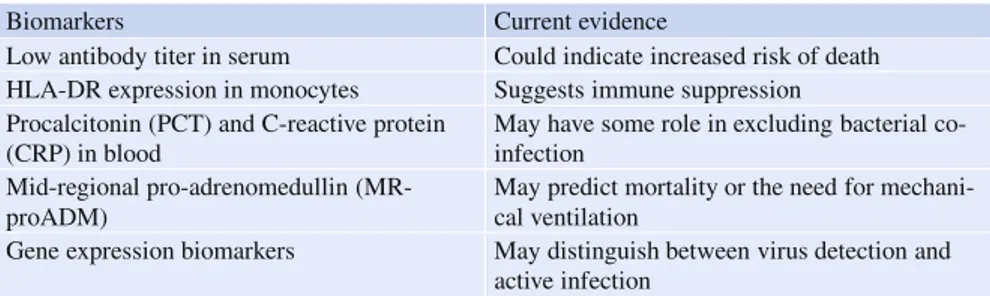
Host response biomarkers should form an important part of the diagnostic eval-uation of an infected patient. Biomarkers assist clinical evaleval-uation by providing additional information that is not available by conventional virus detection assay. This additional information includes an improved ability to distinguish between co-incidental ‘bystander’ virus and true infection, to predict clinical risk for further de-terioration and to monitor treatment response. Table2summarizes the host response biomarkers that have been recently investigated in the literature.
Gene expression biomarkers are the most recent development in biomarker re-search. These biomarkers differ from conventional biomarkers (e. g., C-reactive protein [CRP] or procalcitonin [PCT]) in that they are much more influenza specific, due to the fact that many of them are interferon derived genes, which are upregu-lated in response to respiratory virus infection. A recently published landmark study
Table 2 Host response biomarkers for influenza infection
Biomarkers Current evidence
Low antibody titer in serum Could indicate increased risk of death HLA-DR expression in monocytes Suggests immune suppression Procalcitonin (PCT) and C-reactive protein
(CRP) in blood
May have some role in excluding bacterial co-infection
Mid-regional pro-adrenomedullin (MR-proADM)
May predict mortality or the need for mechani-cal ventilation
showed that these biomarkers could address several important clinical questions si-multaneously (whereas conventional biomarkers could address only one question at a time) [10]. First, these biomarkers could assist clinicians to identify patients most likely to have infection (bacterial and viral) in a heterogeneous population of patients with undifferentiated respiratory illnesses. Second, among infected pa-tients, the biomarkers could distinguish between bacterial and viral infection. Third, among infected patients, the biomarkers could prognosticate and predict clinical outcomes. In addition, the biomarkers could be easily measured in most clinical settings due to the ease of sampling (only 2.5 ml of whole blood is required) and the wide availability of PCR machines (to measure gene-expression). Importantly, because these biomarkers reflect changes in the immune pathway during influenza infection, they provide additional diagnostic information not offered by conven-tional pathogen detection assay (e. g., virus nucleic amplification). Although further validation studies are necessary before these biomarkers can be widely adopted in clinical practice, it is highly likely that they will be incorporated into the diagnostic armamentaria of modern laboratories in the future.
Management
The management of severe influenza infection is mainly supportive. Standard mea-sures should include those used for the management of acute respiratory distress syndrome (ARDS). Therapeutic agents for severe influenza infection are limited, with oseltamivir being the most commonly used anti-viral agent. A recent meta-analysis showed that oseltamivir could reduce symptom duration and the risk of developing lower respiratory tract complications (e. g., viral pneumonia) [11]. How-ever, its efficacy is dependent on oseltamivir being administered in the early phase of the illness. This may pose difficulty in the management of ICU patients, because these patients often present in the late phase of their illness. Regardless of the tim-ing of presentation, oseltamivir should be considered in all high-risk patients. The current recommendation by the World Health Organization (WHO) indicates that it should be administered in immunocompromised patients, patients with severe comorbidities or underlying chronic lung diseases, age < 2 or > 65 years, morbid obesity, nursing home residents, women who are pregnant or post-partum, and pa-tients with signs of severe respiratory disease.
corticosteroids in severe influenza infection is not recommended in routine clinical care and should be restricted to patients in the setting of clinical trials.
Future Therapy
Although conventional treatment for severe influenza infection is limited, novel therapeutic agents have shown great promise. These novel agents consist of mainly two classes: immune agents that modulate host response and anti-viral agents that inhibit viral replication.
Immune Agents that Modulate Host Response
Host Factors that Control Viral RNA Replication
In order to replicate successfully, the influenza virus mRNA undergoes transcrip-tion. Initiation of primary viral RNA transcription depends on the activity of host RNA polymerase. Inhibition of this transcription process provides a therapeutic op-portunity to halt the commencement of viral RNA replication. Inhibitors of this process, such as CDK9 inhibitor, have undergone preclinical evaluation.
Host Signaling Pathways Influenced by Redox Balance
The influenza virus hijacks the host cell signaling pathway to benefit its own prop-agation. Phosphorylation of the mitogen-activated protein kinase (MAPK) pathway has been shown to facilitate viral nucleoprotein trafficking [13]. Therefore, inhibi-tion of the MAPK pathway could potentially reduce spread of the influenza virus. Of particular relevance to the intensivist is the fact that the activity of the MAPK pathway is determined by the oxidative-reductive state of the host cell. N-acetyl-cysteine, a well-established drug already commonly used in ICU patients, could modulate the oxidative-reductive state of the host cell, thereby affecting influenza virus propagation. A recent study has demonstrated the potential efficacy of this agent in treating severe influenza infection in an animal model [14]. Other anti-oxidant agents, such as p38 inhibitor or glutathione, are also potential new host-based therapeutic agents that modulate the redox balance within the host cell. In addition to the MAPK pathway, the PI3K pathway is also sensitive to the effect of redox balance. PI3K is a signaling pathway implicated in influenza infection [15]. An in vitrostudy showed that inhibition of this pathway could reduce influenza virus replication [16]. Importantly, PI3K inhibitors have already been approved as anticancer drugs. Therefore, the possibility of extending their use as anti-influenza agent offers a promising new avenue for future investigation.
Host Factors that Regulate Inflammation
path-way inhibitors, such as acetyl-salicylic acid, could block influenza virus replication and propagation. Other pathway inhibitors, such as SC75741, also decrease viral replication. This agent has the unique feature of having a low potential in selecting viral resistant variants, therefore making it unlikely to result in anti-viral resistance [17]. Furthermore, SC75741 has recently been shown to reduce viral replication and cytokine expression in highly pathogenic strains (e. g., H5N1 and H7N7), making it a potential candidate for further investigation in severe influenza infection [18].
The cyclooxygenase (COX) pathway is another pro-inflammatory pathway that has been implicated in influenza virus infection. Highly pathogenic influenza strains, such as H5N1, strongly upregulate COX-2 mediated pro-inflammatory sig-naling that causes hypercytokinemia during severe H5N1 infection. A non-steroidal COX-2 inhibitor has been shown to inhibit H5N1 infection in human macrophages, making it another potential agent for severe influenza infection [19].
Host Interferon Pathway
The interferon pathways (type I and type II) are the most potent defense of the host cell against influenza virus infection. Activation of interferon pathways leads to upregulation of more than 300 interferon-stimulated genes. Many of these in-terferon-stimulated genes have potent anti-influenza activity, such as MX1 (anti-influenza),ISG15 (inhibits influenza virus replication),OAS1, OAS2, OAS3 (de-grades viral RNA),EIF2AK2(inhibits viral replication),HERC5(positive regulator of anti-viral response) andIFIT2(inhibits expression of viral mRNA). In addition, these genes activate the adaptive immune response and induce programmed cell death of virally infected cells.
Novel therapeutic strategies take advantage of this endogenous anti-influenza defense by identifying trigger points that activate the interferon pathway. Several molecular pathways are known to trigger the interferon pathway. For example, Toll-like receptor (TLR) 3 and 7 are known to activate the interferon pathway in lung epithelium and immune cells. In plasmacytoid dendritic cells, TLR7 activa-tion produces massive interferon release at 1,000 times that of any other immune cell in the human host. Ligands that selectively target TLR7 in plasmacytoid den-dritic cells could be potential therapeutic targets. Other TLR ligands, such as CpG oligodeoxynucleotides (TLR9), have been shown to protect against lethal influenza infection in experimental settings [20]. In lung epithelium, TLR3 is the dominant pathway leading to interferon pathway activation. A large number of TLR3 and TLR9 agonists are currently in clinical trial phase for the treatment of autoim-mune conditions, cancer and viruses. It is possible to extend the application of these agents to treat severe influenza infection. Further investigation on these promising new agents may open the door for developing new treatments in severe influenza infection.
Host Factors Implicated in Virus Entry into Human Cells
S1 (TMPRSS) that belongs to the type II transmembrane serine protease family. This enzyme is located in the human airway epithelium and plays an important role in permitting influenza virus to gain entry into the host cell. Consequently, a protease inhibitor that binds to the TMPRSS molecule is a potential drug tar-get in the treatment of influenza infection. Recent studies have identified three TMPRSS molecules, namely TMPRSS2, TMPRSS4 and TMPRSS11D, as poten-tial drug targets [21]. These molecules have been detected in multiple locations within the human respiratory tract, including nasal mucosa, the trachea, the distal airway and the lung. Aprotinin, a drug familiar to most intensivists, is a protease inhibitor and has been shown to reduce influenza virus replication. In addition to reducing viral replication, aprotinin has also been shown to reduce inflammatory cytokines, suggesting a further benefit other than its impact on viral replication. So far, findings with the TMPRSS molecule have been derived mainly fromin vitro models. Further studies in animal models and human clinical trials are needed in the future.
Anti-Viral Agents that Inhibit Viral Replication
Neuraminidase
Neuraminidase is a glycoside hydrolase that removes a sialic acid residue of the host cellular receptor recognized by influenza virus hemagglutinin. Therefore, it is an essential component of a process that allows virus penetration through mu-cosal barriers and subsequently to gain entry into the host cell. In addition, after virus replication, neuraminidase detaches the virion from the infected cells, thereby facilitating release and subsequent spread of the viral progeny. Consequently, raminidase is essential for viral infectivity to host cells. Therefore, inhibiting neu-raminidase is the primary therapeutic strategy currently used in clinical practice. Most clinicians will be familiar with two neuraminidase inhibitors, zanamivir and oseltamivir.
Table 3 Drugs that block the two critical processes in hemagglutinin function
Virus interacting with cell surface Virus fusion with cell membrane Carbohydrate-binding agents that recognize
glycosylation sites on hemagglutinin
Molecules that inhibit confirmation change in hemagglutinin
Peptides against hemagglutinin Neutralizing antibodies directed against the stem region of hemagglutinin
Decoy receptor or sialic acid-containing in-hibitors
Neutralizing monoclonal antibodies directed against the globular head domain of hemag-glutinin
Hemagglutinin
Hemagglutinin is pivotal for the interaction between influenza virus and the sialic acid on the surface of the host cells. In addition, it is required for the fusion be-tween the viral envelop and the endosomal membrane of the host cell, which is the final step in the virus’s entry into the host cell. Inhibiting hemagglutinin could be achieved by two methods: (1) preventing the interaction between viral surface molecules and the host cell surface receptor; and (2) blocking the fusion of the viral envelop with the host cell membrane. Table3summarizes the recent development in the new drugs that utilize the above two strategies.
M2 Ion Channel
The M2 protein is a proton channel inside the influenza virus. After gaining entry into the host cell, the influenza virus activates the M2 protein by sensing a drop in the pH value inside the enveloped vesicle (the endosome). The activation of the M2 proton channel results in a proton flux from the endosome into the virion core. Acidification of the virus interior leads to dissociation of the viral ribonucleopro-tein complexes. Subsequent membrane fusion releases the ribonucleoproribonucleopro-tein into the cytoplasm. This release allows the virus to be imported into the nucleus to start viral replication. Other important functions of the M2 protein are: formation of the filamentous strains of the virus; release of the budding virion; and stabilization of the virion budding site. Due to these important functions, inhibition of M2 protein represents an ideal therapeutic target. A well-known licensed antiviral drug, aman-tadine, is an M2 blocker that binds the N-terminal channel lumen of the M2 pore resulting in repulsion of protons and subsequently prevent virus uncoating. Unfor-tunately, this class of drug is not active against all strains of influenza virus (e. g., influenza B). In addition, the emergence of drug-resistant virus variants has been reported. These drawbacks have significantly limited the use of M2 blockers.
Conclusion
un-changed over the last few decades, due mainly to a lack of effective new therapies with which to treat such patients.
However, we have gained a much better understanding of the mechanisms of the disease in recent years. This improved understanding points to the pivotal roles played by immune dysregulation in causing severe disease. Furthermore, our ability to diagnose influenza infection, to stratify high-risk patients and to prognosticate clinical outcomes has also improved thanks to recent advances in genomic sci-ence. Importantly, a large number of novel therapeutic agents are currently under investigation. These novel agents target multiple critical points of the host response pathway. Agents that modulate the host response hold particularly great promise since dysregulated immunity is the main driver towards more severe infection. In the future, clinical trials will be an important next step to demonstrate the efficacy of these novel agents.
References
1. Perrone LA, Plowden JK, Garcia-Sastre A, Katz JM, Tumpey TM (2008) H5N1 and 1918 pandemic influenza virus infection results in early and excessive infiltration of macrophages and neutrophils in the lung of mice. PLoS Pathog 4:e1000115
2. Peteranderl C, Morales-Nebreda L, Selvakumar B et al (2016) Macrophage-epithelial paracrine crosstalk inhibit lung edema clearance during influenza infection. J Clin Invest 126:1566–1580
3. Hogner K, Wolff T, Pleschka S et al (2013) Macrophage-expressed IFN-beta contributes to apoptotic alveolar epithelial cell injury in severe influenza virus pneumonia. PLoS Pathog 9:e1003188
4. Soloff A, Weirback H, Ross T, Barratt-Boyes SM (2012) Plasmacytoid dendritic cell depletion leads to an enhance mononuclear phagocyte response in lungs of mice with lethal influenza infection. Comp Immunol Microbiol Infect Dis 35:309–317
5. Moseman EA, Liang X, Dawson AJ et al (2004) Human plasmacytoid dendritic cells acti-vated by CpG oligodeoxynucleotides induce the generation of CD4+CD25+regulatory T cells. J Immunol 173:4433–4442
6. Wang Z, Wan Y, Qiu C et al (2015) Recovery from severe H7N9 disease is associated with diverse response mechanisms dominated by CD8+ T cells. Nat Commun 6:6833
7. Chartrand C, Leeflang MM, Minion J, Brewer T, Pai M (2012) Accuracy of rapid influenza diagnostic tests: a meta-analysis. Ann Intern Med 156:500–511
8. Metersky ML, Masterton RG, Lode H et al (2012) Epidemiology, microbiology and treatment considerations for bacterial pneumonia complicating influenza. Int J Infect Dis 16:e321–e331 9. Robinson KM, Kolls JK, Alcorn J (2015) The immunology of influenza virus-associated
bac-terial pneumonia. Curr Opin Immunol 34:59–67
10. Sweeney TE, Wong HR, Khatri P (2016) Robust classification of bacterial and viral infections via integrated host gene expression diagnostics. Sci Transl Med 6:346ra91
11. Dobson J, Whitley RJ, Pocock S, Monto AS (2015) Oseltamivir treatment for influenza in adults: meta-analysis of randomised controlled trials. Lancet 385:1729–1737
12. Zhang Y, Sun W, Svendsen ER et al (2015) Do corticosteroids reduce the mortality of influenza A (H1N1) infection? A meta-analysis. Crit Care 19:46
14. Geiler J, Michaelis M, Naczk P et al (2010) N-acetyl-L-cysteine (NAC) inhibits virus repli-cation and expression of pro-inflammatory molecules in A549 cells infected with highly pathogenic H5N1 influenza A virus. Biochem Pharmacol 79:413–420
15. Ayllon J, Garcia-Sastre A, Hale BG (2012) Influenza A viruses and PI3K; are there time, place and manner restrictions? Virulence 3:411–414
16. Shin YK, Liu Q, Tikoo SK, Babiuk LA, Zhou Y (2007) Effect of the phosphatidylinositol 3-kinase/Akt pathway on influenza A virus propagaton. J Gen Virol 88:942–950
17. Ehrhardt C, Ruckle A, Hrincius ER et al (2013) The NF-kappaB inhibitor SC75741 efficiently blocks influenza virus propagation and confers a higher barrier for development of viral resis-tance. Cell Microbiol 15:1198–1211
18. Haasbach E, Reiling SJ, Ehrhardt C et al (2013) The NJ-kappaB inhibitor SC75741 protects mice against highly pathogenic avian influenza A virus. Antiviral Res 99:336–344
19. Lee SM, Gai WW, Cheung TK, Peiris JS (2011) Antiviral effect of a selective COX-2 inhibitor on H5N1 infection in vitro. Antiviral Res 91:330–334
20. Cheng WK, Plumb AW, Lai JCY, Abraham N, Dutz JP (2016) Topical CpG Oligodeoxynu-cleotide adjuvant enhances the adaptive immune response against influenza A infections. Font Immunol 7:284
21. Yamaya M, Shimotai Y, Hatachi Y, Homma M, Nishimura H (2016) Serine proteases and their inhibitors in human airway epithelial cells: effects on influenza virus replication and airway inflammation. Clin Microbiol 5:238
in Critical Care: A Practical Guide
J. Schouten and J. J. De Waele
Introduction
Management of infections is an important issue in many health care settings, but severe infections are most prevalent and antimicrobial use is most abundant in the intensive care unit (ICU). Not surprisingly, antimicrobial resistance has emerged primarily in the intensive care setting, where multiple facilitators for the develop-ment of resistance are present: high antibiotic pressure, loss of physiological bar-riers and high transmission risk. ‘Intensive care’ had higher proportions of treated patients, combination therapy, hospital-acquired infections and parenteral adminis-tration of antibiotics in a point prevalence survey on antimicrobial prescription by the ESAC (European Surveillance of Antimicrobial Consumption) in 172 European hospitals across 25 countries [1].
Antimicrobial prescription is a complex process influenced by many factors. The appropriateness of antimicrobial use in hospitals varies among physicians, hospitals and countries due to differences in professional background, clinical experience, knowledge, attitudes, hospital antibiotic policies, collaboration and communication among professionals, care coordination and teamwork, care logistics, and differ-ences in sociocultural and socioeconomic factors [2].
One can imagine that changing professional practice is a major challenge. The scientific literature is full of examples from which it would appear that patients are not given the care that, according to recent scientific or professional insight as summarized in guidelines, is desirable. A multitude of studies has shown that 30–
J. Schouten (
)Dept. of Intensive Care Medicine, Canisius Wilhelmina Ziekenhuis Weg door Jonkerbos 100, 6532 SZ Nijmegen, Netherlands IQ healthcare, Radboud University Medical Center Nijmegen, Netherlands
e-mail: j.schouten@cwz.nl
J. J. De Waele
Dept. of Intensive Care Medicine, University Hospital Gent 9000 Gent, Belgium
15 © Springer International Publishing AG 2017
40% of patients do not receive care according to guidelines and the findings for antimicrobial prescribing are similar [3]. This renders changing ICU antimicrobial use into a challenge of formidable complexity. Given that many influencing factors play a part, the measures or strategies undertaken to improve antimicrobial use need to be equally diverse.
Many interventions and programs have been designed to improve appropriate antimicrobial use in terms of choice of drugs, dosing, timing, de-escalation and discontinuation. Such interventions are collectively known as antimicrobial stew-ardship programs. An ICU antimicrobial stewstew-ardship program can be thought of as a menu of interventions that is adapted and customized to fit the infrastructure and organization of ICUs [4].
In this chapter, we will review the rationale for antimicrobial stewardship pro-grams and take a step by step approach on how to implement such propro-grams in the critical care setting and how to optimize compliance to relevant antibiotic steward-ship recommendations in the ICU.
Rationale for Antibiotic Stewardship in Critical Care
Health care institutions have adopted antimicrobial stewardship programs as a mechanism to ensure more appropriate antimicrobial use. Antimicrobial stew-ardship programs can have a significant impact in the ICU, leading to improved antimicrobial use and resistance patterns and decreased infection rates and costs, due to the inherent nature of infections encountered and the high and often inap-propriate antibiotic utilization in the ICU setting.
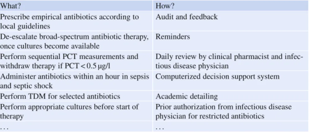
Stewardship programs are composed of two intrinsically different sets of inter-ventions (Table1). A first set of interventions describes recommended professional care interventions that define appropriate antimicrobial use in individual patients, regarding indication, choice of drug, dose, route or duration of treatment. For exam-ple, these may address ‘de-escalation of therapy’ in individual ICU patients. A sec-ond set of interventions describes recommended strategies to ensure that profession-als apply these professional care interventions in daily practice. These include both restrictive (e. g., formulary restriction) and persuasive (e. g., education, feedback) strategies to improve appropriate antimicrobial use in patient care. The second set of interventions is therefore used to ensure that the first set of interventions is appropri-ately applied in patients [5]. These behavioral change interventions either directly or indirectly (through interventions targeting the system/organization) target the pro-fessional and, overall, restrict or guide towards the more effective propro-fessional use of antimicrobials.
Table 1 Antibiotic stewardship interventions: what are the recommendations and how can we ensure adherence?
What? How?
Prescribe empirical antibiotics according to local guidelines
Audit and feedback
De-escalate broad-spectrum antibiotic therapy, once cultures become available
Reminders
Perform sequential PCT measurements and withdraw therapy if PCT < 0.5 µg/l
Daily review by clinical pharmacist and infec-tious disease physician
Administer antibiotics within an hour in sepsis and septic shock
Computerized decision support system
Perform TDM for selected antibiotics Academic detailing Perform appropriate cultures before start of
therapy
Prior authorization from infectious disease physician for restricted antibiotics
. . . .
PCT: procalcitonin;TDM: therapeutic drug monitoring
available. While the impact of antimicrobial stewardship programs on appropriate-ness of antibiotics, utilization and costs is fairly consistent across studies, there is no convincing evidence that there is an effect on individual patient outcomes. The absence of such direct evidence does not imply that antimicrobial stewardship strategies are not beneficial in the ICU setting: effects may especially affect future patients by reducing emergence of resistance [6,7]. As an example of a persuasive intervention, audit and feedback by an antimicrobial stewardship program pharma-cist to reduce broad-spectrum antibiotic prescribing was shown to be effective in an interrupted time interval analysis in a single center ICU, reducing both cost and development of Gram-negative resistance to carbapenems [8].
Restrictive strategies, such as formulary restrictions and prior authorization of rescue antibiotics by a pharmacist, infectious disease physician or clinical microbi-ologist, are considered effective at reducing antibiotic use and curbing development of resistance. However, there is a concern of a ‘squeezing the balloon’ phenomenon for this type of approach: restriction of certain classes of antibiotics may result in a reduction in their use and resistance rates, but it may also result in a shift to a higher usage of other antibiotics, thus negatively affecting the resistance rates for those alternative antibiotics. Restriction may be effective in an outbreak setting where there is a strong relationship between increased resistance and the use of a particular class of antibiotics, rather than a longterm solution [9].
Education, distributing evidence-based guidelines and using computer decision support systems or a combination of any of these interventions, generally have a positive impact on appropriate prescribing patterns. However, results on clinically relevant outcomes or resistance patterns are variable [10].
all advise the ICU physician on the optimal use of antibiotics. Recent evolution in the organization of ICUs, increasingly reverting from an open to a closed for-mat (where intensivists are primarily responsible for patient care and provide 24/7 cover) may contribute to a reluctance of ICU physicians to accept outside interfer-ence.
An ICU antimicrobial stewardship program may exhibit some very ICU-specific goals and strategies, but an ICU is still located within the walls of a hospital and a large number of its admissions come through the wards. Resistance patterns in the ICU mimic those in the wards and antibiotic use patterns are usually similar. ICU physicians should thus be actively involved in hospital antibiotic stewardship teams and the responsibilities of infectious disease physicians, microbiologists and phar-macists within the ICU should be clearly defined in an antimicrobial stewardship program. Influencing the use of antibiotics in the ICU can be a challenging path for infectious disease physicians, clinical pharmacists and clinical microbiologists.
A Systematic Approach Towards Improving Antibiotic
Prescribing in the ICU
Step 1. How Do You Measure Appropriate Antibiotic Prescribing in the ICU: Developing Quality Indicators for Your Unit
It is not only important to define what ‘appropriate antimicrobial use in ICU pa-tients’ is but also how it can be validly and reliably measured. The development of so-called quality indicators can help to define and measure recommended pro-fessional performance in individual patients. A quality indicator is ‘a measurable element of practice performance for which there is evidence or consensus that it can be used to assess the quality, and hence change the quality of care provided’ [11]. While quality indicators for hospital stewardship programs have been well described, they may not all be so relevant for the ICU setting (e. g., antibiotic in-travenous-oral switch therapy) or they may represent recommendations that are particularly relevant in an ICU setting (e. g., adequate measurement of antibiotic concentration levels: percentage of patients in whom a level was performed timely and for the correct indication)
Table 2 Evidence-based recommendations to increase the appropriate usage of antibiotics in ICU patients: a 5-day bundle
1st
The clinical rationale for antibiotic start should be documented in the medical chart at the start of therapy
Appropriate microbiological culture according to local and/or international guidelines should be collected
The choice of empirical antibiotic therapy should be performed according to local guidelines
2nd
Review of the diagnosis based on newly acquired microbiological cultures De-escalation therapy (the narrowest spectrum possible) according to available microbiological results
3rd–5th
Review of the diagnosis based on newly acquired microbiological cultures De-escalation therapy (the narrowest spectrum possible) according to available microbiological results
Interruption of treatment should be considered according to local and/or interna-tional guidelines
From [21] with permission.
the isolate; and (6) short duration of therapy, according to international guidelines, should be considered in patients with a definitive diagnosis.
Comparable sets of quality indicators, oriented at national or local settings and culture, have been developed. To evaluate the effectiveness of an antimicrobial stew-ardship program, ideally, these qualitative data are reported together with quantita-tive data: antibiotic usage data and local patterns of resistance to the most relevant causative microorganisms, specific for the ICU. Regular (e. g., quarterly) feedback on the use of restricted (or rescue) antibiotics, such as carbapenems, glycopeptides, linezolid, colistin – expressed in defined daily dose (DDD)/100 patient days – may add to awareness of intensive care physicians and will facilitate discussions at ICU patient meetings.
Step 2. Stewardship Interventions
Stewardship interventions aim to improve antimicrobial prescribing so that patients receive the appropriate antibiotic for the indication, at the right time, with the ap-propriate dose and dosing interval, via the apap-propriate route and for the apap-propriate duration. These interventions intend to alter the behavior of individual prescribers and, as a final goal, to improve patient outcomes and ecological outcomes (curb the development of antimicrobial resistance).
between studies that compared similar stewardship interventions. If any behav-ioral stewardship intervention can improve antimicrobial use, how can you then choose – from the menu of potentially effective interventions – those that lead to improvement in a very specific setting (such as an ICU)?
Step 3. Understanding Key Drivers
of Current Antibiotic Prescribing Behavior
Looking at change models and theories derived from various disciplines and scien-tific areas, the essential principle for successful behavior change is to link the choice of intervention as closely as possible to the results of an analysis of barriers [17]. Extensive assessment of an inventory of barriers and facilitators to change may lead to a tailored mix of interventions that is most likely to be effective.
So, to select those behavioral stewardship interventions that might work best in your own ICU from all the potentially effective interventions available in the litera-ture, key drivers of current prescribing behavior must be understood: “It is not only microbes that we need to investigate: equally important is a better understanding of our own actions” [1].
Determinants of a prescribing practice are factors that may hinder or help im-provements in that practice. The assessment of these determinants, both barriers and facilitators, should inform the choice of behavioral stewardship interventions, e. g., education should be chosen as a strategy to address a lack of knowledge with prescribers, or reminders if ‘forgetting to apply the recommended prescribing prac-tice’ is the problem. An understanding of the determinants for change is crucial to the selection of effective interventions. In daily practice, however, the chosen inter-ventions to improve health care are mostly based on implicit personal beliefs about human behavior and change [16]. For example, Charani et al. concluded, in their re-view on optimizing antimicrobial prescribing in acute care, that although qualitative research showed the influence of social norms, attitudes and beliefs on antimicro-bial prescribing behavior, these behavioral determinants were not considered when developing improvement interventions [17].
For most changes in health care, a wide range of determinants influences whether appropriate care is provided or not. Flottorp et al. synthesized using a systematic review various frameworks and taxonomies of factors that help or hinder improve-ments in health care [18]. They developed an overview of 57 potential determinants categorized in seven domains. This can be a helpful stewardship tool to facilitate an inventory of factors that influence a specific prescribing practice in a specific hospital or ward. The following categories of determinants are distinguished:
1. guideline factors (e. g., the clarity of the recommendation, the evidence support-ing the recommendation);
2. individual health professional factors (e. g., awareness and familiarity with the recommendation, or the skills needed to adhere);
4. professional interactions (e. g., opinions and communication among profession-als or referral processes);
5. incentives and resources (e. g., availability of necessary resources, or extent to which the information system influences adherence);
6. capacity for organizational change (e. g., capable leadership, or the relative pri-ority given to making necessary changes); and
7. social, political, and legal factors (e. g., payer or funder policies).
There are various recommended antimicrobial prescribing practices, e. g., optimize antimicrobial dosing or de-escalation of antimicrobial therapy. The relevance and importance of determinants can vary across different recommendations. Therefore, when various prescribing practices need to be improved in daily practice, it is necessary to consider each determinant in relationship to each separate recommen-dation.
Flottorp et al. [18] developed various worksheets that can be used to help priori-tize the prescribing practices that warrant stewardship team efforts and a worksheet to help measure determinants. Stewardship teams that aim to measure determi-nants of a specific antimicrobial prescribing practice can apply various methods to identify factors that help or hinder improvement of that practice: semi-structured interviews with individual professionals involved in the prescribing practice, group interviews, questionnaires, and observation.
Step 4. The Selection and Development of Effective Behavioral Stewardship Interventions: Intervention Mapping
To systematically link behavioral stewardship interventions to the various determi-nants, a structured approach should be followed. An important example of a theory-based approach is the Intervention Mapping approach [19]. Intervention mapping is a protocol for the design of intervention programs, which guides developers through a series of steps that assists them in theory-based and evidence-based program de-velopment. Following a needs assessment and a specification of determinants, the-ory-based methods are selected from the literature, translated into practical strate-gies, operationalized into plans, implemented and evaluated.
For a more pragmatic approach, Flottorp et al. also developed tools that can be used to support the selection of tailored behavioral stewardship interventions, i. e., a worksheet and a ‘definitions questions examples’ checklist to help the stewardship team to select and develop one or more tailored behavioral stewardship interven-tions in a pragmatic way [18].
to address a lack of knowledge, educational theories would suggest that profession-als should be involved in finding solutions for the prescribing practice problem, and that personal targets for improvement and individual learning plans related to the recommended practice should be defined.
On the other hand, it is important to determine whether systematic reviews of the effectiveness of the interventions chosen have been published, for example by checking the Prospero database or the Cochrane Effective Practice and Organiza-tion of Care (EPOC) website (http://epoc.cochrane.org/). EPOC focuses on state-of-the-art reviews of interventions (i. e., various forms of continuing education, quality assurance projects, financial, organizational, or regulatory interventions) designed to improve professional practice and the delivery of effective health services. Until now they have published over a hundred systematic reviews in the Cochrane Li-brary. One of the reviews included is a review by Ivers et al. ‘Audit and feedback: effects on professional practice and health care outcomes’ [20]. They conclude that the effect of using audit and feedback varied widely across the included studies, ranging from little or no effect to a substantial effect on professional behavior and on patient outcomes. Multivariable meta-regression indicated that effectiveness could, among others, be augmented when it is delivered by a supervisor or col-league, it is delivered in both verbal and written formats, and when it includes both explicit targets and an action plan. All these success ingredients could be included in a feedback stewardship intervention if, of course, feedback was selected as a tai-lored intervention to address a lack of awareness and to make people conscious of problems in current care routines.
There is no behavioral change intervention – or magic bullet – that works in all circumstances: the challenge lies in systematically building an intervention on the careful assessment of determinants and on a coherent theoretical base, while linking determinants to interventions, taking the lessons regarding the effectiveness of various behavioral interventions into account.
Practical Application
In a single ICU setting, using relatively simple methods these challenges can be met. A point prevalence study in your ICU once a year will provide a good impression of which areas of care are most in need of improvement. A point prevalence study can be carried out together with infectious disease physicians/clinical microbiologists and the clinical pharmacy.
Once the stewardship recommendation that needs to be tackled most urgently is known, a well-structured group discussion focused on barriers and facilitators that influence appropriate performance of the recommendation can lead to surprising in-sights. Based on these insights and the supporting literature linking specific barriers to effective interventions, these can be selected and carried out.
(Plan-Do-Study-Act) cycles can be used to target one relevant aspect of antibiotic care at a time, preferably going for the ‘low hanging fruit’ first.
A Practical Example
Until recently there has been little attention to the decision-making in the final phase of antibiotic therapy. As it is clear that the duration of antibiotic therapy is an im-portant determinant of acquiring multidrug resistance (MDR), limiting antibiotic exposure at the end of the therapy is probably as important as making the correct empirical choice.
Although many guidelines recommend a duration of 7 days for many common ICU infections, in real life the duration of therapy is often longer. Therefore, du-ration of antibiotic therapy is certainly an attractive target if we want to reduce antibiotic exposure in critically ill patients. Multiple studies in different types of infections have demonstrated that prolonged duration – 10–14 days of antibiotic therapy or longer – is not necessary for most infections in the ICU, including hos-pital-acquired pneumonia.
A small audit on 40 patients in two ICUs (ICU A and ICU B) shows that the average duration of antibiotic therapy is > 10 days.
Barrier Analysis
In both ICUs a focus group session is performed with ICU physicians, their junior doctors, an infectious disease physician or clinical microbiologist and a clinical pharmacist.
In ICU A, ICU physicians indicate that they forget to discuss antibiotic therapy during ward rounds and do not feel it is an important part of their daily work.
In ICU B, ICU physicians indicate that often the patient may be improving but not yet fully recovered from organ dysfunction; traditional biomarkers do not offer much support to guide antibiotic therapy; and cultures from samples collected in the days after the start of antibiotic therapy may suggest persisting infection leading to prolongation of antibiotic therapy. Clinical criteria to discontinue antibiotic therapy are not helpful either so they feel left in the dark as to when to stop antibiotic therapy. They feel afraid that re-infection might occur if they withdraw antibiotic therapy early.
Choosing an Intervention
Pitfalls
Apart from choosing the intervention, it is important to optimize the delivery of the intervention. For ICU A, the literature suggests that education is more effective if personal targets for improvement and individual learning plans are established.
Other pitfalls in this context include failing to consider prior adequate antibiotic therapy after de-escalation in calculating the total duration of therapy, and ignoring the impact of exposure to small(er)-spectrum antibiotics. Predetermined duration of therapy upon initiation of the antibiotic, automatic stop orders and computerized physician order entry (CPOE) systems may provide easy tools to achieve this goal. Similarly, as antibiotics are often continued in the ward after a patient has been discharged from the ICU, clear instructions about the envisaged duration of therapy are mandatory.
Conclusion
In conclusion, antibiotic stewardship programs are an indispensable tool for im-proving antibiotic prescription in the critically ill patient. In the era of increasing antibiotic resistance the importance of this approach will only increase. Implemen-tation of the different components of antibiotic stewardship programs is, however, very important, and often overlooked by critical care physicians. There is no su-perior behavioral change intervention – or magic bullet – that works in all circum-stances: the challenge lies in systematically building an intervention based on the careful assessment of determinants and on a coherent theoretical base, while linking determinants to interventions, and taking the lessons regarding the effectiveness of various behavioral interventions into account.
References
1. European Academies Science Advisory Council (2016) Infectious diseases and the future: policies for Europe. A non-technical summary of an EASAC report.http://www.easac.eu/ fileadmin/Reports/Infectious_Diseases/Easac_11_IDF.pdf. Accessed 1 November 2016 2. Hulscher ME, Grol RP, van der Meer JW (2010) Antibiotic prescribing in hospitals: a social
and behavioural scientific approach. Lancet Infect Dis 10:167–175
3. Grimshaw JM, Thomas RE, MacLennan G et al (2004) Effectiveness and efficiency of guide-line dissemination and implementation strategies. Health Technol Assess 8:iii–iv (1–72) 4. Bartlett JG (2011) Antimicrobial stewardship for the community hospital: Practical tools &
techniques for implementation. Clin Infect Dis 53(suppl 1):S4–S7
5. De Waele JJ, Schouten J, Dimopoulos G (2015) Understanding antibiotic stewardship for the critically ill. Intensive Care Med 42:2063–2065
6. Kaki R, Elligsen M, Walker S, Simor A, Palmay L, Daneman N (2011) Impact of antimicrobial stewardship in critical care: a systematic review. J Antimicrob Chemother 66:1223–1230 7. Mertz D, Brooks A, Irfan N, Sung M (2015) Antimicrobial stewardship in the intensive care
setting, a review and critical appraisal of the literature. Swiss Med Wkly 21:145
9. Burke JP (1998) Antibiotic resistance – squeezing the balloon? JAMA 280:1270–1271 10. Dellit TH, Owens RC, McGowan JE Jr et al (2007) Infectious Diseases Society of America and
the Society for Healthcare Epidemiology of America guidelines for developing an institutional program to enhance antimicrobial stewardship. Clin Infect Dis 44:159–177
11. Lawrence M, Olesen F (1997) Indicators of quality in health care. Eur J Gen Pract 3:103–108 12. De Angelis G, De Santis P, Di Muzio F et al (2012) Evidence-based recommendations to
in-crease the appropriate usage of antibiotics in ICU patients: a 5-day bundle. Poster presentation at 22nd European Congress of Clinical Microbiology and Infectious Diseases, London, 31/03– 03/04/2012.
13. Wagner B, Filice GA, Drekonja D et al (2011) Antimicrobial stewardship programs in inpatient hospital settings: a systematic review. Infect Control Hosp Epidemiol 35:1209–1228 14. Patel D, Lawson W, Guglielmo BJ (2008) Antimicrobial stewardship programs: interventions
and associated outcomes. Expert Rev Anti Infect Ther 6:209–222
15. Patel SJ, Larson EL, Kubin CJ, Saiman L (2007) A review of antimicrobial control strategies in hospitalized and ambulatory pediatric populations. Pediatr Infect Dis J 26:531–537 16. Grol R (1997) Beliefs and evidence in changing clinical practice. BMJ 315:518–521 17. Charani E, Edwards R, Sevdalis N et al (2011) Behavior change strategies to influence
antimi-crobial prescribing in acute care: a systematic review. Clin Infect Dis 53:651–662
18. Flottorp SA, Oxman AD, Krause J et al (2013) A checklist for identifying determinants of practice: a systematic review and synthesis of frameworks and taxonomies of factors that pre-vent or enable improvements in healthcare professional practice. Implement Sci 8:35 19. Bartholomew LK, Parcel GS, Kok G (1998) Intervention mapping: a process for developing
theory- and evidence-based health education programs. Health Educ Behav 25:545–563 20. Ivers N, Jamtvedt G, Flottorp S et al (2012) Audit and feedback: effects on professional
prac-tice and healthcare outcomes. Cochrane Database Syst Rev CD000259
Activated Coagulation System
G. F. Lehner, A. K. Brandtner, and M. Joannidis
Introduction
The intimately linked inflammation and coagulation systems are considered to play a key role in the pathogenesis of the sepsis syndrome. The overwhelming systemic inflammatory host response to infection is frequently complicated by devastating coagulation disturbances leading to disseminated intravascular coagulation (DIC). A typical feature of sepsis is the release and elevated levels of different cytokines [1]. Several of these cytokines are capable of promoting the release of extracellular vesicles, such as microvesicles, from cells [2].
Microvesicles carry a wide range of receptors and signaling molecules on their surface. The composition and density of these molecules is dependent on the type and level of activation of the cells releasing the microvesicles. Levels of microvesi-cles are elevated in a broad range of diseases including malignancies, autoimmune diseases, metabolic and anaphylactic syndromes as well as in septic conditions [3]. The level of circulating microvesicles can reflect the extent of cellular stress or indicate progression of pathologies. Vesiculation induced by cellular stress and apoptosis creates a pool of bio-effectors, which likely mediate various transcellu-lar communication mechanisms in homeostasis or orchestrate the observed host response in disease [4].
Microvesicles in Sepsis
Several studies have analyzed the amount of circulating microvesicles in sepsis to elucidate their role in the pathophysiology of this syndrome or to test them as potential biomarkers. A frequently used method to determine counts of distinct
mi-G. F. Lehner A. K. Brandtner M. Joannidis (
)Division of Intensive Care and Emergency Medicine, Department of Internal Medicine, Medical University Innsbruck
Anichstrasse 35, 6020 Innsbruck, Austria e-mail: michael.joannidis@i-med.ac.at
29 © Springer International Publishing AG 2017
crovesicle subtypes is quantification by flow cytometry. Techniques that use solid-phase capturing-assays or functional assays to determine levels and characteristics of microvesicles are also employed. The origin of the microvesicles can be deter-mined by labeling microvesicles with antibodies directed against epitopes that are specific for the parental cell. The determination of a total microvesicle count is difficult, maybe even impossible at present. Some studies try to use exposed phos-phatidylserine (e. g., annexin V+ or lactadherin+ microvesicle) as a surrogate for the total microvesicle amount. However, there is evidence that not all microvesicles carry phosphatidylserine on the surface [5,6]. Thus, this approach might underes-timate the actual microvesicle count. Nevertheless, it is a common measure used to approximate total microvesicle counts [7].
The following section provides an overview of the most relevant findings con-cerning counts of different microvesicle subtypes in septic populations compared to controls, i. e., mostly healthy volunteers or intensive care unit (ICU) patients with-out infection. Reflecting the fundamental difficulty of microvesicle quantification, published data on microvesicles in sepsis are often contradictory.
Total Amount of Microvesicles
With the intention of quantifying microvesicles in septic patients, two studies found higher counts of annexin V+ microvesicles in patients with severe sepsis compared to healthy controls [8,9]. Similarly, Oehmcke et al. reported higher levels of phos-phatidylserine equivalents (annexin V+ microvesicles) in septic patients, although the increase in annexin V+ microvesicle count was not statistically significant [10]. In contrast, Forest et al. reported a lower level of annexin V+ microvesicles in pa-tients with infections fulfilling at least one of the systemic inflammatory response syndrome (SIRS) criteria compared to controls without infection [11].
No significant differences in annexin V+ microvesicles were detected in stud-ies by Mostefai et al. [12]. These latter findings are in accordance with data from animal models in which there was no difference in annexin V+ microvesicles in septic rats [13] or in a mouse endotoxemia model [14]. Remarkably, Zhang et al. [15] reported increased counts of all microvesicle subtypes analyzed in a study in septic patients compared to healthy controls, using lactadherin positivity to deter-mine phosphatidylserine harboring microvesicles, which is considered to be more sensitive in flow cytometry analysis [7].
Platelet-derived Microvesicles
Interestingly, Woth et al. observed increased counts of one specific platelet-derived microvesicle subtype (CD42A+/annexin V+) only in patients with mixed fungal sepsis [8].
Erythrocyte-derived Microvesicles
Most studies did not report alterations in counts of erythrocyte-derived microvesi-cles (e. g., CD235a+ microvesimicrovesi-cles) [11, 12, 16, 18, 19]. Intriguingly, only the subtype of annexin V negative erythrocyte-derived microvesicle (CD235a+/annexin V) was increased in a study by Joop et al. [19]. Higher counts of erythrocyte-de-rived microvesicles were also detected in septic rats in a study by Mortaza et al. [13].
Leukocyte-derived Microvesicles
Overall, leukocyte-derived microvesicles can be analyzed using a common leuko-cyte marker (e. g., CD45). Two studies using this marker revealed opposite results – elevated levels in septic rats [13] and lower levels in humans when compared to ICU controls without infection [12]. However, in humans, Mostefai et al. reported ele-vated counts of CD62L+ microvesicles, a marker also known as L-selectin, which is considered a surrogate of leukocyte activation [12].
Two studies tried to measure counts of lymphocyte subsets (CD4+, CD8+, CD20 or CD38). Their numbers, however, were either not altered or below the detection limit [16,19]. No differences were found in monocyte-derived microvesicles (i. e., CD14+ or CD11b+ microvesicles) [9,12,16]. Several studies detected increased counts of granulocyte-derived microvesicles, such as CD66b+ [16,19], and neu-trophil-derived [20,21] microvesicles. Only Mostefai et al. found no significant differences in counts of granulocyte-derived microvesicles [12].
Endothelial-derived Microvesicles